Intra- and interspecifi c shell variability of the genus Urocythereis Ruggieri, 1950 (Ostracoda: Hemicytheridae)
in the La Strea Bay (Ionian Sea, Italy)
Giuseppe AIELLO¹,*, Diana BARRA², Roberta PARISI3
1,2,3 Dipartimento di Scienze della Terra, dell’Ambiente e delle Risorse, Università degli Studi di Napoli Federico II, Largo San Marcellino 10, 80138 Napoli, Italy.
*Corresponding author: aie64llo@hotmail.com
² Email: dibarra@unina.it
3 Email: robyparisi@alice.it
1urn:lsid:zoobank.org:author:DBCEA76E-42E4-4899-ABFC-9884160E6FB1
2urn:lsid:zoobank.org:author:6D83D636-3FAF-4247-BF51-ABF7AAD01434
3urn:lsid:zoobank.org:author:15FA09D2-9ED6-4314-87EA-8750D4F75BF4
Abstract. The variability of the reticulum pattern, ornamentation and outline of the Urocythereis populations of the La Strea Bay is analysed. The results show that the shell features of the form U. distinguenda (Neviani, 1928) (= U. oblonga Brady, 1866) have to be included within the high variability range of U. margaritifera (G.W. Müller, 1894). Consequently, it is suggested that in the upper infralittoral waters of the inlet two (and not three, as stated in previous investigations) species of the genus Urocythereis presently live. A second form, displaying a relatively low variability, is described as a new species, U. ilariae sp. nov.
Keywords. Intraspecifi c variability, shell morphology, Urocythereis, celation, outline analysis.
Aiello G., Barra D. & Parisi R. 2016.Intra- and interspecifi c shell variability of the genus Urocythereis Ruggieri, 1950 (Ostracoda: Hemicytheridae) in the La Strea Bay (Ionian Sea, Italy). European Journal of Taxonomy 193:
1–35. http://dx.doi.org/10.5852/ejt.2016.193
Introduction
During a study of the Recent ostracods of the La Strea Bay more than two thousand valves of Urocythereis were collected and assigned to three species (Aiello et al. 2006). Most specimens were assigned to U. margaritifera (G.W. Müller, 1894) and U. distinguenda (Neviani, 1928), while a third form was left in open nomenclature and named Urocythereis sp. 1.
The presence of shells exhibiting transitional characters between the former two species (then included in U. margaritifera) indicated an unsolved taxonomic issue. For a correct evaluation of the degree of intraspecifi c variability in Urocythereis, we have re-examined sub-recent specimens recovered in this relatively small (length: 2,5 km, maximum width: 1 km) inlet, also known as Porto Cesareo Lagoon. It http://dx.doi.org/10.5852/ejt.2016.193 www.europeanjournaloftaxonomy.eu 2016 · Aiello G. et al.
This work is licensed under a Creative Commons Attribution 3.0 License.
R e s e a r c h a r t i c l e
urn:lsid:zoobank.org:pub:046A4DBB-702E-4261-86E3-DA0DB690B162
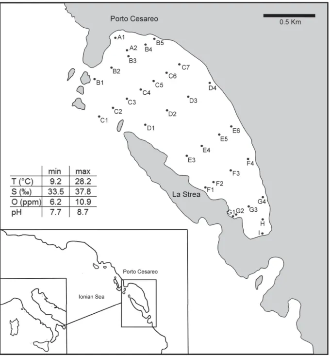
is in its northern part connected with the Ionian Sea, and is characterised by a subtropical biocoenosis (Parenzan 1976, 1983, 1984). The environmental setting is entirely included in the upper part of the infralittoral zone, with water depth not exceeding fi ve meters. Ecological parameters vary over seasons and sectors of the bay (Fig. 1).
It can be supposed that environmental factors infl uence, at least in part, the development of different shell morphs. Consequently, the studied specimens may represent slightly different ecomorphotypes belonging to dead assemblages.
Fig. 1. Location map of the study area and sampling stations and range of selected physico-chemical parameters of the La Strea Bay (data from Belmonte & Rubino 1988; Mercurio et al. 2000; Gherardi et al. 2001).
With the aim to minimize the unavoidable taxonomist’s subjectivity, we have employed two morphological methods of discrimination: the fi rst is the comparison of ornament morphology, taking into account the features of the reticulation especially; the other is the morphometric analysis of the outline by means of the computer program Morphomatica (Linhart et al. 2006).
To be more precise, our intent is: 1) to defi ne intrademic, non-polytypic (sensu Sylvester-Bradley 1976) variations of shell features, namely the morphological variability of the Urocythereis demes - panmictitc populations (freely interbreeding, sharing the same gene pool, relatively confi ned) of the La Strea Bay;
2) to assess how many Urocythereis species are present in the Recent/sub-Recent thanatocoenoses of the bay; we consider this attempt as a step toward a better understanding of the complex morphology of the genus from a paleontological viewpoint; 3) to characterize groups of fossae, stable within species and variable among species (e.g., Al-Furaih 1977).
Intraspecifi c variability in ostracod shells
The studies on intraspecifi c morphological variations include investigations on soft parts (i.a., Rossetti
& Martens 1996; Martens et al. 1998; Yin et al. 1999), internal features of smooth valves (i.a., Aiello et al. 2007, and references therein), surface ornamentation (i.a., Keen 1982; Peypouquet et al. 1988;
Neil 2000) and outline (Baltanas & Danielopol 2011, and references therein). Intraspecifi c variability can be continuous or discontinuous, and continuous variations may seem very simple to analyse if compared with polymorphism sensu Ford (1940), that is “the occurrence together in the same locality of two or more discontinuous forms of a species in such proportions that the rarest of them cannot be maintained merely by recurrent mutation” (see Clark 1976 for discussion and extensive references).
Yet, when we deal with morphologically highly variable taxa, also non-polymorphic species highlight complex taxonomic problems.
Here, we focus on the continuous variation of shape and ornament in Urocythereis, with special regard to reticulation. Urocythereis species may show all fi ve types of continuous variation of fi ne sculpture (in position, form, strength of expression, size and number of elements) described by Liebau (1971).
High-degree variability in ostracod shell sculpture, derived from both genetic and environmental factors, is a recurrent problem experienced by authors dealing with studies on shallow marine assemblages (e.g., Hartmann 1982), which always include some phenotypically plastic species. Phenotypic plasticity, that is the ability of an organism to express different phenotypes under different environmental conditions (see, e.g., West-Eberhard 1989), may represent a fundamental resource for effective adaptation to coastal environments, characterized by wide ranges and rapid changes of ecological parameters, and is a major drawback for systematic investigations. Seemingly distinct features occurring in some infra-littoral ostracod taxa lead taxonomists to propose a large number of specifi c names, which frequently have not stood the test of time. In other cases, taxonomists “lumped” together very different forms (see, e.g., a brief discussion on the genus Carinocythereis in Aiello & Szczechura 2001).
The genus Urocythereis
The genus Urocythereis was originally described by Ruggieri in 1950, with the intent to accommodate three strictly related hemicytherid species, with subrectangular valves and an amphidont hinge provided with a reniform posterior tooth in the right valve. These species were Cytherina favosa Roemer, 1838, designated as type species, Cythereis margaritifera G.W. Müller, 1894 and Cythereis distinguenda Neviani, 1928, from the Pliocene to Recent of the Mediterranean area. Since then, dozens of species have been included in Urocythereis, especially from the Neogene to Recent of the Mediterranean area and the eastern Atlantic. Aiello et al. (2004) assigned the south-western Atlantic species, previously included in Urocythereis, to the new genus Ruggiericythere, due to the different structure of the reticulation-ridges system. The allied genus Yezocythere Hanai & Ikeya, 1991 includes eastern Asian
species with a low subcentral tubercle, lacking in Urocythereis. Even though the authors recognized a second distinctive feature, the smaller size of the median frontal scar, the value of this character at genus level remained questionable. The Neogene to Recent Eastern African and Asian records of Urocythereis (e.g., U. pohangensis Huh & Whatley, 1997; U. salebrosa Ahmad, Neale & Siddiqui, 1991), as well as its presence in Paleogene deposits (e.g., U. bertelsae Kielbowicz, 1988), are, in our opinion, questionable.
In our present state of knowledge, Urocythereis is a Cenozoic genus possibly restricted to the infralittoral and uppermost circalittoral zone (0–40 m, Athersuch 1977) of the Eastern Atlantic - Mediterranean area.
The taxonomy of this genus is complex due to the unusually wide variability of the shell ornamentation and, consequently, species limits within the genus remain partly undefi ned.
A major contribution to the knowledge of the genus is the study by Athersuch (1977), who provided a revision of all the then known living and some fossil species, in addition to an excellent iconography.
He described detailed ornamental patterns of fi ve allied species (U. distinguenda (Neviani, 1928), U. margaritifera, U. favosa (Roemer, 1838), plus U. neapolitana and U. britannica Athersuch, 1977, described as new species) based on “nine arbitrary groups” of fossae. The author used these patterns to demonstrate differences among groups of species, one being later described by Ruggieri & Russo (1980) as a new genus, Nonurocythereis. These groups are not effective for discrimination at species level. The outline of the muri, as well as the shape and dimension of the fossae, and their degree of anastomosis, are rarely steady, so that reticulation varies “even between members of the same species” (Athersuch 1977). Uncertainty is increased due to the frequent occurrence of celation (Sylvester-Bradley & Benson 1971), a morphological noise able to conceal, in some cases almost completely, ornamental patterns.
In a similar way, the relationship between normal pores and reticulation, used by authors for ostracod morphology studies (e.g., Hunt & Yasuhara 2010), is blurred by various degrees of development of shell calcifi cation. Normal pores are well distinct in internal view, not in external view, and consequently the relationship between pores and reticulum pattern is diffi cult to establish.
Reticulation
Shell reticulation represents a complex of features that, since the early contributions of Pokorny (1969a, 1969b), Liebau (1969, 1971) and Benson (1971, 1972), has been considered by ostracod workers a fascinating object of study for taxonomy, evolution and related research fi elds. Ornament pattern defi nition has been carried out especially on hemicytherid and trachyleberid taxa, frequently showing a well-defi ned and more or less stable reticulation. Interspecifi c variations, in related species, are sometimes limited to a few fossae and highly consistent in each species (Okada 1982b). The analysis of the fossal pattern has been used for species discrimination (e.g., Al-Furaih 1977) and in studies on the evolution and ontogeny of Trachybeleridoidea (i.a., Hunt 2007a, 2007b; Hunt & Yasuhara 2010; Tanaka et al. 2011). Jones (1988) showed the possibility to extend the reticulation analysis to Paleozoic taxa.
Okada (1981, 1982a, 1982b) extensively studied the relationship of the epidermal cells to the reticulation pattern and he recognized a direct correspondence between fossae and the underlying epidermal cells. Since the muri represent the boundary of adjacent cells, the mesh arrangement refl ects cell lineages, commonly considered as genetically determined (i.a., Liebau 1971; Benson 1972; Irizuki 1993; Hunt 2007a).
Reticulated species frequently show a pattern of ridges where homologous fossae can be recognized by their constancy in shape, number and arrangement and by the presence of landmarks such as pores, ridges or spines (i.a. Benson 1972; Liebau 1991; Hunt 2007b; Hunt & Yasuhara 2010). In Urocythereis, the number of fossae is relatively stable within species, while their shape and arrangement vary widely, pores are frequently obscured by calcifi cation and the features of anatomical landmarks, for example muscle scars, can be diffi cult to defi ne. Furthermore, alae, carinae or tubercles are not present in Urocythereis. Consequently, an unambiguous identifi cation of fossae and an accurate quantitative study
of their variations (e.g., Hunt & Yasuhara 2010 on Poseidonamicus) has proved unattainable. Ruiz et al. (2006) hypothesized a direct infl uence of environmental factors on the variability of Urocythereis reticulation.
Celation as a taxonomic issue
The development of an outer layer of calcite, the tegmen, overlapping the ornament of the valves, has been named “celation” by Sylvester-Bradley & Benson (1971). The authors considered the tegmen as a secondary thickening paving over the outer surface and thus obscuring, to some degrees, primary reticulation. Benson (1972) hypothesized that the development of the tegmen represents the architectural response to the requirement of a high shell strength. In deep and cold waters, this lightweight solution may represent a case of parsimony in design balancing the scarcity of available skeletal material with the development of high resistance to compression. Athersuch (1977) observed that the development of celation (not recorded in juveniles) in Urocythereis distinguenda is related neither to depth nor to other known ecological parameters, and suggested that the growth of the tegmen depends from the age of the individuals: an intense celation would be typical of older adults. Another hypothesis considers celation as a response to calcium saturated bottom waters, pertaining to the “environmentally cued polymorphism” described by Peypouquet et al. (1980, 1981, 1988). Anyway, because celation varies continuously, we agree with Neil (2000) on preferring the term “environmentally-cued variation”. In this particular case the terms “celation” and “agradation” (“aggradation” in Neil 2000) are equivalent.
These two hypotheses are not mutually exclusive.
The ecological demands of an infralittoral genus can hardly be compared with the bathyal and abyssal taxa studied by Benson (1972), who supposed that the secondary development of the top of the muri in shallow water species, such as Cythere lutea O.F. Müller, 1785 and Hemicythere villosa (Sars, 1866), has to be an adaptation to high energy sedimentary regimens. The attested preference of Urocythereis for sandy bottoms (e.g., Athersuch et al. 1989; Aiello et al. 2006) and relatively agitated waters supports this interpretation. It is understood that celation may be a consequence of simple genetic variation, or a mixture of genetic and environmental effects. In any case, the occurrence of specimens with more or less celated shells poses a taxonomic problem, whether we are dealing with two species (in this case U. margaritifera and U. distinguenda), with individuals of the same species that have possibly built their valves in different physico-chemical conditions, or, alternatively, with younger and older adults of the same species.
How many Urocythereis species occur in the La Strea Bay?
The assignment of the Urocythereis specimens of the La Strea Bay to the above mentioned three species by Aiello & Barra (in Aiello et al. 2006) followed the original description and fi gures of G.W. Müller (1894) and subsequent literature for U. margaritifera, and Athersuch (1977) for U. distinguenda.
Cythereis margaritifera was described by G.W. Müller in his monograph on the ostracods living in the Gulf of Naples, and later recorded from Pliocene to Recent in the Mediterranean area. Cythereis distinguenda is the name proposed by Neviani (1928) to replace the pre-occupied Cythere oblonga Brady, 1866 (non C. oblonga M’Coy, 1844). Athersuch (1977) discussed nomenclatural problems and designated a neotype for U. distinguenda from the shallow waters of Cyprus. This author fi gured U. margaritifera specimens from Müller’s collection, showing individuals with rounded fossae as well as with fossae reduced to punctae and foveolae (sensu Athersuch et al. 1989) because of celation. In the same paper the specimens assigned to U. distinguenda show a marked celation with small-sized irregular punctae-foveolae.
A third form present in the La Strea Bay bottom samples was left in open nomenclature and named U. sp. 1 because of the resemblance with the specimens from the beach sands of Tripoli fi gured by
Barra (1997). This previously undescribed species has a marked affi nity with U. exedata Uliczny, 1969, especially in the very distinct ocular riblet, a feature homologous to the prominent “ocular ridge”
described by Benson (1972) on some Bradleya species. The new species shows a distinct ornamental pattern and specifi c features. Conversely, it seems very diffi cult to defi ne an unambiguous boundary between U. distinguenda and U. margaritifera, necessitating the examination of “intermediate” morphs, in order to verify the existence of two separate species rather than a single highly variable species.
Material and methods
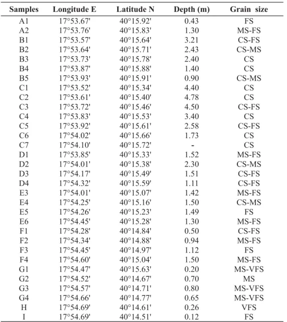
All the 32 samples were taken within the depth range of 0.12–4.78 m bsl (Aiello et al. 2006), from the Recent bottom sediments of the La Strea Bay (Table 1). The samples contained 2133 valves and 128 carapaces, including both adults and young instars. All the specimens were studied and 150 shells were
Samples Longitude E Latitude N Depth (m) Grain size
A1 17°53.67' 40°15.92' 0.43 FS
A2 17°53.76' 40°15.83' 1.30 MS-FS
B1 17°53.57' 40°15.64' 3.21 CS-FS
B2 17°53.64' 40°15.71' 2.43 CS-MS
B3 17°53.73' 40°15.78' 2.40 CS
B4 17°53.87' 40°15.88' 1.40 CS
B5 17°53.93' 40°15.91' 0.90 CS-MS
C1 17°53.52' 40°15.34' 4.40 CS
C2 17°53.61' 40°15.40' 4.78 CS
C3 17°53.72' 40°15.46' 4.50 CS-FS
C4 17°53.83' 40°15.53' 3.40 CS
C5 17°53.92' 40°15.61' 2.58 CS-FS
C6 17°54.02' 40°15.66' 1.73 CS
C7 17°54.10' 40°15.72' - CS
D1 17°53.85' 40°15.33' 1.52 MS-FS
D2 17°54.01' 40°15.38' 2.30 CS-MS
D3 17°54.17' 40°15.49' 1.51 CS-FS
D4 17°54.32' 40°15.59' 1.11 CS-FS
E3 17°54.01' 40°15.07' 1.42 MS-FS
E4 17°54.25' 40°15.16' 1.50 CS-MS
E5 17°54.26' 40°15.23' 1.49 FS
E6 17°54.45' 40°15.28' 1.30 MS-FS
F1 17°54.28' 40°14.84' 0.50 CS-FS
F2 17°54.34' 40°14.88' 0.94 MS-FS
F3 17°54.45' 40°14.97' 1.12 FS
F4 17°54.60' 40°15.04' 1.50 MS-FS
G1 17°54.47' 40°15.63' 0.20 MS-VFS
G2 17°54.52' 40°14.67' 0.70 MS
G3 17°54.57' 40°14.71' 0.80 MS-VFS
G4 17°54.66' 40°14.77' 0.65 MS-VFS
H 17°54.69' 40°14.61' 0.26 VFS
I 17°54.69' 40°14.51' 0.12 FS
Table 1. Coordinates, depth and granulometry of the studied samples. Abbreviations: VSF = Very Fine Sands, FS = Fine Sands, MS = Medium Sands, CS = Coarse Sands.
selected for analyses and iconography. This study deals exclusively with adult specimens. Young instars were considered only to evaluate the possible occurrence of celation in juveniles.
The analysis of the reticulation pattern and celation variations is based on scanning electron microscopy (SEM) micrographs (carried out at Cisag, Università di Napoli Federico II). The above mentioned high variability of the reticulation in Urocythereis species lead us to select some areas of the shell, where the fossae-muri pattern was detectable with a minimum degree of uncertainty.
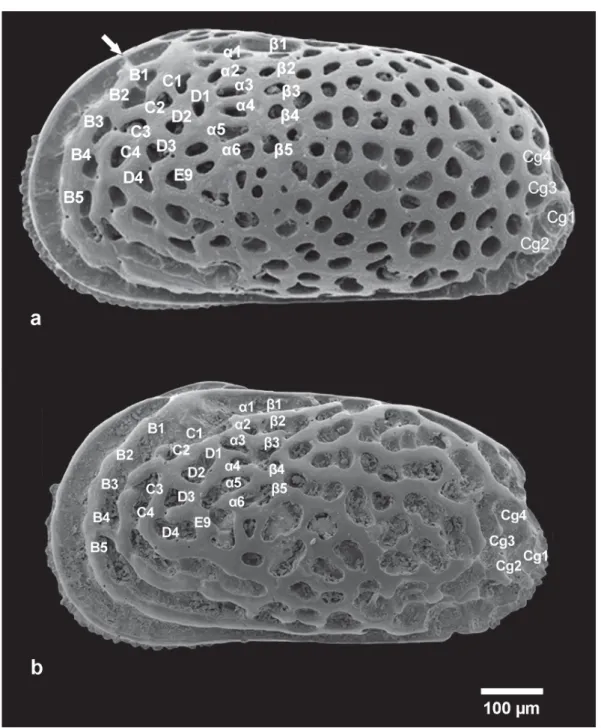
Fig. 2. Scheme of the fi ve fossal groups used for reticulation pattern analysis. White arrow indicates the pre-ocular bridge. A. Urocytheris margaritifera (G.W. Müller, 1894), ABMC 2014/057. B. Urocytheris ilariae sp. nov., ABMC 2014/047.
The Morphomatica test was performed on drawings in transmitted light. They were carried out by means of a Visopan Reichert, with a magnifi cation of 201 times. The pictures were drawn with a technical pen (thickness of the line: 0.30 mm) on tracing paper. The studied specimens are housed in the Aiello Barra Micropaleontological Collection (ABMC), Distar, Università degli Studi di Napoli Federico II.
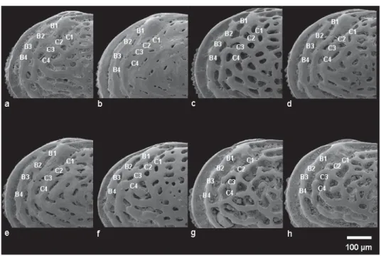
Fig. 3. Detail of the antero-dorsal area in different morphs. — A–F. U. margaritifera (G.W. Müller, 1894). A. ABMC 2014/045. B. ABMC 2014/053. C. ABMC 2014/057. D. ABMC 2014/059. E. ABMC 2014/075. F. ABMC 2014/083. — G–H. U. ilariae sp. nov. G. ABMC 2014/038. H. ABMC 2014/047.
Table 2. Catalogue number and sampling stations of the specimens used in the Morphomatica analysis.
Catalogue number Samples Catalogue numbers Samples
ABMC 2014/125 C4 ABMC 2014/142 C5
ABMC 2014/126 C4 ABMC 2014/143 C5
ABMC 2014/127 D3 ABMC 2014/144 D3
ABMC 2014/128 D3 ABMC 2014/145 D3
ABMC 2014/129 E6 ABMC 2014/146 D3
ABMC 2014/130 E6 ABMC 2014/147 D3
ABMC 2014/131 E6 ABMC 2014/148 E6
ABMC 2014/132 E6 ABMC 2014/149 E6
ABMC 2014/133 E6 ABMC 2014/150 E6
ABMC 2014/134 H ABMC 2014/151 G2
ABMC 2014/135 H ABMC 2014/152 G2
ABMC 2014/136 A2 ABMC 2014/153 G2
ABMC 2014/137 A2 ABMC 2014/154 H
ABMC 2014/138 A2 ABMC 2014/155 H
ABMC 2014/139 B3 ABMC 2014/156 H
ABMC 2014/140 C4
Reticulation pattern
In a highly variable genus such as Urocythereis the purpose of naming the position of all the meshes seems diffi cult to achieve (in Urocythereis especially in the ventral area and in the posterior half of the shell). Consequently, we identifi ed fi ve arbitrary fossal groups where the pattern can be recognized, both in different species and specimens of the same species, as homologous without uncertainties (Fig. 2).
These groups were selected with the aim to defi ne the structure of inter- and intra-specifi c reticulation pattern variations. We chose muri/fossae sets that could be reliably considered homologous in U. margaritifera and U. exedata. The two species are quite different and we hope that these groups may be used for future comparisons among other species of Urocythereis.
Fossal groups are defi ned as follows:
a) Pre-ocular fossa (Pre-ocular bridge): presence of a preocular connection between the anterior marginal rim and the ocular riblet, as fi gured in Fig. 2.
b) Anterodorsal group: a group of fossae located in the subocular area (B1, B2, B3, B4, C1, C2, C3, C4) as numbered in Figs 2 and 3.
c) D3-D4-E9 group: these fossae form a triangular area situated in the anterocentral area, behind the fourth concentric anterior ridge as fi gured in Figs 2 and 4.
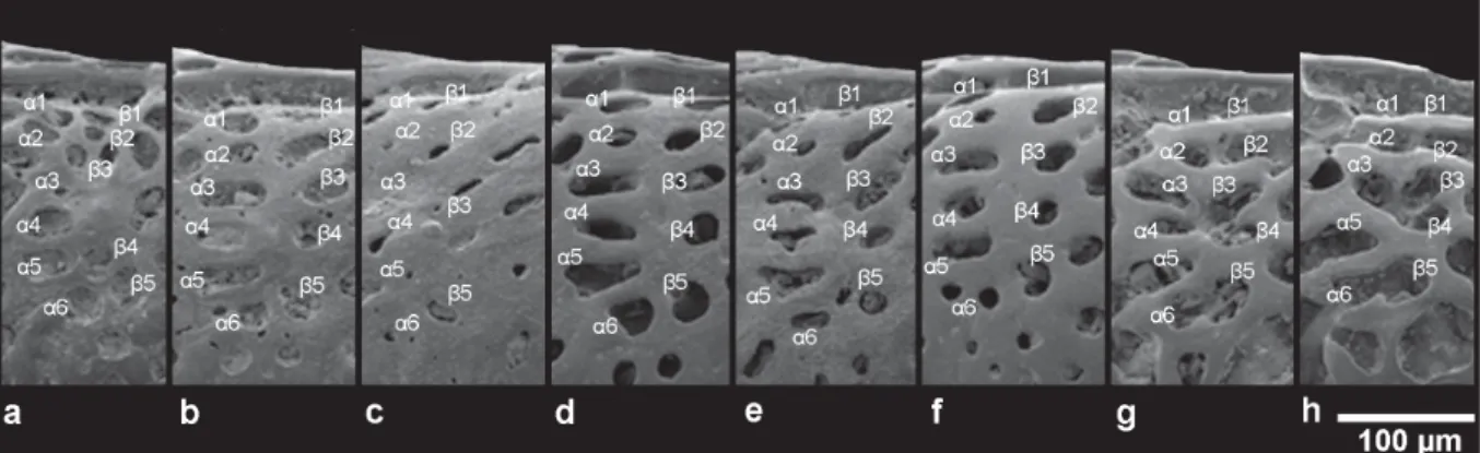
d) Dorsal median group: Athersuch (1977) defi ned this group as “two vertical rows of fossae directly beneath the post-ocular sinus and directed towards the sub-central area” (α1–α6, β1–β5 in Figs 2 and 5);
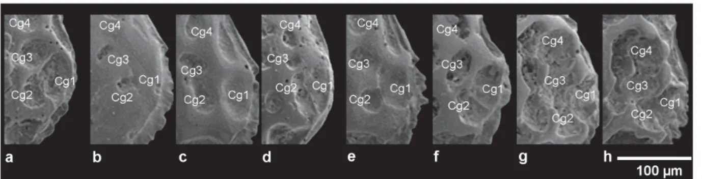
e) Caudal group: the group of fossae (Cg1–Cg4) located on the caudal process area as fi gured in Figs 2 and 6.
Fig. 4. Detail of the antero-central area showing the fossal group D3-D4-E9 in different morphs. — A–F. U. margaritifera (G.W. Müller, 1894). A. ABMC 2014/045. B. ABMC 2014/50. C. ABMC 2014/053. D. ABMC 2014/057. E. ABMC 2014/075. F. ABMC 2014/083. — G–H. U. ilariae sp. nov.
G. ABMC 2014/047. H. ABMC 2014/069.
Fig. 5. Dorsal median group in different morphs. — A–F. U. margaritifera (G.W. Müller, 1894).
A. ABMC 2014/037. B. ABMC 2014/04. C. ABMC 2014/053. D. ABMC 2014/057. E. ABMC 2014/075.
F. ABMC 2014/083. — G–H. U. ilariae sp. nov. G. ABMC 2014/047. H. ABMC 2014/069.
Outline analysis
A quantitative analysis of ostracod morphology has been carried out by means of different methods, including techniques of outline analysis (for a partial review, see Baltanás & Danielopol 2011). In recent years, ostracod researchers seem leaning towards using the computer program Morphomatica, based on a B-spline algorithm specifi cally adapted to ostracod valve outline (Baltanás et al. 2003; Linhart et al.
2006), which provides valuable results in specifi c discrimination (i.a., Iepure et al. 2007; Danielopol et al. 2008; Ligios & Gliozzi 2012; Mazzini et al. 2014). The method has been described, for example, in Gross et al. 2008. This technique has been used mostly on smooth or weakly ornamented ostracod taxa, whereas we tested its effectiveness by using it to discriminate strongly reticulated forms. The material consists of 20 left valves of the Urocythereis margaritifera-distinguenda group and 11 left valves of U. ilariae sp. nov. (Table 2). On the basis of the shell features a clear discrimination between female and male valves is not possible. The selection of the valves showing a higher h/l ratio would be, in absence of discontinuity, a mere example of circular reasoning. Consequently, we selected the best-preserved valves, with no regard for their supposed sex, well-aware of the resulting limitations. Due to the presence of unevenly preserved marginal denticulation we chose, unlike the usual procedure using transmitted light microscope photographs, to analyse and digitize drawings generated using Visopan Reichert at a magnifi cation of ×201. The results of Morphomatica were processed using Cluster analysis, non- metric multidimensional scaling (n-MDS) and analysis of similarities (One-way ANOSIM Pairwise Test) by means of the program PRIMER 6 (Clarke & Gorley 2006).
Results
Celation, variability and specifi c assignment of Urocythereis sp. 1
The morphological variations in Urocythereis sp. 1 appear limited when compared with the U. margaritifera-distinguenda group. Celation is rare and the fossae are never hidden by secondary calcifi cation; the structure of reticulation is somewhat constant. The basic pattern shows a close resemblance to the Pliocene form described by Uliczny (1969) as Urocythereis favosa exedata and elevated to the rank of species by Mostafawi & Matzke-Karasz (2006). Some stable shell features of U. sp. 1 are different from those of the Pliocene taxon, leading us to describe a new species, named U. ilariae sp. nov. as discussed in the systematic section.
Fossae shape and size variations and celation in the U. margaritifera-distinguenda group
Brady (1866) described a species from Recent sponge-sands of the Eastern Mediterranean as Cythere oblonga, characterized by “somewhat distant, oblong, pittings”. The name C. oblonga was, however, preoccupied by C. oblonga M’Coy, 1844; therefore, in 1928 Neviani assigned a new name, Cythereis distinguenda, recognized by Athersuch (1977, 1982) as the “next available name for Brady’s species”.
Fig. 6. Detail of the caudal group in different morphs. — A–F. U. margaritifera (G.W. Müller, 1894). A. ABMC 2014/045. B. ABMC 2014/053. C. ABMC 2014/059. D. ABMC 2014/062. E. ABMC 2014/075. F. ABMC 2014/083. — G–H. U. ilariae sp. nov. G. ABMC 2014/038. H. ABMC 2014/069.
In previous investigations, the present authors used the size of the fossae to discriminate between U. margaritifera and U. distinguenda, being relatively large in the former species and very small in the latter one. The fi nding of specimens exhibiting transitional features makes their specifi c attribution diffi cult. The following morphs can be defi ned on the basis of the size and shape of the fossae:
Morph a (commonly assigned to U. margaritifera): medium-sized fossae, equidimensional, except in the anterodorsal zone where they are medium- to small-sized; generally rounded, elongated or fused in the anteroventral area (e.g., Fig. 16A, H);
Morph b (commonly assigned to U. margaritifera): medium-sized fossae rounded and equidimensional on the whole surface of the shell (e.g., Figs 2A; 16B);
Morph c (“transitional” form): mesh size from medium to very small, generally rounded (e.g., Fig. 17E);
Morph d (“transitional” form): fossae mainly elongated, from small to medium-sized (e.g., Fig. 17B);
Morph e (“transitional” form): the anterior area shows fossae very small or completely celated; the remaining fossae are small and rounded (e.g., Fig. 17D);
Morph f (commonly assigned to U. distinguenda): small-sized fossae; narrow and elongated in the anterior part of the valve, rounded in the posterior part and mixed in the central area (e.g., Fig. 16D);
Morph g (commonly assigned to U. distinguenda): small-sized fossae, rounded or elongated in the posterior half of the shell; very small (reduced into puncta) to completely celated in the anterior half (Fig. 17C);
Morph h (commonly assigned to U. distinguenda): size of fossae ranging from small to very small;
elongated or rounded (Fig. 16G).
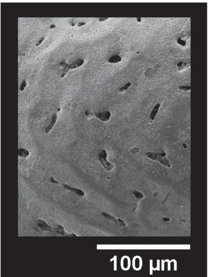
It has to be noted that in this latter group the muri are generally densely or sparsely papillate; in specimens showing intense celation, papillae occur on the tegmen and are very sparse or lacking in correspondence with the underlying muri, revealing the reticulation pattern (Fig. 7). A similar feature was observed by Benson (1972) in the species Poseidonamicus nudus Benson, 1972.
In our opinion, these data suggest that U. margaritifera and U. distinguenda are morphotypes of the same species.
Reticulation variability
The fossal groups show the following variations:
a. Pre-ocular fossa (pre-ocular bridge)
In U. margaritifera/distinguenda, a murus located in the antero-dorsal corner, in pre-ocular position, generally connects the anterior marginal rim and the ocular riblet, which delimitate the marginal furrow (Figs 3A, C–E; 19D), forming a pre-ocular fossa. The pre-ocular bridge is more or less well defi ned in U. margaritifera/distinguenda, including celated specimens, where it is highly developed and the pre- ocular fossa is reduced (Fig. 3B). In some specimens of U. margaritifera/distinguenda, the bridge is absent and the furrow is continuous up to the eye tubercle (Fig. 3F).
The pre-ocular fossa never occurs in U. ilariae sp. nov. (Fig. 3G–H).
b. Anterodorsal group. Fossal pattern B1–B4 – C1–C4
In U. ilariae sp. nov., the presence of a murus between the fossae B5 and B4, connecting the second and the third marginal riblets, is a stable feature and a possible landmark when comparing species
of Urocythereis. It is subdued in some very rare celated specimens (Fig. 18G). A composite fossa, including the anastomized fossae B1, B2, B3, B4, C1 and C2, shows slight intraspecifi c variability (Figs 2B; 3G–H); in some specimens (Fig. 18C, F) B4 and C4 are connected.
In U. margaritifera/distinguenda, B and C fossae never connect. The fossae B1 and B2 may be divided (Fig. 17G), partially fused (Figs 2; 16B) or merged (Fig. 3E–F). Fossae B2-B3-B4 are never joined, C1 and C2 are usually divided (Fig. 3C, F) and can show some degrees of anastomosis (Fig. 3D–E).
In some specimens celation may simulate mesh subdivision. For example, in Fig. 3B the C2 mesh is almost completely closed by tegmen and small foveolae mirror the underlying fossa. Fossae C3 and C4 are never fused.
c. D3-D4-E9 group
In U. margaritifera/distinguenda, D4 and E9 are usually distinct (Fig. 4B–F), rarely joined together (Fig. 4A). In a few specimens, D4 is partly divided, and D4 and E9 appear as an elongated subhorizontal fossa (Fig. 4A). D3 is constantly distinct.
Fig. 7. Detail of papillae in heavily celated specimen of U. margaritifera (G.W. Müller, 1894), ABMC 2014/071.
In U. ilariae sp. nov., the fossae D3-D4-E9 form a steady triangular pattern with D4-E9 fused and D3 is situated above them. In very rare celated specimens, D4 and E9 are distinct (Fig. 18G).
d. Dorsal median group. Fossal pattern α1–α6, β1–β5
In U. margaritifera/distinguenda, an ideal scheme of this group can be represented as two rows: the anterior row consists of six fossae, the posterior row includes fi ve fossae, named α1–α6 and β1–β5 (Fig. 5A–F), respectively. This is seemingly in contrast with the scheme of Athersuch with 5+4 fossae;
in fact, the upper two fossae (α1 and β1), just below the post-ocular sinus, may be clearly observed in dorsal view (Fig. 19D). In lateral view they appear very reduced or even completely hidden (Figs 5E–F; 16H; 17B). In the posterior row, between the 4th (β4) and 5th (β5) fossa, a small smooth area, the “focus” of Athersuch (1977), is present. The lowermost fossae, herein named α6 and β5, pertain, following Liebau’s scheme (1971), to the E-ring encircling the muscle-scar node.
We observed the following variations:
- fossa α3 divided in two subfossae (Fig. 16F);
- fossa α5 divided in two (Fig. 16F, H) or three (Fig. 17F) subfossae;
- fossa α6 divided in two (Figs 5F; 17G);
- fossa β3 partially connected with the centrodorsal fossae (Figs 16C; 17A).
The shape of the meshes shows a wide variability, partly due to celation, ranging from large rounded fossae to small pits, including narrow foveolae (Fig. 5A–F).
In U. ilariae sp. nov., the dorsal median group shows a different pattern (Fig. 5G–H). In comparison with the ideal scheme, the uppermost fossae (α1, β1) are absorbed in the post-ocular sinus; α2 and β2 are located under the post-ocular sinus; α2 is alternatively isolated (Figs 5G; 19B) or fused with β2 (Fig. 18B). The fossa β2, in turn, is fused with a posterior fossa (Figs 5G; 19B). The α3-β3/α4-β4 meshes may coalesce in part, generally forming a butterfl y shaped quadruple fossa (Fig. 5G); in some specimens α4 is fused with an anterior fossa (Fig. 5H).
e. Caudal group
In U. margaritifera/distinguenda, it consists of a basic pattern of three well-defi ned fossae, two marginal and one internal with various shapes in different specimens (Fig. 6A–F). They may appear as a single triple fossa (Fig. 17H) or as a single plus one double fossa (Fig. 2A). In the latter case, anastomosis partially hides the identity of the fossae. Celation may completely obscure this pattern (Figs 6B; 17C).
In U. ilariae sp. nov., a fourth upper fossa (Cg4) merges with the caudal group, forming a multiple L-shaped or elongated fossa; it can be more or less fused with another internal elongated multiple fossa (Fig. 6G–H).
Our observations indicate that the reticulation pattern in the forms previously attributed to U. margaritifera and U. distinguenda are identical. In U. ilariae sp. nov., well distinct patterns of homologous fossae can be recognized.
Outline analysis
The valve outline of U. margaritifera (i.e., U. margaritifera “morph margaritifera” plus U. margaritifera
“transitional morph”), U. distinguenda (i.e., U. margaritifera “morph distinguenda”) and U. ilariae sp.
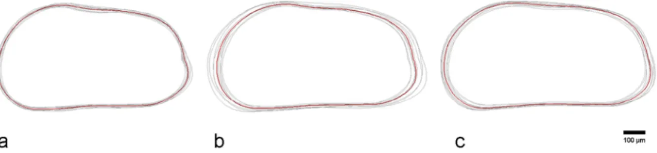
nov. were analysed and compared. In Fig. 8A–C the “non normalized area” (size dependent) mode results show that the variability of U. ilariae sp. nov. (Fig. 8A) is very limited. By contrast, in U. margaritifera (Fig. 8B) the variability is relatively wide, linked to the length of the valves. Conversely, with the
“normalized area” (size independent) mode, a low variability is observed in U. margaritifera (Fig.
9B) while it is relatively high in U. ilariae sp. nov. (Fig. 9A). U. distinguenda shows an intermediate variability in both modes (Figs 8C; 9C).
Comparisons of the mean outline of the three forms indicate that U. ilariae sp. nov. is positively separated from U. margaritifera and U. distinguenda, while the discrimination of the latter forms is diffi cult. In
“normalized area” mode (Fig. 10B) U. ilariae sp. nov. shows a shorter length and a different shape in the caudal and in the anterodorsal regions. Its shorter length is more evident in the mean outlines deriving from “non normalized area” mode analysis (Fig. 10A).
Fig. 8. Comparison of valve outlines obtained by Morphomatica analysis. Results for “non-normalized area” mode. A. U. ilariae sp. nov. B. U. margaritifera (G.W. Müller, 1894). C. U. distinguenda (Neviani, 1928).
Fig. 9. Comparison of valve outlines obtained by Morphomatica analysis. Results for “normalized area”
mode. A. U. ilariae sp. nov. B. U. margaritifera (G.W. Müller, 1894). C. U. distinguenda (Neviani, 1928).
Fig. 10. Comparison of mean outlines of U. ilariae sp. nov. (green), U. margaritifera (G.W. Müller, 1894) (light blue) and U. distinguenda (Neviani, 1928) (dark blue) obtained by the Morphomatica analysis. A. Results for “non-normalized area” mode. B. Results for “normalized area” mode.
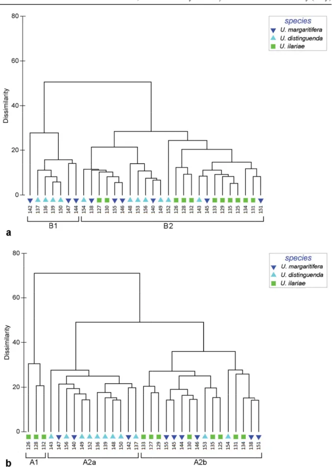
Fig. 11. Cluster analysis performed on valve outlines obtained by Morphomatica analysis. A. Dendrogram for “non-normalized area” mode. B. Dendrogram for “normalized area” mode.
Cluster analysis for “normalized area” mode reveals two main clusters, the fi rst (A1) includes three valves of U. ilariae sp. nov., the second (A2) consists of all the remaining specimens (Fig. 11B). This second cluster is subdivided into two subclusters: A2a, including nine valves pertaining to U. distinguenda and three specimens of U. margaritifera and A2b, where all the three forms are present (eight U. ilariae sp. nov., two U. distinguenda and six U. margaritifera valves).
The “non normalized area” mode analysis (Fig. 11A) discriminates two main clusters: B1, consisting of three valves of U. margaritifera and four of U. distinguenda, and B2 including the remaining specimens.
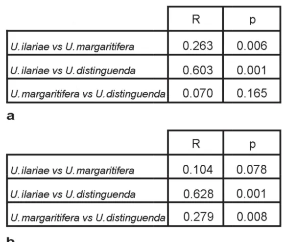
The ANOSIM Pairwise Test applied both to the “non normalized area” (Fig. 12A) and “normalized area”
mode (Fig. 12B) displays that U. ilariae sp. nov. and U. distinguenda are separated on the basis of the outline analysis (R = 0.603; R = 0.628). Conversely, U. margaritifera cannot be discriminated from U. ilariae sp. nov. or from U. distinguenda.
N-MDS analysis provided comparable results (Fig. 13). The distribution areas of U. ilariae sp. nov. and U. distinguenda are well defi ned for both “non normalized area” mode (Fig. 13A) and “normalized area” mode (Fig. 13B), whereas the distribution of U. margaritifera specimens overlaps both U. ilariae sp. nov. and U. distinguenda.
Fig. 12. Analysis of similarities (One-way ANOSIM Pairwise Test). A. R statistic and p value for “non- normalized area” mode. B. R statistic and p value for “normalized area” mode.
Fig. 13. Non-metric multidimensional scaling (n-MDS) analysis. A. Results for “non-normalized area”
mode. B. Results for “normalized area” mode.
Taxonomy
Classifi cation follows Liebau (2005) and, for the attribution to subfamily, Hartmann & Puri (1974).
Subclass Ostracoda Latreille, 1802 Superorder Podocopomorpha Kozur, 1972
Order Podocopida Sars, 1866 Suborder Cytherocopina Gründel, 1967 Infraorder Nomocytherinina Liebau, 1991 Superfamily Trachyleberidoidea Sylvester-Bradley, 1948
Family Hemicytheridae Puri, 1953
Subfamily Urocythereidinae Hartmann & Puri, 1974 Genus Urocythereis Ruggieri, 1950
Urocythereis ilariae sp. nov.
urn:lsid:zoobank.org:act:607A810F-773C-4F8F-91E7-D2EB5223CB7D Figs 2B; 3G–H; 4G–H; 5G–H; 6G–H; 14; 18A–J; 19A–C, E–J Urocythereis favosa (Roemer) n. ssp. Bassiouni, 1965: pl. 40, fi gs 8–9.
Urocythereis favosa (Roemer) subsp. – Wouters 1973: 385, pl. 2, fi g. 7.
Urocythereis sp. – Bonaduce, Ciampo & Masoli 1976: 46, pl. 22, fi g. 9.
? Urocythereis sp. – Athersuch 1977: pl. 17, fi g. 2.
Urocythereis aff. U. favosa (Roemer) – Arbulla, Pugliese & Russo 2001: fi g. 3t.
? Urocythereis sp.1 – Barra 1997: 82, pl. 4, fi g. 6.
Urocythereis sp.1 – Aiello et al. 2006: tables 3, 10. — Aiello, Barra & Parisi 2013: fi g. 1d.
Diagnosis
A large reticulate species of Urocythereis, subrectangular in lateral view, infl ated-ovate in dorsal view.
Reticulum with large polygonal-rounded, frequently coalescing, large fossae separated by broad muri.
In the anteroventral area the muri form distinct riblets running parallel to the margin.
Etymology
In honour of our friend and collegue Ilaria Mazzini, in recognition of her important contribution to ostracodology.
Type material (4 carapaces, 43 valves: 29 adults and 14 juveniles) Holotype
IONIAN SEA: ABMC 2014/03 Paratypes
IONIAN SEA: ABMC2 014/026–036, ABMC 2014/038, ABMC 2014/042, ABMC 2014/044, ABMC 2014/046–049, ABMC 2014/063–064, ABMC 2014/069, ABMC 2014/072–073, ABMC 2014/080, ABMC 2014/097–103, ABMC 2014/120–135.
Stratum typicum Recent.
Locus typicus
La Strea Bay (Porto Cesareo Lagoon), Southern Italy, Ionian Sea, sampling station E4, 17°54'25" N, 40°15'59" E, depth 1.5 m bsl.
Description
Measurements (holotype): LV: L = 0.85 mm, H = 0.44 mm (Fig. 18A).
Large (L = 0.85–0.90 mm) species of Urocythereis, characterized by large fossae and strongly developed muri, subrectangular in lateral view, infl ated-ovate in dorsal view. Valves strongly calcifi ed and thick.
Dorsal margin gently, unevenly convex, ventral margin weakly sinuous; anterior end broadly rounded, denticulate in the lower part; upper part of the posterior margin concave, lower part of the posterior margin convex, variably denticulate, forming short blunt caudal process located below mid-height. Maximum height at anterior cardinal angle, greatest length below mid-height. Surface of valves coarsely reticulate.
Fossae, showing subrounded or irregular shape, coalesce, especially in marginal areas, forming both multiple anastomized elongated fossae and deep sulci parallel to margin. The corresponding muri tend to form a system of concentric riblets. Marginal rim starts from anterior part of dorsal margin, behind eye tubercle (Fig. 19C), and ends in posteroventral angle. Second riblet, constantly well developed, runs parallel to margin of valve except posterior end. This ocular riblet is connected with eye tubercle and rises above dorsal margin. Marginal rim and second riblet not connected. Third riblet, irregularly developed, delimits anteriorly the reticulum stricto sensu from subocular area to posterior part of ventral area and is connected with second riblet anteriorly, at mid height, through single radial murus; in the ventral area second and third riblets converge and, in lateral view, they seem apparently to be connected, but ventral view (Fig. 19A) shows they remain separate. The fossae between second and third riblet mainly anastomized. Fourth riblet fully part of reticulum, and shows a rather regular parallel trend only in anterocentral area. Surface of central area irregularly reticulate with subrounded/polygonal fossae with a low degree of anastomosis. Conversely, fossae located in proximity of caudal process coalesce following a longitudinal trend. Rare specimens show celation, never fully developed. Muri smooth, not papillate (Fig. 19E).
Hinge holamphidont (sensu Scott 1961): in left valve posterior hinge socket elongate and curved;
anterior element formed by ovate-rounded (or elongate) tooth and elongate socket; median bar smooth;
its posterior thickening forms, in some cases, barely defi ned toothlet; right valve hinge complementary, with faintly crenulate teeth (Figs 18I–J; 19G–J).
Inner lamella, marginal pore canals and muscle scar pattern (Fig. 19F) characteristic of genus (details in Athersuch 1977).
Distribution
The species occurs in the Recent of the Mediterranean: Gulf of Naples (Bassiouni 1965), Sardinia (Arbulla et al. 2001), South Adriatic Sea (Bonaduce et al. 1976) and possibly Libya (see section Remarks); it has previously been recorded in fossil associations from the Tyrrhenian (upper Pleistocene) of Tunisia only (Wouters 1973). Distribution data are summarized in Fig. 14.
Remarks
U. ilariae sp. nov. has previously been assigned to U. favosa (Bassiouni 1965; Wouters 1973), type species of the genus Urocythereis (neotype fi gured by Athersuch 1977). The reticulation of U. favosa differs from that of U. ilariae sp. nov. in the different style of fossal anastomosis. This is mostly evident, for example, in the anterodorsal zone, where the continuous depressed area formed by the fossal pattern C1-C2/B1-B4 is present in U. ilariae sp. nov. and absent in the Pliocene species.
The shell characters of U. exedata, described by Uliczny (1969) as a subspecies of U. favosa (SEM micrographs in Mostafawi & Matzke-Karasz 2006), show a close resemblance to those of U. ilariae sp.
nov., especially in the structure of the ocular riblet, homologous to Bradleya’s “ocular ridge” (Benson 1972). The Pliocene species probably represents an ancestor of the living form. The two species differ in some reticulum features. In the anteroventral area of U. ilariae sp. nov. the third and the fourth riblets are connected ventrally and anteriorly; consequently they delimit the merged C fossae, forming an anteroventral furrow enclosed by muri. Conversely, in U. exedata the anteroventral area is characterized
Fig. 14. Distribution of U. ilariae sp. nov. and U. margaritifera (G.W. Müller, 1894). Circles = Recent;
triangles = Pleistocene; squares = Pliocene; fi lled = certain; empty = uncertain.
by a segment of the third riblet encircled by an elongated ring made up of B and C anastomized fossae, anteriorly and ventrally connected. In the anterodorsal area of U. ilariae sp. nov., the third concentric riblet is more or less developed in different specimens (Figs 2B; 18D, F), while in U. exedata in the anterodorsal area the fossae of the B group coalesce with C and D fossae, the muri follow a radial trend and consequently the third concentric riblet is virtually absent.
The assignment of the North-African form, fi gured by Athersuch (1977) as Urocythereis sp. and by Barra (1997) as Urocythereis sp. 1, to U. ilariae sp. nov. needs further investigation. At the current state of knowledge we are inclined to interpret the morphological differences between the central Mediterranean species and the Lybian deme as the beginning of an allopatric speciation.
Urocythereis margaritifera (G.W. Müller, 1894) Figs 2A; 3A–F; 4A–F; 5A–F; 6A–F; 16A–K; 17A–J; 19D Cythere oblonga Brady, 1866: 353, pl. 59, fi gs 5a–d (non C. oblonga M’Coy, 1844).
Cythereis margaritifera G.W. Müller, 1894: 368, pl. 32, fi gs 26, 29, 32, 35–37.
Cythereis (Auris) distinguenda Neviani, 1928: 105 (synonymy only) (non p. 105 description and pl. 2, fi gs 91–93).
Urocythereis margaritifera alba Uliczny, 1969: 65, pl. 15, fi g. 9.
Urocythereis sp. Athersuch, 1977: pl. 17, fi g. 5.
Urocythereis sp. 2 Barra, 1997: 82-83, pl. 4, fi g.8.
Urocythereis sp. 3 Barra, 1997: 83, pl. 4, fi g. 11.
Hemicythere (Urocythereis) margaritifera – Ruggieri 1953: 94, pl. 6, fi g. 1.
Urocythereis britannica Athersuch – Kubanc 1995: 32–33, pl. 8, fi gs 4a–b.
Urocythereis crenulosa (Terquem) – Mostafawi & Matzke-Karasz 2006: pl. 6, fi g. 9 (non pl. 8, fi g. 1;
non Cythere crenulosa Terquem, 1878).
Urocythereis distinguenda – Athersuch 1977: 257, 259, pl. 7, fi gs 1–6; pl. 8, fi gs 1–6; pl. 9, fi gs 1–5;
pl. 12, fi gs 5–6; fi gs 3c–d. — Athersuch 1979: fi g. 2.19. — Aiello et al. 2006: tabs. 3, 7, 10. — Aiello, Barra & Parisi 2013: fi g. 1b.
Urocythereis favosa (Roemer) – Barbeito-Gonzalez 1971: 279, pl. 13, fi gs 1b, 3b, 4b, 6b, pl. 46, fi gs 24- 27 (non pl. 13, fi gs 2b, 5b, pl. 46, fi gs 28–29). — Doruk 1974: pl. 38, fi g. 3, pl. 40, fi gs 1–3 (non pl. 34, fi gs 1-2, pl. 38, fi gs 1-2). — Puri 1974: pl. 13, fi g. 3. — Tunoglu 1999: pl. 7, fi g. 1.
Urocythereis aff. U. favosa – Bonaduce, Ciampo & Masoli 1976: 45, pl. 22, fi g. 8 (sic fi g. 7).
? Urocythereis favosa – Triantaphyllou, Tsourou, Koukousioura & Dermitzakis 2005: pl. 3, fi g 11.
Urocythereis margaritifera – Athersuch 1977: 260, 262, pl. 12, fi gs 1–4; pl. 13, fi gs 1–6; pl. 14, fi gs 1–5; fi gs 3e–f. — Tsapralis 1981: 100, pl. 1, fi g. 1. — Lachenal 1989: 175–176, pl. 3, fi g. 14. — Kubanç 1995: 31–32, pl. 8, fi gs 3a–c. — Aiello et al. 2006: tabs. 3, 5. — Perçin-Paçal & Balkis 2012: pl. 2, fig. 3. — Aiello, Barra & Parisi 2013: fi g. 1a.
? Urocythereis margaritifera – Aranki 1987: 72, pl. 19, fi gs 5–7. — Stancheva 1989: pl. 2, fi g. 9. — Şafak, Avşar & Meriç 1999: pl. 3, fi g. 12.
Urocythereis cf. U. margaritifera – Arbulla, Pugliese & Russo 2001: fi g. 3s.
Urocythereis ? margaritifera – Aiello, Barra & Parisi 2013: fi g. 1c.
Urocythereis margaritifera alba – Breman 1976: 63-64, pl. 9, fi g. 124. — Aiello, Barra, De Pippo &
Donadio 2012: pl. 2, fi g. 8.
? Urocythereis margaritifera alba – Uffenorde 1972: 79, pl. 8, fi g. 9.
Urocythereis margaritifera margaritifera – Uliczny 1969: 65, pl. 15, fi g. 8.
? Urocythereis margaritifera margaritifera – Sissingh 1972: 128, pl. 10, fi g. 8.
Urocythereis seminulum (Seguenza) – Şafak, Avşar & Meriç 1999: pl. 3, fi g. 11.
Urocythereis sp. – Mostafawi, 1994: 107, pl. 7, fi g. 6.
Distribution
The species is widely distributed in the infralittoral waters of the Eastern Mediterranean (Brady 1866;
Barbeito-Gonzalez 1971; Doruk 1974; Athersuch 1977, 1979; Kubanç 1995; Tunoglu 1999; Perçin- Paçal & Balkis 2012), the Tyrrhenian Sea (G.W. Müller 1894; Puri 1974) and the southern Mediterranean (Athersuch 1977; Lachenal 1989; Barra 1997). Recordings from the Black Sea are uncertain: the specimen fi gured by Stancheva (1989) is a young instar, and Schornikov (1969) reported Müller’s original drawings. The species is present in the southern part of the Adriatic Sea; the fi ndings in the central and northern Adriatic are doubtful (Uffenorde 1972; Bonaduce et al. 1976; Breman 1976).
Fossil specimens have been reported from the Upper Pleistocene–Holocene of the Gulf of Gabès (Lachenal 1989), the Pleistocene of Southern Italy (Ruggieri 1953; Aiello et al. 2012), Zakynthos (Tsapralis 1981), the Northern Peloponnesus (Mostafawi 1994) and, possibly, Rhodes (Sissingh 1972) and from the Pliocene of Cephalonia (Uliczny 1969). Distribution data are summarized in Fig. 14.
The presence of the species in Miocene sediments (Şafak et al. 1999) has to be confi rmed by further studies.
Remarks
The analysis of the shell features of the Urocythereis population in the La Strea Bay and comparisons with the literature have convinced us that U. margaritifera and U. distinguenda (= U. oblonga) are two morphotypes of the same species. In particular, we consider the latter “species” as the celated variation of the former. Celation is not expressed homogeneously on the valves in all the specimens; consequently, also “transitional” shells show different morphs.
The original illustration by Müller (1894: pl. 32, fi g. 26) shows anteroventral fossae horizontally merged;
the lectotype reported by Athersuch (1977: pl. 13, fi g. 2) and the Libyan specimen fi gured by Barra (1997, as U. sp. 2) shows the same feature. Presently, we do not regard this character as diagnostic, due to the observed variability.
Fig. 15. Scheme of the hidden reticulation evidenced in a celated specimen, LV, sample E6, ABCM 2014/053. Same specimen as Fig. 16G.
Athersuch (1977) fi gured some appendages, including the right copulatory appendage, of U. distinguenda and the left copulatory appendage of U. margaritifera. Regarding the discrimination of the two species, the author stated that in U. margaritifera the ductus ejaculatorius is short and is contained within the area of the appendage, whereas in U. distinguenda the duct is much longer and passes beyond the ventral margin. Examination of Athersuch’s illustrations (fi gs 4.d; 5.i) and a comparison with Müller’s drawing of the male copulatory appendage of Cythereis margaritifera (pl. 32, fi g. 32) show that only very subtle differences are present and they represent, in our opinion, intraspecifi c variations.
The maximum mesh size is reached in the subspecies Urocythereis margaritifera alba Uliczny, 1969.
This form, in which celation is not developed, does not occur in the La Strea Bay. In some specimens the SEM micrographs revealed a feeble trace of the muri underlying the secondary calcifi cation, as shown in Fig. 15. The comparison of this hidden reticulation with the specimens fi gured by Uliczny (1969), Breman (1976) and Aiello et al. (2012) suggests that U. m. alba is a non-celate morphotype of U. margaritifera. The North Adriatic form fi gured by Uffenorde (1972) (very similar to the Pliocene specimen fi gured by Şafak et al. 1999 as U. margaritifera) shows some tiny differences in the reticulation pattern and the assignment to U. margaritifera is queried.
The Libyan form fi gured by Athersuch (1977: pl. 17, fi g. 5) as U. sp. and by Barra (1997) as U. sp. 3 fi ts the U. margaritifera morph c (Fig. 17E) well.
The left valve, fi gured by Aranki (1987) from the western Mediterranean shallow waters, shows some minor differences in the reniform outline and in some details of the reticulum. The relationships between Mediterranean and Atlantic forms (i.e., between U. margaritifera and U. britannica, frequently reported as U. oblonga) need further investigations.
Ruggieri (1953) hypothesized that U. favosa and U. margaritifera might be conspecifi c, the latter species representing a subspecies of the former. In spite of the similarity of the two forms, some features of the reticulum seem to allow a separation of the two species. In U. favosa (neotype fi gured in Athersuch 1977) the fossae B3 and B4 are merged, as well as C3 and C4; in U. margaritifera they are distinct; in U. favosa the fossae D2-D1 and α4 coalesce while in U. margaritifera they are distinct. In the caudal group the fossa Cg4 in U. margaritifera is separate, whereas in U. favosa the arrangement of the fossae is similar to that in U. ilariae sp. nov. In the La Strea Bay, and possibly in the Recent of the Mediterranean, U. favosa s.s. is not recorded and we prefer to retain them as separate species.
Some Urocythereis spp. from the Pliocene of Rhodes have been described by Terquem (1878) and fi gured by Mostafawi (1989). They are distinct from U. margaritifera in some characters of the reticulum. By contrast, the specimen from Cephalonia fi gured by Mostafawi & Matzke-Karasz (2006) as U. crenulosa fi ts well with some specimens of U. margaritifera from Porto Cesareo.
The Atlantic forms reported as U. britannica and (erroneously) as U. oblonga (e.g., Guillaume et al. 1985; Ruiz et al. 2006) show a high variability and a complex affi nity with U. margaritifera and U. favosa and they have not been considered in the present study.
Discussion
The comparison of fi ve selected fossae-muri homologous groups convinced us that at present two species of Urocythereis live in the Porto Cesareo Lagoon and not three as previously reported by Aiello et al. (2006, 2013). Reticulum analysis demonstrates that the ornamentation patterns of U. margaritifera and U. distinguenda (sensu Aiello et al. 2006) are basically identical, and that the differences between morphs are similar, for example, to the continuous (non-polymorphic s.s.) variations described by Neil (2000) for the hemicytherid species Chapmanella fl exicostata (Chapman, 1914).
Fig. 16. U. margaritifera (G.W. Müller, 1894). A. LV, sample E4, ABCM 2014/037. B. RV, sample E4, ABCM 2014/040. C. LV, sample A2, ABCM 2014/083. D. RV, sample E4, ABCM 2014/039. E. LV, sample D3, ABMC 2014/075. F. RV, sample E4, ABCM 2014/041. G. LV, sample E6, ABCM 2014/053.
H. RV, sample H, ABCM 2014/065. I. LV (A-1 instar), sample E6, ABCM 2014/054. J. LV (A-2 instar), sample D3, ABCM 2014/114. K. RV (A-1 instar), sample A2, ABCM 2014/056.
Fig. 17. U. margaritifera (G.W. Müller, 1894). A. LV, sample E6, ABCM 2014/045. B. LV, sample B3, ABCM 2014/059. C. LV, sample E6, ABCM 2014/050. D. LV, sample B3, ABCM 2014/062. E. LV, sample A2, ABCM 2014/055. F. LV, sample H, ABCM 2014/070. G. LV, sample B2, ABCM 2014/058.
H. RV, sample H, ABCM 2014/066. I. LV, sample H, ABMC 2014/071. J. RV, internal view, sample E6, ABMC 2014/052.
Fig. 18. U. ilariae sp. nov. A. Holotype, LV, sample E4, ABMC 2014/038. B. Paratype, RV, sample E4, ABMC 2014/044. C. Paratype, LV, sample E6, ABMC 2014/028. D. Paratype, RV, sample E4, ABMC 2014/036. E. Paratype, LV, sample H, ABMC 2014/069. F. Paratype, RV, sample E5, ABMC 2014/030. G. Paratype, LV, sample E5, ABMC 2014/032. H. Paratype, RV, (A-1 instar), sample D3, ABMC 2014/121. I. Paratype, RV, internal view, sample E4, ABMC 2014/033. J. Paratype, LV, internal view, sample E4, ABMC 2014/034.
Fig. 19. — A–C, E–J. U. ilariae sp. nov. A. Paratype, C in ventral view, sample G2, ABCM 2014/26.
B. Paratype, C in dorsal view, sample D3, ABCM 2014/80. C. Paratype, C in dorsal view, sample F3, ABCM 2014/27. E. Paratype, LV, sample E6, ABMC 2014/028. F. Paratype, LV, detail of muscle scars, sample E4, ABMC 2014/034. G. Paratype, RV, detail of the anterior part of the hinge, sample E4, ABMC 2014/033. H. Paratype, LV, detail of the posterior part of the hinge, sample E4, ABMC 2014/034.
I. Paratype, RV, detail of the posterior part of the hinge, sample E4, ABMC 2014/033. J. Paratype, LV, detail of the anterior part of the hinge, sample E4, ABMC 2014/034. — D. U. margaritifera (G.W.
Müller, 1894), C in dorsal view, sample C1, ABCM 2014/060.