Methods for development and characterization of HIV-1 envelope immunogens
D
ISSERTATION ZURE
RLANGUNGDES
D
OKTORGRADES DERN
ATURWISSENSCHAFTEN(D
R.
RER.
NAT.)
DER
F
AKULTÄT FÜRB
IOLOGIE UNDV
ORKLINISCHEM
EDIZINDER
U
NIVERSITÄTR
EGENSBURGVorgelegt von
Benjamin Zimmer
aus
Lauingen (Donau)
im Jahr
2019
Das Promotionsgesuch wurde eingereicht am:
28.02.2019
Die Arbeit wurde angeleitet von:
Prof. Dr. Ralf Wagner
Benjamin Zimmer
Meiner Familie
Content declaration
The work presented in this thesis was undertaken in the laboratory of Prof. Dr. Ralf Wagner at the Institute of Medical Microbiology and Hygiene, University Hospital Regensburg, Germany, between 2014 and 2018. I, Benjamin Zimmer, hereby declare that all the work presented was done by my own with the following exceptions. All MicroScale Thermophoresis experiments and their evaluation were performed by the 2bind molecular interactions GmbH (Am Biopark 11, 93053 Regensburg, Germany). The production of virus- like particles (see 4.1.3) and their analysis by ELISA (see 4.1.6 and 4.1.10) was done by Tobias Fischer during his doctoral thesis, performed under my experimental supervision.
Poly(lactic acid) particles for MST and ELISA analyses were produced and characterized in the laboratory of Bernard Verrier (Institute of Biology and Chemistry of Proteins (IBCP), University of Lyon, France). The design and development of the BG505 SOSIP gp160 alanine substitution library (see 4.2) was performed together with Dr. David Peterhoff (laboratory of Prof. Dr. Ralf Wagner). Mapping experiments with the BG505 SOSIP gp160 alanine substitution library (see 4.2.3) were performed by Iris Ganser during her master`s thesis, which was supervised by Dr. David Peterhoff. Fluorescence-activated cell sorting was performed at the Central FACS Facility (University Hospital Regensburg) of the Regensburg Center for Interventional Immunology (RCI). Next generation sequencing was performed in the laboratory of Prof. Dr. Gunter Meister (Department of Biochemistry I, University of Regensburg, Germany) under the instruction of Norbert Eichner. NGS data were evaluated by an analyzer tool, programmed by Dr. Benedikt Asbach (laboratory of Prof. Dr. Ralf Wagner).
This work has not been submitted for any other degree at this university or any other institute of learning. Where required, permission from individual journals for usage of figures was obtained.
Contents
Abstract ... 10
Zusammenfassung ... 12
1. Introduction ... 14
1.1 The HIV-1 pandemic ... 14
1.2 HIV-1 vaccine clinical trials ... 18
1.3 The HIV-1 envelope glycoprotein ... 21
1.3.1 Biosynthesis and structure of the HIV-1 envelope protein ... 22
1.3.2 Function of the HIV-1 envelope ... 23
1.3.3 Immune evasion mechanisms of the HIV-1 envelope ... 25
1.4 Env directed antibody responses ... 27
1.4.1 Antibody responses during acute and chronic infection ... 27
1.4.2 Broadly neutralizing antibodies... 28
1.5 Advanced Env vaccine design and efforts towards the induction of cross-neutralizing antibody responses ... 31
1.5.1 Stabilized trimers to focus immune responses towards bNAb epitopes ... 32
1.5.2 Sequential immunization to guide B-cells towards bNAb responses... 34
1.5.3 Approaches to improve durability and quality of Env-directed antibody responses ... 36
1.6 Biophysical and biochemical characterization of Env proteins ... 38
2. Objectives ... 40
3. Materials and methods ... 41
3.1 Human codon optimization ... 41
3.2 Molecular biology ... 41
3.2.1 General molecular biology ... 41
3.2.2 Preparation of plasmid DNA for transfection of human cells ... 42
3.2.3 Cloning of the BG505 SOSIP alanine substitution library... 43
3.2.4 Magnetic beads purification ... 47
3.2.5 Next generation sequencing ... 48
3.3 Cell biology ... 50
3.3.1 Cultivation of human cells ... 50
3.3.2 Transient transfection of human cells ... 52
3.3.3 Stable transfection of Flp-In™ T-REx™ 293 CMV_Furin+ cells ... 53
3.3.4 Induction of Env expression in stable cell lines ... 53
3.3.5 Generation of the BG505 SOSIP gp145 glycan-knockout library expressed by stable cell lines ... 54
3.3.6 Flow cytometry ... 55
3.3.7 Flow cytometry-based antibody titration ... 56
3.3.8 FACS analysis of the BG505 SOSIP gp145 glycan-knockout library ... 57
3.4 Protein biochemistry ... 58
3.4.1 Env constructs ... 58
3.4.2 Expression and purification of soluble Env proteins... 58
3.4.3 Production and purification of HIV-1 Gag VLPs ... 60
3.4.4 Blue Native PAGE ... 61
3.4.5 SDS-PAGE, Western Blot and Slot blot analysis ... 61
3.4.6 Anti-Env antibodies ... 63
3.4.7 Protein labeling ... 64
3.4.8 Poly(lactic acid) particles ... 65
3.4.9 ELISA ... 66
3.5 MicroScale Thermophoresis ... 68
4. Results ... 70
4.1 MicroScale Thermophoresis and ELISA analysis of Env proteins ... 70
4.1.1 MicroScale Thermophoresis... 70
4.1.2 The Env/V3 model system ... 72
4.1.3 Production of soluble and VLP-presented Env/V3 chimeras for MST and ELISA analysis ... 73
4.1.4 Interactions between mAb 447-52D and soluble, monomeric gp120 Env/V3 chimeras in buffer ... 75
4.1.5 Interactions between mAb 447-52D and soluble, trimeric gp140 Env/V3 chimeras in buffer ... 76
4.1.6 Interactions between mAb 447-52D and VLP-presented gp145 Env/V3 chimeras in buffer ... 78
4.1.7 Summary: Interactions between Env/V3 chimeras and mAb 447-52D in buffer .. ... 80
4.1.8 Interactions between mAb 447-52D and soluble, monomeric gp120 Env/V3 chimeras in 50 % human serum ... 82
4.1.9 Interactions between mAb 447-52D and soluble, trimeric gp140 Env/V3
chimeras in 50 % human serum ... 83
4.1.10 Interactions between mAb 447-52D and VLP-presented gp145 Env/V3 chimeras in 50 % human serum ... 84
4.1.11 Summary: Interactions between Env/V3 chimeras and mAb 447-52D in 50 % human serum ... 86
4.1.12 The BG505 SOSIP.664 model system ... 88
4.1.13 Production and purification of BG505 SOSIP.664 trimers ... 89
4.1.14 Interactions between BG505 SOSIP.664 trimers and quaternary structure- characterizing antibodies in buffer and 50 % human serum ... 89
4.1.15 Interactions between PLA-adsorbed BG505 SOSIP.664 trimers and quaternary structure-characterizing antibodies in buffer and 50 % human serum ... 93
4.2 Generation of a BG505 SOSIP alanine substitution library for cell-based antibody epitope mapping ... 96
4.2.1 Impact of stabilizing SOSIP modifications and cytoplasmic tail truncation on expression levels of cell-membrane expressed BG505 Env ... 97
4.2.2 Features of BG505 SOSIP alanine substitution library ... 99
4.2.3 Antibody epitope mapping with the BG505 SOSIP gp160 alanine substitution library ... 102
4.2.4 Summary ... 105
4.3 High-throughput analysis of a cellular BG505 SOSIP gp145 glycan-knockout library by FACS in combination with NGS ... 106
4.3.1 Overview of the mammalian cell display-based FACS selection system ... 106
4.3.2 Comparative analysis of stable cell lines and transiently transfected cells regarding Env expression and antigenicity ... 108
4.3.3 Generation and quality control of a cellular BG505 SOSIP gp145 glycan- knockout library ... 113
4.3.4 FACS of the BG505 SOSIP gp145 glycan-knockout library with bNAb PGT135 ... 116
4.3.5 Summary ... 119
5. Discussion ... 120
5.1 MicroScale Thermophoresis and ELISA analysis of Env proteins ... 121
5.1.1 Interactions between Env/V3 chimeras and mAb 447-52D in buffer and 50 % human serum ... 121
5.1.2 Interactions between BG505 SOSIP.664 trimers and quaternary structure- characterizing antibodies in buffer and 50 % human serum ... 124
5.1.3 Interactions between PLA-adsorbed BG505 SOSIP.664 trimers and quaternary structure-characterizing antibodies in buffer and 50 % human serum ... 125
5.1.4 Conclusion and potential applications for MST ... 126
5.2 Generation of a BG505 SOSIP alanine substitution library for cell-based antibody epitope mapping ... 129
5.2.1 Antibody epitope mapping with the BG505 SOSIP gp160 alanine substitution library ... 129
5.2.2 Conclusion ... 131
5.3 High-throughput analysis of a cellular BG505 SOSIP gp145 glycan-knockout library by FACS in combination with NGS ... 133
5.3.1 Advantages of the applied stable cell line based mammalian cell display system and comparison to the previously reported approach ... 133
5.3.2 FACS of the BG505 SOSIP gp145 glycan-knockout library with bNAb PGT135 ... 135
5.3.3 Future directions ... 136
5.3.4 Conclusion ... 138
6. References ... 139
7. Appendix ... 154
Abbreviations ... 180
Danksagung ... 182
10
Abstract
Currently, most preventive vaccine strategies against the human immunodeficiency virus-1 (HIV-1) focus on the induction of broadly neutralizing antibodies (bNAbs) targeting the viral envelope glycoprotein (Env). However, all attempts to elicit bNAbs by vaccination remained unsuccessful so far. The development of stabilized Env trimers is considered as first success on the road towards broader neutralizing antibody responses. Characterized by a favorable antigenicity profile, meaning the efficient presentation of bNAb epitopes and occlusion of non-neutralizing epitopes, these trimers for the first time elicited autologous tier 2 neutralization in animal models. Novel approaches, including particle-based Env delivery and development of improved Env immunogens are considered important steps to broaden humoral immune responses. This thesis investigated methods for the characterization and development of improved Env immunogens. The first project of this thesis investigated the potential of the biophysical in-solution method MicroScale Thermophoresis (MST) for the analysis of soluble and particle-presented Env proteins under artificial (buffer) and physiological conditions (serum). Using two model systems, 1) a five-member Env/V3 chimeric library with distinct binding capacities towards the monoclonal antibody 447-52D, and 2) stabilized BG505 SOSIP.664 trimers and a panel of quaternary structure-characterizing antibodies, it has been demonstrated that MST is useful tool for the analysis of soluble and particle-presented Env immunogens under artificial and near-physiological conditions (50 % serum). Thereby, results were in general agreement with data obtained from enzyme-linked immunosorbent assay (ELISA), validating MST as a fast, low-sample consuming method for the analysis of Env immunogens. The second project focused on the development of a mammalian cell display approach for mapping of bNAb epitopes, providing valuable information for rational vaccine design. For this purpose, a BG505 SOSIP gp160 based alanine substitution library was developed and applied to proof-of-principle screenings with well-characterized bNAbs. Here, the library demonstrated its capacity for epitope mapping.
However, further improvements are necessary to turn the library into a powerful mapping tool. The third project aimed to investigate the influence of single glycan deletions on binding of bNAbs. For this purpose, a cellular BG505 SOSIP gp145 glycan-knockout library was generated and applied to fluorescence-activated cell sorting (FACS). An initial screening with the glycan-dependent bNAb PGT135 resulted in a 35-fold enrichment of the Env variant
11 N332A in the applied low-affinity gate. This demonstrates that the library could be a useful to tool to identify glycans that contribute to binding of bNAbs.
12
Zusammenfassung
Aktuell zielen die meisten präventiven Impfstrategien gegen das humane Immundefizienz- Virus 1 (HIV-1) darauf ab breit-neutralisierende Antikörper (bNAbs) gegen das virale Hüllprotein (Env) zu induzieren. Trotz aller Anstrengungen die diesbezüglich unternommen wurden, blieb die Entwicklung eines Schutz-vermittelnden Vakzins bisher erfolglos. Die Entwicklung von stabilisierten Env Trimeren, welche sich durch ein vorteilhaftes Immunogenitätsprofil auszeichnen – effektive Präsentation von bNAb Epitopen bei gleichzeitiger Vermeidung der Exposition von nicht-neutralisierenden Antikörper Epitopen – wird als erster Erfolg auf dem Weg zu einem schützenden Vakzin angesehen. In Tierstudien, in denen stabilisierte Env Trimere als Immunogene verabreicht wurden, konnte erstmals eine Neutralisation von autologen tier 2 Viren erreicht werden. Basierend auf diesen Erkenntnissen sollen neue innovative Ansätze, wie beispielsweise eine Partikel-vermittelte Env Präsentation und die Entwicklung von neuartigen Env Immunogenen, dazu beitragen eine breitere humorale Immunantwort zu induzieren. Die Ziele dieser Arbeit waren, zu untersuchen welchen Beitrag biophysikalische und Zytometer-basierte Methoden zur Charakterisierung und Entwicklung von verbesserten Env Immunogenen leisten können. Im ersten Teil der Arbeit sollte untersucht werden, ob sich die biophysikalische Messmethode Microscale Thermophoresis (MST) für die Charakterisierung von Antikörper-Env Interaktionen eignet.
Hierbei sollte insbesondere überprüft werden, ob die MST zur Analyse von Partikel- präsentierten Env Proteinen herangezogen werden kann. Zudem sollte die Anwendbarkeit der MST unter physiologischen Bedingungen (Serum) getestet werden. Hierzu wurden zwei Modellsysteme verwendet: 1) Eine Modell-Bibliothek bestehend aus fünf chimären Env Proteinen (Env/V3) mit definiertem Bindungsprofil zum monoklonalen Antikörper 447-52D und 2) stabilisierte BG505 SOSIP.664 Trimere und ein Set von Quartärstruktur- charakterisierenden Antikörpern. Die Ergebnisse zeigten, dass die MST in der Lage ist Interaktionen zwischen löslichen und Partikel-präsentierten Env Proteinen und Antikörpern zu analysieren. Dies gilt sowohl für Messungen unter artifiziellen Bedingungen (Puffer) als auch für Messungen unter nahezu physiologischen Bedingungen (50 % Serum). Die Ergebnisse aus den MST Analysen stimmten dabei im Großen und Ganzen mit Ergebnissen aus enzyme- linked immunosorbent assay (ELISA) Experimenten überein, wodurch die MST als schnelle und zugleich sparsame Methode zur Analyse von Env Immunogenen validiert werden konnte.
Der zweite Teil der Arbeit konzentrierte sich auf die Entwicklung eines auf Säugetierzellen
13 basierenden Display Verfahrens zu Kartierung von bNAb Epitopen. Zu diesem Zweck wurde eine auf dem BG505 SOSIP gp160 Env basierende Alanin-Substitutionsbibliothek entwickelt, welche mit einem Set von bekannten bNAbs charakterisiert wurde. Hierbei zeigte sich sowohl, dass die Bibliothek prinzipiell für Kartierungsexperimente herangezogen werden kann, als auch, dass noch einige Verbesserungen nötig sind, um aus der Bibliothek in ein robustes und präzises Instrument zur Antikörperkartierung zu generieren. Der dritte Teil der Arbeit zielte darauf ab, den Einfluss einzelner Glykan-Deletionen auf die Bindung von bNAbs zu untersuchen. Zu diesem Zweck wurde eine zelluläre BG505 SOSIP gp145-Glykan- Knockout-Bibliothek generiert. Diese wurde mittels fluoreszenz-aktivierter Zellsortierung (FACS) auf ihre Bindung zum glykan-abhängigen bNAb PGT135 hin untersucht. Hierbei konnte eine 35-fache Anreicherung der Env Variante N332A im gate mit niedriger Affinität zum PGT135 Antikörper beobachtet werden. Dieses Ergebnis zeigt, dass die Bibliothek zukünftig dazu verwendet werden könnte, Glykane zu identifieren die einen Beitrag zur Bindung von bNAbs leisten.
14
1. Introduction
1.1 The HIV-1 pandemic
Since its identification in the 1980`s HIV-1, a retrovirus in the genus Lentiviridae and causative agent of the acquired immunodeficiency syndrome (AIDS), spread all over the world and became one of the most devastating infectious diseases in recent history1,2. The HIV-1 infection is characterized by a progressive depletion of T lymphocytes, carrying the cluster of differentiation 4 (CD4) receptor. The loss of CD4+ T lymphocytes, impairing the cellular immune response, is in close association with an increased susceptibility of infected individuals to opportunistic infections, defining AIDS3.
The origins of the pandemic are located in west central Africa, were HIV-1 emerged from four cross-species transmission events of simian immunodeficiency viruses (SIVs) to humans, most likely by exposure to infected ape body fluids. The four independent transmission events resulted in four HIV-1 linages, termed groups M (major), N (non-M/non-O), O (outlier) and P (pending). Phylogenetic tree analysis showed, that group M and N are closely related with SIVcpzPtt strains, indicating that they are of chimpanzee origin. Origins of groups P and O are not fully understood, but phylogenetic data support a gorilla origin of both, group P and O1,4. Out of the four groups described above, M is responsible for the HIV-1 pandemic.
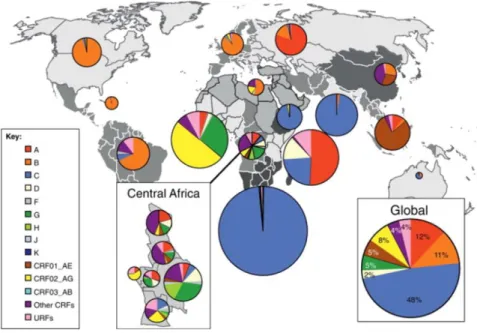
Phylogenetic and statistical analyses indicate that group M emerged in the beginning of the twentieth century, most likely in the area around Kinshasa and it is assumed that early diversification of group M also took place in this area, from which the infection spread globally. Currently, group M lineages are classified by nine subtypes (A–D, F–H, J, K), nowadays predominating in different geographic areas, resulting from a number of founder events1, see Figure 1. Furthermore, there exists a growing number of currently about 100 circulating recombinant forms (CRFs), resulting from multiple infections with different subtypes in the same population5,1. Within group M, subtype C accounts for almost 50% of the infections, and thus represents the dominant subtype in the actual HIV-1 pandemic, followed by subtypes A (12 %) and B (11 %), CRF02_AG (8 %), CRF01_AE (5 %), subtypes G (5 %) and D (2 %)6, as shown in Figure 1.
15
Figure 1. Geographic distribution of HIV-1 subtypes and recombinants. Pei charts illustrate the regional percentage distribution of HIV-1 subtypes, circulating recombinant forms (CRFs) and unique recombinant forms (URFs), represented by different colors. The distribution was calculated according to data collected from 2004-2007. Figure adapted from Hemelaar, J. The origin and diversity of the HIV-1 pandemic. Trends Mol. Med. 18, 182–92 (2012)7, with permission from Elsevier, license number 4536020267052.
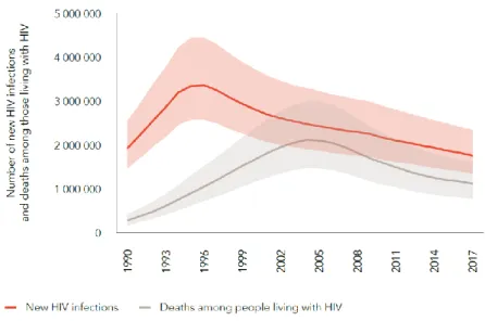
According to the Joint United Nations Programme on HIV/AIDS (UNAIDS) 77.3 million [59.9–100 million] people have become infected with HIV-1 since the start of the epidemic from who 35.4 million [25.0–49.9 million] died due to AIDS-related illness. In 2017, there were 36.9 million [31.1 million–43.9 million] people living with HIV, with sub-Saharan Africa accounting for about 70 % of the global burden of infection. With major efforts, the number of new infections has been steadily reduced from 3.4 million [2.6–4.4 million] in 1996 (peak of new infections) to 1.8 million [1.4–2.4 million] in 2017, meaning a reduction of 47 %. Furthermore, AIDS-related deaths have been reduced by more than 51 % since the peak in 20048, see Figure 2.
16
Figure 2. Number of new HIV infections and deaths among the HIV population. Global data from 1990-2017, adapted from UNAIDS9.
Ambitious prevention programs, significant progress in the development of antiretroviral drugs and modern treatment strategies went hand in hand and contributed to this success.
However, the story of success in anti-retroviral therapy included three important therapeutic revolutions. The first revolution was the introduction of new classes of antiretroviral drugs in 1995/1996, including protease inhibitors and non-nucleoside reverse-transcriptase inhibitors.
These newly introduced anti-retroviral drugs in combination with nucleoside-analog reverse- transcriptase inhibitors, which until then were the only treatment option available, greatly improved the prognosis of an otherwise lethal disease. The combined antiretroviral therapy is nowadays referred to as cART and in 2018 there were about 40 antiretroviral drugs available, including 7 drug classes, which were approved by the US Food and Drug Administration (FDA)10,11,12. The second revolution was marked by the realization that HIV-1 treatment contributes to prevention, since studies showed that treatment in serodiscordant couples (one HIV seronegative and one HIV seropositive partner) reduced the risk of sexual transmission by 96 %. The third revolution arised when first studies showed that early cART treatment, also if the number CD4+ cells is more than 500 cells per cubic millimeter, is associated with a significant clinical benefit in terms of a 57 % reduced risk in AIDS and non-AIDS morbidity and mortality13,14,11. The findings of these studies had a lasting impact on treatment guidelines. For example, while in the 2013 World Health Organisation (WHO) guidelines a CD4+ cell count of less than or equal to 500 cells per cubic millimeter was one of the criteria for starting therapy, recommendations changed in the 2016 WHO guidelines where therapy for all infected individuals is recommended, regardless of the CD4+ cell count. This is known
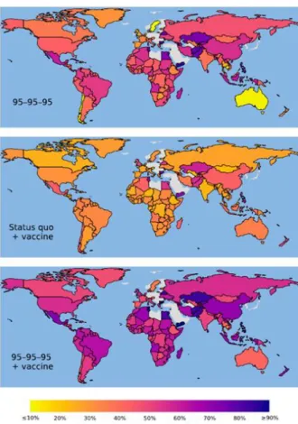
17 as the test and treat strategy15,16,11. Further, in 2014 UNAIDS launched the 90-90-90 targets meaning that by 2020 90 % of all people living with HIV know their HIV status, 90 % of people with diagnosed HIV infection will receive sustained cART, and 90% of those on cART will be virally suppressed. These targets were extended to 95-95-95 by 2030. Modeling analyses suggest that achieving these ambitious goals by 2020, enables ending of the epidemic in 203017,18. However, another study predicts that if current diagnosis, treatment and suppression levels are maintained (53-75-77, when the study was published in 2017) a global median of 49 million new infections will occur between 2015 and 2035. The same study predicts that from the 49 million infections 22 million can be averted by reaching 90-90-90 and a further 3.3 million by reaching 95-95-95targets. Furthermore, the study predicted that a 50 % efficacy vaccine alone, rolled out in 2020 with an annual scale-up of 25 % coverage to a maximum coverage of 70 %, and under current diagnosis and treatment levels, would avert 17 million new infections19, see Figure 3. This highlights that even a moderate efficacy vaccine could significantly contribute to the end the HIV-1 pandemic.
Figure 3. Calculated averted infections between 2015 and 2030 compared with maintaining status quo diagnosis, treatment and viral suppression levels.
Averted infections by reaching UNAIDS 95-95-95 targets (95 % of infected people know their HIV status, 95 % with diagnosed infection receive cART, 95% of people receiving cART are virally suppressed) (Top), rollout of a vaccine in 2020 (Middle) and 95-95-95 targets combined with vaccine enrollment in 2020 (Bottom). Data are based on diagnosis, treatment and viral suppression levels of 53-75-77. Figure from Medlock, J. et al. Effectiveness of UNAIDS targets and HIV vaccination across 127 countries. Proc. Natl.
Acad. Sci. U. S. A. 114, 4017–4022 (2017)19.
18 In summary, major efforts in prevention programs and significant progress in anti-retroviral therapy contributed reduce the number of new infections and AIDS-related death. This is also reflected by the current UNAIDS global data, since in 201775 % of people living with HIV know their status, 79 % of people living with HIV who know their status are accessing antiretroviral therapy and 81 % of people accessing treatment had suppressed viral loads8. However, a strategy with focus on anti-retroviral therapy has some drawbacks since it requires daily medication, a high level of compliance and medical monitoring, still representing major challenges in low-income countries. As described above, a prophylactic vaccine could significantly contribute to prevent further infections with a still not curable disease and therefore the development of such a vaccine is of utmost importance.
1.2 HIV-1 vaccine clinical trials
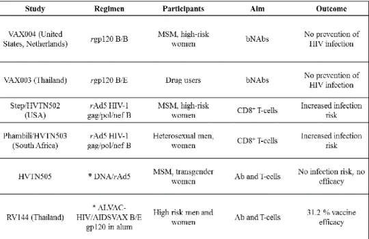
Soon after the discovery of HIV it was expected that an effective vaccine would be rapidly developed but so far only six HIV-1 vaccine candidates have been tested in clinical efficacy trials, with no liscenced vaccine yet, see Figure 4. However the story of vaccine paradigms and clinical trials can be separated in three overlapping “waves“20,21.
The first wave of HIV vaccines was based on the concept that neutralizing antibody responses would be sufficient to protect from infection. Since the viral envelope glycoprotein is the mediator of host cell infection and therefore a main target of a neutralizing antibody response, vaccine development in late 1980s to mid-1990s focused on recombinant Env immunogens.
These first recombinant Env immunogens were primarily produced as glycoprotein (gp) 120 proteins, representing soluble monomeric subunits of the Env protein, that is naturally present as trimer of heterodimers on the surface HIV virions, see 1.3.120,22. From 1998 to 2003, two phase 3 efficacy trials with recombinant bivalent gp120 vaccines were carried out. However, these studies known as VAX004 (gp120 subtype B/B) and VAX003 (gp120 subtype B/E) failed to show efficacy, see Figure 4. Although both vaccines induced high titers of neutralizing antibodies (NAbs), these were of limited specificity and therefore could not prevent HIV infection20,21,23,24.
19
Figure 4. HIV vaccine clinical trials conducted until today. Abbreviations: recombinant (r), men who have sex with men (MSM), broadly neutralizing antibodies (bNAbs), prime-boost regimen (*), antibodies (Ab). Freely reproduced from Trovato, M. et al. HIV Vaccination: A Roadmap among Advancements and Concerns. Int. J. Mol. Sci. 19, 1241 (2018)21.
The second wave of HIV vaccines started in the early 2000s when the failure of recombinant Env vaccines and new findings, supporting the importance of CD8+ T-cells in controlling viral replication, focused the field towards cytotoxic T-lymphocytes (CTLs)20. Two phase 2b vaccine efficacy trials were conducted to explore if cell-mediated immune responses can confer protection from infection or reduce plasma viral load after infection. In both, the Step study (HIV Vaccine Trials Network (HVTN) 502, started in 2004) and the Phambili trial (HVTN503, started in January 2007), see Figure 4, vaccinees were immunized with the MRKAd5 HIV-1 vaccine. This vaccine, consisting of a mixture of replication-defective recombinant adenovirus type 5 (Ad5) vectors expressing HIV-1 group-specific antigen (gag), polymerase (pol) and negative regulatory factor (nef) subtype B genes, induced cell-mediated immune responses in a phase I clinical trial25,26,27. However, after the first interim analysis of the Step study in September 2007 both studies were stopped, since, despite of eliciting interferon (IFN)-γ enzyme-linked immuno spot assay (ELISPOT) responses in 75 % of the vaccinees, the vaccine did no prevent HIV infection. Instead, analysis of available data revealed an increased risk of infection for male vaccinees who were uncircumsized or Ad5 seropositive pre-vaccination21,28,29,30.
The third wave began with vaccine efficacy trials that attempted to stimulate both arms of the immune response to ideally induce both, protective antibody and protective cell-mediated immune responses. Until today, two of those studies were conducted and evaluated. From
20 2009 to 2013 the HVTN enrolled a phase 2b efficacy trial called HVTN505, see Figure 4. To adress the viral diversity, the vaccine regimen was based on a heterologous prime/boost approach including immunogens from multiple HIV-1 subtypes. The vaccine regimen consisted of a DNA prime (six DNA plasmids expressing HIV-1 clade B Gag, Pol, and Nef and Env proteins from clades A, B, and C) and a rAd5 boost (four rAd5 vectors expressing a HIV-1 clade B Gag-Pol fusion protein and Env proteins from clades A, B, and C). However, despite of inducing both, cellular and humoral immune responses, the vaccine regimen did not show efficacy in preventing HIV infection21,31,32. The RV144 phase 3 clinical trial, conducted in Thailand between 2003 and 2005 and enrolling a total of 16,402 volunteers, was the only clinical trial that demonstrated an, albeit modest, efficacy in preventing HIV infection, see Figure 4. The study tested a heterologous prime/boost regimen consisting of two vaccines, matching the in Thailand predominantly circulating HIV-1 subtypes B and CRF01_AE: 1) ALVAC-HIV (vCP1521), a canarypox vector expressing HIV-1 CRF01_AE gp120 Env (linked to the transmembrane-anchoring portion of subtype B gp41) and HIV-1 subtype B Gag and protease was used for priming; and 2) AIDSVAX B/E (same vaccine as in the VAX003 study) formulated with alum adjuvant was used for boosting. Prior to RV144 study, this prime/boost regimen was tested in a phase 2 trial were it induced both, cellular and humoral immune responses and was therefore qualified for advanced testing in the RV144 trial33,34. The results of the RV144 trial, published in 2009, demonstrated 31.2 % vaccine efficacy in preventing HIV aquisition after 3.5 years. Further, post-hoc analyses reported that even higher vaccine efficacies (60.5 %) occurred in the first year following vaccination and declined over time33,35,36. Analysis of immune correlates, reviewed in36,37, revealed that the protective effect primarily correlated with non-neutralizing immunoglobulin G (IgG) antibodies targeting the variable 1/variable 2 (V1/V2) regions of the Env protein (see 1.3.1).
The protective effect of the antibody response was most likely mediated by Fc-receptor functions, including antibody-dependent cellular cytotoxicity (ADCC)38,37. Further, Env- specific IgG3 immune responses correlated with a decreased risk of HIV transmission.
Recently, a study comparing matched Env IgG1 and Env IgG3 antibodies reported that the IgG3 subclass is more potent than the IgG1subclass in mediating virion internalization. This maybe a mechanism by which non-neutralizing Env-specific IgG1 and IgG3 antibody responses contributed to vaccine efficacy in the RV144 trial39,36. Polyfunctional CD4+ T-cell responses also correlated with a decreased risk of HIV infection. Two T-cell subsets were identified, significantly correlating with a decreased risk of infection. One subset expressed CD40 ligand (CD40L), interleukin (IL)-2, IL-4, IFN-γ and tumor necrosis factor (TNF)-α,
21 while the other expressed CD40L, IL-2 and IL-4. Notably, both subsets include CD40L and IL4, which are involved CD4+ T-cell/B-cell interactions, indicating that these T-cell subsets may contribute T-cell help necessary for antibody production40,36. Next to correlates of protection in RV144 described above, there was also a strong direct correlate increasing risk of infection. Studies reported that high levels of serum IgGA, binding to specific envelopes increased risk of infection. Further, it has been shown that the Env-specific IgA/IgG ratio directly correlates with infection risk, most likely due to interfering events, since the same study reported that Env specific IgA can do both, inhibit Env binding and block ADCC activity of vaccine-induced IgG targeting the same epitope41,42,37,36. Based on the promising results from the RV144 trial a number of follow-on trials were designed, to test if the RV144 regimen can induce protective immune responses in South Africa. For this purpose the RV144 regimen was adapted to subtype C using ALVAC-HIV (vCP2438) in combination with recombinant bivalent subtype C gp120 formulated in MF59 adjuvant. This combination is currently tested in a phase 2b/3 efficacy trial (HVTN702) which was launched in 2016 in South Africa43,21.
When the results of the RV144 study were published in 2009, the focus of vaccine design was redirected from cell-mediated immune responses towards the humoral immune response, since antibodies, although not neutralizing, were correlated with protection from HIV infection. In parallel, studies of humoral immune responses of HIV-positive individuals using novel high-throughput screening methods resulted in the identification of scores of Env- directed broadly neutralizing antibodies (bNAbs) since beginning of 200944. BNAbs are characterized by potent and broad neutralizing activity (described in detail in 1.4.2), and the attempts to induce a bNAb response by vaccination further focused vaccine design towards the Env protein and dominate the field until today.
1.3 The HIV-1 envelope glycoprotein
The viral envelope glycoprotein (Env) plays a key role in HIV-1 infection, since it mediates the infection of CD4+ host cells, and thus enables viral replication. In addition, Env is the only viral protein on the surface of HIV virions and therefore the only target of the humoral immune response. Thus, the Env protein represents a key component in HIV-1 vaccine design45,46,47.
22
1.3.1 Biosynthesis and structure of the HIV-1 envelope protein
The HIV-1 Env protein is expressed as gp160 precursor polypeptide that gets extensively glycosylated during synthesis in the endoplasmatic reticulum (ER). Thereby, glycans account for about the half of the molecular mass of the protein. Glycosylation of Env proteins occurs as N-linked glycosylation at the asparagine of NXT/S sequons, with X being any amino acid (AA) except possibly proline or aspartic acid48,49. In addition, there are studies reporting some rare O-linked glycosylations of Env proteins, which can occur at serine or threonine residues.
But contrary to N-linked glycosylation sites, there is no clear-cut motif in the amino acid sequence which defines their glycosylation50,51,52. The gp160 precursor is proteolytically processed by Golgi-associated serin proteases of the Furin family, cleaving at the C-terminus of the amino acid consensus sequence REKR, into non-covalently linked gp120/gp41 subunits, called protomers53,54. Three gp120 / gp41 protomers assemble into Env trimers that are transported to the plasma membrane, and thus incorporated into virions. From there, they can mediate host cell entry by a series of complex receptor-triggered conformational changes55,56, see 1.3.2.
During the infection of host cells, gp120 and gp41 have different functions, which are defined by their structure. The gp120 subunit, including CD4 and co-receptor binding sites, is heavily glycosylated and has a highly variable surface that can be subdivided in five variable loops (V1-V5) and five constant regions (C1-C5), see Figure 5A to C 46,57,58. Structurally, gp120 can be separated in an inner- and outer domain that are connected by the bridging sheet, consisting of four anti-parallel β-sheets. While the inner domain non-covalently connects gp120 to the gp41 subunit, the outer domain is involved in receptor binding58. The gp41 subunit anchors the Env protein in the viral membrane and contains the fusion machinery, required for fusion of host and viral membranes. In comparison to gp120, the gp41 subunit is more conserved in its amino acid sequence. The gp41 subunit consists of the fusion peptide (FP), two heptad repeats (HR1, HR2), a conserved disulfide loop (DSL), the membrane- proximal external region (MPER), the transmembrane domain (TM) and the cytoplasmic tail (CT), see Figure 5A to C46,59,60.
23
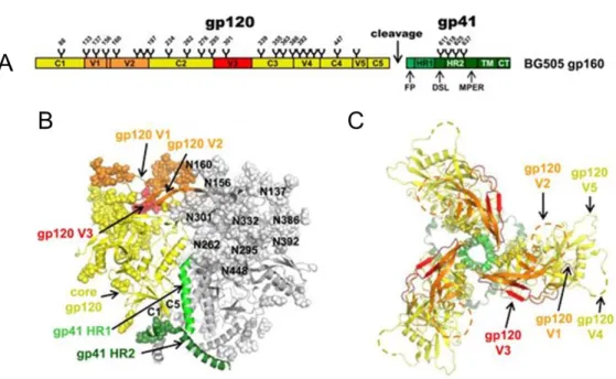
Figure 5. Schematic representation and overall architecture of the HIV-1 Env trimer. (A) Schematic view of the BG505 full-length gp160 consisting of gp120 and gp41 subunits. Gp120 includes constant regions 1-5 (C1-C5) and variable loops 1- 5 (V1-V5), while gp41 contains the fusion peptide (FP), heptad repeats 1 and 2 (HR1/HR2), the disulfide loop (DSL), the membrane-proximal external region (MPER), the transmembrane domain (TM) and the cytoplasmic tail (CT). Glycan trees and the Furin cleavage site are also indicated. (B), (C) Structure of the soluble, cleaved, recombinant HIV-1 Env BG505 SOSIP.664, representing a soluble native-like Env trimer61. Note, since the BG505 SOSIP.664 is a soluble, MPER deleted protein, ending at amino acid position 664 (HXB2 numbering), the MPER, TM, and CT are not part of the crystal structure.
(B) Side view of the Env trimer. For one out of the three protomers, the gp120 core is highlighted in yellow while V3 and V1/V2 loops are colored in red and orange, respectively. Gp41 HR1 and HR2 are labeled in different shades of green. Protein components are depicted according to their secondary structure, while glycans are shown as spheres and are numbered according to their asparagine (N) residues (HXB2 numbering). (C) Top view on the Env trimer with variable loops V1-V5 projecting into the periphery. The localizations of V2 and V4 loops are indicated by dashed lines as they are disordered in the structure. Figure adapted from Julien, J.-P. et al. Crystal structure of a soluble cleaved HIV-1 envelope trimer. Science (80-.
). 342, (2013)60, reprinted with permission from AAAS, license number 4536030553645.
1.3.2 Function of the HIV-1 envelope
HIV-1 can infect all cells that express its primary receptor CD4 and co-receptors of the chemokine receptor family, including the C-C chemokine receptor type 5 (CCR5) or C-X-C chemokine receptor type 4 (CXCR4). Thus, CD4+ T-cells as well as macrophages are susceptible to HIV infection47,62. The metastable nature of the pre-fusion state of the protein plays a crucial role in host cell infection, since it allows the protein to undergo irreversible conformational changes required for the fusion process63.
24
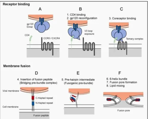
Figure 6. HIV entry into host cells. HIV entry into host cells involves two major events, receptor binding and membrane fusion. In the first step, the HIV Env trimer, consisting of three gp120 (blue spheres) and three gp41 (blue bars) subunits comes in proximity to host cells (A). After engagement of the CD4 receptor (green spheres) by the gp120 subunit, containing the CD4 binding site (CD4bs), structural rearrangements result in the exposure of the co-receptor binding site (B). Followed by co-receptor binding (C), resulting in further structural rearrangements, the fusion peptide (FP) is inserted into the host cell membrane (D). The formation of a stable six-helix bundle (6HB) brings the viral and host cell membrane in close proximity.
This process is mediated by N and C-terminal α-helical regions of the gp41 subunits, termed heptad repeats 1 and 2 (HR1/HR2), shown in blue and red. During this process, the N-terminal heptad repeats of the three gp41 subunits fold into a central triple-stranded coiled coil and the three C-terminal heptad repeats pack antiparallel into the three groves of the coiled- coil. The process involves a prefusion intermediate (E) and results in the formation of the 6HB and the establishment of the fusion pore (F), required to release the viral core into the cytoplasm. Figure from Lobritz, M. A., Ratcliff, A. N. & Arts, E. J.
HIV-1 Entry, Inhibitors, and Resistance. Viruses 2, 1069–105 (2010)47.
The initial contact is mediated by the gp120 subunit that binds with its CD4 binding site (CD4bs), consisting of discontinuous residues in the constant regions of gp120, to the N- terminus of CD4, Figure 6A, and B. This results in the rearrangement of the gp120 subunit and exposure of the highly conserved co-receptor binding site, consisting of regions of C4, β- sheets of V1/V2 and residues of the V3 loop, see Figure 6B. The interaction of the co- receptor binding site with CCR5 or CXCR4 co-receptors leads to further structural rearrangements, resulting in the exposure of the FP. The FP, consisting of 15 hydrophobic amino acids located at the N-terminus of gp41, inserts into and destabilizes the host cell membrane (Figure 6C and D). After insertion of the FP, viral and host cell membrane are brought in close proximity and membrane fusion occurs. This process involves a re-folding event within gp41 that is mediated by N and C-terminal α-helical regions, termed heptad repeat 1 and 2 (HR1/HR2). HR1 and HR2 of each gp41 subunit fold into each other, forming a stable six-helix bundle (6HB). In this configuration, the FP and the transmembrane domain
25 of gp41 are located at the same end of the molecule. The re-folding event and release of free energy finally results in membrane fusion and formation of a fusion pore (Figure 6E and F).
Lipid mixing results in enlargement of the fusion pore and when a critical size is reached the viral core is released into the cytoplasm47,64,65. From there, subsequent steps in HIV replication cycle occur, reviewed in2,66.
1.3.3 Immune evasion mechanisms of the HIV-1 envelope
The viral Env protein is the only viral component on the surface of HIV-1virions, and thus exposed to the selection pressure of the humoral immune response. In order to avoid antibody-mediated neutralization, HIV has evolved a number of escape mechanisms:
1) HIV-1 variability (Figure 7B): The high replication rate (~109 virions are generated per day in an infected individual) and the error-prone reverse transcriptase HIV-1( approximately one nucleotide substitution per genome during a single replication cycle) are responsible for the extensive genetic diversity of HIV-1 and contribute to the adaptation of both, antiretroviral therapy and immune responses67. Thereby the envelope is the most variable of all HIV-1 genes68. Studies have shown that Env proteins can exhibit 35 % amino acid diversity between subtypes and 20 % within a subtype. Thereby most sequence variation occurs in the gp120 subunit, including variable loops69,70. The high variability in these domains is driven by the need of the virus to continuously escape hosts neutralizing antibody responses. The Env variable loops 1-5 (V1-V5) contribute to masking of more conserved Env g120 regions lying underneath, and can tolerate high mutation rates, including insertions and deletions. For example, the length of the most variable loop V1/V2 can range from 50 to 99 amino acids, while V4 and V5 loop lengths range from 19 to 44 and 14 to 36 amino acids, respectively. The length of the V3 loop is more or less constant (34-35 amino acids), since V3 is involved in important trimer stabilizing contacts and in co-receptor engagement. Next to their diversity in amino acid composition and length, variable loops exhibit also high levels of glycosylation and flexibility, and thus contribute to the viral defense to protect conserved Env regions from neutralizing antibody responses 46,71,72,73. 2) Env glycanshield (Figure 7A):
HIV-1 Env trimers contain a median of 93 N-linked glycans, comprised of a mixture of mainly high-mannose-type and further processed complex-type glycans that account for approximately 50 % of the mass of the protein. The glycan shield consists of a network of interlocked oligosaccharides that occludes most of the polypeptide surface of Env. Thus, it enables HIV-1 to avoid most antibody-mediated neutralization. The glycan shield appears to be evolving by coupling the high HIV mutation rate with high numbers of N-linked glycans
26 on the surface of Env trimers. This seems to be the primary mechanism by which HIV escapes humoral immune responses since despite of high titers of HIV-reactive antibodies, a sustained viremia is frequently observed during chronic infection74,75,76,77.
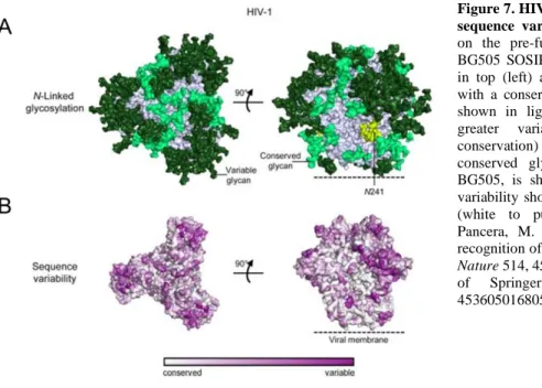
Figure 7. HIV-1 envelope glycan shield and sequence variability. (A) N-linked glycans on the pre-fusion closed Env strucure of BG505 SOSIP.664 (PDB ID: 4TVP), shown in top (left) and side view (right). Glycans with a conservation of more than 90 % are shown in light green, while glycans with greater variability (less than 90 % conservation) are depicted in dark green. A conserved glycan residue, not present in BG505, is shown in yellow. (B) Sequence variability shown from conserved to variable (white to purple). Figure adapted from Pancera, M. et al. Structure and immune recognition of trimeric pre-fusion HIV-1 Env.
Nature 514, 455–61 (2014)78, with permission of Springer Nature, license number 4536050168052.
3) Conformational masking: Immune evasion is also mediated by conformational masking of Env conserved functional sites such as the co-receptor binding site in gp120 that is not fully exposed until the CD4 binding site has bound to the CD4 receptor. Similarly, the fusion machinery in the MPER of gp41 becomes only accessible when the co-receptor binding site is occupied and viral fusion is already initiated. Thus, conformational masking restricts the access of antibodies to these relatively conserved regions of Env and therefore contributes to immune evasion79,80,81. 4) Conformational flexibility (Figure 8): HIV Env pre-fusion trimers are highly dynamic metastable fusion machineries that alternate between closed and more open conformations. This is referred to as “breathing”. It has been shown that trimer opening results in the exposure of some non-neutralizing epitopes, possibly misleading humoral immune responses. However, the relative proportion of pre-fusion conformational states varies within different isolates. For example, in easy-to-neutralize tier 1 isolates a larger proportion of trimers adopt an open conformation, compared to neutralization-resistant tier 2 isolates. In addition, CD4 engagement of the gp120 subunit results in dissociation of the Env trimer, and thus to exposure of epitopes that induce non-neutralizing antibody responses, since these epitopes are occluded in pre-fusion state trimers82,83,84.
27
Figure 8. Conformational states of HIV-1 pre-fusion trimers. Biophysical data suggest that pre-fusion Env trimers can fluctuate between closed (left) and more open states (right) in a dynamic equilibrium. This is referred to as trimer breathing. Figure adapted with modifications from Ozorowski, G. et al. Open and Closed Structures Reveal Allostery and Pliability in the HIV-1 Envelope Spike. Nature 547, 360–363 (2017)85, with permission of Springer Nature, license number 4536060148887.
5) Low spike density: HIV presents only a low number of Env proteins on its surface (~14/per virion), thereby impeding bivalent antibody binding by inter-spike crosslinking. The primarily monovalent binding suffers from reduced potency, compared to bivalent binding.
Thus, the low spike density contributes to immune evasion of Env86. In addition, virions bear non-functional gp120-gp41 monomers, most likely resulting from trimer dissociation along the axis of trimerization, and gp41 stumps that result from shedding of non-covalently linked gp120 subunits. Both, gp120-gp41 monomers and gp41 stumps expose non-functional Env epitopes, and thus contribute to immune evasion by misleading antibody responses87,88. The features and mechanisms described above enable HIV-1 to continuously escape from host humoral immune response. Further, they represent major hurdles that have to be considered within the development of a protective vaccine targeting the HIV-1 Env protein, see 1.5.
1.4 Env directed antibody responses
1.4.1 Antibody responses during acute and chronic infection
Env-directed antibody responses can be classified into three groups that occur at different timepoints during the course of HIV infection89. The first group includes Env-binding but non-neutralizing antibodies that arise within weeks after transmission. Virion-antibody immune complexes are the first form of detectable B-cell responses that occur about one week after plasma virus detection, followed by first free plasma anti-gp41 antibodies a few days later. In contrast, anti-gp120 antibodies primarily targeting V3 can be detected about four weeks after onset of viremia. However, these early antibodies have no detectable effect on plasma viral load and there is also no evidence of antibody-induced Env selection90. The antibodies described above, typically bind to immunodominant Env epitopes on HIV virions that are not present on native functional trimers, such as gp41 stumps, resulting from gp120 shedding. Therefore, they are not able to neutralize functional trimers. Nevertheless, the may
28 have some antiviral activity, since antiviral activity is not restricted to neutralization (meaning viral inactivation by binding and blocking of functional Env trimers, and thus preventing infection of host cells) but also includes other antibody functions such as ADCC or antibody- dependent cell-mediated virus inhibition (ADCVI)89,91.
The second group includes antibodies that can neutralize HIV-1 in a strain specific manner (NAbs). These antibodies typically arise after several weeks to months after infection. The reasons for the slow development of neutralizing antibodies remain elusive. Presentation of immunodominant epitopes (for example gp41 stumps) that divert antibody responses towards non-neutralizing epitopes, and impaired CD4+ T cell help may contribute to the delayed induction of neutralizing responses. As their name implies NAbs are capable to neutralize the infecting (autologous) strain but cannot neutralize most other (heterologous) strains. NAbs typically target epitopes in variable loops or other regions with relatively high sequence variation, which is also the reason why HIV can easily escape by amino acid substitution, insertions/deletions or an evolving glycan shield, see 1.3.3 89,91.
However, in some individuals the sustained co-evolution between NAbs and viral escape after years results in the development antibodies with increased neutralization breadth and potency.
These antibodies are termed broadly neutralizing antibodies and represent the third group of Env-directed antibody responses, which is described separately in the following chapter.
1.4.2 Broadly neutralizing antibodies
During HIV-1 infection between 10 % and 50 % of all infected individuals develop broadly neutralizing antibodies that can neutralize diverse viral isolates. A much smaller fraction (around 1 %), referred to as elite neutralizers, develops bNAbs with exceptional breadth and potency (bNAbs in the context of this thesis). Some of these bNAbs were capable to neutralize over 90 % of isolates tested in pseudovirus assays92,93. However, even infected individuals with broadly neutralizing serum activity do not benefit from the existing bNAbs as the autologous virus continues to escape from neutralization94.The reasons why only a small proportion of infected individuals develop potent broadly neutralizing responses are rather complex and involve a number of factors. For example, high viral loads, low CD4+ counts, a higher frequency of T-follicular helper (Tfh) cells in the periphery and the duration of infection have been associated with the development of bNAb responses92,93,95.
29 Typically, bNAbs arise between two and four years after infection in the chronic phase of disease and display a number of unusual features96,95. 1) High degree of somatic hypermutation: A high level of somatic hypermutation (SHM) is a common feature shared by bNAbs. Whereas most affinity matured human antibodies carry about 15-20 somatic mutations, bNAbs are more extensively mutated with up to 40-110 somatic mutations.
Typically, mutations accumulate in the complementarity determining region (CDR) loops of the antibodies since they usually contact the antigen. In addition, mutations in the canonical framework regions (FWRs), required for scaffolding of CDRs and commonly less tolerant to mutations, seem to play a critical role in both, breadth and potency of most bNAbs79,97. 2) Long HCDR3 loops: Some, but not all bNAbs exhibit an unusual long heavy chain complementarity-determining region 3 (HCDR3) with loop lengths between 20 to 34 amino acids (average in humans: 16 amino acids). Long HCDR3 loops are typical features that enable V1/V2 and V3 directed bNAbs to penetrate the Env glycan shield. Similar is observed for MPER directed bNAbs, where long HCDR3 loops help to reach highly conserved hydrophobic residues of gp41. However, the lower frequency of human B-cells encodes for long HCDR3s. In addition, antibodies with long HCDR3s have the potential of auto- reactivity, thus B-cells encoding for this antibodies are frequently deleted by negative selection. Taken together, it is seen as challenging to induce antibodies with long HCDR3s by vaccination98,79. 3) Poly-/Autoreactivity: Despite poly-/autoreactivity is normally associated with negative B-cell selection, some bNAbs also display poly-/autoreactivity. Polyreactivity likely contributes to increase the overall affinity via heteroligation. In addition, lipid binding seems to essentially contribute to the general neutralization mechanism of MPER-directed bNAbs. Thereby, the initial low affinity contact to the viral lipid membrane seems to result in the exposure of lipid-immersed MPER epitopes that are subsequently bound with high affinity79,99.
Until 2009 only a limited amount of bNAbs was described in literature. The first generation bNAbs were isolated in the 1990s by phage display and from human hybridomas and resulted in the identification of conserved Env epitopes (see Figure 9), such as the CD4bs (bNAb:
b12100,85), the MPER region (bNAbs: 4E10101,102, 2F5101,103,104) and a glycan epitope at the outer domain (OD) of gp120 (bNAb: 2G12101,105). However, all epitopes of the bNAbs described above are present within a single gp120/gp41 protomer, resulting from monomeric antigens used for selection and screening79,92. Since 2009, the development of new screening procedures and technologies, including single B-cell culture and functional screenings with tier 2 neutralization as primary selection criterion, resulted in the identification of scores of
30 bNAbs, which reflect the second generation of bNAbs. Compared to first generation bNAbs, second generation bNAbs were in general of greater breadth and potency44,92. Since a screening with neutralization as primary selection criterion is not restricted to antibody- binding to already existing antigens, like phage display or B-cell sorting, some of the isolated antibodies defined new bNAb epitopes (see Figure 9). These include the gp120 V2-glycan site at the trimer apex, a region at the gp120/gp41 interface and the gp120 V3-glycan epitope, overlapping the 2G12 epitope. Since the V3-glycan epitope has a central glycan at position 332 it is also referred to as N332-glycan supersite44,92,89,106. Notably, multiple epitopes of the newly identified bNAbs were characterized/identified using BG505 SOSIP.664 trimers61,56 (described in detail in 1.5.1). BG505 SOSIP.664 trimers have been proven valuable for structural studies since using these trimers in cryo electron microscopy and x-ray crystallography approaches resulted in the first authentic high-resolution structures of a HIV-1 Env trimer. For example, a new cluster of quaternary structure-specific antibodies (including PGT151107,108) was mapped to the gp120-gp41 interface of BG505 SOSIP.664 trimers56.
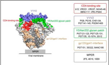
Figure 9. bNAb epitopes on the Env trimer. Major epitopes of broadly neutralizing antibodies (bNAbs) on the surface of BG505 SOSIP.664 (PDB ID: 4TVP.
Epitopes are shown in different colors: CD4 binding site (red);
V1/V2 (V2-glycan) (blue); V3- glycan (green); gp120/gp41 interface (wheat); MPER (illustrated as grey rectangle). Unspecified regions of gp120 and gp41 moieties are shown in light and dark grey, respectively.
Representative bNAbs for each epitope are listed in boxes (right).
Figure from Zhang, Z. et al. Antiviral Therapy by HIV-1 Broadly Neutralizing and Inhibitory Antibodies. Int. J. Mol. Sci. 17, 1901 (2016)109.
In summary, several findings described above greatly impacted HIV vaccine design and focused vaccine design towards the Env protein. These findings include that 10 % to 50 % of all infected individuals can develop a broadly neutralizing antibody response, with a smaller number developing bNAbs with the capacity to neutralize over 90 % of tested isolates. This shows that the human immune system is basically able to generate broadly neutralizing antibody responses91. Further, a large number of bNAbs have been isolated since 2009 and mapping of these antibodies to newly available immunogens, such as the BG505 SOSIP.664,
31 resulted in the discovery of new bNAb epitopes. The knowledge, gained from the discovery and mapping bNAbs, was and is of utmost importance in development of next generation Env immunogens, described in 1.5. That the induction of bNAbs is a desirable goal in HIV vaccine development is highlighted by passive immunization and challenge experiments. It has been shown that passive immunization of bNAbs can protect macaques from infection with chimeric simian-human immunodeficiency viruses (SHIVs) encoding HIV-1 envelope genes in an SIV backbone110,111,112. This indicates that bNAbs, if present in sufficient titers prior to infection, can protect from infection.
1.5 Advanced Env vaccine design and efforts towards the induction of cross-neutralizing antibody responses
The first HIV vaccines were monomeric gp120 subunit vaccines that failed to confer protection by neutralization in phase 3 efficacy trials23,24, see 1.2. Although gp120 monomers may display epitopes of potent bNAbs such as the CD4 binding site, they failed to induce neutralizing antibody responses. The presentation of immunodominat non-neutralizing epitopes, such as gp120 epitopes normally occluded by inter-subunit interaction, and the inability to present potent quaternary structure and gp41 epitopes may have contributed to this outcome56. However, the only efficacy trial demonstrating modest efficacy (RV14433) was based on a gp120 subunit vaccine. Notably, a primary correlate of protection was a non- neutralizing antibody response directed against V1/V2, see 1.2. However, to overcome limitations of gp120 described above, HIV Env vaccine design focused on the production of soluble trimeric proteins. A general strategy for the production of soluble trimers involved a stop codon before the gp41 transmembrane domain to generate soluble proteins consisting of the ectodomain of gp41 (gp41ECTO) and gp120 subunits. Further, they included the inactivation of the Furin cleavage site (e.g. REKR to SEKS) to produce covalently linked gp120 and gp41 subunits that otherwise would rapidly dissociate into gp120 subunits and a trimerized form of gp41. When tested in humans, these Env trimers elicited only weak neutralizing antibody responses, limited to easy-to-neutralize tier 1 strains56,113,114,115. The limitations of the first soluble Env trimers were revealed more than a decade after their development, when studies have shown that these trimers adopt non-native conformations with gp120 subunits dangling from trimerized gp41 thereby exposing non-neutralizing epitopes56,116,117.
32
1.5.1 Stabilized trimers to focus immune responses towards bNAb epitopes
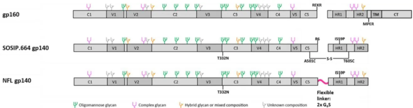
A hallmark in HIV Env vaccine design was the development of the soluble cleaved BG505 SOSIP.66461 trimer, based on the clade A transmitted/founder isolate BG505118. Many modifications in the wild-type BG505 sequence (see Figure 10), resulted in BG505 SOSIP.664 gp140, representing trimers with improved stability, homogeneity and solubility.
The sequence modifications included an optimized Furin cleavage site, REKR to RRRRRR (R6), to enhance cleavage and therefor proper protein folding, disulfide-bond forming cysteines at amino acid positions 501 and 605 (referred to as “SOS”) for covalent linkage of gp120-gp41ECTO subunits and a helix-breaking point substitution isoleucine to proline at amino acid position 559 (I559P), allowing gp41ECTO subunits to remain in the pre-fusion ground state. Furthermore, the MPER region was deleted to improve homogeneity and solubility (C-terminal residue at amino acid position 664) and a glycan was introduced at amino acid position 332 (T332N), since the binding of several bNAbs is depending on the presence of this glycan. Negative stain electron microscopy confirmed that affinity and size exclusion chromatography (SEC) purified BG505 SOSIP.664 trimers were highly homogeneous with more than 95 % adopting a tri-lobed propeller shape, nowadays defining the characteristics of native-like soluble trimers56,61. With melting temperatures of about 67
⁰C, the trimers were highly stable and different biochemical and biophysical assays (ELISA, surface plasmon resonance spectroscopy (SPR), biolayer interferometry (BLI), isothermal titration calorimetry (ITC)) confirmed that the BG505 SOSIP.664 trimers have desirable antigenic properties since they show binding to all known bNAbs (with the exception of MPER directed bNAbs) but no or very poor binding to non-neutralizing antibodies61,119,120,56. In an immunization study with rabbits, BG505 SOSIP.664 trimers elicited strong and consistent NAb responses against the autologous tier 2 virus BG505.T332N, whereas BG505 gp120 monomers and uncleaved gp140 trimers induced weaker or undetectable NAb responses. Notably, autologous neutralization was observed the first time for Env-based immunogens. In an immunization study with rhesus macaques BG505 SOSIP.664 trimers also induced a NAb response against the autologous virus but antibody titers were lower than in rabbits. However, heterologous neutralization of a tier 2 clade C isolate was not achieved121,122. Although both, rabbits and macaques, developed autologous neutralization, the epitopes mainly contributing to autologous neutralization differed between the species as it was reported in a very recent published study123. According to the study, a previously
33 described glycan-hole site in BG505 SOSIP.664 (amino acid positions 241/289) accounts for about 50 % of autologous neutralization in rabbits. However, the study identified another important epitope in the C3 region of Env (termed C3/465) that accounts for about 25 % of the neutralizing responses in rabbits. Remarkably, the same study was able to show that the neutralizing response in macaques is dominated by the newly identified C3/465 epitope123,124. Meanwhile, the SOSIP.664 design was successfully transferred to other isolates with properties similar to BG505. In addition, different strategies were applied to further stabilize BG505 SOSIP trimers and to reduce the conformational flexibility in order to avoid/reduce undesired exposure of non-neutralizing epitopes related to trimer breathing (for example non- neutralizing V3 epitopes) and CD4-induced open conformations56. These strategies include chemical crosslinking of trimers as well as further stabilization by applying further stabilizing modifications125,56. Further, findings from the SOSIP design resulted in alternative designs for trimer stabilization, like the native-flexible linker design (NFL). The NFL design essentially reflects the SOSIP design, with the major difference that gp120 and gp41 subunits are covalently linked by a flexible glycine-serine (GS) linker instead of a disulfide bond. Thus, NFL trimers do not require Furin cleavage to adopt a native-like conformation126,127. By direct comparison in an immunization study with macaques, BG505 NFL trimers and BG505 SOSIP.664 trimers induced approximately equivalent neutralizing antibody responses128. In addition, the same study investigated the immunogenicity of advanced BG505 SOSIP.664 trimers designed for further stabilization, reduced V3 loop exposure and minimized CD4 reactivity/CD4-induced non-nAb epitope exposure129,130,131. Interestingly, despite of reduced V3 responses, there was no difference in absolute tier 2 NAb responses between further modified versions and the basis version of BG505.SOSIP.664 trimers128.