The Role of Type I Interferon in Regulating Macrophage Responses to Salmonella Typhimurium Infection
I n a u g u r a l D i s s e r t a t i o n
zur
Erlangung des Doktorgrades Dr. nat. med.
der Medizinischen Fakultät und
der Mathematisch-Naturwissenschaftlichen Fakultät der Universität zu Köln
vorgelegt von
Nina Judith Hos
aus Nürnberg
Hundt Druck, Köln
2019
Referenten: Prof. Dr. Stefan Höning Prof. Dr. Adam Antebi
Datum der mündlichen Prüfung: 21.05.2019
STATEMENT
Parts of this work, including Material and Methods, Results, and Discussion section, have been published previously in the Journal of Cell Biology with the title “Type I interferon enhances necroptosis of Salmonella Typhimurium-infected macrophages by impairing anti- oxidative stress responses.” (Hos NJ, Ganesan R, Gutierrez S, Hos D, Klimek J, Abdullah Z, Krönke M, Robinson N. 2017; 216(12):4107-4121. doi: 10.1083/jcb.201701107).
TABLE OF CONTENTS
SUMMARY ... 1
ZUSAMMENFASSUNG ... 2
ABBREVIATIONS ... 3
1. Introduction ... 5
1.1 Salmonella Typhimurium infection ... 5
1.1.1 Microbiology and epidemiology of S. Typhimurium ... 5
1.1.2 Pathogenesis of S. Typhimurium ... 6
1.2 Interaction of S. Typhimurium with macrophages ... 7
1.2.1 Activation of inflammatory pathways ... 7
1.2.2 Induction of oxidative stress ... 8
1.2.3 Interference with autophagy ... 9
1.2.4 Induction of cell death ... 11
1.3 Type I interferon (IFN-I)-dependent immune responses to S. Typhimurium ... 13
1.3.1 Induction of IFN-I synthesis ... 13
1.3.2 Activation of the IFN-I receptor pathway ... 14
1.3.3 IFN-I-mediated cell death ... 14
HYPOTHESIS... 16
OBJECTIVES OF THIS WORK ... 16
2. Material and Methods ... 17
2.1 Material ... 17
2.1.1 List of instrumentation ... 17
2.1.2 List of software ... 17
2.1.3 List of chemicals and reagents ... 17
2.1.4 List of antibodies ... 19
2.2 Methods ... 20
2.2.1 Cell culture and bacterial infection ... 20
2.2.2 Protein and enzymatic assays ... 21
2.2.3 Mitochondrial and cell viability assays ... 22
2.2.4 Microscopic analyses ... 24
2.2.5 mRNA assays ... 25
3. Results ... 28
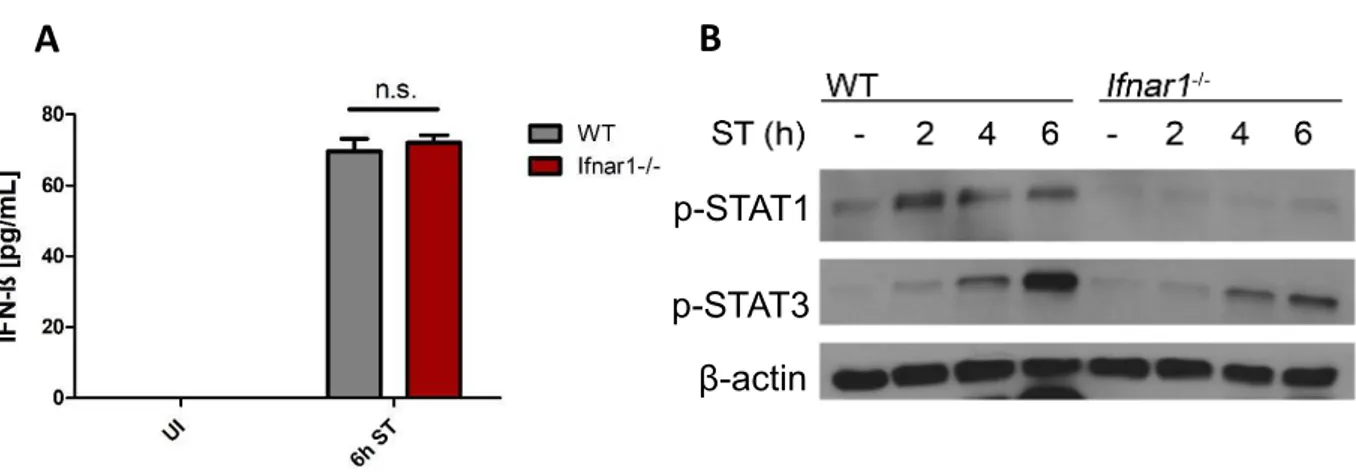
3.1 S. Typhimurium induces IFN-I signaling pathways in macrophages ... 28
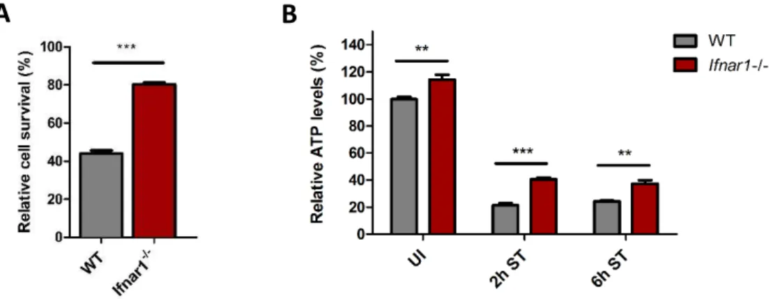
3.2 S. Typhimurium induces IFN-I-mediated cell death of macrophages ... 29
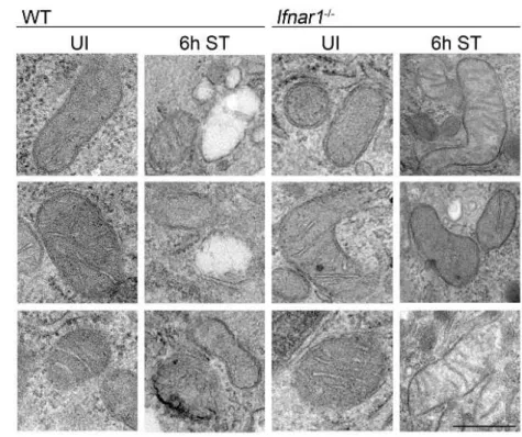
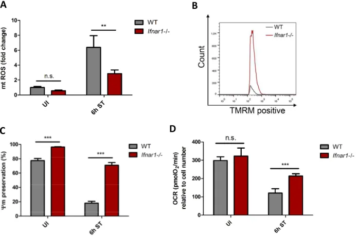
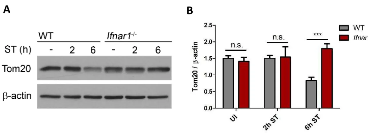
3.3 IFN-I signaling exacerbates S. Typhimurium-induced mitochondrial damage ... 30
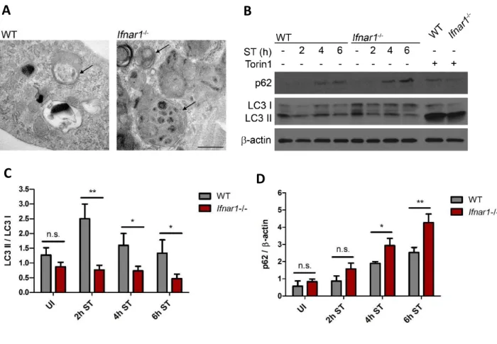
3.4 IFN-I-mediated mitochondrial damage and energy depletion transiently triggers autophagy ... 33
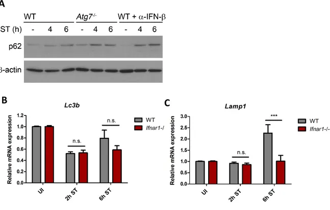
3.5 Reduced p62 levels contribute to IFN-I-mediated cell death ... 37
3.6 IFN-I signaling attenuates Nrf2 activation and anti-oxidative responses ... 41
3.7 Impaired Nrf2 function mediates S. Typhimurium-induced cell death ... 47
3.8 Nrf2 function is regulated by RIP3 ... 50
3.9 Pgam5 prevents Nrf2 activation downstream of IFN-I/RIP3 ... 52
4. Discussion... 56
4.1 IFN-I signaling exacerbates S. Typhimurium-induced mitochondrial dysfunction and oxidative stress ... 57
4.2 IFN-I signaling transiently triggers autophagy ... 58
4.3 IFN-I signaling reduces p62 levels through autophagic degradation ... 60
4.4 IFN-I signaling disrupts Nrf2-dependent anti-oxidative stress responses ... 61
4.5 S. Typhimurium infection activates Pgam5 downstream of IFN-I/RIP3 ... 63
4.6 S. Typhimurium infection induces cell death through impaired Nrf2 function ... 64
CONCLUSION ... 65
FUTURE PERSPECTIVES ... 66
ACKNOWLEDGEMENTS ... 67
BIBLIOGRAPHY ... 68
DECLARATION ... 77
SUMMARY
Salmonella Typhimurium (S. Typhimurium) is a Gram-negative, facultative intracellular bacterium that exploits the host’s type I interferon (IFN-I) response to induce cell death in macrophages. We have previously demonstrated that activation of the IFN-I receptor (Ifnar1) results in recruitment of RIP1 and subsequent formation of a RIP1/RIP3 complex, leading to a specific form of programmed cell death, termed necroptosis, in S. Typhimurium- infected macrophages (Robinson et al. 2012). Despite our detailed knowledge on IFN- I/RIP1/RIP3-dependent necroptosis execution, the IFN-I-mediated pathways that determine whether infected macrophages will undergo necroptosis remain elusive. This work therefore sought to identify the IFN-I-mediated events that sensitize S. Typhimurium-infected macrophages to cell death.
Here, we demonstrate that S. Typhimurium infection causes mitochondrial damage and impairs the host´s anti-oxidative stress response through upregulation of the mitochondrial phosphatase Pgam5 downstream of IFN-I/RIP3. Pgam5 subsequently interacts with the transcription factor Nrf2, which sequesters Nrf2 in the cytosol thereby repressing the transcription of Nrf2-dependent anti-oxidative genes. The impaired ability to respond to S. Typhimurium-induced oxidative stress results in ROS-mediated mitochondrial damage, ATP depletion, transient induction of autophagy, and autophagy-mediated degradation of p62, which impairs p62-Keap1 interaction. Consequently, Keap1 interacts more with Nrf2, which further represses Nrf2 function and additionally impairs anti-oxidative stress responses to S. Typhimurium infection thereby sensitizing macrophages to cell death.
Taken together, we identify impaired Nrf2-dependent redox homeostasis of S. Typhimurium-infected macrophages as an important mechanism that drives cell death downstream of IFN-I/RIP3.
ZUSAMMENFASSUNG
Salmonella Typhimurium (S. Typhimurium) ist ein Gram-negatives, fakultativ intrazelluläres Bakterium, welches die Typ-I-Interferon (IFN-I)-Antwort des Wirts ausnutzt, um den Zelltod in Makrophagen zu induzieren. Unsere Vorarbeiten zeigen, dass die Aktivierung des IFN-I- Rezeptors (Ifnar1) zur Rekrutierung von RIP1 und zur anschließenden Bildung eines RIP1/RIP3-Komplexes führt, was zu einer bestimmten Form des programmierten Zelltods, der sogenannten Nekroptose, in mit S. Typhimurium infizierten Makrophagen führt (Robinson et al. 2012). Trotz unseres detaillierten Wissens über die Mechanismen der IFN-I/RIP1/RIP3- abhängigen Nekroptose sind die durch IFN-I vermittelten Signalwege, die darüber bestimmen, ob infizierte Makrophagen ihr Nekroptose Programm ausführen, weiterhin unklar.
Ziel dieser Arbeit ist es daher, die durch IFN-I vermittelten Ereignisse zu identifizieren, die mit S. Typhimurium infizierte Makrophagen für den Zelltod sensibilisieren.
Unsere Ergebnisse zeigen, dass die Infektion mit S. Typhimurium eine Schädigung der Mitochondrien verursacht und die anti-oxidative Stressreaktion des Wirts beeinträchtigt, indem die mitochondriale Phosphatase Pgam5, welche IFN-I/RIP3 nachgeschaltet ist, hochreguliert wird. Pgam5 interagiert anschließend mit dem Transkriptionsfaktor Nrf2, wodurch Nrf2 im Cytosol sequestriert und die Transkription von Nrf2-abhängigen anti- oxidativen Genen beeinträchtigt wird. Die eingeschränkte Fähigkeit auf S. Typhimurium- induzierten oxidativen Stress zu reagieren, führt zur ROS-vermittelten Schädigung von Mitochondrien, einem ATP Mangel, der vorübergehenden Induktion von Autophagie und der autophagievermittelten Degradation von p62, welches die p62-Keap1-Interaktion beeinträchtigt. Folglich interagiert Keap1 vermehrt mit Nrf2, wodurch die Funktion von Nrf2 weiter eingeschränkt wird und die anti-oxidative Stressantwort gegen S. Typhimurium zusätzlich beeinträchtigt wird, wodurch Makrophagen für den Zelltod sensibilisiert werden.
Zusammenfassend haben wir die gestörte Nrf2-abhängige Redox-Homöostase als einen wichtigen Mechanismus identifiziert, der den Zelltod von mit S. Typhimurium infizierten Makrophagen stromabwärts von IFN-I/RIP3 vermittelt.
ABBREVIATIONS
AREs Antioxidant response elements
Atg7-/- Autophagy-related protein 7 deficient macrophages
ATP Adenosine triphosphate
BMDMs Bone marrow-derived macrophages
CFUs Colony forming units
ConcA Concanamycin A
Ctrl Control
Drp1 Dynamin-related protein 1
Gapdh Glyceraldehyde 3-phosphate dehydrogenase
Gclc Glutathione synthesis enzyme glutamate cysteine ligase
Hmox-1 Heme oxygenase-1
IFN-I Type I interferon
IFN-β Interferon beta
Ifnar1-/- Type I interferon receptor deficient macrophages
IgG Immunoglobulin G
Keap1 Kelch-like ECH-associated protein 1
LC3 Microtubule-associated proteins 1A/1B light chain 3B
LDH Lactate dehydrogenase
LPS Lipopolysaccharide
MFI Mean fluorescence intensity
MLKL Mixed lineage kinase domain-like protein
MOI Multiplicity of infection
mRNA Messenger RNA
mTORC1 Mechanistic target of rapamycin complex 1 mtROS Mitochondrial reactive oxygen species
Ψm Mitochondrial membrane potential
Nqo1 NAD(P)H quinone oxidoreductase 1
Nrf2 Nuclear factor (erythroid-derived 2)-like 2
OCR Oxygen consumption rate
Pgam5 Phosphoglycerate mutases family member 5
p.i. Post infection
RIP3 Receptor-interacting protein kinase 3
RIP3-/- Receptor-interacting protein kinase 3 deficient macrophages
ROS Reactive oxygen species
S. Salmonella
SCV Salmonella containing vacuole
SD Standard deviation
SPI Salmonella pathogenicity island
Sqstm1/p62 Sequestosome-1
ST Salmonella Typhimurium
S6K S6 kinase
TLR Toll like receptor
TMRM Tetramethylrhodamine methyl ester
TNF Tumor necrosis factor
Tom20 Translocase of outer membrane subunit 20
T3SS Type 3 secretion system
UI Uninfected
WT Wildtype
1. Introduction
1.1 Salmonella Typhimurium infection
1.1.1 Microbiology and epidemiology of S. Typhimurium
Salmonellae are Gram-negative, rodshaped bacteria that infect or colonize a wide range of mammalian hosts worldwide. Salmonellae are motile, facultative intracellular and facultative anaerobe bacteria that belong to the large family of Enterobacteriaceae. The genus Salmonella consists of two species, Salmonella enterica and Salmonella bongori.
Salmonella enterica is further divided into six different subspecies. In total, about 2,500 different Salmonella serotypes have been identified with most of the clinically relevant serotypes belonging to subspecies I (Salmonella enterica, subspecies enterica) (Tindall et al.
2005).
Humans infected with Salmonella typically present with symptoms of gastroenteritis or typhoid fever (systemic infection with fever and abdominal symptoms). Gastroenteritis can be caused by numerous different Salmonella serotypes (~ 500 serotypes) that are collectively known as nontyphoidal Salmonellae (NTS), whereas typhoid fever is predominantly caused by Salmonella enterica serovar Typhi and Paratyphi (Robert Koch Institute 2019). The global burden of NTS gastroenteritis has been estimated at about 94 million cases and 155,000 deaths per year (Majowicz et al. 2010). In Germany, more than 12,000 cases of NTS were reported in 2018 according to the Robert Koch Institute (Robert Koch Institute 2019). Children below the age of five years, elderly people and patients with immunosuppression are more susceptible to Salmonella infection (Ryan and Ray 2004). Among NTS, Salmonella enterica serovar Enteritidis (S. Enteritidis) and Salmonella enterica serovar Typhimurium (S. Typhimurium) are of particular interest, as they are the most commonly isolated serotypes worldwide (Lan et al. 2009; World Health Organisation 2019).
1.1.2 Pathogenesis of S. Typhimurium
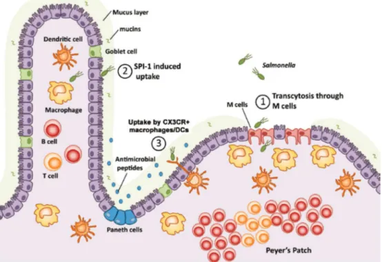
S. Typhimurium infection is usually acquired by oral ingesting of contaminated food, such as dairy products, eggs or meat. Approximately 104 to 106 bacteria are sufficient to cause infection in humans (Robert Koch Institute 2019). After overcoming gastric acidity, S. Typhimurium penetrates the epithelial barrier of the small intestine (Fig. 1). It actively invades intestinal epithelial cells or it induces its phagocytic uptake by microfold cells (M cells) that overlie the lymphoid follicles of Peyer's patches (Jones et al. 1994). In humans, S. Typhimurium infection usually does not proceed beyond the lamina propia. In mice and immunocompromised patients, however, S. Typhimurium gains access to the basolateral side of the intestine, where it is sensed and phagocytosed by monocytic-derived cells of the gut-associated lymphatic tissue (GALT), predominantly macrophages and dendritic cells (Broz et al. 2012). In susceptible hosts, S. Typhimurium enters the lymphatic system and the bloodstream via infected monocytes, allowing S. Typhimurium to spread to the liver and the spleen thereby establishing systemic infection (Vazquez-Torres et al. 1999).
Figure 1. Schematic representation of S. Typhimurium invading the intestinal mucosa.
(1) The major route of Salmonella invasion is via transcytosis through M cells at the Peyer´s Patches followed by (2) Salmonella pathogenicity island 1 (SPI-1)-mediated invasion of enterocytes. Alternatively, (3) Salmonella can
also be taken up by CX3CR+ macrophages or dendritic cells (DCs) located at the basolateral intestinal side. Figure taken from (Broz et al. 2012).
Following epithelial cell invasion or phagocytic uptake, S. Typhimurium is engulfed in a membrane‐derived compartment called Salmonella‐containing vacuole (SCV) (Bakowski et al. 2008). In a strict sense, SCVs are only formed in epithelial cells, whereas in professional phagocytes S. Typhimurium is taken up by phagosomes. SCVs show certain characteristics of late stage endosomes and express lysosomal markers that include lysosome-associated membrane proteins (LAMPs), the GTPase Rab7 and vacuolar ATPases (LaRock et al. 2015).
However, further maturation of the SCV does not occur as S. Typhimurium actively inhibits SCV fusion with the lysosome thereby preventing its enzymatic degradation (LaRock et al.
2015). The ability of S. Typhimurium to persist in host cells, including macrophages, is crucial for its pathogenesis, as strains lacking this ability are non-virulent (Bakowski et al. 2008).
S. Typhimurium virulence is highly dependent on the expression of two distinct type III secretion systems (T3SSs) and several effector proteins that are encoded on large chromosomal clusters termed Salmonella pathogenicity island 1 (SPI‐1) and SPI‐2. The T3SS apparatus, which is highly conserved among Gram-negative bacteria, forms a needle-like complex that mediates the injection of bacterial translocon and effector proteins into the host cell`s cytoplasm (Aizawa 2001). SPI-1-encoded T3SS effector proteins, such as SipA, SopB and SopE, are essential for epithelial invasion by inducing actin cytoskeleton rearrangements leading to membrane ruffling and subsequent uptake of S. Typhimurium (LaRock et al. 2015).
A major function of the SPI‐2-encoded T3SS effector proteins, such as SifA and SsrB, is the modification of the SCV to enable intracellular survival and replication of S. Typhimurium within the inhospitable milieu of the SCV (Hensel et al. 1998).
1.2 Interaction of S. Typhimurium with macrophages 1.2.1 Activation of inflammatory pathways
Once S. Typhimurium has passed the epithelial barrier of the small intestine, it is
sense S. Typhimurium through their pattern recognition receptors (PRRs), which mediate recognition of pathogen-associated molecular pattern molecules (PAMPs). Toll-like receptors (TLRs) function as the PRRs in mammals and play a major role in the defense against extracellular and intracellular S. Typhimurium. TLRs can be activated by a variety of S. Typhimurium-derived ligands, such aspeptidoglycan and lipoproteins (TLR2), LPS (TLR4), flagellin (TLR5) and CpG-rich repeats of bacterial DNA (TLR9) (Broz et al. 2012). TLR activation engages the signaling adaptors MyD88 and TRIF that culminate in the activation of nuclear factor- κB (NF-κB) and interferon regulatory factor 3 (IRF3), which control the expression of several pro-inflammatory cytokines as well as type I interferon (IFN-I) (Medzhitov 2001). The induction of inflammatory signaling pathways is necessary for the generation of a robust antimicrobial milieu and for the proper activation of adaptive immune responses.
1.2.2 Induction of oxidative stress
Reactive oxygen species (ROS) are essential components of the innate immune response against invading pathogens. Upon S. Typhimurium infection, ROS contribute to the efficient control of intracellular S. Typhimurium, as ROS increases the phagosomal bactericidal activity of macrophages (West et al. 2011). Macrophages have been shown to generate ROS through the phagosomal NADPH oxidase and the mitochondrial oxidative phosphorylation (OXPHOS) machinery (Lambeth 2004). ROS comprises oxygen free radicals, such as superoxide anion radical (O2·−) and hydroxyl radical (·OH), and non-radical oxidants, such as hydrogen peroxide (H2O2) and singlet oxygen (1O2), which can be interconverted from one to another (Zorov et al. 2014). The majority of mitochondrial ROS (mtROS) are derived from complex I or complex III of the electron transport chain (Murphy 2009). Although mtROS have been recognized as important signaling hubs linking mitochondrial to cytoplasmic signals, extensive mtROS production results in oxidative DNA damage and cell death (Murphy 2009). Therefore, mtROS levels have to be tightly controlled by a versatile antioxidant system.
Nuclear factor-erythroid 2-related factor 2 (Nrf2) is a key transcription factor that neutralizes cellular ROS by inducing multiple genes whose products have anti-oxidative functions. Kelch-like ECH-associated protein 1 (Keap1), a redox regulated substrate adaptor for a cullin-based ubiquitin ligase, is the major negative regulator of Nrf2 (Lo and Hannink 2008). Under resting conditions, Keap1 sequesters Nrf2 in the cytoplasm and mediates Nrf2 ubiquitination and its subsequent degradation via the proteasomal pathway (Kobayashi et al.
2004; Zhang et al. 2004).In response to oxidative stress, however, Nrf2 is released from Keap1 and translocated into the nucleus, where it binds to conserved antioxidant response elements (AREs) in the promoter regions of target genes, such as the detoxification enzyme NAD(P)H quinone oxidoreductase 1 (Nqo1), glutathione synthesis enzyme glutamate cysteine ligase (catalytic subunit, Gclc), and cell stress protein heme oxygenase-1 (Hmox-1) (Ishii et al. 2002; Rushmore et al. 1991; Wasserman and Fahl 1997).
Nrf2 function is negatively regulated by the mitochondrial phosphatase Pgam5, which has originally been identified as a protein bound to Keap1 (Lo and Hannink 2006). Pgam5 contains an N-terminal mitochondrial-localization sequence that mediates its recruitment to the outer mitochondrial membrane, where Pgam5 forms a ternary complex with Keap1 and Nrf2, in which the dimeric Keap1 simultaneously binds Pgam5 and Nrf2 (Lo and Hannink 2008). Nrf2 is consequently sequestered in this inhibitory complex leading to impaired activation of Nrf2-dependent anti-oxidative genes.
Recent studies suggest that the autophagy adaptor protein Sqstm1/p62 (hereafter, p62), is a positive regulator of the Nrf2 pathway. Upon oxidative stress, p62 directly interacts with the Nrf2-binding site of Keap1. Consequently, Nrf2 is released from Keap1 and translocated to the nucleus leading to the transcription of Nrf2-dependent anti-oxidative enzymes (Ichimura et al. 2013; Komatsu et al. 2010; Lau et al. 2010).
1.2.3 Interference with autophagy
Autophagy is a highly-conserved catabolic process that involves the complex interplay
large intracellular components, such as protein aggregates and long-lived or damaged organelles. Autophagy is usually induced by nutrient starvation through the inhibition of mammalian target of rapamycin complex 1 (mTORC1) leading to the engulfment of endogenous cargo into double-membrane vesicles called “autophagosomes”, which are subsequently degraded by the lysosomal pathway (Deretic et al. 2013; Mizushima et al.
2008).
Furthermore, autophagy is known to mediate the degradation of ubiquitinated cargo, such as misfolded proteins and protein complexes. Autophagy of ubiquitinated proteins requires the adaptor protein p62, which contains multiple protein-protein interaction sites, including a C-terminal ubiquitin-associated (UBA) domain and a LC3 interaction region (LIR).
These domains allow p62 to bind simultaneously to ubiquitinated cargo and LC3 thereby targeting proteins and organelles for autophagosomal degradation (Bjorkoy et al. 2005;
Itakura and Mizushima 2011; Pankiv et al. 2007). Beyond its importance for maintaining cellular energy and protein homeostasis, autophagy has also been shown to have more diverse functions, such as selective removal of proteins or clearance of intracellular pathogens (Sumpter and Levine 2010).
Selective autophagy of invading pathogens (xenophagy) has been recognized as an important component of cell-autonomous immunity. While several studies underscore the importance of xenophagy during infection with Mycobacterim tuberculosis, Listeria monocytogenes, Shigella spp., and S. Typhimurium (Cemma and Brumell 2012), the exact mechanism by which intracellular bacteria are recognized by the autophagic machinery remain poorly understood. It has been suggested that approximately 25% of intracellular S. Typhimurium escape from the SCV following infection of HeLa cells (Birmingham et al.
2006). Once in the cytosol, S. Typhimurium becomes coated with ubiquitin and associates with p62 and other adaptor proteins, such as nuclear dot protein 52 (NDP52), which mediate the LC3-dependent autophagic degradation of S. Typhimurium (Levine et al. 2011; Zheng et al. 2009). By contrast to epithelial cells, our recent work has demonstrated that xenophagy is
of minor relevance during S. Typhimurium infection of macrophages. While S. Typhimurium initially co-localizes with LC3-positive structures, it escapes from these structures during the further course of infection (Ganesan et al. 2017), indicating that it manipulates autophagy.
Probably more important than targeting intracellular bacteria for autophagic degradation, is the interplay between autophagy and innate immune signaling pathways.
Indeed, there is increasing evidence that engagement of TLR4 by LPS in macrophages induces autophagy through interaction of TRIF and MyD88 with the autophagy protein beclin- 1 (Shi and Kehrl 2008; Travassos et al. 2010). Furthermore, autophagy has been shown to fine-tune innate immune responses through modulation of IFN-I production and inflammasome activation (Sumpter and Levine 2010) as well as mediating cell death (Fitzwalter and Thorburn 2015).
1.2.4 Induction of cell death
For many years, apoptosis has been implicated as the major form of programmed cell death, whereas necrosis has mainly been considered as an accidental cell death process that occurs in response to different pathophysiological stimuli (Vanden Berghe et al. 2014).
However, recent genetic evidence and the discovery of specific pharmacological inhibitors of necrosis have redefined necrosis as a highly regulated form of programmed cell death.
Necrosis includes many different cell death modalities, such as necroptosis, ferroptosis, pyroptosis, and cell death associated with the release of (neutrophil) extracellular traps, called NETosis or ETosis (Pasparakis and Vandenabeele 2015).
Among these, necroptosis is probably the most extensively studied form of necrosis.
Necroptosis is a form of programmed cell death dependent on receptor-interacting Ser/Thr protein kinase (RIP) 1, RIP3 and its substrate mixed lineage kinase like (MLKL). Although many inflammatory signals, including TLR4 and IFN-I receptor activation as well as FAS/TRAIL signaling (Pasparakis and Vandenabeele 2015), can activate necroptotic pathways, the induction of necroptosis has been best exemplified by tumor necrosis factor
receptor (TNFR)-mediated signaling. Engagement of the TNFR induces the formation of a complex containing RIP1, RIP3, TNFR-associated death domain (TRADD), caspase-8, and FAS-associated protein with a death domain (FADD). While activation of caspase-8 results in apoptosis, inhibition of caspase-8, for instance by bacterial or viral proteins, triggers the formation of the necrosome. Autophosphorylation of RIP1 and RIP3 recruits the pseudokinase MLKL, which is itself phosphorylated by RIP3 leading to membrane permeabilization and subsequent cell death (Declercq et al. 2009).
Despite our detailed knowledge on TNF-mediated necroptosis execution, the events pre-ceding necrosome formation remain less understood. Mitochondria have been recognized as important mediators of several cell death modalities (Galluzzi et al. 2012) and ATP depletion by mitochondrial damage has been shown to be a trigger for necrosis (Vanlangenakker et al. 2008). In response to infection, mitochondria can change from a source of energy into organelles that promote cell death by releasing mtROS and toxic proteins that are usually retained within the mitochondrial intermembrane space (Kroemer et al. 2007). Furthermore, the mitochondrial phosphatase Pgam5 has recently been implicated to mediate TNF-induced necroptosis. Mechanistically, Pgam5 is recruited to RIP1- and RIP3- containing protein complexes at the outer mitochondrial membrane, where Pgam5 induces Drp1-mediated mitochondrial fragmentation resulting in necroptotic cell death (Wang et al.
2012).
The induction of cell death is a crucial virulence strategy employed by S. Typhimurium (Lindgren et al. 1996). Following invasion of host cells, S. Typhimurium requires the temporary niche of the SCV for intracellular adaption and replication, while at later stages of infection S. Typhimurium induces cell death to escape from the host cell and to infect surrounding cells. In macrophages, S. Typhimurium was found to induce different cell death modalities, including apoptosis-like cell death requiring caspase-8 activation (Lindgren et al.
1996), caspase-1/caspase-11-dependent pyroptosis resulting in IL-1β secretion (Brennan and Cookson 2000), and RIP3 and MLKL-dependent necroptosis via activation of IFN-I
1.3 Type I interferon (IFN-I)-dependent immune responses to S. Typhimurium 1.3.1 Induction of IFN-I synthesis
IFN-I are immunomodulatory cytokines that are secreted by various mammalian cell types in response to viral and bacterial infection. The family of IFN-I consists of about 20 members, with IFN-α and IFN-β being the major IFN-I expressed upon infection (Bogdan et al. 2004). Most of our knowledge on IFN-I signaling is derived from viral infection, but recent studies have also highlighted the importance of IFN-I for the cell-autonomous defense against bacterial infection (Bogdan et al. 2004).
Following infection, IFN-I synthesis is predominantly induced through activation of the stimulator of interferon genes (STING) pathway. Pathogen-derived DNA is sensed in the cytosol by cyclic GMP–AMP synthase (cGAS), which induces the cyclic dinucleotide (CDN) second messenger cGAMP (cyclic GMP-AMP) to activate the signaling adaptor STING.
STING together with TANK-binding kinase 1 (TBK1) is translocated to the perinuclear region, where TBK1 phosphorylates the transcription factor interferon regulatory factor 3 (IRF3).
Subsequently, IRF3 translocates to the nucleus and induces the transcription of numerous innate immune genes, including IFN-β, the main IFN-I in mice (Barber 2015).
Several bacteria have been shown to activate the STING pathway, such as Legionella pneumophila, Francisella tularensis, Streptococcus pyogenes, Brucella abortus and Mycobacterium tuberculosis (Barber 2015; Marinho et al. 2017). STING activation by S. Typhimurium, however, has not been reported to date (Marinho et al. 2017) and IFN-I production by S. Typhimurium is mainly thought to be mediated through LPS-mediated TLR4 activation (Owen et al. 2016; Sing et al. 2000).Gram-negative infection or binding of LPS to TLR4 engages TIR-domain containing adaptor protein inducing IFN-β (TRIF) and TRIF- related adaptor molecule (TRAM) (Yamamoto et al. 2003). This mediates activation of tank- binding kinase 1 (TBK1) and the inhibitor of nuclear factor-κB kinase (IKK-i), which leads to dimerization and nuclear translocation of IRF3 resulting in the transcription of IFN-βand auto- and paracrine signal transduction through the IFN-I receptor (Decker et al. 2005).
1.3.2 Activation of the IFN-I receptor pathway
IFN-I binds to a heterodimeric transmembrane receptor consisting of two subunits, IFN-I receptor (Ifnar) 1 and Ifnar2, respectively (Platanias 2005). Canonical Ifnar activation engages the receptor-associated protein tyrosine kinases Janus kinase 1 (JAK1) and tyrosine kinase 2 (TYK2), which phosphorylate the cytoplasmic transcription factors signal transducer and activator of transcription 1 (STAT1) and STAT2 (Stark and Darnell 2012). After dimerization and nuclear translocation, STAT1 and STAT2 form a complex with IRF9, which is termed IFN-stimulated gene factor 3 (ISGF3). ISGF3 subsequently binds to DNA sequences called IFN-stimulated response elements (ISREs), leading to the transcription of several hundred IFN-I stimulated genes (ISGs), which initiate antimicrobial pathways that limit the spread of infectious agents (Gough et al. 2012).
1.3.3 IFN-I-mediated cell death
Heterologous activation of the IFN-I pathways by various bacterial stimuli results in paradoxical infection outcomes, e.g. the production of IFN-I protects against some intracellular pathogens, such as Mycobacterium tuberculosis (Desvignes et al. 2012), while it enhances the susceptibility to other intracellular pathogens, such as Listeria monocytogenes (O'Connell et al. 2004).
We have previously demonstrated that S. Typhimurium exploits the host´s IFN-I response to induce cell death within macrophages. Activation of the Ifnar1 lead to the recruitment of RIP1, promoting the formation of a RIP1-RIP3 complex, which mediated necroptosis of infected macrophages (Robinson et al. 2012). As a result, mice lacking the Ifnar1 (Ifnar1-/-) survived significantly better (~30 to 60 days), whereas wildtype (WT) mice died within seven days after S. Typhimurium infection (Fig. 2).
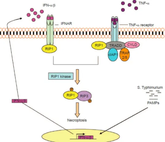
Figure 2. IFN-I-mediated necroptosis in S. Typhimurium-infected macrophages.
Following infection of macrophages, S. Typhimurium induces TLR4-mediated transcription of IFN-I (predominantly IFN-β). IFN-I binds to its cognate receptor (IFNAR) in an autocrine manner. RIP1 is subsequently engaged to the IFNAR resulting in the formation of a RIP1/RIP3 containing complex that triggers necroptosis of infected macrophages. In bone marrow-derived macrophages, IFNAR signaling is required for TNF-mediated necroptosis.
Figure taken from (Du et al. 2013).
Importantly, better control of S. Typhimurium and enhanced survival of Ifnar1-/- mice was specifically mediated by macrophages, because macrophages of Ifnar1-/- mice increased survival when transferred into WT hosts (Robinson et al. 2012).
HYPOTHESIS
Although our previous work has identified IFN-I-mediated activation of RIP1/RIP3 by S. Typhimurium as a novel pathway leading to necroptosis of macrophages, the mechanisms prior to necrosome formation largely remain unknown. We hypothesize that S. Typhimurium infection likely induces the same damage in WT and Ifnar1-/- macrophages. However, IFN-I signaling impairs the response to S. Typhimurium-induced damage in WT macrophages and the inability of WT macrophages to balance cellular homeostasis probably results in necroptosis execution. This work therefore aims to determine the harmful events mediated by IFN-I signaling that precede S. Typhimurium-induced cell death of macrophages.
OBJECTIVES OF THIS WORK
1. To determine the role of IFN-I signaling upon S. Typhimurium infection of macrophages.
2. To unravel the harmful events mediated by IFN-I signaling leading to cell death of S. Typhimurium-infected macrophages.
3. To identify the IFN-I-dependent pathways resulting in cell death of S. Typhimurium- infected macrophages.
2. Materials and Methods
2.1 Materials
2.1.1 List of instrumentation
Instrumentation Company
Countess II cell counter life technologies
Curix60 developing machine Agfa
EM109 electron microscope Zeiss
Enspire multimode plate reader PerkinElmer
FACSCanto flow cytometer BD Biosciences
IX81 inverted confocal microscope Olympus
PCR Cycler CFX96 Bio-Rad
Seahorse XF-96 analyzer Agilent
2.1.2 List of software
Software Company
FLOWJO v10.1 Tree Star
Fluoview FV10-ASW v4.2 Olympus
ImageJ NIH
Primer3web v4.1.0 ELIXIR
Prism v5 GraphPad
2.1.3 List of chemicals and reagents
Chemicals and reagents Company Cat. Number
A/G agarose magnetic beads Merck Millipore 16-663
Albumin Fraction V Carl Roth 8076.4
BCA Protein Assay Kit Pierce 23227
CDDO Biomol Cay81035
Cell Titer-Glo Luminescent Cell Viability Assay Promega G7571 CytoTox 96® Lactate Dehydrogenase Assay Promega #G1780
Enhanced chemiluminescence (ECL) Western Blotting Detection Reagent
GE Healthcare RPN2106
Ethanol absolute Merck 100983
Fetal bovine serum (FBS) Thermo Fisher
Scientific
10270-106
Formaldehyde, 16% Thermo Fisher
Scientific
28908
Gentamicin Life Technologies 15750037
Glycerol Carl Roth 4626.4
IL-1β ELISA kit R&D Systems DY401
Image-iT® R FX signal enhancer Invitrogen I36933
LEAF Purified anti-mouse IFN-β Antibody Biolegend 508104 LEAF Purified Syrian Hamster IgG Isotype
Control Antibody
Biolegend 400916
Lipofectamine 3000 transfection reagent Thermo Fisher Scientific
L3000-008
Methanol Carl Roth 4627.6
MitoSOX Mitochondrial ROS Detection Kit Thermo Fisher Scientific
M36008 NE-PER Nuclear and Cytoplasmic Extraction Kit Thermo Fisher
Scientific
78833
Normal goat serum Life Technologies PCN5000
Opti-MEM Reduced Serum Medium, GlutaMAX Supplement
Thermo Fisher Scientific
51985034 Phosphate-buffered saline (PBS) Merck Millipore L1825 ProLong Gold antifade containing DAPI Life Technologies P36935 Protease & Phosphatase Inhibitor Cocktail Thermo Fisher
Scientific
1861280
RNeasy Mini Kit Qiagen 74106
Seahorse XF Cell Mito Stress Test Kit Agilent 103015-100
Sodium dodecyl sulfate (SDS) Carl Roth 0.183.3
SsoFast EvaGreen Supermix Bio-Rad 1725201
SuperScript III reverse transcriptase Thermo Fisher Scientific
18080-044 Tetramethylrhodamine methyl ester (TMRM) Thermo Fisher
Scientific
T-668
Trigonelline hydrochloride Merck T5509
Tris-HCl Carl Roth 9090.2
Triton X-100 Carl Roth 3051.3
Trypan blue stain 0.4% Thermo Fisher
Scientific
15250061
Tween 20 Carl Roth 9127.1
VLE RPMI 1640 cell culture medium Merck Millipore FG1415
2.1.4 List of antibodies
Antibody Company Cat. Number
Alexa Fluor 488 anti-mouse IgG Invitrogen A-11017
Alexa Fluor 488 anti-rabbit IgG Invitrogen A-11008
Alexa Fluor 594 anti-rabbit IgG Invitrogen A-11072
Anti-mouse IgG HRP-conjugated R&D Systems HAF007
Anti-rabbit IgG HRP-conjugated R&D Systems HAF008
β-actin Santa Cruz sc-47778
GAPDH Santa Cruz sc-47724
Keap1 Proteintech 10503-2-AP
Lamin B Santa Cruz sc-374015
LC3 Sigma Aldrich L7543
MLKL Santa Cruz sc-165025
Nrf2 Santa Cruz sc-722
p70 S6 kinase Cell Signaling 9202
Pgam5 Abcam ab126534
phospho-p70 S6 kinase Cell Signaling 9205
phospho-MLKL Abcam ab196436
phospho-RIP3 Abcam ab195117
RIP3 Santa Cruz sc-135171
SQSTM1/p62 Cell Signaling 5114
Tom20 Santa Cruz sc-17764
2.2 Methods
2.2.1 Cell culture and bacterial infection
Mice and generation of bone marrow-derived macrophages
Bone marrow was extracted from 8 to 12 weeks old wildtype C57BL/6 (WT), type I interferon receptor (Ifnar1-/-), autophagy-related protein 7 (Atg7-/-) and receptor-interacting serine/threonine-protein kinase 3 (RIP3-/-) deficient mice. Bone marrow was differentiated into macrophages in RPMI medium supplemented with 10% of fetal bovine serum (FBS) and 20%
of L929 cell culture supernatant for 7 days in a humidified incubator at 37°C and 5% CO2. Non-adherent cells were removed on days 3 and 5 and adherent bone marrow-derived macrophages (BMDMs) were used for experiments from day 7 to 9. All animal studies were performed according to institutional guidelines on animal welfare and were approved by the North Rhine-Westphalian State Agency for Nature, Environment, and Consumer Protection (Landesamt für Natur, Umwelt and Verbraucherschutz [LANUV] Nordrhein-Westfalen; File no: 84-02.05.40.14.082 and 84-02.04.2015.A443) and the animal care committee of the University of Cologne.
S. Typhimurium infection of macrophages
Salmonella enterica serovar Typhimurium (SL1344) was grown to the late-exponential phase in Brain Heart Infusion (BHI) broth at 37°C with constant agitation. Bacteria were then harvested, resuspended in 10% of sterile glycerol and aliquots were stored at -80°C until further use. For all experiments, BMDMs were infected with a multiplicity of infection (MOI) of 10 for 10 min at room temperature and 30 min at 37°C. Subsequently, extracellular bacteria were removed by three times washing with RPMI medium containing 50 µg/ml gentamicin.
Afterwards, infected BMDMs were incubated with RPMI medium containing 10% FBS and 50 μg/ml gentamicin at 37°C. After 2 h of infection, gentamicin was diluted to 10 µg/ml for the remainder of the experiment. At the desired time points, BMDMs were washed once with PBS and samples were collected for experiments.
Bacterial colony formation assay
BMDMs (0.5 x 10⁶ cells/well) were seeded in 6-well plates 24 h prior to infection with S. Typhimurium. At 0 h and 24 h post infection, BMDMs were lysed with 1% Triton X-100 and 0.01% SDS/PBS solution and samples were collected in 1.5 ml Eppendorf tubes. Lysates were serially diluted with sterile PBS and spread on BHI agar plates, which were incubated overnight at 37°C. The next day, colony forming units (CFUs) were counted to determine the number of intracellular bacteria.
2.2.2 Protein and enzymatic assays Immunoblot analysis
BMDMs (1.0-2.0 x 10⁶ cells/well) were lysed with radio-immunoprecipitation assay (RIPA) buffer containing protease and phosphatase inhibitors and the protein amount of each sample was quantified using the BCA Protein Assay Kit according to the manufacturer`s instructions. Equal amounts of protein were mixed with SDS-PAGE sample loading buffer, boiled, resolved by SDS-PAGE and proteins were transferred to a PVDF membrane. Next, membranes were blocked with either 5% milk or 5% bovine serum albumin (BSA) in tris buffered saline (TBS) containing 0.05% Tween 20 (TBS-Tween) for 1 h at room temperature.
Membranes were then incubated overnight at 4°C with primary antibodies against LC3, p70 S6 kinase, phospho-p70 S6 kinase, SQSTM1/p62, Tom20, Nrf2, β-actin, Keap1, MLKL, phospho-MLKL, RIP3, phospho-RIP3, or Pgam5 diluted in TBS-Tween containing either 5%
milk or 5% BSA. After three times washing with TBS-Tween, membranes were incubated with the appropriate secondary antibody conjugated to horseradish peroxidase (HRP) for 1 h at room temperature. Membranes were washed three times with TBS-Tween and incubated with an enhanced chemiluminescence (ECL) substrate for 1 min at room temperature and exposed to a X-ray film. X-ray films were automatically developed using the Curix60 machine (Agfa). Densitometric quantification of western blotting was performed using ImageJ software (NIH).
Immunoprecipitation
BMDMs were plated in 10 cm dishes at 5.0 x 10⁶ cells per dish 24 h before the experiment. BMDMs were infected with S. Typhimurium and samples were lysed with RIPA buffer containing protease and phosphatase inhibitors. After preclearing the cell lysate with protein A/G agarose magnetic beads for 1 h, beads were removed by placing the tube on a magnetic rack. The whole cell lysate (approximately 500 μg of protein) was incubated with 4 µg of an antibody against Nrf2 overnight at 4°C. Protein A/G agarose beads were added again and incubated for an additional 1 h at room temperature. The immunoprecipitated proteins along with the agarose beads were collected by placing the tube on a magnetic rack.
The collected beads were washed five times with RIPA buffer. Samples were mixed with SDS-PAGE sample loading buffer, boiled and resolved on a 10% SDS-polyacrylamide gel.
The respective proteins precipitated were identified by immunoblot analysis.
Subcellular fractionation analysis
Nuclear fractions from BMDMs (1.0 x 10⁶ cells/well) seeded in 6-well plates 24 h prior to infection were prepared using the NE-PER Nuclear and Cytoplasmic Extraction Kit according to the manufacturer’s instructions. Samples were subjected to SDS-PAGE and analyzed by immunoblotting with antibodies against Nrf2, Lamin B, or GAPDH.
Enzyme-linked immunosorbent assay (ELISA)
BMDMs (0.5-1.0 x 10⁶ cells/well) were seeded in 6-well plates 24 h prior to infection.
For ELISA, cell culture supernatants were collected 6 h and 24 h post infection and were kept at -80°C until assayed for mouse IL-1β according to manufacturer´s instructions. Absorbance was measured with a multimode plate reader at 450 nm (PerkinElmer).
2.2.3 Mitochondrial and cell viability assays Mitochondrial ROS detection
Mitochondrial reactive oxygen species (mtROS) were determined using MitoSOX according to the manufacturer´s instruction. BMDMs (0.5 x 10⁶ cells/well) were seeded in 12-
well plates 24 h prior to the experiment and were infected with S. Typhimurium for 6 h.
Samples were subsequently fixed with 1% (wt/vol) formaldehyde in PBS for 10 min at room temperature and were then collected in FACS tubes. Fluorescence intensity of 50,000 cells per sample was analyzed by gating on live cells using the BD FACSCanto flow cytometer and FLOWJO software (Tree Star).The fluorescence intensity of BMDMs treated with MitoSOX was normalized to the intensity of BMDMs without MitoSOX treatment.
Measurement of mitochondrial membrane potential
Mitochondrial membrane potential (Ψm) was analyzed by TMRM staining, which is a fluorescent dye that accumulates in active mitochondria with intact mitochondrial membrane potentials. BMDMs (0.5 x 10⁶ cells/well) were seeded in 12-well plates 24 h prior to the experiment and BMDMs were subsequently infected with S. Typhimurium. After 6 h of infection, medium was exchanged to fresh RPMI medium containing 10% FBS and 10 µM of TMRM. BMDMs were incubated with TMRM for 10 min at room temperature. Next, cells were washed three times with PBS, samples were fixed with 1% (wt/vol) formaldehyde in PBS for 10 min at room temperature and were then collected in FACS tubes. Fluorescence intensity of 50,000 cells per sample was analyzed by gating on live cells using the BD FACSCanto flow cytometer and FLOWJO software (Tree Star). The fluorescence intensity of BMDMs treated with TMRM was normalized to the intensity of BMDMs without TMRM treatment.
Seahorse assay
Oxygen consumption rate (OCR) was analyzed using the Seahorse XF Cell Mito Stress Test Kit according to manufacturer’s instruction. Briefly, BMDMs were seeded at 0.2 x 10⁵ cells/well in a 96-well Seahorse plate 24 h before the experiment. A utility plate containing equilibration solution was placed in a CO2-free incubator at 37°C overnight. The following day, media was removed and cells were infected with S. Typhimurium as described before.
After 2 h of infection, medium was replaced with glucose, glutamine and sodium pyruvate- supplemented XF assay buffer and the plate was placed in a CO2-free incubator for at least
[FCCP], 2-Deoxy-D-glucose [2DG], Rotenone) were added to the appropriate port of the injector plate. After calibration with the utility plate, the sample plate was run on the Seahorse XF-96 machine.
ATP measurement
BMDMs were seeded at 0.1 x 10⁶ cells/well in a 96-well plate 24 h before infection with S. Typhimurium. After 2 h and 6 h of infection, ATP levels were determined using the Cell Titer-Glo Luminescent Cell Viability Assay according to manufacturer’s instructions.
Luminescence was measured with a multimode plate reader (PerkinElmer).
Cell viability assay
BMDMs were seeded into a 96-well plate at 0.1 x 10⁶ cells/well 24 h prior to the experiment. BMDMs were infected with S. Typhimurium and cell viability was measured at 2 h and 6 h post infection using the CytoTox 96® Lactate Dehydrogenase Assay (LDH) according to the manufacturer’s instructions. Luminescence was determined with a multimode plate reader (PerkinElmer).
2.2.4 Microscopic analyses
Immunofluorescent staining and confocal microscopy
BMDMs (0.1 x 10⁶ cells/well) were seeded in 24-well plates containing 12 mm round coverslips 24 h before infection. After infection with S. Typhimurium, BMDMs were fixed with 4% (wt/vol) formaldehyde in PBS for 15 min at room temperature. BMDMs were washed with PBS and permeabilized with 0.3% Triton X-100 in PBS for 5 min at room temperature. Next, BMDMs were washed with 0.03% Triton X-100 in PBS followed by incubation with Image-iT®
R FX signal enhancer for 30 min and blocking with 5% BSA, 5% normal goat serum and 0.03% Triton X-100 in PBS (blocking buffer) for 1 h at room temperature. After washing with 0.03% Triton X-100 in PBS, BMDMs were incubated overnight at 4°C with primary antibodies against SQSTM1/p62, Tom20, Nrf2, Keap1, or Pgam5 diluted in blocking buffer. BMDMs were then washed three times with 0.03% Triton X-100 in PBS and incubated with appropriate
fluorescent secondary antibodies diluted in blocking buffer for 1 h at room temperature. After three times washing with 0.03% Triton X-100 in PBS, coverslips were mounted on glass slides using ProLong® Gold antifade containing DAPI and stored at 4°C until image acquisition.
Images were acquired with a 60X oil PlanApo objective, numerical aperture 1.4 at room temperature on an Olympus IX81 inverted confocal microscope equipped with PMT detectors for imaging. Olympus Fluoview FV10-ASW 4.2 software was used for acquisition and calculating Pearson’s correlation.
Electron microscopy
BMDMs (1.5 x 10⁶ cells/well) were fixed (2.5% glutardehyde, 2.5% sucrose, 3 mM calcium chloride, 100 mM HEPES, pH 7.4) for 1 h at room temperature. After washing with 0.1 M cacodylate buffer (pH 7.2), cells were scraped and collected in cacodylate buffer. Cells were centrifuged (5 min, 500 x g, room temperature) and resuspended in 500 µl of warm 3%
low melting agarose (diluted in cacodylate buffer). Cell suspension was transferred into a sealed 1 ml tip, placed into a 15 ml falcon tube and centrifuged at 1.500 x g for 10 min at room temperature to concentrate cells. The agarose pellet was removed from the tip and collected into 0.1 M cacodylate buffer. After fixation, cells were dehydrated in graded ethanol series (50%, 70%, 90%, 100%) for 3 min each and were then incubated with ethanol and propylenoxid. Next, cells were embedded using Epon medium. Sections were cut at a thickness of 70 nm (Ultracut UC6, Leica), stained with 1.5% aqueous uranyl acetate for 15 min at 37°C and contrasted using lead nitrate solution. Samples were examined with an EM109 electron microscope (Zeiss) and images were recorded at 7,000 x or 50,000 x magnification.
2.2.5 mRNA assays
Transfection experiments
BMDMs (1.0 x 10⁶ cells/well) were seeded in a 6-well plate 24 h prior to transfection and were incubated with either 50 nM of non-targeting siRNA (#SR-CL000-005, Eurogentec)
or 50 nM of siRNA specific for Nfe2I2 (Nrf2; #L-040766-00-0005, Dharmacon), Sqstm1 (p62;
#L-047628-01-0005, Dharmacon), or Pgam5 (#L-052506-01-0005, Dharmacon) together with the transfection reagent Lipofectamine 3000 according to manufacturer’s instructions. Briefly, for each well 0.5 µl of siRNA (100 µM) was diluted in 50 µl of Opti-MEM medium and was added to 4 µl of Lipofectamine transfection reagent diluted in 50 µl of Opti-MEM medium.
Samples were gently mixed and incubated for 10 min at room temperature. Next, 100 µl of the transfection mix was added drop by drop to each well, which contained 900 µl of RPMI medium supplemented with 10% of FBS. After 5 to 6 h of incubation at 37°C, the medium was exchanged to RPMI containing 10% of FBS. At 48 h post transfection, BMDMs were infected with S. Typhimurium. Knockdown efficiency was individually assessed by immunoblot analyses using antibodies against Nrf2, p62, or Pgam5.
Quantitative real-time PCR (qRT-PCR)
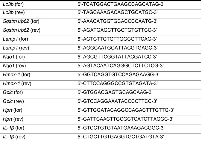
Total RNA from BMDMs (1.0 x 10⁶ cells/well) was isolated by RNeasy Mini Kit and cDNA (500 ng) was synthesized with random hexamers using reverse transcriptase (SuperScript III). Primers were designed using Primer3 software and Basic Local Alignment Search Tool (National Center for Biotechnology Information, Bethesda, MD). PCR reactions (25 µL) contained 10 ng of cDNA, 0.4 µmol/L of each forward and reverse primer, and master mix (SsoFast EvaGreen Supermix). Real-time PCR was performed on a PCR Cycler CFX96 (Bio-Rad) under the following conditions: initial denaturation step of 95°C for 2 min, 40 cycles of 95°C for 5 sec and 60°C for 15 sec, followed by a denaturation step of 95°C for 60 sec and subsequent melt curve analysis to check amplification specificity. Results were analyzed by the comparative threshold cycle method with hypoxanthine-guanine phosphoribosyltransferase (Hprt) as the endogenous reference gene for all reactions. The relative mRNA levels of uninfected BMDMs were used as normalized controls for infected BMDMs. All assays were performed in triplicates, and a non-template control was included in all experiments to exclude DNA contamination. Primers were obtained from Invitrogen and are listed in Tab. 1.
Gene Primer sequence
Lc3b (for) 5’-TCATGGACTGAAGCCAGCATAG-3’
Lc3b (rev) 5’-TAGCAAAGACAGCTGCATGC-3’
Sqstm1/p62 (for) 5’-AAACATGGTGCACCCCAATG-3’
Sqstm1/p62 (rev) 5’-AGATGAGCTTGCTGTGTTCC-3’
Lamp1 (for) 5’-AGTCTTGTGTTGGCGTTCAG-3’
Lamp1 (rev) 5’-AGGCAATGCATTACGTGAGC-3’
Nqo1 (for) 5’-AGCGTTCGGTATTACGATCC-3’
Nqo1 (rev) 5’-AGTACAATCAGGGCTCTTCTCG-3’
Hmox-1 (for) 5’-GGTCAGGTGTCCAGAGAAGG-3’
Hmox-1 (rev) 5’-CTTCCAGGGCCGTGTAGATA-3’
Gclc (for) 5’-GTGGACGAGTGCAGCAAG-3’
Gclc (rev) 5’-GTCCAGGAAATACCCCTTCC-3’
Hprt (for) 5’-GTTGGATACAGGCCAGACTTTGTTG-3‘
Hprt (rev) 5’-GATTCAACTTGCGCTCATCTTAGGC-3‘
IL-1β (for) 5’-GTCCTGTGTAATGAAAGACGGC-3’
IL-1β (rev) 5’-CTGCTTGTGAGGTGCTGATGTA-3’
Table 1. List of primers used in this study. for, forward. rev, reverse.
2.2.6 Statistical analyses
Statistical analyses were performed using Prism software (GraphPad, version 5).
Differences between groups were assessed by two-tailed unpaired Student´s t test or One- way ANOVA with repeated measures when more than two groups were analyzed. Every single experiment was repeated at least three times. Unless otherwise specified, results are presented as mean ± standard deviation (SD). Differences were considered statistically significant when p ≤ 0.05 (∗), very significant when p ≤ 0.01 (∗∗), and highly significant when p
≤ 0.001 (∗∗∗).