LEF-1 is a potential therapeutic target in the treatment of Chronic lymphocytic
leukemia
Inaugural - Dissertation zur
Erlangung des Doktorgrades
der Mathematisch-Naturwissenschaftlichen Fakultät der Universität zu Köln
vorgelegt von
Rajesh Kumar Gandhirajan aus
Chennai, Indien
Köln, Oktober 2009
1. Berichterstatter:
2. Berichterstatterin:
3. Berichterstatter:
Prof. Dr. Guenter Plickert
Prof. Dr. Angelika Anna Noegel Prof. Dr. Jürgen Dohmen
Tag der mündlichen Prüfung: 15
thOktober 2009
T o m y P arents,
B rother, Sister
& G ow ri
Table of Contents
1. SUMMARY ... 1
2. ZUSAMMENFASSUNG ... 3
3. INTRODUCTION ... 6
3.1.HEMATOPOIESIS ... 6
3.2.THE ROLE OF ANTIGENS IN B-CLL ... 8
3.3.BIOLOGY OF CHRONIC LYMPHOCYTIC LEUKEMIA ... 9
3.3.1. Overview of Wnt signaling mechanism ... 11
3.3.2. Molecular Mechanism of Canonical Wnt signaling ... 12
3.3.3. The LEF/TCF protein family ... 14
3.3.4. The Role of Wnt signaling in B-cell development ... 16
3.4.WNT SIGNALING AND DISEASES ... 18
3.4.1. Role of Wnt signaling in human cancers and its therapy ... 19
3.4.2. Role of Wnt signaling in B Chronic lymphocytic leukemia (B-CLL) ... 19
3.5.CLINICAL PERSPECTIVE OF B-CHRONIC LYMPHOCYTIC LEUKEMIA (B-CLL) ... 20
3.5.1. Epidemiology ... 21
3.5.2. Clinical manifestations ... 21
3.5.3. Diagnostic criteria ... 22
3.5.4. Disease staging ... 23
3.5.5. Prognostic factors... 24
3.5.5.1. Cytogenetic abnormalities ... 24
3.5.5.2. IgVH mutational status ... 25
3.5.5.3. Zeta-associated protein (ZAP-70) ... 26
3.5.5.4. CD38 expression... 26
3.6.CURRENT THERAPEUTIC STRATEGIES ... 27
3.6.1. Criteria for patient treatment ... 27
3.6.2. Alkylating agents as single agents and in combination therapy ... 28
3.6.3. Purine analogs as single agents and in combination therapy ... 29
3.6.4. Chemoimmunotherapy ... 29
3.6.5. Allogeneic transplant ... 30
3.6.7. Other Targeted therapies ... 30
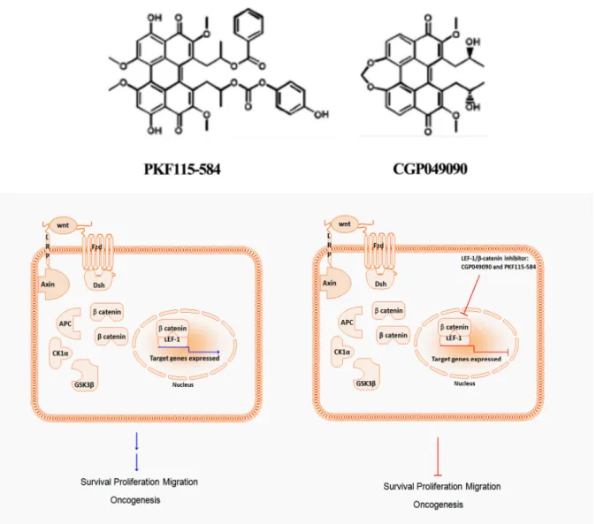
3.6.8. Novel TCF/LEF-1/β-catenin Inhibitors CGP049090 and PKF115-584 ... 31
3.7.PROJECT OBJECTIVES ... 33
4. RESULTS ... 35
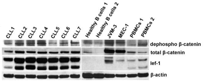
4.1.ACTIVATION OF WNT SIGNALING IN B-CLL ... 35
4.1.1. Expression of downstream signaling components of Wnt signaling in B-CLL and healthy B cells. .. 35
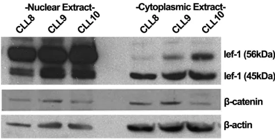
4.1.2. Nuclear localization of LEF-1 and β-catenin in primary B-cells ... 36
4.2.LEF-1 PLAYS A CENTRAL ROLE IN FOR SURVIVAL OF B-CLL CELLS ... 37
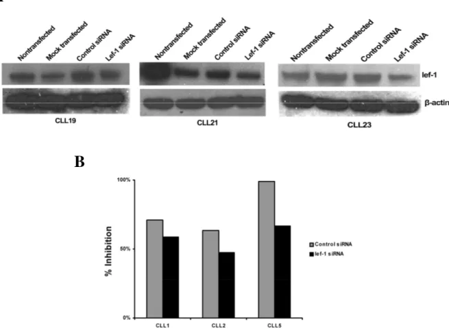
4.2.2. siRNA mediated LEF-1 knockdown induces apoptosis in primary CLL cells... 39
4.2.3. Knockdown of LEF-1 in JVM-3 cell lines by siRNA leads to down regulation of target genes and
reduced proliferation ... 40
4.3.IN VITRO CYTOTOXICITY OF SMALL MOLECULES (CGP049090 AND PKF115-584) ... 41
4.3.1. Cell lines ... 41
4.3.2. Healthy B cells and Primary CLL cells ... 42
4.3.3. Patient Sample Characteristics ... 43
4.3.4. CGP049090 & PKF115-584 induces apoptotic cell death in B-CLL ... 44
4.3.5. CGP049090 & PKF115-584 induced apoptotic cell death is dependent on activation of caspase pathway ... 45
4.3.6. CGP049090 & PKF115-584 suppress the expression of anti-apoptotic proteins ... 47
4.3.7. β-catenin levels are down regulated upon initiation of apoptosis by CGP049090 and PKF115-584 48 4.4.CGP049090 AND PKF115-584 INHIBIT THE INTERACTION BETWEEN LEF-1/Β-CATENIN ... 49
4.4.1. CGP049090 and PKF115-584 down regulate LEF-1/β-catenin target genes in Primary CLL cells 50 4.4.2. CGP049090 and PKF115-584 down regulate LEF-1/β-catenin target genes in JVM-3 cell lines .... 51
4.4.3 Apoptosis induced by CGP049090 and PKF115-584 is inhibited by Pan Caspase inhibitors ZVAD.FMK ... 52
4.4.4 Apoptosis induced by CGP049090 and PKF115-584 leads to cleavage of LEF-1 target proteins .... 53
4.5.ENHANCED AUTO-FLUORESCENCE EXHIBITED BY PKF115-584 ... 54
4.5.1. LEF-1 Overexpression and Intracellular Co-localization with PKF115-584 in CLL Cells. ... 55
4.6.IN VIVO EFFICACY OF CGP049090 AND PKF115-584 IN PRE-CLINICAL MOUSE MODEL. ... 56
4.6.1. CGP049090 and PKF115-584 inhibit tumor growth in vivo ... 57
4.6.2. CGP049090 and PKF115-584 increases the median survival of the treated mice... 59
4.6.3. Tumor growth cessation by CGP049090 and PKF115-584 in vivo is mediated by inhibition of cellular proliferation and apoptosis ... 60
4.6.4. LEF-1 is downregulated upon treatment with CGP049090 and PKF115-584 in vivo ... 61
4.6.5. In Vivo Inhibition of Proliferation (PCNA) and Increase of Apoptosis (cleaved PARP) after CGP049090 and PKF115-584 treatment. ... 62
5. DISCUSSION ... 65
5.1.EXPRESSION OF LEF-1 AND Β-CATENIN IN CLL ... 66
5.2.LEF-1 AND B-CLL SURVIVAL ... 67
5.3.IN VITRO EFFICACY OF CGP049090 AND PFK115-584CLL ... 68
5.4.MECHANISM OF APOPTOTIC INDUCTION BY CGP049090 AND PKF115-584 ... 69
5.5SPECIFICITY OF THE CGP049090 AND PKF115-584 ... 71
5.6.IN VIVO EFFICACY OF CGP049090 AND PKF115-584 ... 72
5.7OFF-TARGET EFFECTS OF CGP049090 AND PKF115-584 ... 74
5.8FUTURE PERSPECTIVES: ... 75
6. MATERIALS & METHODS ... 77
6.1.MATERIALS ... 77
6.1.1. Instruments ... 77
6.1.2. Consumables ... 78
6.1.3. Chemical and Reagents ... 78
6.1.4. Reagent/ Kits ... 80
6.1.5. Antibodies ... 80
6.1.6. Inhibitor stock Solutions ... 81
6.1.7. Cell lines ... 82
6.2.METHODS ... 82
6.2.1. Culture Conditions ... 82
6.2.2. Culture of primary CLL samples ... 82
6.2.2.1. Isolation of PBMCs by Ficoll gradient ... 82
6.2.2.2. Isolation of B-cells by Rosette Sep ... 83
6.2.3. Maintenance of mammalian cells ... 83
6.2.3.1. Culture of suspension cells ... 83
6.2.3.2. Culture of adherent cells ... 83
6.2.3.3. Freezing and thawing cells ... 83
6.2.3.4. Co-culturing cells ... 84
6.2.4. Cytotoxcity Assay ... 84
6.2.5. Quantification of apoptosis flow cytometry ... 85
6.2.6. Immunofluorescence ... 86
6.2.7. siRNA mediated gene knockdown ... 87
6.2.8. Methods in Protein chemistry ... 88
6.2.8.1. Preparation of cell lysates. ... 88
6.2.8.2. Preparation of nuclear and cytoplasmic fractions ... 88
6.2.8.3. Quantification of proteins ... 89
6.2.8.4. Co-immunoprecipitation ... 90
6.2.8.5. SDS PAGE ELECTROPHORESIS ... 91
6.2.8.6. Protein transfer (Western Blot) ... 92
6.2.8.7. Immunoblot ... 92
6.2.9. Generation of JVM-3 Xenograft Subcutaneous Tumor Mouse Model ... 94
6.2.9.1. Evaluation of in vivo antitumor activity ... 95
6.2.10. Histochemistry ... 96
6.2.11. Immunohistochemistry ... 97
7. REFERENCES ... 101
ABBREVIATIONS ... 121
INDEX OF FIGURES ... 123
INDEX OF TABLES ... 124
ACKNOWLEDGEMENTS ... 125
EHRENWÖRTLICHE ERKLÄRUNG ... 127
DECLARATION ... 127
CURRICULUM VITAE ... 128
1. Summary
B-Chronic lymphocytic leukemia (B-CLL) is characterized by accumulation of apoptotic resistant CD5+ B lymphocytes. There is an increased secretion of Wnt ligands indicating an autocrine loop leading to the extended survival of B-CLL cells. Lymphoid enhancer factor 1 (LEF-1) is a potent transcription factor regulating the expression of several Wnt induced target genes. A comprehensive gene expression profiling from two independent studies revealed that LEF-1 mRNA was ~3000 fold overexpressed in B-CLL when compared to its healthy counterpart.
The objective of this present study is to demonstrate the therapeutic benefit of inhibiting LEF-1 expression in B-CLL cells using novel small molecule inhibitors CGP049090 and PKF115-584 in vivo and in vitro. In order to explore the anti-leukemic potential of CGP049090 and PKF115-584 we tested its effects on freshly isolated B-CLL cells, prolymphocytic cell line (JVM-3 & MEC-1) and in a subcutaneous mouse xenograft model.
The present study shows that, in freshly isolated B-CLL cells there was high protein expression and nuclear localization of LEF-1 and β-catenin indicating active LEF-1 mediated transcription whereas LEF-1 remained undetectable in healthy B cells. Preliminary experiments of LEF-1 inhibition using siRNAs resulted in increased apoptosis indicating LEF-1 plays an important role in the survival of B-CLL cells. This observation was extended using CGP049090 and PKF-115584 as they induce dose dependent cytotoxicity in B-CLL, whereas the healthy B cells are not significantly affected. The half maximal inhibitory concentration (IC50) was less than 1 µM in primary B-CLL cells and cell lines whereas it was more than 5 µM in healthy B cells. CGP049090 and PKF-115584 induced apoptotic cell death in primary B-CLL cells and cell lines by cleavage of caspases 8, 9, 3 and 7 and subsequent cleavage of Poly (adenosine diphospate-ribose) polymerase (PARP). Both inhibitors also altered the expression of several anti-apoptotic proteins like X-linked Inhibitor of Apoptosis
Protein (XIAP), Mantle cell lymphoma-1 (Mcl-1) and B cell lymphoma-2 (Bcl-2). Co- Immunoprecipitation experiments revealed that both the inhibitors effectively disrupt the β- catenin/LEF-1 interaction, resulting in the down regulation of LEF-1 target genes such c-myc, cyclin D1 and LEF-1. Furthermore, when the inhibitors were tested in an in vivo JVM-3 subcutaneous xenograft nude mouse model, more than 70% inhibition of tumor growth and an increase in the median survival of the treated group without leading to systemic toxicity was observed. Immunohistochemistry analysis of the tumor sections revealed LEF-1 down regulation and subsequent inhibition of proliferation by down regulation of Proliferating Cell Nuclear Antigen (PCNA) and increase in apoptosis (cleaved PARP).
In summary, the data showed that LEF-1 is a potential therapeutic target in the treatment of B-CLL. Both CGP049090 and PKF115-584 showed potent inhibitory effects on the survival of CLL cells in vitro and in vivo without affecting the healthy cells. Both CGP049090 and PKF115-584 are hence, potential anti-cancer agents in B-CLL and other neoplastic malignancies with aberrant LEF-1/ T cell factor (TCF) transcriptional activity.
Further investigations are warranted to determine the feasibility of these small molecules for therapeutic approach in humans.
2. Zusammenfassung
Die chronische lymphatische B-Zell Leukämie (B-CLL) ist durch eine Akkumulation von apoptose-resistenten CD5-positive B-Lymphozyten charakerisiert. In letzter Zeit wurden verschiedene Faktoren beschrieben, welche zu einer aberranten Aktivierung der Wnt- Signalkaskade in B-CLL Zellen beitragen. Eine vermehrte Sekretion von Wnt-Liganden weist auf einen autokrinen Rückkopplungsmechanismus hin, welcher eine Rolle im gesteigerten Überleben der B-CLL Zellen spielt.
LEF-1 ist ein potenter Transkriptionsfaktor, welcher die Expression verschiedener Wnt-induzierter Zielgene reguliert. LEF-1 wurde in zwei unabhängigen Genexpressionsanalysestudien als Gen mit etwa 3000-facher Überexpression in der B-CLL, relativ zum Expressionslevel in gesunden B-Zellen, beschrieben. Der Transkriptionsfaktor LEF-1 ist damit exklusiv auf B-CLL Zellen exprimiert.
Das Ziel der vorliegenden Studie ist es, den therapeutischen Nutzen einer gezielten Inhibierung von LEF-1 in B-CLL Zellen, durch die Verwendung zweier sogenannter „small molecule“ Inihibitoren CGP049090 and PKF115-584, sowohl in vivo als auch in vitro zu zeigen.
Um den anti-leukämischen Effekt dieser Substanzen zu zeigen, wurden deren Effekte auf frisch isolierte B-CLL Zellen und prolymphozytischen Zelllinien ((JVM-3 & MEC-1) untersucht. Zudem wurden beide Substanzen auf Ihre Wirksamkeit in einem JVM3-Xenograft Mausmodel getestet.
In frischen B-CLL Zellen konnten wir große Proteinmengen von LEF-1 detektieren, welche größtenteils im Zellkern zu finden waren. Auch β-Catenin war vorrangig im Zellkern vorhanden, was darauf schließen lässt, dass LEF-1 in B-CLL Zellen transkriptionell aktiv ist.
LEF-1-Protein war in gesunden B-Zellen nicht detektierbar. Eine Runterregulierung von LEF1 mittels siRNA hat zu einem vermehrten Zellsterben durch Apoptoseinduktion geführt, welches die wichtige Funktion von LEF-1 für das Überleben der B-CLL Zelle zeigt. Diese
Beobachtung wurde dadurch erweitert, dass auch CGO049090 und PKF-115584 sowohl zeit- als auch konzentrationsabhängig zytotoxisch auf B-CLL Zellen wirken, während gesunde B- Zellen in signifikant geringerem Maße betroffen sind. Die IC50 in primären B-CLL Zellen war <1 µM und >5 µM in gesunden B-Zellen. CGO049090 und PKF-115584 führten zu apoptotischem Zelltod durch eine Spaltung der Caspasen 8, 9, 3 und 7 und eine darauffolgende Spaltung von poly (adenosine diphospate-ribose) polymerase (PARP). Beide Inhibitoren führten zudem zu einer veränderten Expression verschiedener antiapoptotischer Proteine wie X-linked Inhibitor of Apoptosis Protein (XIAP), Mantle cell lymphoma-1 (Mcl- 1) und B cell lymphoma-2 (Bcl-2). Co-Immunoprezipitationsexperimente zeigten, dass beide Inhibitoren effektiv den β-Catenin/LEF-1 Komplex trennen, was zur Herunterregulation der LEF-1 Zielgene c-myc, cyclin D1 und LEF-1 führt. Darüber hinaus wurden beide Inhibitoren in einem subkutanen JVM-3-Xenograft Nacktmausmodel getestet und erzielten eine Tumorinhibitionsrate von >70%. Zudem erhöhte sich die mittlere Überlebensrate der Mäuse durch die Behandlung ohne erkennbare systemisch toxische Effekte. Eine Gewebeuntersuchung der Tumore ergab eine Herunterregulation von LEF-1 und eine darausfolgende Herunterregulation des Proliferating Cell Nuclear Antigens (PCNA), welches auf eine erniedrigte Proliferationsrate hinweist. Zudem wurde eine verstärkte Aktivität von PARP detektiert, wodurch auf eine erhöhte Apoptoseinduktion im Tumorgewebe behandelter Mäuse geschlossen werden kann.
Zusammenfassend lässt sich sagen, dass LEF-1 einen potentiellen therapeutischen Ansatzpunkt in der B-CLL darstellt. Die beiden Substanzen CGP049090 and PKF115-584 weisen eine hohe Wirksamkeit bei der Zelltodinduktion B-CLL Zellen gegenüber auf, während sie gesunde B-Zellen kaum beeinträchtigen. CGP049090 and PKF115-584 eignen sich deshalb als potentielle Substanzen in der B-CLL Therapie und auch in anderen neoplastischen Erkrankungen, die eine aberrierende transkriptionelle Aktivität von LEF-
1/TCF aufweisen. Weitere Untersuchungen sind wünschenswert um die Anwendbarkeit dieser zwei Substanzen für therapeutische Zwecke im Menschen genauer zu bestimmen.
3. Introduction
During normal lymphocyte differentiation B cells jeopardize their genomic integrity through the formation and revision of their antigen receptors. A second potentially dangerous event is the response to antigens, which under normal circumstances, is a tight homeostatic regulation of clonal expansion of B cells. A compromise in either of the two events would result in oncogenic genomic hits that block differentiation, prevent apoptosis and/ or promote proliferation leading to several types of lymphoma and leukemia. B-Chronic lymphocytic leukemia (B-CLL) is one such disease which results due to a prolonged and unregulated antigenic stimulation contributing to clonal expansion of leukemic B cells.
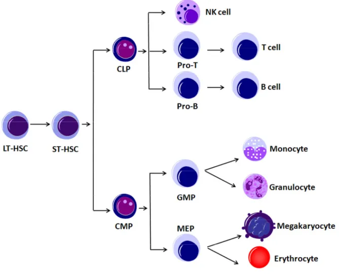
3.1. Hematopoiesis
To comprehend the origin of B-CLL we have to understand the mechanism of generation of different cell types of the lymphoid and myeloid systems (Figure 1). All the mature blood cells in the body are generated from a relatively small number of hematopoietic stem cells (HSCs) and progenitors (Weissman 2000). In the mouse, a single HSC can reconstitute the entire hematopoietic system for the natural lifespan of the animal (Osawa 1996). HSCs generate the multiple hematopoietic lineages through a series of intermediate progenitors. Those are the common lymphoid progenitors (CLPs) that give rise to natural killer cells (NK), T cells, and B cells, and the common myeloid progenitors (CMPs), which can generate monocytes, granulocytes, megakaryocytes, and erythrocytes (Kondo 1997;
Akashi 2000). Out of the CMPs develop more specialized progenitors, that are further restricted to a number and type of cell lineages that they can generate. These are the granulocyte/monocyte progenitors (GMP), which give raise to the granulocytes and monocytes, and the megacarythrocyte/erythrocyte progenitors (MEP), which can develop to megakaryocytes and erythrocytes (Akashi 2000). The terminally differentiated cells produced cannot divide any longer and undergo apoptosis after days to decades depending on their cell type.
During the process of B cell differentiation the B cells generate the B cell receptors (BCRs) by V(D)J recombination involving double strand DNA breaks initiated by recombination activated genes (RAG1 and RAG2) that are resolved by the non-homologus end joining repair apparatus (Fugmann et al. 2000). In this process, the cells are often susceptible to chromosomal translocations replacing the usual regulatory sequences of a gene with heterologous regulatory elements which lead to inappropriate gene expression at the breakpoints, leading to lymphomas (Pelicci et al. 1986).
Figure 1: Hematopoiesis
Long term hematopoietic stem cells (LT-HSC) give rise to short term (ST) HSCs. Due to different stimuli they either become common lymphoid progenitors (CLP) or common myeloid progenitors (CMP). Downstream of CLPs the cells either develop to natural killer cells (NK), to B or T cells. The CMPs give rise to more specialized progenitors, granulocyte/monocyte progenitors (GMP) and megakaryocyte/erythrocyte progenitors (MEP), which finally differentiate to granulocytes, monocytes, megakaryocytes and erythrocytes.
On the other hand when a differentiated but naïve B cell encounters an antigen, the naïve B cell gets activated and interacts with follicular dendritic cells, T cells and the antigen within the microenvironment of the Germinal Centre (GC). Here the B cell undergoes Class Switch Recombination (CSR) via DNA breaks and extensive remodeling of the DNA and somatic hypermutation (SHM) using point mutations of the immunoglobulin genes. This again makes the B cell genome prone to genetic alterations leading to gross chromosomal mutations (Boehm et al. 1989; Honjo et al. 2002).
Interestingly, B-CLL does not represent any of these typical gross chromosomal translocations generated by the above mechanisms (Montserrat & Rozman 1995), but represent in two distinct subtypes i.e. B-CLL cells which have undergone SHM (IgVH
mutated) in the germinal centre and those cells which did not undergo SHM (IgVH
unmutated) (Shaffer et al. 2002). Hence the origin of this disease is still a mystery. The current hypothesis favors the role of an antigen in the pathogenesis of B-CLL.
3.2. The Role of Antigens in B-CLL
The role of antigens have been implicated in some lymphomas such as Marginal Zone Lymphoma (MALT) where the 70% of the patients present an infection with helicobacter pylori leading to gastric ulcers (Wotherspoon et al. 1993; Wotherspoon et al. 1994).
Helicobacter pylori specific T cells stimulate the proliferation of MALT lymphoma cells in culture and these patients can be cured of their MALT simply by antibiotics (Hussell et al.
1993).
Several gene expression studies have been carried out in determining the difference between the IgVH unmutated and IgVH mutated forms of B-CLL (Rosenwald et al. 2001;
Klein et al. 2001). These evidences indicate a potential role of an antigen in the clonal expansion of B-CLL cells. CLL B cells use a biased VH repertoire and have non-random combinations of V, D and J segments that are not characteristic of normal blood B cells.
Furthermore, certain VH genes are used differentially by immunoglobulin-unmutated and -
mutated forms of CLL. For example, the VH1-69 gene is associated almost exclusively with immunoglobulin-unmutated CLL, whereas other VH genes, such as VH4-34, VH1-07 and VH3-21, are over-represented in immunoglobulin-mutated CLL, indicating role of antigen in the expansion of these cells (Damle et al. 1999; Fais et al. 1998; Tobin et al. 2002;). However the nature of this antigen is still unknown, it is predicted that it is presumably auto antigens such as single- and double-stranded DNA, and IgG. (Sthoeger et al. 1989; Borche et al. 1990).
3.3. Biology of chronic lymphocytic leukemia
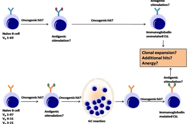
The ability of one cell to influence the behavior of another cell is achieved through cell communication, termed cell signaling. Over the time several different signaling pathways have been developed, as for example the Notch-, the Phosphatidylinositol-3 kinase (PI3K), the BMP-, and the Wnt signaling pathway which are crucial in early development and maintenance of the cells. At the end of each intracellular signaling pathway are target proteins, which when altered changes the behavior of the cell. Moreover, a crosstalk between the different pathways occurs, which leads to a big network of signals and a tight regulation within a cell. Disturbance of the balanced systems often leads to diseases, as for example cancer. B-CLL is such a condition wherein there is a deregulation of several signaling pathways, which leads to extended survival. We will discuss some of the important signaling defects in the pathogenesis of CLL but will mainly focus on the role of Wnt signaling pathway in B cell development and pathogenesis of CLL. Figure 2 shows an overview of the pathogenesis of B-CLL during early stages of differentiation and junctures where additional oncogenic hits/antigenic stimulation might occur resulting in its activation of variety of pathways involved in prolonged survival (Shaffer et al. 2002).
Signaling defects in CLL is considered to be one of the factors prolonging the survival of the B cells than the normal lymphocytes (Hamblin & Oscier 1997). The BCR signaling is important in determining the B-cell fate, and the level of BCR engagement is modulated by the antigenic valency, epitope density and epitope organization. Upon BCR engagement,
receptor aggregation induces phosphorylation of the immunoreceptor tyrosine-based
Figure 2: An overview of the pathogenesis of B-CLL (Adapted from A. L. Shaffer 2002) The two subtypes of chronic lymphocytic leukemia (CLL) — immunoglobulin-mutated and immunoglobulin-unmutated CLL — are distinguished by the presence or absence of immunoglobulin variable (V)-region mutations, by differences in gene expression and by their clinical courses. This model emphasizes the potential role of antigenic stimulation in the progression of this disease. Immunoglobulin-unmutated and -mutated forms have a different repertoire of heavy-chain V-region (VH) gene rearrangements, which indicates that the type of CLL that results is dictated by the specificity of the B-cell receptor (BCR).
Many studies have implicated an antigen or autoantigen indirectly in the pathogenesis of CLL. Antigenic stimulation might occur before and/or after the B cell acquires a genetic change (oncogenic hit) and becomes a CLL cell. It is also possible that early stages of CLL occur without oncogenic hits to the B cell. Immunoglobulin-unmutated CLL most probably originates from a pre-germinal centre (pre-GC) B cell. Immunoglobulin-mutated CLL might originate from a post-GC B cell. Alternatively, immunoglobulin-mutated CLL might originate from a pre-GC B cell that is nevertheless driven by antigen through a GC reaction. The clinical manifestations of CLL might be related to whether antigen drives continued clonal expansion or induces an anergic state. Disease progression might be influenced by the accumulation of additional oncogenic hits.
activation motifs (ITAMs) by Src-family tyrosine kinases (Pierce 2002). This leads to recruitment of other kinases triggering activation of intracellular signaling cascades. Low expression of BCR is the hallmark of CLL (Payelle-Brogard et al. 2002; Vuillier et al. 2005) and its stimulation is considered to be important for survival and proliferation of CLL (Stevenson & Caligaris-Cappio 2004).
PI3K and mitogen activated protein kinases (MAPK) are activated through membrane receptor tyrosine kinases and are involved in several signal transduction pathways in B cells such as CD40 signaling, BCR signaling and signaling of variety of cytokines (Ringshausen et al. 2002). Studies have shown the constitutive activation of these pathways in CLL cells and their requirement in the maintenance of CLL viability (Ringshausen et al. 2004; Longo et al.
2007; Plate 2004).
Failed programmed cell death or apoptosis is a characteristic feature of CLL (Reed 1998). The Bcl-2 family members (bcl-2 bax, bclxL) are over-expressed in CLL (Hanada et al. 1993). The imbalance in the ratio of major pro- and anti- apoptotic family proteins is associated with the treatment response of CLL (Kitada et al. 1998; Pepper et al. 2001). Mcl-1, another anti-apoptotic protein is also overexpressed in CLL cells and contributes to failure of cytotoxic therapy (Kitada et al. 1998; Saxena et al. 2004). There is also evidence that indicate Mcl-1, Bcl-xL and XIAP are regulated by Akt and Erk pathway but only Mcl-1 is essential for CLL survival (Longo et al. 2008).
Most recently great emphasis is being placed on the role of aberrant Wnt signaling in malignant diseases and the different mechanisms, major players of Wnt signaling and its role in B cell development will now be elaborated further.
3.3.1. Overview of Wnt signaling mechanism
The Wnt signaling consists of three different pathways. The classical Wnt/β-catenin pathway, termed canonical Wnt pathway, the frizzled regulated planar cell polarity pathway
(PCP), and the Wnt/Ca2+ pathway (Kuhl et al. 2000; Wang & Malbon 2003). The PCP pathway involves the small GTPases - rho and cdc42 as well as the Jun N-terminal kinase (JNK) (Weber et al. 2000) and regulates Drosophila development independently of β-catenin (Tree et al. 2002). The mechanism is not completely understood, but it seems that it is not a linear signaling pathway from the receptor frizzled (Fzd) through a downstream cytoplasmic protein Dishevelled (Dsh), to tissue specific proteins, but that the signaling involves asymmetric distribution of Fzd and Dsh and this pathway is functioning through a feedback loop (Tree et al. 2002).
In the Wnt/Ca2+ pathway, Fzd appears to act through heterotrimeric guanine nucleotide-binding proteins (G proteins) (Slusarski et al. 1997) and seems to activate phospholipase C (PLC) and phosphodiesterase (PDE) (Ahumada et al. 2002), which lead to increased concentrations of free intracellular calcium and to decreased intracellular concentrations of cyclic guanosine monophosphate (cGMP).
The canonical Wnt cascade plays a critical role in many developmental processes. It has been implicated in the development of B and T cells (Okamura et al. 1998; Reya et al.
2000) and in the self-renewal of hematopoietic stem cells (HSC) (Reya et al. 2003). The transcription factors LEF/TCF mediate a nuclear response to Wnt signals by interacting with β-catenin. Following a Wnt signal, β-catenin is stabilized and transported to the nucleus, and is binding to the LEF/TCF proteins to turn on target genes.
3.3.2. Molecular Mechanism of Canonical Wnt signaling
A schematic overview of the canonical Wnt signaling pathway in the presence and absence of stimulation by Wnt ligand is depicted in Figure 3. In unstimulated cells, β-catenin is present in the cytoplasm together with the tumor suppressor adenomatous polyposis coli (APC), the constitutively active Glycogen synthase kinase 3β (GSK-3β), Casein kinase α-1 (CSKA-1) and Axin (Kimelman & Xu 2006). In this complex, any free β-catenin is captured
and subjected to phosphorylation by GSK-3β at four N-terminal serine and threonine residues (Ikeda et al. 1998). The phosphorylation of β-catenin is recognized by different proteins like (Slimb/TrcP) and get conveyed to ubiquitin conjugating enzymes, which mark β-catenin for degradation (Jiang & Struhl 1998; Marikawa & Elinson 1998). The β-catenin is then rapidly degraded via the ubiquitin-proteasome pathway (Aberle et al. 1997).
The proteins of the Wnt family stimulate the cells via binding of the Wnt ligands to the Fzd family of serpentine receptors (Bhanot et al. 1996). Wnt proteins comprise a large family of 19 identified family members till date, that have been found in round worms, insects, and vertebrates (Sidow 1992). Wnt proteins are secreted glycoproteins that have been shown to be associated with the cell surface receptors. (Mcmahon & Bradley 1990; Papkoff & Schryver 1990). They are involved in a number of developmental and physiologic processes. The Low- density lipoprotein receptor-related proteins (LRP) can bind together with Fzd to the Wnt proteins, thus activating the Wnt cascade (Pinson et al. 2000; Tamai et al. 2000). As a
Figure 3: Schematic overview of the canonical Wnt signaling pathway. In the absence of wnt ligand β-catenin is phosphorylated in the destruction complex and tagged for ubiquitination. But in the presence of wnt ligand the destruction complex of β-catenin is inactivated and hence β-catenin is translocated in to the nucleus where it binds with LEF- 1 and plays a role of transcriptional co-activator expressing β-catenin/LEF-1 target genes.
consequence of this Wnt signal, GSK-3β is inhibited by the cytoplasmic protein Dishevelled (Dsh) (Noordermeer et al. 1994; Kishida et al. 1999; Smalley et al. 1999; Itoh et al. 2000), preventing the phosphorylation of β-catenin and its degradation. The cytoplasmic pool of free β-catenin, thus stabilized, translocates into the nucleus where it can interact with the nuclear
mediators of Wnt signaling, the LEF/TCF proteins. This interaction leads to activation of the Wnt target genes by interaction with the mediators (Hsu et al. 1998). In the absence of a Wnt signal, LEF/TCF proteins cannot activate target genes.
3.3.3. The LEF/TCF protein family
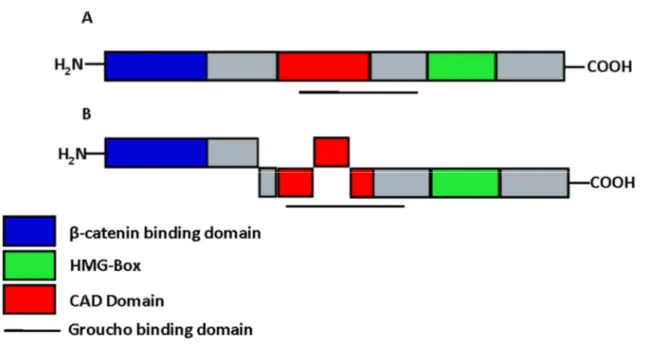
The first members of the LEF/TCF family to be identified were T cell factor 1 (TCF-1) (Oosterwegel et al. 1991; Vandewetering et al. 1991) and Lymphoid enhancer factor 1 (LEF- 1) (Travis et al. 1991; Carlsson et al. 1993). Schematic representations of the structure of LEF-1 isoforms have been illustrated in figure 4. Proteins of the LEF/TCF family share an 80- amino-acid high mobility group (HMG) box. It was shown that the HMG box can bind to DNA as a monomer in a sequence specific manner (Travis et al. 1991; Giese et al. 1991).
Other features of LEF-1 are the β-catenin binding domain (βBD), through which the interaction with β-catenin is achieved, and the context dependent activation domain (CAD) that can interact for example with the Ally of AML-1 and LEF-1 (ALY), an ubiquitously expressed nuclear protein that was shown to be necessary for the T cell receptor α (TCRα) enhancer function (Bruhn et al. 1997) The LEF/TCF family members are expressed in a great variety of tissues such as immature T and B cells of adult mice and in the neural crest, mesencephalon, tooth germs, whisker follicles, and other sites during embryogenesis. It was shown that LEF-1 has an architectural function and can interact with different proteins that result in either activation or repression of target genes. For the activating effect, the LEF/TCF
family members mostly interact with β-catenin to turn on Wnt target genes what makes them a member of the Wnt signaling pathway for this regulation. In a distinct number of cases, LEF-1 can also positively regulate target genes without the help of β-catenin, thus acting independently of the Wnt pathway, as shown for example in the regulation of TCRα by LEF-1 (Travis et al. 1991).
On the other hand LEF/TCF proteins can also actively repress transcription. This was first observed with experiments in Drosophila and Xenopus, showing that in the absence of a Wnt/Wg signal the repression of Ultrabithorax (Ubx) and Siamois is released by mutating the LEF/TCF consensus sites in their transcriptional control elements (Brannon et al. 1997; Riese et al. 1997; Bienz 1998). There are some co-repressors known to directly interact with LEF/TCF proteins that help to repress target genes. Groucho is once such protein that binds to a part of the CAD domain of LEF-1, thereby allowing its binding to β-catenin at the same time leading to a repressive effect in the context of a Wnt signal. Repression of E-cadherin
Figure 4: LEF-1 Isoforms (A) Schematic representation of LEF-1 splice variants and their most conserved domains. (B) Short forms of Lef-1 lack the N-terminal domain, which interacts with β-catenin. The CAD domain in Lef-1 is required for context-dependent activation of the TCR enhancer. The HMG box mediates sequence-specific DNA binding.
through LEF-1 and β-catenin interaction without the help of any co-repressors has also been shown (Jamora et al. 2003). Nevertheless it seems to be more likely, that the main mechanism for repression is mediated without the help of β-catenin. However the reports about the mechanism of repression through LEF-1 are contradictory and no main pathway was discovered yet. Thus, for the repressive effect of LEF/TCF proteins there are still a lot of questions to be answered.
3.3.4. The Role of Wnt signaling in B-cell development
LEF-1 is known to be expressed in transformed pre-B cell lines but rapidly down regulated in mature B cells (Travis et al. 1991). The other family members of the LEF/TCF family are not found to be expressed in any stage of the B cell development. Only little is known about the influence of Wnt signaling on B cell development. The first evidence that Wnt might play a critical role came from the finding, that some leukemic B cell lines overexpress a novel Wnt protein, Wnt 16 (McWhirter et al. 1999). The exact expression pattern of the Wnt proteins and LEF-1 in B cells and the role of Wnt signaling and LEF-1 in the development remained unclear. Recently the effects of LEF-1 on B cell development were subject to intensive analysis (Reya et al. 2000). First the precise pattern of LEF-1 in developing B cells was studied. It could be shown by lacZ reporter assay that LEF-1 is expressed during early B cell development in the fetal liver and adult bone marrow (Galceran et al. 2000). The upregulation occurs in fraction B pro-B cells and LEF-1 can also be detected in fraction C cells. There is no LEF-1 expression found in IgM-positive B cells from the adult spleen or adult bone marrow. To test for a correlation between the expression pattern and the function, fetal liver of Lef1-/- embryos and perinatal bone marrow was analyzed, as an analysis of older mice is not possible due to the early death of Lef1-/- mice. The number of B220+ cells was reduced by more than two fold and was even more obvious after excluding the dying and dead cells. To specify the stage of the cells, B220+ positive cells were tested for
other surface markers and it could be shown, that the majority of the B220 cells were also CD43+, placing them in the pro-B cell compartment. To test if LEF-1 deficiency results in a differentiation defect, they tested bone marrow of mice at postnatal day 13 (P13) for the ratio of IgM- to IgM+ B lymphocytes. Although the total number of the cells was reduced as shown before, the ratio remained still the same and there were no defects in rearrangement of the immunoglobulin heavy and light chains occurring. These findings were also confirmed with adoptive transfer experiments where the mutant B cells behaved like wildtype cells in a wildtype environment. On further analysis the reduced number of B220+ cells was found to be due to reduced cell survival. With TUNNEL assay and Annexin V staining it could be shown that indeed the B220+ cells of Lef-1-/- mice die up to a 20-fold higher frequency. As a cause for the reduced survival, Reya and coworkers analyzed the expression level of several genes known to be involved in apoptosis. The levels of Bcl-2, Bcl-x, and p53 remained unchanged in sorted pro-B cells (fraction B) of Lef1-/- mice compared to wildtype, whereas the expression of Fas and c-myc was elevated. A second defect that can contribute to the reduced size of the B cell compartment is the diminished proliferation of the B cells. With a thymidine incorporation assay it could be shown, that in addition to the increased apoptosis, the decreased proliferation of B cells signifies that LEF-1 has an important function for the proliferation of B cells. As LEF-1 and β-catenin together are members of the Wnt signaling pathway, they went on testing the responsiveness of B cells to Wnt stimuli. While Wnt10B, Wnt3A, and Wnt5A were found to be expressed in bone marrow, only Wnt5A was expressed in the stromal cells of the bone marrow, indicating that the other family members are produced by the hematopoietic cells themselves. It could be shown that proliferation of wildtype pro-B cells is increased after LiCl stimulation and that the soluble Wnt3A could stabilize β-catenin in the cells. Furthermore comparing the responsiveness of wildtype and Lef1-/- cells to Wnt3A stimulation revealed the LEF-1 dependence. Only a small proportion of cells deficient for LEF-1 started to proliferate after Wnt3A addition whereas the majority of
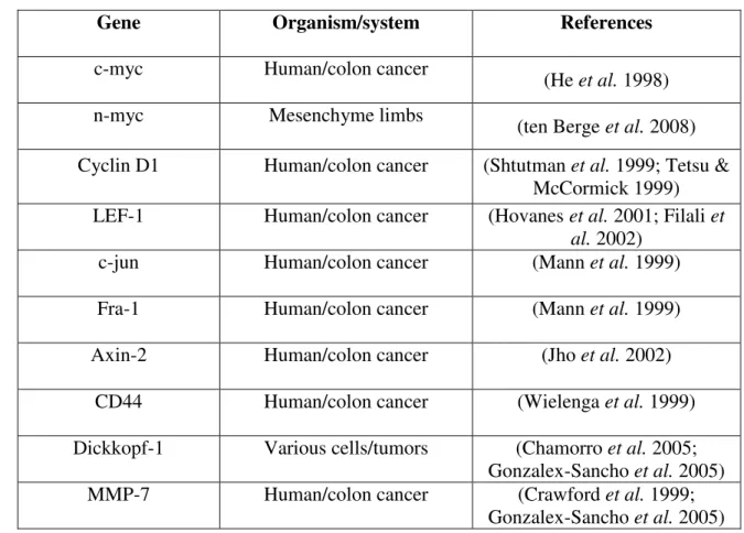
the wildtype cells were found to start dividing. These findings taken together strongly indicate an essential role of Wnt signaling and LEF-1 expression for B cell development. Table 1 shows the list of few validated target genes of TCF/LEF-1 based on the consensus binding sequence (5'-CCTTGAA-3’) in the promoter region of the gene.
Table 1 List of target genes of Wnt/β-catenin/TCF/LEF-1 signaling
Gene Organism/system References
c-myc Human/colon cancer (He et al. 1998)
n-myc Mesenchyme limbs (ten Berge et al. 2008)
Cyclin D1 Human/colon cancer (Shtutman et al. 1999; Tetsu &
McCormick 1999) LEF-1 Human/colon cancer (Hovanes et al. 2001; Filali et
al. 2002)
c-jun Human/colon cancer (Mann et al. 1999)
Fra-1 Human/colon cancer (Mann et al. 1999)
Axin-2 Human/colon cancer (Jho et al. 2002)
CD44 Human/colon cancer (Wielenga et al. 1999)
Dickkopf-1 Various cells/tumors (Chamorro et al. 2005;
Gonzalex-Sancho et al. 2005)
MMP-7 Human/colon cancer (Crawford et al. 1999;
Gonzalex-Sancho et al. 2005)
3.4. Wnt Signaling and Diseases
The Wnt signaling being an important pathway in the development of B cells and in the delicate balance between apoptosis, survival and proliferation of B cells clearly implies that deregulation of this pathway would lead to manifestation of pathological conditions. Besides considerable advances in investigating the mechanisms of Wnt signaling and their role in development, recent studies implicate Wnt signaling in cancer and other clinical conditions (Moon et al. 2004; Polakis 2000; Giles et al. 2003; Lustig & Behrens 2003).
3.4.1. Role of Wnt signaling in human cancers and its therapy
There have been numerous reports describing overexpression or under-expression of Wnt genes in cancer. A transgenic mouse model has established that tumor growth is dependent on Wnt-1 expression (Gunther et al. 2003). Studies of Wnt expression in human breast tumors have tended evidence to support Wnt signaling in development of breast cancer (Lane & Philip 1997; Huguet et al. 1994; Smalley & Dale 2001; Howe & Brown 2004). Cells expressing Wnt1 were resistant to cancer therapy mediated apoptosis. Wnt1 signaling inhibited cytochrome c release and the subsequent caspase-9 activation that was induced by chemotherapeutic drugs, including both vincristine and vinblastine. Further research showed that Wnt1 signaling inhibited apoptosis by activating β-catenin/Tcf mediated transcription (Chen et al. 2001). It was recently demonstrated that inhibition of Wnt2 mediated signaling induced apoptosis in both malignant melanoma cells and non-small-cell lung cancer cells (You et al. 2005; You et al. 2004). These studies show that activation of the β- catenin/TCF/LEF-1 signaling pathway by Wnt ligands not only provides a growth advantage to cancer cells, but also significantly affects the clinical outcome by inhibiting chemotherapy- induced apoptosis. Blocking Wnt signaling, for instance with a monoclonal antibody or development of small molecules can be useful to inhibit the Wnt/β-catenin signaling pathway for treatment of cancer patients and may improve the efficacy of chemotherapy by enhancing apoptosis in cancer cells.
3.4.2. Role of Wnt signaling in B Chronic lymphocytic leukemia (B-CLL)
As previously mentioned Wnt signaling is active during early B cell development in a LEF-1 dependent mechanism (Reya et al. 2000). However some studies exist that indicate factors governing aberrant activation of Wnt signaling in mature B-CLL cells. A comprehensive gene expression profiling from two independent studies revealed that LEF-1 mRNA was ~3000 fold overexpressed in B-CLL when compared to its healthy counterpart
(Gutierrez et al. 2007; Howe & Bromidge 2006). Later series of studies demonstrated autocrine activation of Wnt signaling in different clinical subsets of CLL. Based on a cDNA microarray study, Wnt-3 was found to be uniformly upregulated in B-CLL (Rosenwald et al.
2001). Lu and co workers demonstrated that B-CLL cells are in an autocrine Wnt action where several Wnts and frizzled (Wnt-3, Wnt-5b, Wnt-6, Wnt-14, Wnt-16, Fzd-3) mRNA were found to be elevated in CLL when compared to healthy B cells. The induction of β- catenin by inhibiting GSK3-β also demonstrated an increased survival of B-CLL cells in vitro, whereas inhibiting Wnt signaling by R-etodolac led to apoptosis in CLL cells indicating that this pathway plays a crucial role in survival of CLL cells. (Lu et al. 2004).
There are also evidences for epigenetic alterations of modulators of Wnt signaling at the genomic level. Secreted frizzled-related proteins (SFRP-1, SFRP-2 SFRP-4 and SFRP-5) was found to be frequently methylated in CLL (Liu et al. 2006; Chim et al. 2008). SFRPs are physiological inhibitors of Wnt pathway, which compete with the Fzd receptors for Wnt binding through the CRD domain or by binding directly to Fzd, resulting in the formation of inactive complexes with the receptor. The Wnt inhibitory factor-1 (Wif-1) which can bind to different Wnts and render them inactive is infrequently methylated in CLL (Chim et al. 2006).
Taken together, there is an autocrine Wnt secretion in CLL cells, with overexpression of LEF-1 and concordant methylation of physiological inhibitors of Wnt pathway. As a result of such activation, LEF-1 target genes such as c-myc cyclin D1 are overexpressed in CLL cells providing extended survival of CLL cells (Faderl et al. 2002; Nagy et al. 2003; Plate et al. 2000).
3.5. Clinical perspective of B-Chronic lymphocytic leukemia (B-CLL)
Given the potential therapeutic value of the Wnt signaling pathway in the treatment of B-CLL, it is critical to understand the clinical perspective of the disease with regard to the epidemiology, symptoms, diagnosis, disease staging, and prognosis in order to develop the most effective therapeutic strategies.
3.5.1. Epidemiology
CLL is the most common type of leukemia in the western countries representing 22- 30% of all leukemia’s with an incidence rate of between 1 and 5.5 per 100,000 people and is considered incurable with currently available therapy (Redaelli et al. 2004; Zent et al. 2001).
Geographically it is most prevalent in Australia, USA, Italy, Switzerland and Ireland, while low incidence is seen among Asian countries and US asian origin and is slightly more common in whites than in blacks, which emphasizes the racial factors in the occurrence of this disease (Dores et al. 2007; Groves et al. 1995; Morton et al. 2006). It mostly affects the elderly (>50yrs of age) however an increase in the incidence among the younger individuals has been reported (de Lima et al. 1998; Derossi et al. 1989; Mauro et al. 1999). CLL is more common in men than women with a sex ratio of about 2:1 (Cartwright et al. 2002).
3.5.2. Clinical manifestations
CLL is initially asymptomatic and often diagnosed during routine examination. Despite its lack of visible manifestation in terms of symptoms it affects the immune system leading to the complications like infections and secondary malignancies (Anaissie et al. 1998). Apart from infection patients can present features like painless lymphoadenopathy (enlargement of lymph nodes), bone marrow infiltration with leukemia cells causing anemia (low RBC levels) and thrombocytopenia (low platelets levels) (Mauro et al. 1999). Autoimmune hematological conditions are frequently associated with CLL, of which autoimmune haemolytic anemia (AHA), idiopathic thrombocytopenic purpura and red cell aplasia are more common (Diehl &
Ketchum 1998; Mauro et al. 2000). Hypogammaglobulinemia (low gamma globulin levels) is found in approximately 50% of CLL patients (Dighiero G. 1988).
3.5.3. Diagnostic criteria
The current diagnosis of CLL is based on the revised guidelines from the national cancer institute working group (NCI-WG) (Cheson et al. 1996; Hallek et al. 2008).
Accordingly CLL can be diagnosed when the following conditions exist:
1) A persistent (>3 month) peripheral blood lymphocyte count > 5 x 10⁹ cells/L of mature lymphocytes in the absence of other causes
2) Distinct immunophenotype of CLL
a) Predominant expression of B cell markets like CD19, CD20, CD23 along with T cell marker CD5 antigen, in the absence of other T cell markers
b) Light chain restriction and FMC7 negative
c) Surface immunoglobulin (sIg) of low density and absence or low expression of CD79b
Several hematological disorders like mantel cell lymphoma (MCL), splenic marginal zone lymphoma (SMZL), B cell prolymphocytic leukemia (PLL), Hairy cell leukemia (HCL) and Waldenstrom’s macroglobulinemia resemble CLL in their clinical presentation and microscopic appearance (Jaffe 2001). Therefore a differential diagnosis is necessary to distinguish CLL from other disorders. MCL differs from CLL by CD23 negativity and presence of t (11; 14) translocation (Sanchez et al. 2002). In addition, PLL and HCL are morphologically and immunophentoypically distinct from CLL. Clinically PLL and HCL have prominent splenomegaly without lymphoadenopathy. The immunophenotypic features of PLL and HCL differ from CLL, since they are negative to CD5 and CD23, while positive for FMC7 (Melo 1986; Sanchez et al. 2002). SMZL cells are morphologically distinct from CLL lymphocytes, with a plasmacytoid basophilic cytoplasm (Catovsky & Matutes 1999).
Waldenstrom’s macroglobulinemia is distinct from CLL, with exception of hyper viscosity syndrome associated with as a result of elevated immunoglobulin M (Chng et al. 2006).
3.5.4. Disease staging
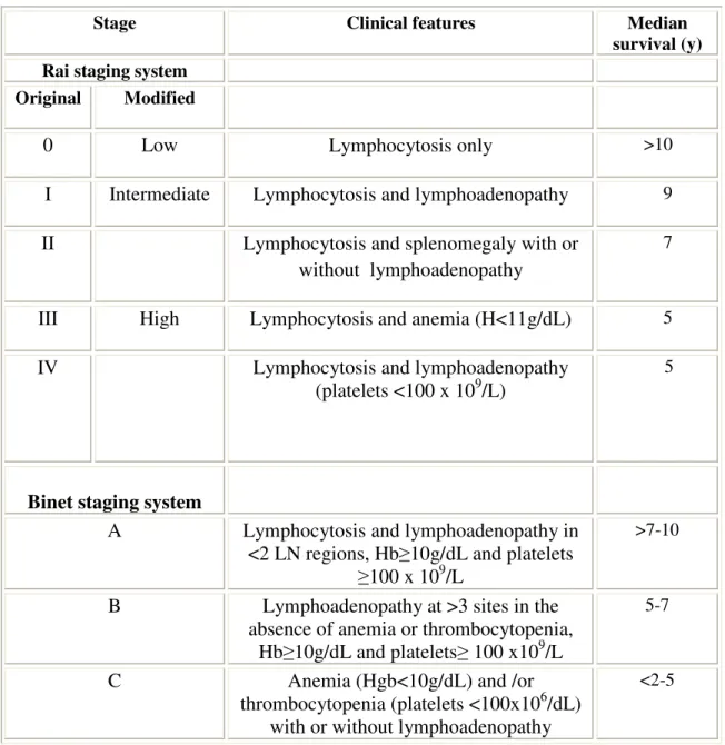
Clinical staging is important in CLL to predict prognosis, which aids in comparison of clinical findings and establishment of therapeutic guidelines. The Rai and Binet staging systems (Table 2 ) are two traditional clinical staging systems which are currently in practice (Binet et al. 2006; Rai et al. 1975).
Table 2: Staging Systems for Chronic Lymphocytic Leukemia
Stage Clinical features Median
survival (y) Rai staging system
Original Modified
0 Low Lymphocytosis only >10
I Intermediate Lymphocytosis and lymphoadenopathy 9
II Lymphocytosis and splenomegaly with or
without lymphoadenopathy
7
III High Lymphocytosis and anemia (H<11g/dL) 5
IV Lymphocytosis and lymphoadenopathy
(platelets <100 x 109/L)
5
Binet staging system
A Lymphocytosis and lymphoadenopathy in
<2 LN regions, Hb≥10g/dL and platelets
≥100 x 109/L
>7-10
B Lymphoadenopathy at >3 sites in the absence of anemia or thrombocytopenia,
Hb≥10g/dL and platelets≥ 100 x109/L
5-7
C Anemia (Hgb<10g/dL) and /or
thrombocytopenia (platelets <100x106/dL) with or without lymphoadenopathy
<2-5
3.5.5. Prognostic factors
With the development of technology and growing insight into the biology of the disease, new prognostic markers became available such as cytogenetics, the mutational status of the immunoglobulin heavy chain variable genes (IgVH), zeta associated protein kinase 70 kDa (ZAP-70) and CD38. These new prognostic markers allow stratifying patients into risk categories at the moment of the initial diagnosis. However further prospective trials are necessary to verify and establish their usefulness in the management of the patients with CLL (Binet et al. 1981).
3.5.5.1. Cytogenetic abnormalities
There is no single characteristic cytogenetic abnormality in CLL (Montserrat &
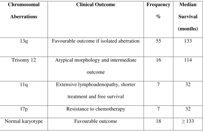
Rozman 1995). With the development of interphase fluorescence in situ hybridization (FISH) techniques, it has become possible to detect numerical and structural chromosomal abnormalities in non dividing cells. Using FISH probes cytogenetic lesions were found in more than 80% of examined CLL cases, which is more than those detected in conventional metaphase cytogenetics and indicates prognostic applicability (Dohner et al. 2000; Krober et al. 2002). The most common cytogenetic alterations include deletion of 13q14.1, deletion of 11q22.3-23.1, trisomy 12 and deletion of 17p13 (Anastasi & Le Beau 1992; Stilgenbauer et al. 1998). Table 3 shows the correlation of specific chromosome aberration with clinical characteristics and outcome in CLL patients (Montillo et al. 2005). A recent study analysed survival of CLL patients in relation to chromosomal abnormalities using FISH. There is increasing evidence that chromosomal abnormalities were associated with prognosis in retrospective and prospective clinical trials (Catovsky 1989; Shanafelt et al. 2006). In a prospective study 13q deletion was correlated with a favourable prognosis and 17p deletion is associated with most unfavourable prognosis among the occurring abnormalities (Grever et al. 2007; Shanafelt et al. 2006). Chromosome 17 abnormalities have been associated with p53
employing alkylating drugs and purine analogues (Byrd et al. 2006). For all these reasons during clinical trials, it is recommended to perform cytogenetics prior to treating a patient (Hallek et al. 2008).
Table 3: Cytogenetics in CLL (Modified from Montillo et al., 2005) Chromosomal
Aberrations
Clinical Outcome Frequency
%
Median Survival (months) 13q Favourable outcome if isolated aberration 55 133
Trisomy 12 Atypical morphology and intermediate outcome
16 114
11q Extensive lymphoadenopathy, shorter treatment and free survival
7 32
17p Resistance to chemotherapy 7 32
Normal karyotype Favourable outcome 18 ≥ 133
3.5.5.2. IgVH mutational status
During recent years, the somatic hypermutation status of the immunoglobulin variable heavy chain (IgVH) genes has been vividly investigated and found to be an important prognostic marker in CLL (Damle et al. 1999; Hamblin et al. 1999). A rearranged clonal IgVH sequences that differ from their germline counter parts by 2% are defined as ‘mutated’
(Dighiero G. 1988; Hashimoto et al. 1995; Schroeder & Dighiero 1994). Based on this subjective threshold value, two subgroups of CLL with clear difference in survival have been
identified as ‘mutated’ CLL, where leukemic cells have rearranged VH genes with 2% or more mutations and ‘unmutated’ CLL with few or no mutations (Fais et al. 1998; Stevenson
& Caligaris-Cappio 2004). Mutational status has been strongly correlated with prognosis of CLL. Patients with unmutated gene sequences had aggressive disease, shorter survival and poorer prognosis than those with mutated genes (Krober et al. 2002; Oscier et al. 2002). Also there are reports indicating that patients with unmutated IgVH genes had a higher risk of relapse after stem cell transplantation (Ritgen et al. 2003). However, there is an exception, where the usage of IgVH gene V3-21 expression has an inferior outcome independent of the mutational status (Thorselius et al. 2006; Tobin et al. 2002).
3.5.5.3. Zeta-associated protein (ZAP-70)
Zeta-associated protein (ZAP-70) is an intracellular tyrosine kinase that is predominantly expressed in T and natural killer cells and functions in transmitting activation signals involved in T-cell receptor signaling and T cell activation (Chan et al. 1992).
Microarray studies reveal that CLL cells share a characteristic gene-expression profile and found that a small number of genes (ZAP-70 and C-type lectin) correlated with the mutational status of IgVH genes (Klein et al. 2001; Rosenwald et al. 2001). Several studies using western blotting, RT-PCR and flow cytometry have confirmed the ability to distinguish between IgVH mutated (ZAP-70- ) and unmutated (ZAP-70+) (Crespo et al. 2003; Orchard et al. 2004; Rassenti et al. 2004). Many groups have validated a threshold of 20% by flow cytometry to separate ZAP-70- from ZAP-70+ cases (Crespo et al. 2003). As IgVH sequencing is laborious and costly, ZAP-70 expression is recommended as surrogate marker for IgVH gene mutational status in CLL (Del Principe et al. 2006; Wiestner et al. 2003).
3.5.5.4. CD38 expression
CD38 is a cell surface molecule that regulates cell activation, proliferation and adhesion and present in various lineages of hematopoietic cells, including B-cells (Cruse et al.
2007). Recently it has been shown to promote proliferation and to prolong survival of CLL cells (Deaglio et al. 2006). CD38 was initially suggested as a surrogate marker for IgVH mutation status in CLL (Damle et al. 1999; Ibrahim et al. 2001). High CD38 expression correlates with unmutated IgVH and is associated with significantly shorter overall survival, progression-free survival times and poor response to chemotherapy (Del Poeta et al. 2001;
Ghia et al. 2003; Jelinek et al. 2001). However other studies found CD38 expression gives discordant results with respect to IgVH mutation status (Hamblin et al. 2000; Krober et al.
2002). This discrepancy is due to the lack of a defined cut-off value for CD38 expression (Boonstra et al. 2006; Krober et al. 2002) and also change in the expression of CD38 with time, especially after initiation of therapy (Hamblin et al. 2002).
3.6. Current Therapeutic strategies
Remarkable progress in elucidating the biology of CLL has been made over the last two decades. Improved understanding of CLL has lead to new prognostic tools and therapeutic options, and holds promise for eventually finding a cure for this disease.
Challenges lie in incorporating the various treatment modalities, including chemotherapy, monoclonal antibodies, immunotherapeutic strategies and novel small molecules, into a comprehensive treatment strategy guided by the biological complexity of CLL.
3.6.1. Criteria for patient treatment
The disease course for CLL is highly variable and the criterion for initiating the therapy is an important consideration in the clinical management of CLL. The decision to treat is guided by the stage of the disease, the presence of symptoms and disease progression (Eichhorst & Hallek 2007). According to the guidelines for diagnosis and treatment (Hallek et al. 2008) set at the recent International Workshop on Chronic Lymphocytic Leukemia (IWCLL) indicate that in routine clinical practice, newly diagnosed patients with asymptomatic early stage disease should be kept under ‘wait and watch’ unless there is evidence of disease progression. Several research groups confirm that the treatment of early-
stage disease with alkylating agents does not prolong the survival (Dighiero et al. 1998).
However symptomatic and progressive disease patients (Rai III and IV or Binet B or C) should be immediately treated to benefit initiation of treatment (Molica et al. 1991; Robak et al. 2006a). Improvement in therapeutic regiments and advances in the understanding the biology of the disease have made it possible to achieve higher percentage of remissions, but never a complete remission of the disease. A complete cure of this disease is still a far-to- reach goal of modern medicine (Ghia et al. 2007).
3.6.2. Alkylating agents as single agents and in combination therapy
For many years alkylating agents like chlorambucil have been the gold standard for the first line therapy in CLL, given daily or intermittently, alone or in association with corticosteroids. Chlorambucil produced overall remission rate of 40-80% in previously untreated patients and all the patients showed a relapse (Binet 1990; Rai et al. 2000; Keller et al. 1986). Despite higher response rates chlorambucil does not prolong survival and use of corticosteroids does not provide any benefit in the treatment (Catovsky et al. 1991; Dighiero et al. 1998; Jaksic et al. 1997). Another alkylating agent, cyclophosphamide is used when chlorambucil is poorly tolerated. A higher response rate was achieved when this drug used in combination therapies with anthracyclines. The best combination therapies were CHOP (cyclophosphamide, adriamycin, vincristine and prednisone), CAP (cyclophosphamide, doxorubicin and prednisone) and COP (cyclophosphamide, vincristine and prednisone) (Keating et al. 1988; Jaksic et al. 1997; Binet 1990). In most randomized studies and meta analyses of randomized trials of these combination chemotherapy regiments showed a higher response rates, but none showed a benefit in terms of survival (Binet 1990; Raphael et al.
1991).
3.6.3. Purine analogs as single agents and in combination therapy
Purine analogs have become the gold standard for first line treatment in CLL replacing monotherapy with alkylating agents (Keating et al. 1991). Three purine analogues are currently used in CLL: fludarabine, pentostatin and cladribine. Fludarabine is most extensively studied and used for therapy in the west and in Europe. Fludarabine monotherapy induces more complete remissions when compared with other treatment regiments containing alkylating agents or corticosteroids (Keating et al. 1991; Rai et al. 2000; Johnson et al. 1996;
Keating et al. 1998; Leporrier et al. 2001). Single agent fludarabine has superior response rates in previously treated patients (Hallek et al. 2001) and also in elderly patients with progressive disease (Eichhorst & Hallek 2007). Randomized studies of cladribine show similar overall and complete remission as with fludarabine as single agent or in combination in CLL (Robak 2002; Montillo et al. 2003).
3.6.4. Chemoimmunotherapy
Monoclonal antibodies and immunotoxins are emerging as attractive agents and have been investigated in clinical trials in patients with CLL (Del Poeta et al. 2008; Robak et al.
2006b; Tam & Keating 2007). Rituximab (Rituxan, Mabthera) is a chimeric human mouse monoclonal antibody that targets CD20 antigen. CD20 antigen is expressed in all B-cell phases except stem cells and plasma cells. Rituximab’s anti-leukemia action includes elimination of B cells includes complement dependent lysis, antibody dependent cell mediated cytotoxicity (ADCC) and direct induction of apoptosis (Voso et al. 2002). Single agent rituximab has limited efficacy at standard FDA approved doses and most studies suggested a higher dose of rituximab in CLL when compared to other lymphomas (Voso et al.
2002; Byrd et al. 2001; Itala et al. 2002; O'Brien et al. 2001). The combination of rituximab with fludarabine (Byrd et al. 2003; Schulz et al. 2002) or fludarabine and cyclophosphamide (Keating et al. 2004) increased the overall and complete remission rates in previously
untreated patients and in the relapsed CLL patients (Wierda et al. 2005). Alemtuzumab (Campath) is a recombinant, fully humanized, monoclonal antibody against CD52 antigen.
Several reports have confirmed significant overall response rate and survival activity alemtuzumab in relapsed or refractory CLL (Keating et al. 2002; Osterborg et al. 2002; Rai et al. 2002). In addition, alemtuzumab showed clinical response in patients with poor prognostic factors, including high-risk genetic markers such as deletions of chromosome 11 or 17 and p53 mutations (Lozanski et al. 2004; Stilgenbauer et al. 2001).
3.6.5. Allogeneic transplant
A recent consensus paper by EMBT (European Group for Bone Marrow Transplantation) provided standard indications for allogeneic transplant in CLL. The authors contend that allogeneic HCT (Hematopoietic cell transplantation) has proven efficacy in poor- risk CLL, for younger patients with 1) non response or early relapse with 12 months after purine analog treatment. 2) Relapse within 24 months after achieving CR (Complete Remission) with a purine analog based regimen. Or 3) patients with 17p- abnormalities with dismal responses with conventional chemotherapy regimens (Dreger et al. 2007). Several nonrandomized trials have evaluated the efficacy of allogeneic stem cell transplantation in CLL. However, most trials were limited by size, absence of long term follow up and high treatment related mortality (Jabbour et al. 2004). A recent Spanish study reported 30 patients with poor-prognosis CLL and or high-risk molecular/cytogenetic characteristics, treated with reduced intensity conditioning (RIC) allogeneic transplant indicate that this therapy may overcome adverse prognostic factors even in patients with 17p-, and provide patients with long term remission suggesting potential cure.
3.6.7. Other Targeted therapies
Apart from immunotherapy, few targeted therapies are being tested in clinical trials for CLL. Oblimersen, an antisense phosphothioate oligonucleotide targets the messenger RNA of