The impact of intragenic CpG content on epigenetic control of transgene expression
in mammalian cells
DISSERTATION ZUR ERLANGUNG DES DOKTORGRADES DER NATURWISSENSCHAFTEN (DR. RER. NAT.)
DER FAKULTÄT FÜR BIOLOGIE UND VORKLINISCHE MEDIZIN DER UNIVERSITÄT REGENSBURG
vorgelegt von
Simone Krinner
aus Straubing im Jahr 2012
Das Promotionsgesuch wurde eingereicht am 6. November 2012 Die Arbeit wurde angeleitet von Prof. Dr. Ralf Wagner
____________________________
Simone Krinner
Prüfungsausschuss:
Vorsitz:
Erstgutachter:
Zweitgutachter:
Drittprüfer:
Prof. Dr. Stephan Schneuwly Prof. Dr. Ralf Wagner Prof. Dr. Christopher Baum Prof. Dr. Gernot Längst
1 ZUSAMMENFASSUNG ... 1
2 ABSTRACT... 3
3 INTRODUCTION ... 5
Eukaryotic gene transcription ... 5
3.1 Chromatin ... 7
3.2 3.2.1 The nucleosome ... 7
3.2.2 Chromatin organization ... 8
Transcriptional control by chromatin ... 10
3.3 3.3.1 Histone modifications ... 10
3.3.2 Chromatin remodeling ... 13
3.3.3 Sequence dependent nucleosome positioning ... 14
Cytosine Guanine Dinucleotides ... 15
3.4 3.4.1 CpG methylation ... 15
3.4.2 Unmethylated CpG dinucleotides... 16
3.4.3 Gene control mechanisms directed by CpG dinucleotides ... 17
Transgene expression... 19
3.5 3.5.1 Viral vector-based transgene expression ... 20
3.5.2 Plasmid-based transgene expression ... 21
Overview of preceding CpG studies ... 23
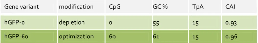
3.6 3.6.1 The model genes hgfp and mmip-1α ... 23
3.6.2 Impact of intragenic CpG content of hgfp and mmip-1α on gene expression ... 25
Aim of the study ...27
3.7 4 RESULTS ... 28
CpG-dependent differential transgene expression using mammalian Flp-In cells ... 28
4.1 4.1.1 Long-term hGFP expression in the presence or absence of selection pressure ... 30
4.1.2 Sorting of CHO Flp-In cells according to hGFP expression levels ... 33
4.1.3 Relative copy number and methylation status of hgfp in correlation to expression levels... 34
4.1.4 Impact of intragenic CpG dinucleotides on chromatin structure ...40
4.1.5 Influence of intragenic CpG dinucleotides on RNAPII occupation ... 47
4.1.6 Impact of intragenic CpG distribution on gene expression in hgfp ... 50
CpG-dependent differential transgene expression in murine embryonic carcinoma cells P19 ... 52
4.2 4.2.1 Generation of SIN-LVs incorporating hgfp variants ... 53
4.2.2 Long-term expression of hGFP variants in P19 cells using different promoters ... 54
4.2.3 Partial prevention of hgfp silencing in P19 cells by DNMT inhibition ... 58
5 DISCUSSION ... 60
Evolution of CpG frequency in the mammalian genome ... 60
5.1 CpG dinucleotide usage is pivotal for transgene expression ... 61
5.2 Intragenic CpG abundance determines expression levels of hGFP and mMIP-1α ... 61
5.3 Intragenic CpG dinucleotides confer no disadvantage for long-term expression 5.4 in mammalian Flp-In cells ... 63
Intragenic CpG dinucleotides cause increased DNA methylation rates, whereas low CpG content 5.5 promotes transgene loss ... 64
Intragenic CpG dinucleotides alter chromatin structure ... 65
5.6 5.6.1 Chromatin density of hgfp transgenes is affected by intragenic CpG dinucleotides . and growth conditions in vivo ... 65
5.6.2 Chromatin density of mmip-1α transgenes is increased upon CpG depletion in vivo... 66
5.6.3 CpG dinucleotides in hgfp affect nucleosome positioning abilities in vitro ... 67
Intragenic CpG dinucleotides increase transcription elongation of mmip-1α ... 68
5.7 Gene expression benefits from TSS-proximity of intragenic CpG dinucleotides ... 68
5.8 CpG frequency and type of promoter determines transgene stability in pluripotent stem cells P19 ... 69
5.9 5.9.1 CMV- and EF-1α-promoter-mediated hGFP expression is gradually silenced in P19 cells ... 70
5.9.2 A2UCOE confers stable hGFP expression in P19 cells and prevents hGFP repression upon intragenic CpG depletion ... 71
5.9.3 DNMT inhibition partly prevents hgfp silencing in P19 cells depending on promoter usage ... 72
Proposed CpG-mediated transcriptional control mechanism and outlook ... 73
5.10 6 MATERIALS... 75
Cell lines ... 75
6.1 Bacterial strains ... 75
6.2 Media and supplements ... 75
6.3 Kits ... 76
6.4 Buffers and reagents... 76
6.5 Plasmids ... 79
6.6 Oligonucleotides ... 79
6.7 Chemicals, enzymes and materials ... 80
6.8 7 METHODS... 81
Cultivation of eukaryotic cells ... 81
7.1 7.1.1 Maintenance of cell lines ... 81
7.1.2 Transient transfections ... 81
7.1.3 Establishment of plasmid-based stable cell lines ... 81
7.1.4 Lentiviral vector (LV) preparation and transduction of cell lines ... 82
Cultivation of prokaryotic cells ... 82
7.2 DNA methods ... 82
7.3 7.3.1 Isolation of genomic DNA ... 82
7.3.2 DNA quantification ... 83
7.3.3 Agarose gel electrophoresis ... 83
7.3.4 DNA purification from agarose gels ... 83
7.3.5 In vitro methylation ... 83
7.3.6 Bisulfite conversion and sequence analysis... 83
Polymerase chain reaction (PCR) ... 84
7.4 7.4.1 Quantitative PCR/real-time PCR ...84
7.4.2 DNA sequencing ... 85
Plasmid construction ... 85
7.5 7.5.1 Ligation ... 85
7.5.2 Transformation of E.coli ... 85
7.5.3 Preparation of plasmid DNA ... 85
7.5.4 Cloning of hgfp chimera ... 86
7.5.5 Cloning of lentiviral transgene vectors ... 86
Protein methods ... 86
7.6 7.6.1 Determination of protein amount according to Bradford ... 86
7.6.2 Enzyme linked Immunosorbent Assay (ELISA) ... 86
7.6.3 Flow cytometry ... 87
Formaldehyde-assisted isolation of regulatory elements (FAIRE) ... 87
7.7 Chromatin Immunoprecipitation (ChIP) ... 88
7.8 Analysis of reconstituted mononucleosomes in vitro ... 89
7.9 7.9.1 Amplification of CpG fragments for nucleosome reconstitutions ... 89
7.9.2 Nucleosome assembly by salt dialysis ... 89
7.9.3 Analysis of mononucleosomes by Native PAGE ... 90
8 REFERENCE LIST ... 91
9 APPENDIX ... 110
List of abbreviations ... 110
9.1 Sequences ... 115
9.2 9.2.1 Murine MIP-1α variants ... 115
9.2.2 Humanized GFP variants ... 116
10 DANKSAGUNG ... 117
Page | 1
1
Die effiziente Produktion rekombinanter Therapeutika in Säugerzellen und Verbesserung gentherapeutischer Verfahren sind bedeutende und expandierende Felder in der medizinischen und pharmazeutischen Forschung. Plasmid-DNA (pDNA)-basierte Vektorsysteme stellen aufgrund ihrer Stabilität, der kostengünstigen Produktion sowie ihres hervorragenden Sicherheitsprofils ein innovatives Gentransfer-System dar. Trotz dieser Vorteile ist der Einsatz von pDNA-Vektoren angesichts begrenzter Transgen-Expressionsraten gegenüber Virus-basierten Verfahren limitiert. Dies erfordert neue Strategien zur Optimierung von pDNA-basierten Genexpressionssystemen, wie beispielsweise durch die gezielte Nutzung transkriptionsregulierender Mechanismen der Zielzelle. CpG Dinukleotide in Transgenen haben sich diesbezüglich als entscheidende Expressions-modulierende Elemente erwiesen.
Anhand der Reportergene codierend für das murine Makrophagen inflammatorische Protein 1 alpha (MIP-1α) und das humanisierte grün fluoreszierende Protein (GFP) konnte bereits in früheren Studien ein proportionaler Zusammenhang zwischen CpG Dinukleotiden im offenen Leserahmen und einem erhöhten Genexpressionslevel gezeigt werden. Dazu wurden die Nukleinsäure-Sequenzen der mip-1α und gfp Gene unter Verwendung alternativer Codons modifiziert. Ausgehend vom mip-1α Wildtyp wurde ein Codon-optimiertes Gen, sowie eine CpG-freie und eine CpG-maximierte Genvariante hergestellt. Weiterhin dienten das für humane Zellen Codon-optimierte gfp Gen und darauf basierend ein CpG-freies gfp Gen als Ausgangskonstrukte für Genexpressionsanalysen. Es konnte gezeigt werden, dass intragenische CpG Dinukleotide einen positiven Einfluss auf die Genexpression in Säugerzellen ausüben, während eine CpG-Depletion zu starken Expressionsverlusten führt. Während keine Hinweise auf veränderte CpG-basierte posttranskriptionelle Regulations- mechanismen zu finden waren, konnte eine deutliche Korrelation zwischen intragenischen CpG Dinukleotiden und gesteigerter de novo synthetisierter mRNA hergestellt werden.
In der vorliegenden Arbeit sollten die durch differenziellen intragenischen CpG-Gehalt hervorgerufenen Regulationsmechanismen von gfp und mip-1α aufgeklärt werden. Das relative Expressionsprofil der CpG-modifizierten gfp Transgene in CHO Flp-In Zellen konnte über den Zeitraum von mindestens einem Jahr durch antibiotischen Selektionsdruck konstant gehalten werden. Die Abwesenheit selektiver Bedingungen resultierte dagegen in sukzessiven Expressionseinbußen, welche sowohl auf Transgenverluste als auch DNA-Methylierung zurückzuführen waren. Während eine hohe intragenische CpG-Frequenz zu gesteigerten Methylierungsraten des Transgen- kontrollierenden Promoters führte, hatte eine intragenische CpG-Depletion einen beschleunigten Transgenverlust zur Folge. Der Genexpressions-Rückgang nach Selektionsrestriktion korrelierte weiterhin bei allen gfp Varianten mit einer höheren Chromatin-Dichte. Interessanterweise ging auch die CpG-Depletion der in Flp-In CHO und HEK 293 stabil und unter Selektionsdruck integrierten gfp und mip-1α
Page | 2
Transgenvarianten mit einer Chromatin-Verdichtung einher. Darüber hinaus bewirkte der variable CpG-Gehalt in gfp eine veränderte in vitro-Positionierung von Nukleosomen. Die Detektion vermehrt aktiv transkribierender RNA Polymerasen II am Gen-Ende CpG-maximierter mip-1α Transgene in stabil transfizierten HEK 293 Flp-In Zellen ließ auf erhöhte Elongationsraten als Folge von CpG-Maximierung schließen.
Expressionsanalysen von gfp Chimären konnten zeigen, dass sich nicht nur die CpG-Frequenz, sondern vielmehr die räumliche Nähe intragenischer CpG Dinukleotide zum Transkriptionsstart (TSS) positiv auf die Expressionseffizienz auswirken.
Um die Effekte intragenischer CpG Dinukleotide auf die Transgenexpression in einem Gentherapie-relevanten Zellsystem zu testen, wurden murine, embryonale pluripotente Stammzellen der Linie P19 mittels lentiviraler Vektoren stabil mit den gfp CpG-Varianten unter verschiedenen Promotoren transduziert. Der Promotor des Cytomegalovirus (CMV) wies in diesem Expressionssystem eine erhöhte Disposition bezüglich gene silencing auf. Im Vergleich zum CMV Promotor führte der Promotor des humanen Elongationsfaktors 1 alpha (EF-1α) zu verzögerten, dennoch deutlichen, Expressionsverlusten. Im Gegensatz dazu verhinderte der bidirektionale, divergent transkribierte Promoter A2UCOE aufgrund seiner ubiquitären Chromatin-öffnenden Eigenschaften eine Transgen-Stilllegung komplett. In Bezug auf den intragenischen CpG-Gehalt konnte auch dieses Expressionssystem trotz hohem gene silencing-Potentials unter bestimmten Bedingungen von der Anwesenheit intragenischer CpG Dinukleotide profitieren. So wies das CpG-angereicherte gfp, exprimiert durch den EF-1α Promotor, auch in P19 Zellen eine deutlich erhöhte Expressionseffizienz auf.
Weiterhin konnte die Gen-Stilllegung des CMV Promotor-kontrollierten gfp durch intragenische CpG Dinukleotide leicht verzögert werden. Die durch den A2UCOE Promotor vermittelte Transkription hingegen wurde durch intragenische CpG Dinukleotide in gfp nicht beeinflusst. Es wird vermutet, dass die Chromatin-öffnende Funktion des A2UCOE Elements eine Chromatin-Kompaktierung als Folge der CpG-Depletion verhindern kann. Mit dieser Eigenschaft scheint A2UCOE die Nachteile der CpG-Depletierung durch Chromatin Verdichtung aufheben zu können.
Insgesamt konnten die anhand der Transgene gfp und mip-1α gewonnenen Daten zeigen, dass sich intragenische CpG Dinukleotide in TSS-Nähe positiv auf die Transkriptionseffizienz auswirken. Die durchgeführten Analysen deuten darauf hin dass dieser Effekt auf die Delokalisierung und Destabilisierung des +1 Nukleosoms durch TSS-proximale intragenische CpG Dinukleotide zurück geht, während eine intragenische CpG-Depletion eine Chromatin-Kondensation zur Folge hat. Diese Veränderungen der Chromatinstruktur werden als Ergebnis epigenetischer Regulationsmechanismen postuliert, die durch die An-, beziehungsweise Abwesenheit intragenischer CpG Dinukleotide hervorgerufen werden. Die genauen Mechanismen dieses Phänomens sind weiterhin nicht vollständig geklärt.
Page | 3
2
The improvement of gene therapy applications and efficient production of recombinant therapeutics in mammalian cells is a growing field of interest in medical and pharmaceutical research. Plasmid-DNA (pDNA)-based vector systems offer an innovative gene transfer strategy due to their high stability, cost efficient production and their excellent safety profile. Despite these advantages, the application of pDNA-vectors is limited compared to viral-vector-based gene transfer regarding transgene expression rates. This requires new strategies to optimize pDNA-based gene expression systems.
The directed utilization of transcription regulating mechanisms in the target cell is a major strategy towards this aim. In this regard, CpG dinucleotides in transgenes have proven to serve as crucial expression-modulating elements.
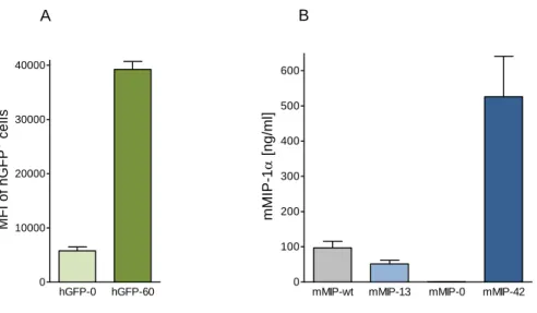
Previous studies have demonstrated a strong correlation between the presence of CpG dinucleotides in transgenes and the level of gene expression by means of the reporter genes coding for the murine macrophage inflammatory protein 1 alpha (MIP-1α) and humanized green fluorescent protein (GFP). The DNA sequence of mip-1α and gfp was modified by using alternative codons. Based on the mip-1α wild type sequence, a codon optimized, CpG-depleted and CpG-enriched mip-1α gene variant were generated.
Additionally, the CpG-rich gfp, optimized for human codon usage, and the CpG-depleted gfp, provided the basis for gene expression analyses. Decreased gene expression was observed as a result of intragenic CpG depletion, whereas the enrichment of intragenic CpG dinucleotides led to a dramatic increase of gene expression. No evidence for CpG-based posttranscriptional regulation mechanisms could be found.
Instead, intragenic CpG dinucleotides clearly correlated with enhanced de novo synthesized mRNA.
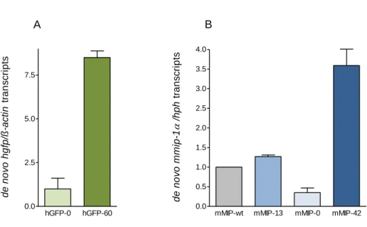
This study aimed to shed light on the CpG-induced mechanisms responsible for expression efficiency variations in gfp and mip-1α. The relative expression profile of CpG-modified gfp transgenes in CHO Flp-In cells could be maintained over at least a year under antibiotic selection pressure. Withdrawal of selective conditions resulted in gradual decrease in gfp expression which was shown to be a consequence of both transgene loss and DNA methylation. While a high intragenic CpG frequency promoted DNA methylation rates of the mediating promoter, intragenic CpG depletion led to accelerated transgene loss. Moreover, gene expression decline upon selection pressure withdrawal correlated with a higher chromatin density in both gfp variants. Notably, chromatin compaction also correlated with intragenic CpG depletion in gfp and mip-1α, stably expressed in Flp-In CHO and HEK 293 cells under selection pressure.
CpG variations in gfp were furthermore shown to influence nucleosome positions in vitro. The detection of increased actively transcribing RNAPII at the gene end of CpG-maximized mip-1α transgenes in stably transfected HEK 293 Flp-In cells indicated enhanced elongation rates resulting from CpG enrichment. Expression analyses of gfp chimera revealed that not only the CpG frequency, but rather the proximity of intragenic
Page | 4
CpG dinucleotides to the transcription start site (TSS) is beneficial for transgene efficiency.
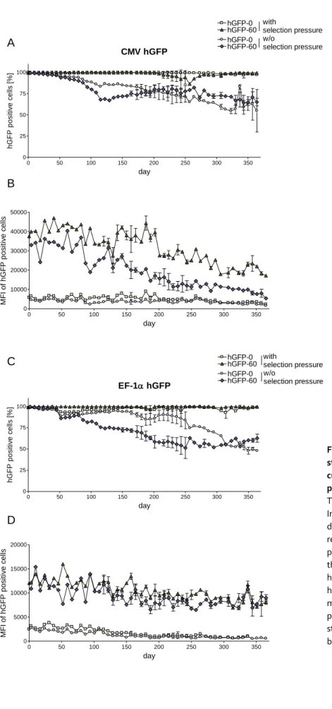
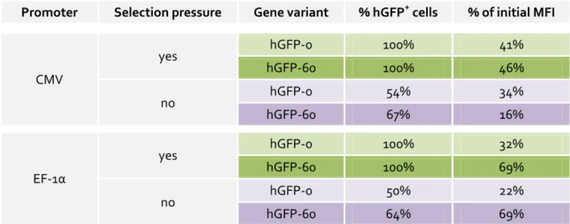
To test the effects of intragenic CpG dinucleotides on transgene expression efficiency in a gene therapy-relevant cell system, murine embryonic pluripotent stem cells of the line P19 were stably transduced with lentiviral vectors (LV) containing the respective gfp variants under different promoters. The promoter of the cytomegalovirus (CMV) revealed a high disposition for gene silencing in this expression system. Compared to the CMV promoter, gfp transcription by the elongation factor 1 alpha (EF-1α) promoter resulted in delayed, yet significant transgene silencing in P19 cells. In contrast, the bidirectional, dual divergently transcribed A2UCOE promoter prevented transgene silencing via its chromatin opening abilities completely. With regard to CpG frequency, the LV-P19 expression system could also benefit from the presence of intragenic CpG dinucleotides under certain conditions, in spite its high gene silencing potential.
EF-1α-promoter-controlled expression of the CpG-maximized gfp variant was clearly increased over the CpG-depleted gfp in P19 cells. CMV promoter-mediated gfp expression revealed slightly delayed gene silencing in CpG-rich compared to CpG-depleted gfp. In contrast, A2UCOE-mediated transcription was not affected by intragenic CpG dinucleotides. It is assumed that A2UCOE can overcome chromatin compaction arising from intragenic CpG depletion due to its chromatin opening property.
The sum of data could show that TSS-adjacent intragenic CpG dinucleotides in gfp and mip-1α transgenes positively influence transcription efficiency. The results gained in this work imply that this effect results from delocalization and destabilization of the +1 nucleosome, whereas intragenic CpG depletion leads to a higher level of chromatin density. These chromatin changes are assumed to result from a complex epigenetic regulation network triggered by intragenic CpG changes. The exact mechanism of this phenomenon remains to be elucidated.
Introduction
Page | 5
3
Eukaryotic gene transcription 3.1
The regulation of gene transcription is fundamental for cellular differentiation, proliferation and the proper response to environmental changes. To achieve the high level of specialization of cells that have a common set of genetic information, gene transcription is subjected to multiple regulatory mechanisms. In prokaryotes, gene regulation allows a single cell to respond to environmental changes by switching genes on and off [1]. In multicellular eukaryotic systems, gene regulation not only serves to adjust to environmental changes. The biologically more important purpose of gene control is to provide the proliferation of many different cell types that compose a multicellular organism. Eukaryotic transcription is an immensely complicated process that is regulated by a large number of proteins (Figure 1) [2]. Sequence-specific binding factors/transcription factors interact with their DNA motifs in response to cellular signals [3]. They recruit transcriptional co-regulators to alter the local chromatin environment and facilitate assembly of the pre-initiation complex (PIC) [4], which is composed of the general transcription factors (GTFs) and Polymerase II (RNAPII) [5].
Among the three eukaryotic Polymerases, RNAPII, consisting of 12 subunits, is responsible for the transcription of protein coding genes [6]. GTFs, comprising TFIIA, TFIIB, TFIID, TFIIE, TFIIF and TFIIH, are essential for exact positioning of RNAPII at the promoter. Associated as the basal transcription machinery, RNAPII and GTFs form a preinitiation complex (PIC) at the core promoter, which is usually located upstream of the translated region [7]. Most core promoters contain a TATA box or equivalent motifs as an essential recognition feature for the basal transcription machinery [8]. TATA-boxes are present in the core promoter region and are typically 30–60 base pairs (bp) upstream of the transcription start site. In addition to these promoter motifs, the initiator (Inr) or downstream promoter element (DPE) interact with various components of the basal transcription machinery [9]. Another feature found at promoters of expressed genes in the yeast genome is the nucleosome-free region (NFR) [10]. What exactly creates an NFR is not fully understood, although some studies could correlate NFRs to poly-dA-dT tracts [11] or CpG islands [12]. Besides promoter regions, enhancers, also termed distal regulatory elements (DREs), contain binding sites for transcription factors. They can be located up to several thousand base pairs away from the actual initiation site [13].
Sequence-specific DNA binding transcription factors act as activators or repressors of transcription. They simultaneously recognize both promoter or enhancer sequences and other co-regulators through their DNA-binding domains and activation domains [4].
Whether a sequence-specific regulator activates or represses gene transcription depends on the genomic context and recruited co-regulators [2]. Co-regulators mainly comprise chromatin-modifying and/or chromatin-remodeling enzymes and the mediator complex [14]. The mediator complex facilitates the interaction between DNA-binding transcription factors, co-regulators and the basal transcription machinery [15].
Introduction
Page | 6 Figure 1 | Regulation of eukaryotic transcription (simplified). Assembly of the PIC,
containing RNAPII (light grey) and GTFs (dark grey) is initiated by binding of TFIID to core promoter elements like TATA box, Initiator (Inr) or downstream promoter element (DPE) (purple). Transcriptional gene regulation involves: the binding of sequence specific binding factors (light green) to distal regulatory elements (DREs) and proximal promoter regions; interactions of DNA-binding factors with co- regulators like mediator (yellow), histone modifying complexes (green), chromatin remodelers (orange) and the basal transcription machinery (grey). The C-terminal domain (CTD) (red wavy line) is unphosphorylated in the PIC and becomes multiply phosphorylated upon initiation. As RNAPII traverses a transcription unit, the phosphorylation pattern changes resulting in the recruitment of different proteins.
The concerted function of all these factors is to express a subset of genes as dictated by a complex interplay of environmental signals.
The C-terminal domain (CTD) of the largest subunit of the eukaryotic RNAPII contains several YSPTSPS heptad repeats (52 in mammals) that are unphosphorylated in the PIC of RNAPII and become multiply phosphorylated upon initiation [16]. As RNAPII traverses a transcription unit, the phosphorylation pattern changes resulting in the recruitment of different proteins to the CTD [17]. Phosphorylation has predominantly been found at serine 2 and serine 5 of the heptad repeats. Phosphorylation of the serine 5 residue occurs during transcription initiation and has been connected to multiple processes of transcription such as promoter clearance for transition from initiation to early elongation and 5′-end capping of pre-mRNA [18]. Modification of serine 2 is found
TATA Inr DPE IID
Introduction
Page | 7
when the polymerase is associated with the coding region and has been implicated in productive elongation and the 3′-end processing of the transcript [19].
Several regulatory proteins specifically recognize the respective phosphorylation pattern of the CTD. Thereby, the CTD of RNAPII coordinates events during the transcription cycle by recruiting co-regulators involved in histone modifications and/or remodeling, transcription elongation, termination and mRNA processing [2].
Chromatin 3.2
Eukaryotic DNA is up to a thousand times longer than the cell’s length [20]. Therefore, an organized packaging system is needed to fit the DNA into the nucleus. The nucleoprotein complex that meets this requirement is called chromatin. The term was first used by Walther Flemming, who discovered a visible cell substance with staining characteristics and therefore named it chromatin, which means “stainable material” [21].
Different states of chromatin, called euchromatin and heterochromatin, are found in the nucleus. They correlate with transcriptional active or repressed genes. Euchromatin undergoes a process of condensation and decondensation during cell cycle. It constitutes the majority of the chromosomal material and contains genes that are actively expressed. Heterochromatin remains highly condensed during the cell cycle. It is mostly found at the centromers and telomers of chromosomes as well as along the entire inactive X chromosome in female mammals [22].
The nucleosome 3.2.1
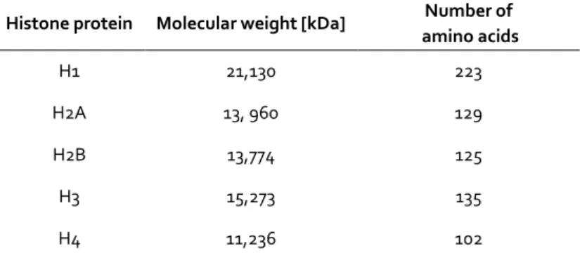
Nucleosomes are the primary structural units of chromatin, composed of DNA and histones. Histones are highly conserved, basic proteins of 11 to 21 kilo Dalton (kDa) (Table 1). In 1997, the structure of a nucleosome core particle could be resolved by X-ray diffraction at a resolution of 2.8 Å (Figure 2) [23]. It shows a nucleoprotein complex of approximately 147 bp of genomic DNA wrapped in a left handed superhelix 1.7 times around a histone octamere which has a diameter of 11 nm in length and 5.5 nm in height.
Table 1 | Molecular weight and size of histones. Values given are derived from bovine histones. Modified from [24].
Histone protein Molecular weight [kDa] Number of amino acids
H1 21,130 223
H2A 13, 960 129
H2B 13,774 125
H3 15,273 135
H4 11,236 102
Introduction
Page | 8
A histone octamere contains two copies each of histones H2A, H2B, H3 and H4. All four histone proteins have a similar structural motif in common. The trihelical histone fold core mediates both binding between histones itself and between histones and DNA.
Each histone has polypeptide extensions with NH2- and/or COOH-terminal ends that stick out from the globular regions. These tails are targets for posttranslational modifications like acetylation and methylation [25]. Different from the rest of the histones, histone H1 is involved in the chromatin packing into a higher-order structure [20].
Figure 2 | Structure of the nucleosome core particle. The model shows the DNA double helix (brown and torquiouse) wound around the central histone octamere, consisting of two copies each of histones H2A, H2B, H3 and H4. Hydrogen bonds and electrostatic interactions between histones and DNA keep the nucleosome in place [23].
Chromatin organization 3.2.2
Nucleosomes are connected by nucleosome-free linker DNA to form a 10-nm fiber, also called the “beads-on-a-string array” [26][27]. The length of linker DNA varies among species, ranging from about 20 to 60bp. The linker region and parts of the nucleosomal DNA are associated with the linker histone H1, which binds to the nucleosome and causes the assembly of nucleosomes into a higher-order structure, the 30-nm filament [25][20]. While the X-ray crystal structure of the nucleosome core particle has early been resolved in atomic detail [23], the structure of the 30-nm chromatin fiber has been an issue of debate. In 1976, Finch and Klug postulated the “solenoidal model for superstructure in chromatin”, which would direct the linker DNA between two nucleosomes into a strong bend [28]. For another model of organization, the so called
Introduction
Page | 9
zig-zag structure, it was assumed that the linker DNA is straight and crosses the center of the 30-nm fiber [29]. X-ray analysis of a tetra nucleosome seems to support the zig-zag structure, which falls into the category of the 'two-start helix' type [30]. By contrast, electron microscope measurements provide evidence for the solenoid model characterized by interdigitated nucleosomes [31]. Both models agree on the function of the linker histone to determine the topology and degree of chromatin compaction [32].
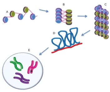
Very recent analyses indicate that the 30-nm fiber involves both zigzag and bent linker motifs, depending on physiological conditions [33]. The 30-nm chromatin fiber results in an approximately 50-fold compaction of DNA. To obtain a higher level of organization, a hierarchical folding of chromatin structure, schematically illustrated in Figure 3, is needed [22]. A series of loops of 30-nm fibers are anchored at their base to the chromatin scaffold to form the 300-nm fiber [34]. The chromatin scaffold consists of non-histone proteins and has the shape of a metaphase chromosome. On average, each loop encompasses 20.000 to 500.000bp of DNA and is about 300nm in length. Tight helical coiling of the 300nm fiber produces the scaffold-associated chromatin structure. This helix is again packed and folded to generate an individual 700nm wide chromatid, two of which compose a metaphase chromosome [22] (Figure 3).
Figure 3 | Hierarchical folding of chromatin. (A) Beads-on-a-string array.
Alternating nucleosomes are depicted with blue and green surfaces; (B) The 30-nm fiber twists further and forms a more compact fiber (C) that is arranged in loops (blue), with some portions attached to a protein scaffold (red) (D); (F) metaphase chromosome. Modified from [35].
A
B C
D
E
Introduction
Page | 10
The mechanism of higher order chromatin formation, ultimately resulting in metaphase chromosome formation, is still poorly understood. Multiple chromatin-associated proteins (CAPs) have been suggested to play an important role in the formation and dissociation of the chromatin structure beyond the 30-nm fiber. H1 is considered to be an important CAP in the organization of higher chromatin structure by stabilizing the folded state as was revealed by electron microscopy [36]. An important process for chromosome organization is the interaction of core histone domain tails which are also targets of multiple modifications in the course of gene transcription [37].
Transcriptional control by chromatin 3.3
Chromatin generally limits the accessibility of specific DNA sequences and inhibits the initiation and progression of the polymerase during transcription. There are basically three different ways by which the chromatin structure can be altered: i)By chromatin remodeling, ii)histone modification and iii) the replacement of core histones by histone variants. Together with DNA methylation and RNA binding, these regulation mechanisms are summarized as epigenetic control [38].
Histone modifications 3.3.1
To date, more than a hundred of histone modifications have been found. Several recent reviews cover this complex topic [39][40][41]. In the following sections, only a selection of modifications controlling gene activity is discussed. Among the many types of histone modifications that have been detected so far, acetylation, methylation and phosphorylation are the most frequently detected and best understood (Table 2). Over 60 different histone residues have been identified to be a target of modification, and in the case of methylation, multiple modifications (mono-, di- and trimethyl) can occur at one lysine or arginine [42][43].
Introduction
Page | 11 Table 2 | Overview of the most important types of histone modifications in mammals. Modified amino acids include Lysine (K), Arginine (R), Serine (S), Threonine and Proline (P). Modified from [44].
Depending on type and position of modification, opposed effects on transcription rate have been observed. The acetylation of histones generally activates a gene cumulatively, whereas methylation can have opposing effects (Table 2). Modifications that have been connected with transcription activation have been described as euchromatin modifications. Those that have been mapped to inactive genes are referred to as heterochromatin modification [45].
Genome-wide studies have revealed that individual histone modifications can be mapped to specific states of gene activity [46] (Figure 4). For example, the modifications H3K4me2/3 (histone H3 lysine4 di- and trimethylation) are mainly found in actively transcribing promoters, and H3K36me3 is frequently found in the body of actively transcribed genes, increasing towards the 3’ end. By contrast, modifications like H3K27me3 and H4K20me3 are mostly mapped to regions where transcription is repressed [39]. Some modifications, such as H3K27me3 and H3K4me3 are however coincident with both activation and repression of gene transcription, respectively (Figure 4).
Modifications Residues Modified Modification Position Impact on Transcription
Acetylation K-ac H3 (9,14,18,56), H4 (5,8,13,16), H2A,
H2B Activation
Methylation (lys) K-me1 K-me2 K-me3 H3 (4,36,79) Activation
H3 (9,27), H4 (20) Repression
Methylation (arg)
R-me1 R-me2a R-
me2s H3 (17,23), H4 (3) Activation
Phosphorylation S-ph T-ph H3 (3,10,28), H2A, H2B Activation
Ubiquitylation K-ub H2B (120) Activation
H2A (119) Repression
Sumoylation K-su H2B (6/7), H2A (126) Repression
Isomerization P-cis > P-trans H3 (30-38) Activation/ Repression
Introduction
Page | 12 Figure 4 | Distribution of histone modifications on active and inactive genes. Modification patterns differ on actively transcribed and silenced genes, which is displayed as a schematic view of modification distribution over the gene. Promoters of actively transcribed genes carry high levels of active modifications such as acetylations and methylation of H3K4. At the transcriptional start site there is a nucleosome-free region (NFR) within the promoter. Inactive genes have a fairly even distribution of silencing modifications, such as H3K9 methylation and H4K20 methylation, whereas H3K27 methylation is enriched in the promoter. Modified from [39].
Strahl and Allis postulated the hypothesis of a histone code, proposing that the combination of histone modifications at a certain genomic locus determines the activity state of the underlying gene [47]. This hypothesis of a histone code is heavily discussed within epigenetic research, arguing that gene regulation by histone modifications might rather reflect a cumulative more than a combinatorial effect [48]. Nevertheless, the frequently made observation of distinct histone patterns demonstrates that histone modifications can indeed serve as indicator for gene activity or inactivity. In what respect these histone distributions are a matter of cause or consequence of gene activity is however not fully understood [39].
Histone modification is carried out by a variety of enzymes, categorized as acetyltransferases, methyltransferases, kinases etc. A detailed list of histone modifying enzymes is reviewed by Kouzarides [44]. The co-presence of both modifying and de- modifying enzymes indicates that histone modification is a highly dynamic process.
Active Gene
Inctive Gene
NFR
Introduction
Page | 13
There are two major functions of histone modifications. First of all, histone modifications result in the weakening of inter- and intranucleosomal as well as histone-DNA interactions, thereby relaxing the chromatin structure. A simple consideration that led to this assumption is the fact that, apart from methylation, histone modifications all result in a net charge change of nucleosomes [39]. The disruption of chromatin contacts allows transcription factors to bind to their targets and is therefore fundamental for transcription. The second purpose of histone modification is the direct recruitment of regulatory proteins or DNA-methyltransferases (DNMTs) to their cognate binding sites [49]. An example for such co-regulators is the SET domain- containing histone methyltransferase enzyme SUV39H1, which is responsible for trimethylation of H3K9 and heterochromatinization of pericentromeric satellite repeats.
These proteins are also required to recruit de novo methyltransferases to methylate CpG dinucleotides in the satellite sequence [50]. In addition to transcription factors and DNA modifying enzymes, histone modification patterns interact with remodeler complexes [43].
Chromatin remodeling 3.3.2
The dynamic property of DNA is maintained by chromatin remodeling complexes. These multi-protein complexes are essential for many chromatin functions such as the proper spacing of nucleosomes during nucleosome assembly, DNA repair or the binding of transcription factors to specific genes in the course of transcription regulation [51]. A broad range of remodeler complexes has been identified. All of them contain an ATPase domain which belongs to the superfamily II (SFII). On the basis of sequence similarities of the ATPases, remodeller complexes can be grouped into a number of subfamilies [52][53]. Most of these subfamilies have been designated to the archetypal member, such as S.cerevisiae Snf2p (Snf2 subfamily), Drosophila melanogaster Iswi (Iswi subfamily), or Mus muculus Chd1 (Chd subfamily). Several of them, e.g. members of the Iswi subfamily, have been reported to possess DNA-translocation activity [54]. Different remodelers affect the structure of the nucleosome array in a particular way and thereby influence a widespread number of nuclear processes, reviewed in [52]. For instance, the members of Iswi, namely the NURF (nucleosome remodeling factor), CHRAC (chromatin accessibility factor) and ACF (ATP-utilizing chromatin-assembly and remodeling factor) predominantly position nucleosomes in a manner to repress transcription [55]. By contrast, RSC (remodels the structure of chromatin), a member of the Swi/Snf family, mediates pathways that both activate and repress transcription [56]. Different than the variety of remodelers with regard to substrate specificity and chromatin product, the mechanism by which nucleosomes are rearranged has been suggested to be uniform.
According to the ‘loop recapture model’, DNA translocation against a histone octamere is achieved by the successive detachment of DNA, starting from the edge of the nucleosome, its bending and recapturing by the octamere to form a loop that is carried along the DNA strand [57].
Introduction
Page | 14
Sequence dependent nucleosome positioning 3.3.3
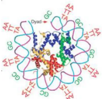
It is now well established that the DNA sequence itself determines the strength of DNA-histone interactions and the bending flexibility of the DNA helix around a histone octamere [58][59]. Poly (A) and poly (T) regions result in conformationally rigid molecules and therefore require high energy to incorporate into nucleosomes. By contrast, dinucleotides form nucleosomes of high stability: AA, TT and TA dinucleotides are favored approximately every 10bp where both DNA strands face towards the nucleosome core. GC dinucleotides are favored approximately every 10bp where both phosphodiester backbones face outward (Figure 5). A study of Gupta et al. has identified a 3bp periodicity of CG and GC dinucleotides to be a highly nucleosome favored sequence [60].
Figure 5 | Three dimensional structure of one-half of a symmetric nucleosome. Bends around the nucleosome core are favored by the dinucleotides AA/TT/TA that oscillate approximately 10bp periodically in phase with each other and out of phase with GC dinucleotides recurring every approximately 10bp as indicated [59].
The sequence preference calculation is based on a thermodynamic model that evaluates the free energy for any nucleosome constellation [59]. This includes the calculation of sterically allowed nucleosome organizations and competition between positions at each dinucleotide. A genome-wide analysis of nucleosome positioning demonstrated that approximately 50% of the in vivo nucleosome organization is solely determined by sequence preferences of nucleosome occupation [61]. By using high-density tiling arrays over the yeast genome, it was shown that a nucleosome-free region (NFR) was a common feature of promoters [10]. The so-called “−1” and “+1” nucleosomes are located in canonical regions upstream and immediately downstream of the NFR, respectively.
These well-positioned nucleosomes encompassing the NFR at promoters have regulatory functions of transcriptional regulation (see chapter 3.1) [62].
Introduction
Page | 15
Cytosine Guanine Dinucleotides 3.4
Nucleosome positioning is influenced by short periodic repeats of cytosines followed by a guanine [60]. These so called CpG dinucleotides are significantly underrepresented throughout the vertebrate genome than would be calculated from base composition.
[63][64][65]. Since cytosines within CpGs are the exclusive targets for methylation in vertebrates, it was anticipated that this deficiency was related to DNA methylation [66].
The selective pressure resulting in this CpG loss was provided by the inherent mutability of methylated cytosine. The deamination of cytosine results in uracil, which is easily recognized and removed by uracil glycosylases. By contrast, the deamination of methyl cytosine gives rise to thymine, which is not recognized as foreign and therefore leads to a transition mutation in the subsequent replication. As a result, methylated CpG dinucleotides in the germ line tend to be lost over time [67]. Organisms with high levels of DNA methylation therefore tend to exhibit the most pronounced CpG deficiency [65].
CpG methylation 3.4.1
DNA methylation patterns among eukaryotes are not uniform. The most frequent pattern found in invertebrate animals is the so-called ‘mosaic methylation’. It is characterized by moderate levels of methyl-CpG dinucleotides accumulated in domains of methylated DNA, interspersed with unmethylated domains. Vertebrates, on the other hand, exhibit high levels of methylated CpG dinucleotides distributed over the entire genome, except for small methylation free regions at transcriptionally active regions.
This pattern is referred to as the ‘global methylation’ [68]. The transition from the ancestral mosaic methylation to the vertebrate global methylation is believed to have evolved in the evolution of CpG DNA immunity. The genomes of most bacteria and DNA viruses are rich in unmethylated CpG dinucleotides. These CpG motifs of several microbial parasites are detected by pattern recognition receptors, such as the Toll-like receptor 9 (TLR9), during the innate immune response in some vertebrates [69]. Since methylated CpGs have no potential to activate this defense, the genome of the host vertebrates prevents an auto immune response. The CpG-poor, globally methylated vertebrate genome is therefore believed to be a prerequisite of the CpG immunity [68].
The DNA methylation patterns in mammalian cells are usually well regulated and tissue- specific [70][71]. DNA methylation patterns of specific cell types are established during mammalian development and maintained in adult somatic cells [72]. In mammalian germ cells and early embryos, dramatic reprogramming with complete removal of methylation occurs, followed by renewed de novo methylation [73]. Not only global methylation changes, but also gene-specific de novo methylation and demethylation have been observed, for example during differentiation of hematopoietic progenitors [74]. DNA methylation in mammalian cells is mostly correlated with gene silencing, which is virtually always the case if this concerns promoter elements [75][76]. However, DNA methylation of gene bodies is also found to be positively correlated with transcription [77][78][79].
Introduction
Page | 16
The majority of methylated DNA in differentiated cells is however harbored by non-coding transposable elements such as SINEs (short interspersed nuclear elements), LINEs (long interspersed nuclear elements) and endogenous retroviruses. These elements encompass approximately 42% of the human genome [80][81].
Methylation occurs at the 5-position of the cytosine residue within CpG dinucleotides, resulting in 5-methylcytosine (m5C). The reaction is catalyzed by DNA methyltransferases (DNMTs), which catalyze the transfer of a methyl group from S-adenosyl-L-methionine to cytosine [82]. There are three enzymatically active DNMTs, which can be divided into de novo and maintenance methyltransferases. De novo methyltransferases act after the replication in unmethylated DNA. Maintenance methyltransferases catalyze the addition of methyl groups to hemi-methylated DNA during replication [82]. DNMT1 is the major maintenance methyltransferase [83].
DNMT3A and DNMT3B are de novo methyltransferases acting on unmethylated DNA.
They are responsible for establishing methylation patterns during early development and each of them has distinct functions [84]. DNMT3L is a protein that is homologous to DNMT3A and DNMT3B but contains no catalytic activity. Instead, DNMT3L assists the methylation during gametogenesis by recruiting de novo methyltransferases [85]. DNA demethylation can be accomplished either passively, by leaving the new DNA strand unmethylated after replication, or actively. Some studies support the existence of active demethylation in zygotes [86] and in somatic cells [87]. So far, the exact mechanism is still not fully understood.
Unmethylated CpG dinucleotides 3.4.2
CpG dinucleotides are largely depleted throughout the mammalian genome as a consequence of their high susceptibility to mutation [66]. The result is that CpGs are relatively rare unless there is selective pressure to keep them or a region is not methylated due to active regulation of gene expression. Those genomic loci are mostly promoter regions of housekeeping genes that comprise at least half of the genes in the human genome [76].
It has been suggested that the unmethylated state of CpG dinucleotides is also dependent on germ line and early embryonic transcription. As a result of this lack of methylation, CpG dinucleotides in these regions are less suppressed and consequently appear relatively CpG-rich compared with the rest of the genome [88]. These stretches of mostly non-methylated CpGs are called CpG islands. CpG islands, defined by Bird in 1986, are on average 100obp of length, have a C+G content of 0.5 or higher and an observed to expected CpG dinucleotide ratio of 0.6 or higher within a range of 200bp or greater [89][90]. CpG Islands are mostly found within the promoter and the first exon of several genes, particularly housekeeping genes [67][91]. In addition to housekeeping promoters, the average of protein coding genes in the human genome display a significant excess of CpG dinucleotides in exons, most pronounced in the first exon, compared to introns [67][92][93][94].
Introduction
Page | 17
Gene control mechanisms directed by CpG dinucleotides 3.4.3
The high frequency of CpG dinucleotides in promoters and gene bodies of constitutively expressed genes versus the low frequency of CpG dinucleotides in mostly non-functional DNA already points to the outstanding role of this element as a transcriptional regulator.
Despite more than 25 years of intensive study on CpG islands/regulatory CpG motifs since their discovery [89], the exact mechanisms by which CpG dinucleotides affect gene transcription are still poorly understood.
Trans acting proteins have been found that interact with unmethylated CpG dinucleotides leading to a unique chromatin architecture [95]. The transcription factor Sp1, for instance, has been demonstrated to bind to unmethylated CpG Islands to protect them from de novo methylation, which ensures active gene transcription [96]. In addition to Sp1, the CRE binding factor (CREB) [97] and CCCTC binding factor (CTCF) [98] contain CpG in their binding recognition site and DNA recognition is impaired upon CpG methylation.
More than 15 years ago, another important factor binding to unmethylated CpG dinucleotides was found in tobacco: the nuclear CpG-binding protein 1 (CGBP-1) binds with high affinity to unmethylated CpG dinucleotides [99]. A human CpG binding protein (hCGBP) was isolated a few years later, revealing specific binding for unmethylated CpG dinucleotides and thereby functioning as a transcriptional activator [100]. Subsequently, this protein was renamed as CXXC finger protein 1 (CFP1) [101]. CFP1 has frequently been localized in nuclear regions that are associated with euchromatin, which underlines its exclusive function as a transcriptional activator [102].
The key feature of CFP1 is a cysteinrich CXXC DNA-binding domain [100]. This zinc- finger like domain is highly conserved and frequently found in proteins involved in epigenetic regulation, such as the DNA methyltransferase 1 (Dnmt1) [103], methyl-CpG binding proteins MBD [104] and histone H3-Lys4 methyltransferase [105]. CFP1 was shown to associate with a histone H3K4 methyltransferase complex (SET1 complex) catalyzing the addition of the tri-methyl modification (H3K4me3) [106]. H3K4me3 coincides with promoters and 5’ end of actively transcribed genes [107] (see also chapter 3.3.1). Histone lysine methylation marks are recognized by specific effector proteins containing plant homeodomain (PHD) finger domains or chromatin organization modifier (chromo) domains. PHD finger proteins can activate gene transcription, such as via TFIID [108] and the nucleosome remodeling factor (NURF) [109]. Another transcription factor binding to unmethylated CpG dinucleotides via the zink finger CXXC domain is the H3K36-specific lysine demethylase enzyme KDM2A. Binding of KDM2A to CpG results in removal of H3K36 methylation, thereby creating a “CpG island chromatin” that is depleted of this repressive modification [110].
The binding of unmethylated CpG dinucleotides by CpG-specific transcription factors, which are able to affect histone modifying activities, suggests that CpG dinucleotides may use chromatin associated processes to provide a transcriptionally active surface [95]. In addition to chromatin mediating abilities, early studies of CpG island chromatin revealed a distinct depletion of Histone H1 at CpG islands [111]. Histone H1 represses transcription [112] due to stabilization of chromatin structure [113].
Introduction
Page | 18
Methylated CpG dinucleotides of regulatory elements have also been found to direct numerous gene control processes. For example, CpGs involved in tumori-genesis [114] or genomic imprinting [115] become methylated during cellular differentiation. DNA methylation has been shown to block the recruitment of zink finger CXXC proteins which then creates a repressive chromatin environment [107][110].
Additionally, methylated CpG dinucleotides provide binding sites for methyl CpG- binding domain proteins (MBDs) that interact with further co-regulators like histone deacetylase (HDAC) eventually leading to inhibition of gene expression [116]. A prominent mediator between DNA and histone modification is the DNMT3A/B homolog DNMT3L. DNMT3L binds to histone H3, and thereby recruits de novo methyltransferases to DNA. Once H3K4 becomes methylated, the interaction between DNMT3L and the nucleosome is inhibited [117]. Histone methyltransferases responsible for trimethylation of H3K9 are simultaneously required for the recruitment of DNMT3A and DNMT3B in order to methylate CpG dinucleotides, eventually leading to heterochromatinization at satellite sequences [50]. This process of heterochromatinization is initiated by a Dicer-mediated mechanism that recognizes RNA duplexes found at satellite sequences. The resulting RNA-induced silencing complex (RISC) is then specifically targeted back to pericentromeric regions where it probably recruits enzymes involved in this heterochromatin pathway [118][119][120].
Apparently, the interactions between histone and DNA modifying events can work in both directions: CpG methylation provides the template for some histone modifications, and histone modifications can recruit DNMTs. It seems that histone modifications provide more labile transcriptional repression, whereas DNA methylation is a rather stable epigenetic mark that is not easily reversed [49].
The mechanisms mentioned above are just a small insight into the many pathways that are directed by unmethylated or methylated CpG dinucleotides, respectively. Their extensive implications in epigenetic mechanisms underpin their role as a key player in transcriptional regulation. Despite recent advances in the understanding of regulatory CpG elements, there are still many gaps in the knowledge of this field that need to be filled to better understand cellular responses to the environment. Further to that, the understanding of CpG-mediated transcriptional control would be useful in the design of optimized transgene expression systems.
Introduction
Page | 19
Transgene expression 3.5
The design of optimized transgene systems is crucial for gene therapy applications and the production of recombinant proteins. Prokaryotic and simple eukaryotic expression systems are inexpensive, fast growing and easy to handle. Nevertheless, these systems lack a suitable native glycosylation machinery and may not fold and secrete the recombinant proteins correctly [121][122]. Due to these limitations, mammalian cell culture has become the standard system for recombinant protein production.
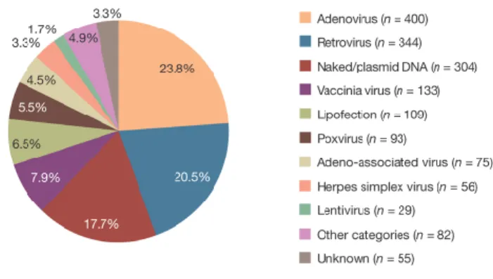
Accordingly, about 60–70% of all recombinant pharmaceuticals are produced in mammalian cells, particularly CHO and HEK 293 cells [123]. The growing demand for therapeutic proteins requires the establishment of highly effective and sustainable expression systems. Besides optimization of the translational or secretory capacity of host cells, the maximization of transgene expression levels is a major attempt to increase protein yields [124]. The first step of successful transgene expression in the target cell is the choice of the appropriate gene delivery system. There are currently two major delivery categories used for transgene expression: plasmid-based and viral vector-based [125] (Figure 6).
Figure 6 I Vectors used in gene therapy trials. Viral vectors, in particular retro- and adenoviruses, are the most frequently used vehicles for gene transfer to human cells. The development of efficient expression systems has made plasmid-based transgene delivery to the third most frequently used vector system in gene therapy trials [125].
Introduction
Page | 20
Viral vector-based transgene expression 3.5.1
Viral vectors are mostly genetically modified, replication deficient viruses. They are able to transduce cells with high delivery efficiency and can be used in a variety of cells [125].
While DNA-based viral vectors, such as adeno and adeno-associated viruses (AAV), usually persist as episomal DNA in the host cell [126], retroviruses have the ability to confer long-term transgene expression through gene integration [127].
3.5.1.1 Retroviral vectors
Retroviral vectors are generated by exchanging replication elements by the gene of interest. Necessary cis-acting RNA regions, primarily the long terminal repeat (LTR), which is necessary for packaging, reverse transcription, integration and transcription regulation, are retained. All viral genes are usually deleted from the viral vector. The production of attenuated retroviral vectors takes place in packaging cells that provide all essential viral proteins in trans. Transgenes are delivered into the cell by receptor mediated fusion of viral and host cell lipid membrane. Upon entrance of the viral vector into the cell, reverse transcription is initiated. The viral genome is converted to a double-stranded DNA provirus, which is then inserted into the host genome [127].
One subclass of retroviruses often used in gene therapy trials comprises lentiviral vectors. In addition to the three essential gag, pol and env gene products, lentiviruses contain accessory viral proteins that regulate viral gene expression and infectivity [128].
These viral proteins interact with the nuclear import machinery to mediate the active transport of the viral preintegration complex through the nucleopore. This ability enables lentiviruses to transduce non-dividing cells [129].
Lentiviruses preferably integrate into or in the proximity of active transcription units [130]. Self-inactivating retroviral vectors (SIN LVs) have a deleted U3 region of the 3’LTR containing the viral enhancer sequence. This ability provides gene transfer with higher safety due to the reduced risk of enhancer-mediated mutagenesis [131]. Transgene expression in LVs has been shown to undergo epigenetic modifications, eventually leading to gene silencing [132][133][134].
3.5.1.2 Ubiquitously acting chromatin opening elements (UCOEs)
An attractive approach to overcome transgene silencing in LVs is the introduction of ubiquitously acting chromatin opening elements (UCOEs). UCOEs are regions containing CpG islands extending over dual divergently transcribed promoters derived from housekeeping gene loci [135][136]. UCOEs have been reported to provide stable transgene expression in cell culture systems even when integrated into heterochromatin regions [135]. This feature confers considerable utility for gene therapy and recombinant therapeutic applications.
Introduction
Page | 21
Plasmid-based transgene expression 3.5.2
Alternative to virus-based delivery systems, which still bear several safety risks, plasmid- based gene delivery has become a common technique in gene therapy, DNA vaccination and the production of recombinant proteins in mammalian cells [137]. Plasmid DNA can be delivered to cells either physically or by synthetic particles. These particles typically consist of DNA complexed with cationic lipids, peptides or polymers capable of efficient gene transfer into the target cell. The easiest physical method of transgene delivery is by needle injection into the target tissue, i.e. muscle cells [138], skin [139], liver [140] or tumor [141]. Needle injection is the major application of DNA vaccination [142]. Other physical methods include electroporation [143], ballistic DNA administration [144] or sonoporation [145][146], just to name the most commonly used physical techniques. For review, see Kamimura et al [147].
Among the synthetic compounds, liposomes, particularly those composed of cationic lipids, have been reported to be most effective for gene delivery [148]. Liposomes are particles consisting of lipid bilayers encompassing an aqueous compartment. They are formed spontaneously when lipids are hydrated in an aqueous solution [147].
Alternative to liposomes, numerous polymer-based compounds such as polyethylenimine (PEI) [149], polyamidoamine [150], polyallylamine [151] and chitosan [152] are being widely employed today. These cationic polymers condense DNA into positively charged particles and prevent DNA fromdegradation. The cellular uptake of these complexes occurs via endocytosis [147].
Besides simplicity of delivery, the advantages of plasmid-based transgene expression are low toxicity and sustainability. The main disadvantage of plasmid-based techniques compared to viral-based methods is the low gene delivery efficiency. Large efforts have been made to modify the carrier or delivery vehicle to achieve higher transfection rates [137]. High transfection rates are however useless if transgene expression is ineffective.
Once inside the cell, plasmid DNA is subjected to the cells regulation mechanisms that can directly be influenced by sequence elements of the plasmid DNA [137].
Plasmid-based vectors have a large capacity for transgene DNA. Rational plasmid design aims for the manipulation of a variety of regulatory factors that impact on gene transfer and gene expression. A plasmid accommodates the expression cassette (EC), which contains the gene(s) of interest and any regulatory sequences required for expression in mammalian cells, such as the promoter and the poly A site. The rest of the plasmid, the bacterial backbone (BB), usually contains an antibiotic resistance gene and an origin of replication required for the production of the plasmid DNA in bacteria [153].
Numerous efforts have been made to establish systems providing efficient plasmid-based transgene expression. One approach to improve transgene expression is to generate minicircles. In minicircles, the BB is removed by site-specific recognition sequences, which results in the generation of two smaller supercoiled minicircles. The minicircle harboring the EC is then separated from the other circle containing unwanted BB elements [154] such as antibiotic resistant genes or elements provoking DNA methylation and heterochromatin-associated histone modifications [137]. Another
Introduction
Page | 22
strategy to avoid transgene silencing is the inclusion of a scaffold matrix attachment region (S/MAR). S/MARs are AT-rich sequences derived from eukaryotic DNA where the nuclear matrix attaches. They have been shown to contain DNA-unwinding elements and binding sites for transcription factors and topoisomerase II. Since S/MARs harbor mammalian origins of replication, they can promote sustainable episomal replication and maintenance in mammalian cells [155]. Another crucial factor for successful transgene expression is the careful choice of an appropriate promoter. Dependent on the type of application and target cell or tissue, different promoters should be selected.
Endogenous housekeeping promoters express at low but constitutive rates. Due to this ability, they are recently preferred over viral promoters that provide high but often unstable transgene expression due to gene silencing [137]. Furthermore, a tissue-specific promoter has the potential of improved specificity and safety [156][157].
The adaptation of the codon usage has proven to be extremely effective in promoting transgene expression [158][159][160]. According to the codon bias of the host cell, the respective protein sequence is translated back into the DNA sequence, selecting only the most frequently used tRNAs of the respective organism. The use of plasmids free of CpG dinucleotides has been reported to minimize inflammation and provide prolonged transgene expression [161]. On the other hand, CpG dinucleotides in the EC have conversely been demonstrated to provide improved transgene expression in mouse tissue [162].
3.5.2.1 Applications of plasmid-based transgene technologies
Optimizing plasmid DNA not only promotes gene therapy applications. It also benefits plasmid DNA vaccination strategies [163] and transfection of mammalian cells providing for recombinant protein production [164]. Conventionally, transient expression or random integration techniques are used for recombinant protein expression. These approaches however usually result in random integration and irreproducible levels of gene expression. To overcome these problems, stable integration systems have been developed that generate stable mammalian cell lines with defined integration sites and reproducible level of protein expression [165]. The Flp-In recombinase system which is based on the site-specific recombinase (Flp) from Saccharomyces cerevisiae offers a single targeted integration site, has been used for applications like the production of antibodies [166][167] or vaccine immunogens [168]. Initially, this site specific integration system was developed for basic research to study and compare transcriptional reporter gene activities as it allows the expression of numerous reporter gene constructs at an identical genomic location [165]. It is therefore a useful tool to investigate the impact of regulatory plasmid vector elements on transgene expression in the host cell.