Analysis of the impact of codon choice on gene expression of HIV-1
DISSERTATION ZUR ERLANGUNG DES
DOKTORGRADES DER NATURWISSENSCHAFTEN (DR. RER. NAT.) DER FAKULTÄT FÜR BIOLOGIE UND VORKLINISCHE MEDIZIN
DER UNIVERSITÄT REGENSBURG vorgelegt von
Thomas Schuster aus
Kronstadt
im Jahr 2019
Das Promotionsgesuch wurde eingereicht am:
26.04.2019
Die Arbeit wurde angeleitet von:
Prof. Dr. Ralf Wagner Unterschrift:
Thomas Schuster
A Contents
A Contents ...- 4 -
B Abstract ...- 8 -
C Introduction ...- 9 -
The human immunodeficiency virus -1 (HIV-1) ...- 9 -
C.1.1 Epidemiology ... - 9 -
C.1.2 Structure of HIV-1 ... - 10 -
C.1.3 The life cycle of HIV-1 ... - 11 -
C.1.4 Genome organization and viral gene expression ... - 12 -
C.1.5 The HIV-1 Gag Polyprotein ... - 13 -
C.1.6 The role of the Rev protein and the Rev-responsive element in the life cycle of HIV- 14 - C.1.7 Usage of a subgenomic gag reporter system to analyze the effects of codon adaptation on viral gene expression... - 16 -
C.1.8 The role of the Tat protein in the transcription of viral genes by the cellular RNA polymerase II . - 16 - RNA export from the nucleus to the cytoplasm ...- 17 -
The genetic code and its role in protein biosynthesis...- 18 -
Codon usage and codon usage bias ...- 20 -
Codon optimization and deoptimization ...- 21 -
Nucleotide composition of HIV ...- 22 -
Objective ...- 23 -
D Material and methods ...- 25 -
Cell lines ... - 25 -
D.1.1 Prokaryotic cell lines ... - 25 -
D.1.2 Eukaryotic cell lines ... - 25 -
Nucleic acids ...- 25 -
D.2.1 Oligonucleotides ... - 25 -
D.2.2 Plasmids ... - 31 -
D.2.3 Antibodies ... - 32 -
D.2.4 Commercial Kits ... - 32 -
D.2.5 Standards ... - 33 -
D.2.6 Computer programs and databases...- 33 -
Cell culture techniques ...- 34 -
D.3.1 Cultivation of eukaryotic cells ... - 34 -
D.3.2 Transfection of eukaryotic cells ... - 34 -
Microbiological methods ...- 35 -
D.4.1 Growth and selection of prokaryotic cells ... - 35 -
D.4.2 Transformation of chemically competent cells... - 35 -
Molecular biology methods ...- 35 -
D.5.1 Photometric quantification of nucleic acid concentrations ... - 35 -
D.5.2 Isolation of plasmid DNA ... - 36 -
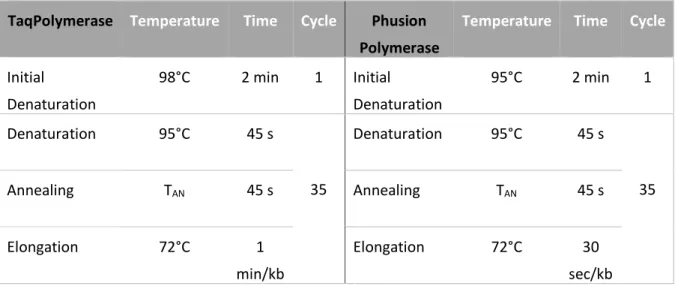
D.5.3 Polymerase chain reaction ... - 36 -
D.5.4 Fusion PCR ... - 37 -
D.5.5 Standard cloning procedures ... - 38 -
D.5.6 Cloning with exocutter BsmBI ... - 38 -
D.5.7 Agarose gel electrophoresis ... - 38 -
D.5.8 Detection of potential cryptic splicing products ... - 39 -
D.5.9 Generation of completely and partially codon adapted gag-variants ... - 39 -
Working with RNA ...- 39 -
D.6.1 Isolation of total RNA... - 40 -
D.6.2 Nuclear Run on assay ... - 40 -
D.6.3 Determination of mRNA half-life ... - 41 -
D.6.4 Reverse transcription (synthesis of copy DNA) ... - 42 -
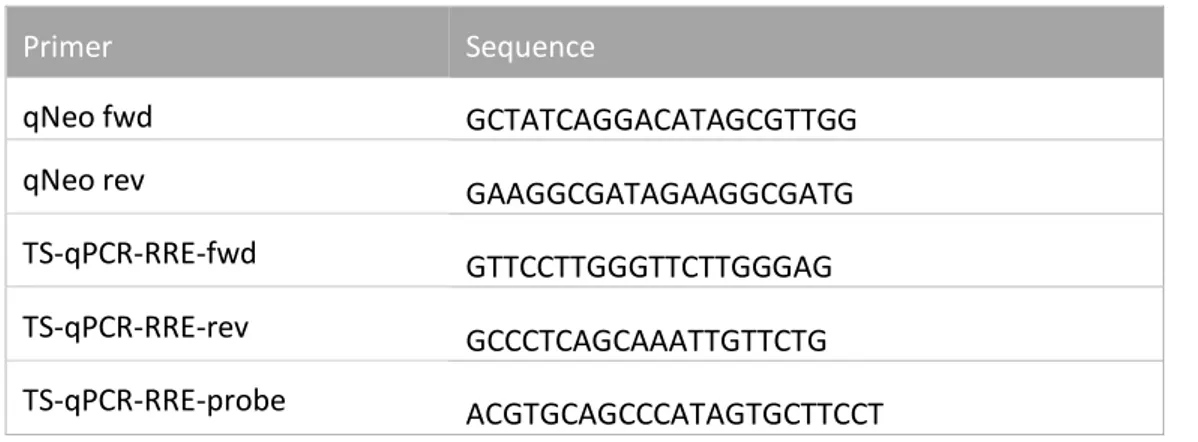
D.6.5 Quantification of mRNA expression levels by quantitative real-time PCR (RT-qPCR) ... - 42 -
Protein biochemistry techniques ...- 47 -
D.7.1 Lysis of HEK293T cells ... - 47 -
D.7.2 Quantification of total protein amount by Bradford analysis ... - 47 -
D.7.3 Quantification of Gag expression by p24-Enzyme-linked Immunosorbent Assay (ELISA) ... ... - 48 -
Analysis of GFP expression by flow cytometry ...- 49 -
DNA Sequencing ...- 50 -
E Results ...- 51 -
Generation of partially humanized gag variants ...- 51 -
Sequence properties of partially humanized gag variants ...- 52 -
Influence of humanization on gag protein expression of 5‘-3’ adapted variants ...- 54 -
Effect of Rev inhibition by addition of Leptomycin B on the gag expression of the 5’- 3’ adapted gag-variants ...- 55 -
Influence of humanization on Gag protein expression of 3‘-5’ adapted variants ...- 57 -
Characterization of the impact of the 5’ end of the HIV-1 gag on gene expression of codon adapted variants ...- 58 -
E.6.1 Analysis of the impact of the first 423 bp at the 5’ end of HIV-1 gag... - 58 -
E.6.2 Analysis of the importance of the first 100 base pairs at the 5’ end of HIV-1 gag ... - 59 -
E.6.3 Localization of a potential inhibitory sequence motif at the 5’ end of HIV-1 gag ... - 61 -
Confirmation of the position of the inhibitory sequence motif ...- 63 -
Influence of humanization on mRNA expression levels of 5’-3’- and 3’-5’-adapted variants ... ...- 64 -
Analysis of transcription efficiency for selected partially humanized variants ...- 66 -
Determination of mRNA stability of selected gag variants ...- 69 -
Detection of potentially generated cryptic splicing variants ...- 72 -
Impact of the Splice Donor SD1 on gag expression ...- 73 -
Analysis of expression of partially humanized gag variants under control of the LTR promoter ...- 75 -
Analysis of the impact of codon adaptation on a quasi-lentiviral GFP reporter ...- 76 -
Influence of the identified inhibitory HIV-1 gag motif on egfp expression...- 79 -
F Discussion ...- 81 -
Generation of partially humanized variants and analysis of gene expression ...- 82 -
F.1.1 Generation and analysis of partially humanized gag variants ... - 82 -
F.1.2 Generation and analysis of partially humanized egfp variants ... - 83 -
Localization and examination of the inhibitory motif within the 5’ part of HIV-1 gag ...- 84 -
Molecular mechanisms that contribute to altered expression of partially codon-adapted genes ...- 85 -
F.3.1 Molecular mechanisms that contribute to the general dependency of protein production from the
a mount of humanized codons ...- 85 -
F.3.2 Molecular mechanisms that contribute to the function of the inhibitory motif on protein production
... - 87 -
F.3.3 Influence of changed mRNA secondary structures on altered gene expression ... - 88 -
F.3.4 Influence of splicing on altered gene expression ... - 91 -
F.3.5 Impact of the promoter on altered gene expression ... - 92 -
Future prospects ...- 92 -
G Appendix ...- 94 -
Northern Blot analysis of 5’-3’-adapted variants ... - 94 -
Comparison of p24 levels in the supernatant and intracellular p24 levels of transfected HEK293T cells. ... - 94 -
H List of References ...- 96 -
I Danksagung ...- 105 -
- 8 -
B Abstract
The degeneracy of the genetic code with its 61 codons encoding for only 20 amino acids is the basis for a phenomenon known as codon usage bias. That means that different organisms show differences in the frequency of occurrence of synonymous codons. Despite the necessity of HIV to use the host’s translational machinery for viral gene expression, the virus exhibits an adenine-rich nucleotide composition which differs clearly from the GC-rich coding regions of humans. Changing the nucleotide composition by choosing synonymous codons at certain positions might therefore impact viral replication by affecting viral gene expression. Previous studies of our group showed that adapting the gag gene to human codon usage (huGag) led not only to a significantly increased protein production but also caused independency of Rev, an accessory protein of HIV that exports intron- containing viral mRNAs. The aim of this work was to gain insight into the effects of codon adaptation, especially regarding length and position, and the associated impact on gag expression. For this, subgenomic gag reporter constructs were generated that systematically varied the humanized part of the gene. Those constructs were then transfected into HEK293T cells. Gag expression was investigated on protein level by p24 ELISA as well as on RNA level by qPCR. Furthermore, transcription efficiency as well as RNA stability were analyzed using nuclear run-on and actinomycin D assays. It was observed that increasing the length of the humanized sequence starting from the 5’ end directly correlated with p24 and gag mRNA levels. Contrary to that, such a correlation was lacking for constructs humanized progressively in 3’ to 5’ direction. It became apparent that humanization of the 5’ end of gag is necessary for enhanced protein production and Rev-independent expression. In addition, it was found that a short sequence, surrounding the nucleotides 61-75 of gag in the 5’-part of the gene contains a specific inhibitory motif which affects transcription rate as well as RNA stability. Moreover, the expression pattern of the different variants was analyzed under the control of a heterologous CMV promoter as well as an LTR promoter of HIV-1. For both systems, comparable expression patterns were observed. By PCR analysis of reverse transcribed RNA from transfected cells, as well as northern blot analysis, the generation of cryptic splicing variants was excluded.
Further, a second reporter gene was used in order to investigate whether the function of the
identified inhibitory motif could be transferred. For this, a quasi-lentiviral system was used to express
egfp after its adaptation to human and HI-viral codon usage. It became apparent that the inhibitory
effect of the identified motif was only transferable when it was embedded in a larger part of wild-
type gag. The influence of the inhibitory motif on HIV gag expression as well as the missing inhibitory
effect on egfp could be based on characteristics of the mRNA secondary structures. For gag, but not
for egfp variants, presence of the motif had a clearly destabilizing effect in mRNA folding predictions,
which might influence protein binding of the RNA degradation machinery. In future experiments an
examination of this hypothesis would be eligible and helpful to gain further insight in the connection
between altered codon usage and gene expression.
- 9 -
C Introduction
The human immunodeficiency virus -1 (HIV-1)
C.1.1 Epidemiology
Despite intensive research for more than 30 years [2] and accompanying major advances in medical treatment, the infection with the immunodeficiency virus (HIV) still represents a life threatening situation for millions of people. Untreated, an HIV infection leads to the Acquired Immune Deficiency Syndrome known as AIDS. The course of this disease is typically characterized by three phases. Firstly, infected people show influenza-like symptoms caused by a high viremia and a rapid decline in CD4- positive T-cells [3]. The second phase is a symptom-free state, often mediated by a T-cell driven immune response against HIV [4]. Lastly, the continuous depletion of CD4-positive T-cells leads to an increase in viral load. This phase is generally linked to opportunistic infections and tumors, which are typically for the disease pattern of AIDS [5].
The adequate availability of Antiretroviral Therapy (ART) still is a problem in some areas of the world.
So, despite constantly increasing ART coverage over time, in 2017 only about 21 million patients of the estimated 37million infected people are receiving an adequate therapy, which accounts for only around 60%. Especially in Western and Central Africa and the Eastern Mediterranean, ART coverage is not satisfactory with 29% or even 18% of HIV positive men and 48% and 19% of HIV positive women receiving ART, as shown in Figure C-1[1].
Figure C-1: Accessibility of antiretroviral treatment to HIV positive individuals
Development of the numbers of ART-receiving people worldwide (left) and ART coverage for males and females in different WHO regions. Modified after [1]. In 2017 only 21.7 million of the estimated 37 million HIV-infected people received ART. Especially people in Western & Central Africa as well as the Eastern Mediterranean were not supplied with an adequate therapy.
- 10 -
HIV-1 can be classified into the four Groups M, N, O and P which arose through zoonotic transmissions of the chimpanzee simian immunodeficiency virus (SIV
CPZ) or gorilla immunodeficiency virus (SIV
GOR) to humans [6]–[8]. Group M, which causes most of the infections, can be divided into nine clades (A-D, F-H, J and K). Besides HIV-1, there is also the human immunodeficiency virus type 2 (HIV-2), which is predominantly found in West African nations, though increasing numbers of infections have been recognized also in other parts of the world. The modes of transmission are equal to those of HIV-1, namely sexual contact, blood-borne exposure (blood transfusion, shared needles), and perinatal transmission [9].
C.1.2 Structure of HIV-1
HIV-1 is a member of the lentiviruses which belong to the viral family retroviridae. Because of the intensive research over the last decades most questions regarding the general structural features as well as the viral replication have been solved and can be reviewed in different excellent publications, books and reviews from which the following information are derived [10]–[15]. A characteristic feature of retroviruses is the reverse transcription of their RNA genome in double-stranded DNA, followed by the integration into the host’s genome. Infectious HIV-1 particles contain a membrane which is derived from the host’s cell membrane and encircles a conic capsid. Furthermore, the viral membrane contains the envelope protein (Env), which is the only viral protein that is present at the particle’s surface and consists of gp120 and gp41. The matrix protein (MA, p17) is bound inside of the membrane by an N-terminally attached myristoyl group. The conic capsid is formed only by the p24 capsid protein (CA) and contains the viral enzymes reverse transcriptase (RT), protease (PR), integrase (IN) as well as the viral genome [10]–[15].
Figure C-2: Schematic structure of an HIV particle (modified after [10])
The lipid membrane of HIV is derived from the host’s plasma membrane by budding of the virus and contains the only viral surface molecules gp41 and gp120. Inside of the lipid membrane the matrix protein (MA, p17) is attached. Inside of the conic capsid which is built by the capsid protein (CA, p24) the two identical RNA copies are complexed with nucleocapsid proteins (NC, p7). Furthermore, the capsid contains the viral enzymes reverse transcriptase (RT), integrase (IN) and protease (PR).
- 11 -
C.1.3 The life cycle of HIV-1
The initial adsorption of the viral particle to the cells is mediated by an interaction of the external glycoprotein gp120 to the CD4 receptor of the target cell e.g. T-helper cells, monocytes, macrophages and dendritic cells. This event is followed by a conformational change of the gp120 protein which allows the interaction with either the CCR5 or CXCR4 chemokine receptor. These processes enhance the binding affinity between virus and cell surface which allows the fusion of the membranes and therefore enables the entry into the cell. This is followed by uncoating and reverse transcription of the viral RNA genome in double-stranded DNA, which generates the long terminal repeats at both ends of the genome (see below). The RNA part gets degraded during this process by the RNase-H function of the reverse transcriptase. The double-stranded DNA molecule is transported into the nucleus as a pre-integration complex (PIC) together with associated cellular and viral proteins like the integrase, Vpr and matrix protein (p17). A phosphorylated form of p17 stays connected with the
pre-integration complex and allows, together with the Vpr protein and cellular nuclear import factors, the transport of the reverse-transcribed genome through the nuclear pores, which enables
Figure C-3: Replication cycle of HIV-1 (from [16])
Initial binding of HIV to the target cell is mediated by interaction of gp120 and the CD4 receptor of the host cell. After a conformational change an additional interaction with the chemokine receptor CXCR4 or CCR5 occurs. This allows the fusion of the membranes of the virus and the host cell, which is followed by uncoating and reverse transcription of the viral genome in double-stranded DNA. The reverse-transcribed genome is transported with cellular and viral proteins as a pre-integration complex into the nucleus. The viral protein integrase mediates the integration of the viral genome into the host's genome, followed by transcription. The transcribed viral RNAs are used for protein synthesis and as viral genomes for progeny virions.
- 12 -
the infection of non-dividing cells. Inside the nucleus the viral integrase mediates the integration of the genome at an unspecific position, followed by transcription of viral RNAs. The different classes of transcripts are used either for protein synthesis or as viral genomes for progeny virions [16]–[20].
C.1.4 Genome organization and viral gene expression
HIV-1 contains two identical copies of (+)-sense, single-stranded RNA molecules in a complex with nucleocapsid proteins (NC, p7). Since the two RNA molecules have a 5’ cap as well as 3’
polyadenylation, they show typical features of a eukaryotic mRNA. The genome of HIV-1 (as shown in Figure C-4) has a size of about 9700 nucleotides which encode for structural genes (gag, pol and env) as well as regulatory and accessory genes (tat, rev, nef, vif, vpr and vpu). After reverse transcription of the RNA into double-stranded DNA, and following degradation of the viral RNA, the proviral DNA is generated by integration into the human genome. During that process, long terminal repeats (LTRs) are generated which contain all cis-active sequences as well as promoter and enhancer elements which control the retroviral gene expression. The viral genes get transcribed by the cellular DNA-dependend RNA polymerase and are expressed from partially overlapping open reading frames.
Lastly, after alternative splicing, the mRNAs get translated by the cellular translation machinery.[10], [21].
Figure C-4: Schematic structure and organization of the HIV-1 genome:
During integration, long terminal repeats (LTRs) are generated. The structural genes gag, pol and env are shown in yellow, whereas regulatory and accessory genes are depicted in grey. The open reading frames of tat and rev are encoded by two exons each.
The produced transcripts of HIV during infection can be classified into three groups. Shortly after
infection, only short, completely spliced mRNAs are produced which encode the regulatory viral
proteins Tat, Nef and Rev. Over time, the transcription rate gets strongly enhanced and incompletely
spliced mRNAs are produced. Those mRNAs encode the envelope protein (Env) as well as the
accessory proteins Vif, Vpr and Vpu. Additionally, the last group of transcripts, the unspliced mRNAs
are used for translation of the Gag-Pol polyprotein and as new genome for progeny virions. This
complex and strongly regulated gene expression pattern is significantly regulated by the two proteins
Tat and Rev. Tat enhances transcription by binding to a regulatory RNA element called TAR and
- 13 -
recruits elongation factors to the transcription complex resulting in strong enhancement of transcription. The viral protein Rev, which role is described in detail in C.1.6, plays an essential role in mediating the cascade-like gene expression [22]–[24]. Among others, HIV Gag that was mainly used to analyze the influence on codon usage on viral gene expression in this work, depends on the Rev-mediated mRNA export.
C.1.5 The HIV-1 Gag Polyprotein
The Gag (group-specific antigen) polyprotein of HIV-1 is the main structural protein of the virus and is translated from an unspliced 9 kb transcript. Gag plays also an important role in the immune response against HIV, since the initial HIV-specific immune response is directed against Tat, Rev and Gag. Because the regulatory and accessory proteins show a higher mutation rate, the immune response against Gag plays a key role to fight the virus.[25], [26]. Intensive research has shown the importance of Gag for the viral life cycle. Initially assumed as a simple scaffold protein, Gag meanwhile is known to specifically recognize genomic RNA as well as viral and cellular proteins [27].
Furthermore, Gag is necessary and sufficient for budding of HIV from the host's plasma membrane [28]. Therefore, Gag has to package two copies of viral genomic RNA per particle [29] and interact with cellular trafficking proteins to hijack the endosomal-sorting complexes required for transport (ESCRT) system [30]–[32].
Figure C-5: Schematic overview of the budding process of HIV-1 mediated by Gag (from [33])
Gag proteins move towards the plasma membrane, interact with each other and induce extrusion of the lipid bilayer, followed by the membrane fusion to pinch-off the virion. Lastly, proteolytic processing by the viral protease leads to the maturation of the viral particle [33], [34].
Structurally, Gag can be divided into four major domains: The matrix, capsid, nucleocapsid and link protein, each playing a different role during the viral life cycle.
The matrix protein consists of 128 amino acids and plays an essential role for plasma membrane
targeting and viral assembly. For both processes, an N-terminal myristoylation at Gly-2 plays a key
role [35], [36], since it allows the binding of Gag to cholesterol- and sphingomyelin-rich micro
domains within the plasma membrane [37].
- 14 -
The capsid protein is responsible for forming the core of the virus and can be divided into a C-terminal and N-terminal domain (CTD and NTD), joined by a flexible linker [38], [39] and a hexameric protein lattice is produced by CTD interaction[40]. Since formation of the mature and immature virus is strongly dependent on the CA-CA interaction, inhibitors targeting different sites of the capsid protein can have severe effects on viral replication, making CA an interesting target for an antiviral therapy [27], [41].
The nucleocapsid protein (NC) has several functions despite being one of the smallest parts of Gag with only 55 amino acids. During viral maturation and assembly, NC is involved in Gag-Gag interaction as well as in the recognition of the nucleic acid [27]. An important feature are two zinc finger motifs which are separated by a functionally important basic domain [42]. Furthermore, NC acts as an RNA chaperone and supports reverse transcription as well as the integration in the host's genome [10].
The last component of Gag is the 52 amino acid long link protein (p6). It gets translated as two different forms, the in-frame Gag p6 and the -1 frameshifted Gag–Pol p6*. It plays an important role for the release of new virions from the plasma membrane because of the so-called late domain of HIV, a sequence which seems to be responsible for the separation of the viral envelope and the cellular plasma membrane [43]–[46]. As mentioned above, the expression of Gag is strongly dependent on the accessory protein Rev and its interaction with a conserved sequence called the Rev-responsive element.
C.1.6 The role of the Rev protein and the Rev-responsive element in the life cycle of HIV
The regulatory protein Rev is a 13-kDa phosphoprotein which allows the export of unspliced and incompletely spliced viral mRNAs. Rev consists of 116 amino acids which are encoded by two exons and consists of several distinct domains [10], [47]–[50]. At the N-terminus an arginine rich motif functions as a nuclear localization sequence (NLS) [51], [52]. The interaction with a conserved RNA secondary structure called Rev- responsive element (RRE), which is present in all unspliced and incompletely spliced HI-viral mRNAs, is mediated by a arginine rich domain between amino acid 35 and 50 [10], [52], [53]. The RRE is a ∼ 350 nucleotide long, highly structured element inside of the env RNA. It forms a conserved secondary structure with five stems around a central loop, according to
Figure C-6: Five stem model of the Rev-responsive element from [34]:
Rev binding sides are shown in grey
- 15 -
the original model. Later, a 4-stem model suggested that stem loop III and IV form a hybrid III/IV stem loop together with the intervening loop region [54]–[56]. Initially Rev binds to the IIB site, followed by a multimerization of Rev by five additional molecules. The bi-directional transport is mediated by a nuclear localization signal (NLS) and a distinct nuclear export signal (NES). The NLS overlaps with the RNA-binding domain, while a leucine-rich sequence acts as the NES, allowing the interaction with cellular mRNA export proteins [47], [57]. Especially the amino acids between position 73 and 84 are essential for the Rev-mediated nuclear export, since they mediate the interaction with exportin 1, also known as CRM1 (chromosomal maintenance gene 1) which binds to the GTP-loaded form of the Ran protein. Thereby, the mRNA-Rev-protein-Crm1-Ran-GTP complex is exported out of the nucleus through the nuclear pores. By hydrolyzation of GTP, Crm1 and Ran disengage themselves from the export complex and Rev dissociates from the mRNA which now gets translated. By this mechanism HIV ensures that all viral gene products can get translated in a highly regulated way (see Figure C-7).
Figure C-7: Role of Rev in the HIV-1 life cycle (from [58])
HIV transcripts can be classified into unspliced, incompletely spliced and fully spliced mRNAs. At the beginning of infection, in the absence of Rev, only fully spliced mRNAs can be exported out of the nucleus. From those mRNAs, Tat, Nef and Rev get translated. Unspliced and incompletely spliced mRNAs are retained inside of the nucleus until Rev migrates back, binds to a secondary structure known as Rev-responsive element, which is present in all variants of unspliced and incompletely spliced transcripts and mediates the export of those mRNAs. For this, Rev interacts with exportin 1, also known as CRM-1 which mediates the export through the nuclear pore [58].
The fully spliced transcripts that get exported even in the absence of Rev use cellular proteins for the
mRNA export. The correct transport of all different classes of RNA in general is a highly regulated
process and is described in the following chapter.
- 16 -
C.1.7 Usage of a subgenomic gag reporter system to analyze the effects of codon adaptation on viral gene expression
To analyze the effects of altered codon usage on gag expression independently of other processes in the life cycle of HIV like infectivity, uncoating, reverse transcription or budding, a simplified gag reporter system was used, which was established in previous studies of our group. For this, the fact was used that HIV-1’s late gene expression depends on cis-acting elements as well as the interaction of Rev with the RRE. For mimicking of this situation the 5’ UTR of HIV-1 carrying the highly functional major splice donor SD1 was fused upstream and a fragment known to carry the RRE was fused down- stream to the gag-encoding open reading frame. Adapting the gag gene to human codon usage resulted in constitutive nuclear export allowing high levels of Gag expression independently of the Rev/Rev-responsive element system because of the modification of intragenic regulatory elements [59]. Later, it was shown that it is possible to transfer this reporter system also to other report genes, like the green fluorescent protein (GFP). Adapting the open reading frame of GFP to HIV-1’s codon usage was sufficient to turn this hivGFP RNA into a quasi-lentiviral message following the rules of late lentiviral gene expression. Again, cis-active elements which are known to influence HIV-1gene expression, like the 5’UTR as well as the RRE were added either upstream (5’UTR) or downstream (RRE) of the open reading frame [60]. This principle was used in several studies of our group and also in this thesis, in order to analyze the influence of partial codon adaptation on viral gene expression.
C.1.8 The role of the Tat protein in the transcription of viral genes by the cellular RNA polymerase II
The transactivator of transcription (Tat) plays a central role in the life cycle of HIV, since it mediates a more than hundred enhanced transcription of viral genes from the LTR promoter by binding to the TAR (transactivation response) element. The TAR element is a secondary structure which is built across the first 59 nucleotides and is present at the 5’ part of all viral mRNA species. The binding of Tat is followed by the recruitment of the positive transcription elongation complex (P-TEFb), which consists of CDK9 and Cyclin T1. CDK9 then phosphorylates the carboxyl terminal domain of RNAP II.
Transcription by RNAP II is a highly complex process from initiation to termination. Transcriptional
initiation requires the assembly of the preinitiation complex (PIC), composed of the general
transcription factors (GTFs) TFIIA, TFIIB, TFIID, TFIIE, TFIIF, and TFIIH; the Mediator complex; and
RNAP II with an unphosphorylated carboxy terminal domain (CTD). But, throughout the process of
transcription, the CTD becomes highly phosphorylated which enhances the transcriptional activity
[61]–[63]. This process is mediated by the Tat protein in the gene expression of HIV-1.
- 17 -
RNA export from the nucleus to the cytoplasm
As already mentioned above, the fully spliced transcripts are exported without the help of viral proteins like Rev. Those mRNAs are transported by the same mechanisms that are responsible for the export of cellular mRNAs. In general, for different RNA species that get exported out of the nucleus through the nuclear pore, different export pathways are used. Small RNAs, like tRNAs and micro RNAs get exported by relatively simple export pathways. Their transport is mediated by a direct binding to their respective export receptors. For example, the export of tRNAs is mediated by the export receptor exportin-t, a member of the karyopherin superfamily, which binds directly to tRNAs in a RanGTP-dependent manner. After transport of the tRNA-exportin-t-RanGTP complex to the cytoplasm, RanGAP stimulates GTP hydrolysis on Ran, inducing the release of the tRNA cargo from its receptor (see below) [64]–[66]. Large RNAs, like rRNAs and mRNAs build quite complex ribonucleoprotein (RNP) particles, including specific adaptor proteins to the given export factors [65].
Figure C-8: Schematic illustration of different RNA export pathways (from [65]
The major routes of RNA export pathways are shown. Those include tRNAs, microRNAs (miRNAs), small nuclear (sn)RNAs, messenger RNAs (mRNAs) and ribosomal RNAs (rRNAs). The primary transcripts are shown at the top as well as the molecules after processing, maturation and assembly with export factors (export adaptors are shown in blue, export receptors are shown in yellow). For the mRNA pathway, metazoan and yeast proteins are listed and additional adaptor proteins and RNA binding factors (orange ovals) are depicted.
- 18 -
The precursor mRNA molecules undergo different processing steps like capping, splicing and cleavage/polyadenylation at the 3' end. For this, the mRNA associates with a variety of proteins to form a ribonucleoprotein particle (mRNP). Furthermore, an interaction with nuclear export factors also occurs already during transcription and processing, which ensures that only completely functional transcripts are provided to the translational machinery [67]. In principle the processes during mRNA export are as follows. Transcription of pre- mRNA molecules is carried out by RNA-polymerase II followed by an immediate assembly into RNPs, containing several heterogeneous nuclear ribonucleoproteins (hnRNPs) and splicing commitment factors. Those mRNAs remain inside the nucleus until the completion of splicing, when commitment factors are released and the nuclear mRNA export factors Tap and p15 are recruited to the mature mRNA.
Subsequently, the RNP docks at the nuclear pore complex (NPC) and enters the cytoplasm.
Nonshuttling hnRNPs have to be released at this point for example by the RNA helicase Dbp5. After the nuclear export, binding of cytoplasmic RNA binding protein occurs followed by translation of the mRNA. The shuttling hnRNPs like hnRNP A1 and nuclear mRNA export factors, including Tap and p15, are released and recycled to the nucleus [68].
The genetic code and its role in protein biosynthesis
The genetic code has a central place in biology, since it defines the rules to translate the 4-letter alphabet of nucleic acids into amino acid sequences, the 20-letter alphabet of proteins [69]. This process, known as protein biosynthesis, starts already in the nucleus with the transcription of DNA into a messenger RNA (mRNA). After the export of the mRNA into the cytoplasm (see above), the given nucleotide sequence gets translated according to the genetic code into an amino acid sequence, generating new proteins. These processes underlie complex and highly regulated mechanisms, themselves, like transcription initiation, elongation and termination, as well as post- transcriptional modification. This is also true for nuclear export processes or translational processes like initiation, elongation, transcription and post-translational modifications. Those mechanisms have been studied intensively over the years and were summarized in different excellent reviews
Figure C-9: Export of eukaryotic mRNA from the nucleus (from [61])
Processes and proteins involved in the nuclear export of mRNAs. Letters A-C stand for hnRNPs, S stands for splicing commitment factors. Explanation given in text.
- 19 -
[70]–[74]. A simplified schematic illustration of the translation of a DNA sequence into an amino acid sequence is shown in Figure C-10.
Figure C-10: Schematic and simplified overview of different processes involved in protein biosynthesis (from [69]) (1) Unwinding of the DNA double helix to allow transcription of the desired sequence into an mRNA molecule (2) which gets transported through the nuclear pore into the cytoplasm (3). After that, the mRNA attaches to the ribosome where transfer RNAs (tRNAs), each carrying a specific amino acid, interact with the mRNA trough the tRNA’s anticodon (4).
Elongation occurs and a growing peptide chain is generated (5) [69].
The genetic code is universal, which means that it is used by all creatures and plays a central role in the processes shown above, since it determines which amino acid sequence gets translated from the codons of the mRNA molecule. After the double helical structure of DNA was deciphered by Watson and Crick in 1953 [75] fundamental findings regarding the genetic code were done during the 1960s.
In 1961 Crick and Brenner performed a historic experiment to demonstrate that the genetic code is made up of a series of three base pairs codons by using mutants in the rII locus of T4 phage [76].
Later, based on the finding that poly-uracil RNA incorporated only phenylalanine [77], Nirenberg was
able to decipher the genetic code [78]. Since the genetic code acts as a fundamental feature all over
in biology, it was and still is subject of intensive research that results in four key points, which should
always be remembered about the genetic code. Firstly, the genetic code is non-overlapping. That
means, consecutive amino acids are determined by consecutive codons. In an overlapping code,
consecutive amino acids would be encoded by codons that share some bases. Secondly, an amino
acid is always encoded by three bases, which are termed codons. Thirdly, the code is read from a
defined starting point to an end point, which determines the coding region. And fourthly, the code is
degenerated. That means some amino acids are encoded by more than one codon. This degeneracy
- 20 -
is mediated by a loose kind of base pairing at one end of the codon and anticodon, called wobble position [79]. So, some amino acids can be transported to the ribosome by several tRNAs with different anticodons, whereas for certain other amino acids this can only be done by one specific tRNA. Figure C-11 shows how 61 codons encode the 20 amino acids and three stop codons.
Figure C-11: The genetic code (from [80])
Shown are the 20 amino acids and their chemical features (indicated by different colors) and how they are encoded by the different codons.
The degeneracy of the genetic code is also the basic principle for codon bias and codon optimization, two points that play a central role in this thesis and will be explained in the following chapters.
Codon usage and codon usage bias
The degeneracy of the genetic code allows the usage of different codons for the same amino acid.
Those codons are called synonymous codons and contrary to the first intuition those codons are not
used in an equal frequency, but some codons are preferred. This phenomenon is known as codon
usage bias and varies between and among species [81]. As explanations for the non-random usage
of different codons a combination of mutation, selection and random drift is proposed [82]. In this
model a balance between those forces favors specific, superior codons. That means that a mutation
in an unpreferred codon could result in the generation of a preferred one when this allows a more
efficient way of gene expression [83]–[86]. Furthermore, highly expressed genes show in general a
stronger bias in synonymous codon usage [87]–[89]. As explanation for this phenomenon, one of the
most intensively supported hypotheses over the years was translational selection. This theory is
generally explained by better fitting codons for the most abundant isoaccepting tRNAs [90]–[92]. A
- 21 -
fact, which might be true for prokaryotes, where frequently used codons correlate with abundant cognate isoacceptor transfer RNAs [93].But especially for eukaryotes a variety of factors like GC- content, recombination rate, RNA stability, codon position and gene length can also influence codon usage bias [83], [94]–[98]. Therefore, translational efficiency by itself is not sufficient to account for effects of synonymous codon usage in higher organism. Especially the perception of the influence of synonymous codons on RNA level rose over the last years. Chen et al. quantified the impact of synonymous codons on mRNA level by analyzing over 3556 variants of a heterologous gene encoding the green fluorescent protein (GFP) and 523 synonymous variants of the endogenous gene TDH3 in yeast. They could show a positive correlation between mRNA levels and codon usage bias, which points to a direct effect of synonymous mutations on transcript concentration, most likely by influencing mRNA degradation rate [99]. Further studies point into the same direction. Kholiswa et al showed in 2008 that transfection of Jurkat cells with codon-optimized gag mRNAs led to a small increase in Gag production, whereas transfection of optimized DNA resulted in a very large enhancement of expression, indicating a higher ranking role of mRNA levels [100]. Furthermore, also our group could show that codon-usage-mediated inhibition of HIV-1 gag expression in mammalian cells occurs independently of translation [101].
Codon optimization and deoptimization
The fact that amino acids can be decoded in most cases by more than one codon facilitates the option to change codon choice as a molecular biological tool in form of codon optimization and deoptimization. Codon optimization and deoptimization describes the possibility to change the natural codon usage in two different directions. So, it is possible to introduce synonymous mutations in a coding region that do not affect the amino acid sequence of a protein but substitute specific nucleotides so that either better or worse fitting codons are generated. For this, a robust method had to be used to quantify codon usage bias. For this problem, different methods exist [102] and are favored by different groups. For the analysis in this thesis, the codon adaptation index (CAI) [103]
was predominantly used, which is defined as the geometric mean of the relative adaptiveness values (w) of individual codons. The relative adaptiveness (w) of a codon is the ratio of the observed frequency of that codon to the frequency of the most abundant codon for the same amino acid. The most abundant codon is calculated for a reference set of highly expressed genes, such as ribosomal protein genes. Thus, 𝑊 =
𝑋𝑖𝑗𝑋𝑖𝑚𝑎𝑥
where X
ijis the observed frequency of the i
thcodon for the j
thamino
acid and X
imaxis the maximal X value for codons for the same amino acid [83]. The probably widest
application for codon optimization is in heterologous gene expression, a well-established method to
produce recombinant protein products like drugs, industrial enzymes or biofuels in foreign organisms
more efficiently [104]–[108]. On the other hand, it is also possible to worsen the codon usage by
using synonymous mutations. As an example, it was already shown that replacement of optimal
codons in viral genomes can lead to attenuated viruses by influencing different features like
- 22 -
dinucleotide frequency, GC-content or codon pairs [109]–[111]. In this thesis an optimization of HIV- gag was performed in order to analyze the effects of altered codon usage on viral gene expression and the thereby mediated Rev-dependency/independency as already described in several earlier studies of our group [59], [60], [112]–[114]. Further The prerequisite to do this is the different nucleotide composition of HIV compared to its human host.
Nucleotide composition of HIV
Despite the necessity of HIV to use the host’s translational machinery for viral gene expression, the virus exhibits an A-rich nucleotide composition with 36.2% A, 23.9% G, 22.2% U and 17.6% C, which differs clearly from the GC-rich coding regions of humans, where e.g. the GC content of isochores can be up to 60% [115]–[118]. The deviating codon usage is not exclusive for HIV, since also other viruses show an A-rich nucleotide composition or a differing codon usage compared to their hosts in general [119], [120]. From an evolutionary point that raises the question whether or how far the viruses benefit from different codon choice, because after all they rely on cellular processes like transcription or translation and therefore need to take over cellular functions and direct them towards the efficient production of new viruses [121]. The fast adaptation of HIV-1 epitopes to specific human MHC-I molecules validates the importance of viral evolution and adjustment to their hosts [122]. Therefore, there must be reasons why the deviating nucleotide composition of HIV-1 has evolved and seems to be maintained. In general, regarding this question, a model consisting of a combination of mutational activity and/or evolutionary selection is often favored [123]. Mutational activity could be caused either by the enzymatic properties of the error prone reverse transcriptase or by the cellular editing activities of the APOBEC enzyme (apolipoprotein B mRNA editing enzyme, catalytic polypeptide-like) [124]. The first point can be explained by a biased guanine (G) to adenine (A) transition induced by an imbalanced dNTP pool during reverse transcription [125]. In addition it was shown that cytidine deamination by the restriction factor APOBEC3G/3F also contributes to the hypermutation [126].
APOBEC describes an evolutionary conserved class of enzymes catalyzing C-to-U editing by
deamination of cytidine. In this reaction, a coordinated zinc ion in an enzyme active site acts as a
Lewis acid to activate a water molecule for hydrolytic, nucleophilic attack of the amide group at the
C4 position of cytidine and a conserved glutamic acid acts as a proton shuttle to convert a cytidine
base to a uridine with an ammonium leaving group [127]. Despite all these facts, the question
remains what effects an altered codon usage would have on viral gene expression and what
molecular mechanisms are involved. To gain insight into this question was a central point of this
thesis.
- 23 -
Objective
The question how an altered codon usage affects viral replication has been addressed in different studies for different viruses, but most of them focused on the influence of synonymous codons on viral replication [109]–[111], [128]. All of these studies provide important findings in the field of synonymous codon usage but rely on the complex interplay of several processes of viral replication but not gene expression per se. To address this direct influence of codon adaptation on gene expression, a simplified system of subgenomic gag reporter variants with varying codon- adapted(humanized) parts should be used to study the effects on gene expression independently from other biological processes of the replication cycle of HIV. For this, subgenomic gag reporter genes with systematically enlarged humanized parts under the control of a heterologous CMV promoter should be generated. In previous studies, our group already established this subgenomic
reporter system in order to analyze the effects of codon usage on HIV gag gene expression [59]. We could show that a complete humanization of HIV gag led to a Rev-independent and constitutive export of gag by elimination of cis-acting sequences [59]. Furthermore, humanization significantly
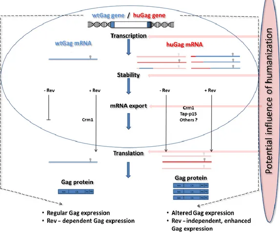
Figure C-12: Schematic illustration of the objective of this thesis:
The Impact of partial humanization (wtGag vs. huGag) should be investigated on different biological stages. Quantification of mRNA and protein levels should be performed as well as experiments analyzing the differences in transcription efficiency, mRNA stability, export pathways and effects on translational processes.
- 24 -
increased gag expression in dependency of the ratio of native and optimized codons [101]. To analyze
positional and additive effects of codon optimization in a more detailed and systematic way, the
subgenomic reporter system should be used in this work to analyze the effects of partially humanized
variants. Therefore, the expression of the partially humanized variants should be analyzed on protein
as well as mRNA levels, either by ELISA or by quantitative real time PCR. To get insight into the
underlying molecular mechanisms, different molecular biological analyses regarding mRNA half-life,
or transcriptional efficiency should be performed, as well as the analysis of potential alternative
splicing effects. To address the question to what extent the results would be transferable to other
genes, egfp should be used as an additional reporter gene. For this, a quasi-lentiviral GFP reporter
system should be used, which our group established in a previous work [59], [60]. Taken together,
the aim of this work should be to address the impact of humanization on the gene expression of HIV-
1 in the context of various biological processes, as shown in Figure C-12.
- 25 -
D Material and methods
Cell lines
D.1.1 Prokaryotic cell lines
Table D-1: Overview of prokaryotic strains
D.1.2 Eukaryotic cell lines
Table D-2: Overview of eukaryotic cell lines used
Nucleic acids
D.2.1 Oligonucleotides
Table D-3: Overview of oligonucleotides used
Primer name Sequence Usage
Gag_c_wt_hu rev CTGCAGTGTACTAGTAGTTCCTGCTATGTCACTTC cloning of gag variants Gag_c_wt_hu fwd CAGGAACTACTAGTACACTGCAGGAACAGATCG cloning of gag variants Gag_d_hu_wt rev GGCCCTGCAGTTCTTGGCAATGTGGC cloning of gag variants Gag_d_hu_wt fwd CCAAGAACTGCAGGGCCCCTAGGAAAAAGGG cloning of gag variants Gag_c_wt_hiv rev CTCTTGCAGTGTACTAGTAGTTCCTGCTATGTCACTTC cloning of gag variants Gag_d_hiv_wt fwd GCAAAGAACTGTAGGGCCCCTAGGAAAAAGGG cloning of gag variants Gag_c_wt_hiv fwd GGAACTACTAGTACACTGCAAGAGCAGATAGGAT cloning of gag variants Gag_d_hiv_wt rev GGGCCCTACAGTTCTTTGCTATGTGCCC cloning of gag variants Gag_b_wt_hu rev GCACCATTTGCCCCTGGAGGTTCTG cloning of gag variants Gag_b_wt_hu fwd CCAGGGGCAAATGGTGCATCAGGCCATC cloning of gag variants
Strain Genotype
DH5α F- supE44 ΔlacU169 (φ80 lacZΔM15) hsdR1 recA1 endA1 gyrA96 thi- 1 relA1 [129]
DH10B F- mcrA Δ(mrr-hsdRMS-mcrBC) Φ80dlacZΔM15 ΔlacX74 endA1 recA1 deoR Δ(ara,leu)7697 araD139 galU galK nupG rpsL λ- [130]
Cell line Description
HEK293T Ad5-transformed human embryonic kidney cell line [131], expressing the “SV40 large T-antigen” [132]
HeLa Human epithelial cell line (derived from cervix carcinoma)
- 26 -
Gag_b_wt_hiv fwd CAGGGGCAAATGGTACACCAGGCAATATCAC cloning of gag variants Gag_f_hu_wt rev GATCTTTACTGGCTGCTAGGATCGC cloning of gag variants Gag_f_hu_wt fwd CCTAGCAGCCAGTAAAGATCTTCAGACCTGGAGGAG cloning of gag variants Gag_f_hiv_wt rev GTCTGAAGATCTTTACTGCTTGGATCGCTTC cloning of gag variants Gag_f_hiv_wt fwd CAAGCAGTCAGTAAAGATCTTCAGACCTGGAGGAG cloning of gag variants Gag_e_hu_wt rev CCTAAAAAATTGGCCTGCCGCTCG cloning of gag variants Gag_e_hu_wt fwd GAGCGGCAGGCCAATTTTTTAGGGAAGATCTGGCCTTCC cloning of gag variants Gag_e_hiv_wt rev TCTGAAGAAAATTTGCCTGTCTCTCTGTGCA cloning of gag variants Gag_e_hiv_wt fwd CACAGAGAGACAGGCAAATTTTTTAGGGAAGATCTGGCCTTCC cloning of gag variants Gag_a_wt_hu rev GGCTCCCATCTCTCTCCTTCTAGCCTCCG cloning of gag variants Gag_a_wt_hiv rev CTCTTGCTCCCATCTCTCTCCTTCTAGCCTCCG cloning of gag variants Gag_a_wt_hu fwd GAAGGAGAGAGATGGGAGCCAGAGCCTC cloning of gag variants Gag_a_wt_hiv fwd GGAGAGAGATGGGAGCAAGAGCAAGC cloning of gag variants Gag_b_wt_hiv rev GGTGTACCATTTGCCCCTGGAGGTTCTG cloning of gag variants Gag_c2_hu_wt fwd CCACCAGCACCCTTCAGGAACAAATAGGATGG cloning of gag variants Gag_c2_hu_wt rev CTGAAGGGTGCTGGTGGTGCCGG cloning of gag variants ß-actin-Exon3-fwd CACTGTGCCCATCTACGAGG cloning of gag variants ß-actin-Exon3-rev CTCTTGCTCGAAGTCCAGGG cloning of gag variants Gag_d_wt_hu rev CTGGGGGCTCTGCAATTTTTGGCTATGTGCCCTT cloning of gag variants Gag_d_wt_hu_fwd CAAAAATTGCAGAGCCCCCAGAAAGAAAGG cloning of gag variants Gag_e_wt_hu_rev CCAGGAAGTTAGCCTGTCTCTCAGTACAATCTTT cloning of gag variants Gag_e_wt_hu_fwd AGAGACAGGCTAACTTCCTGGGCAAGATCTG cloning of gag variants TS - A Erw1 (A5-A6)
Rev TGCTGGGCCTTTTTCTTACTTTTGTTTTGCTCTTCCTCTATC cloning of gag variants TS - A Erw1 (A5-A6)
For AAAACAAAAGTAAGAAAAAGGCCCAGCAGGCTGC cloning of gag variants TS - A Erw2 (A4-A6)
Rev TGCACACAATAGAGGACTGCTATTGTATTATATAATGATC cloning of gag variants TS - A Erw2 (A4-A6)
For GCAGTCCTCTATTGTGTGCACCAGCGGATCG cloning of gag variants
TS - A Erw3 (A3-A6)
Rev GCTGGCCCAGTATTTGTCTACAGCCTTCTGATGTC cloning of gag variants TS - A Erw3 (A3-A6)
Fwd GTAGACAAATACTGGGCCAGCTGCAG cloning of gag variants
TS - A Erw4 (A2-A6)
Rev CTGGCCCACACGATATGTTTTAGTTTATATTGTTTCTTTCCCCCTGG cloning of gag variants
- 27 -
TS - A Erw4 (A2-A6)
Fwd CTAAAACATATCGTGTGGGCCAGC cloning of gag variants
TS - A Erw5 (A2-A6)
Rev GACAGCACCGACGCTCTCGCACC cloning of gag variants
TS - A Erw5 (A2-A6)
Fwd GAGCGTCGGTGCTGTCTGGCGGC cloning of gag variants
TS - A Erw6* (A1-A2)
Rev GCTTAATACAGAGGCTCTGGCTCCC cloning of gag variants
TS - A Erw6* (A1-A2)
Fwd AGCCAGAGCCTCTGTATTAAGCGGGGGAGAATTAGATAAATGG cloning of gag variants TS-BsmBI-MK3-
vorne_fwd (105) GAGAGAGAGACGTGGGTGCGAGAGCG cloning of gag variants TS-BsmBI-MK3-
vorne_rev (105) CACCCACGTCTCTCTCTCTCCTTCTAGCCTCC cloning of gag variants TS-BsmBI-MK1_fwd
(hinten_285) GACAAATACCGTCTCTGGGCCAGCTGCAG cloning of gag variants TS-BsmBI-MK1_rev
(hinten_285) GCCCAGAGACGGTATTTGTCTACAGCCTTCTGATGT cloning of gag variants TS-BsmBI-MK1_fwd
(vorne_225) AGCTAGAGAGACGACGATTCGCAGTTAATCCTGGC cloning of gag variants TS-BsmBI-MK1_rev
(vorne_225) CGAATCGTCGTCTCTCTAGCTCCCTGCTTGCC cloning of gag variants TS-BsmBI-MK2_rev
(hinten_225) CTTTCCAGGAGACGGCTCTCTGCTGGCCCAC cloning of gag variants TS-BsmBI-MK2_fwd
(hinten_225) AGAGAGCCGTCTCCTGGAAAGATTCGCCGTGAAC cloning of gag variants TS-BsmBI-MK2_rev
(vorne_165) TGGCCGTCTCCTTAACCGAATTTTTTCCCATTTATCTAATTCTCCC cloning of gag variants TS-BsmBI-MK2_fwd
(vorne_165)
CGGTTAAGGAGACGGCCAGGGGGAAAGAAACAATATAAACTAAA
AC cloning of gag variants
TS-BsmBI-MK3_rev
(hinten_165) CGCAGTGAGACGCTGATCTTCTCCCACTTGTCC cloning of gag variants TS-BsmBI-MK3_fwd
(hinten_165) ATCAGCGTCTCACTGCGGCCTGGC cloning of gag variants
pCMV-LTR_NL4-3 fwd GTACACGCGTTGGAAGGGCTAATTTGGTCCC cloning of gag variants
pCMV-LTR_NL4-3 rev GCCGAGTCCTGCGTC cloning of gag variants
pCMV-LTR_NL4-3 fwd
(PCR1b) GACGCAGGACTCGGC cloning of gag variants
TS-wt15-
90_PCR1a_Rev CTTAATACCGAGGCTCTGGCTCCCATC cloning of gag variants TS-wt15-
90_PCR1b_Fwd AGCCAGAGCCTCGGTATTAAGCGGGGGAG cloning of gag variants
- 28 -
TS-wt30-
90_PCR1a_Rev CTAATTCTCCGCCAGACAGCACAGAGG cloning of gag variants TS-wt30-
90_PCR1b_Fwd
GCTGTCTGGCGGAGAATTAGATAAATGGGAAAAAATTCGGTTAA
GG cloning of gag variants
TS-wt45-
90_PCR1a_Rev GAATTTTTTCCCACTTGTCCAGCTCGCCG cloning of gag variants TS-wt45-
90_PCR1b_Fwd GAGCTGGACAAGTGGGAAAAAATTCGGTTAAGGCCAG cloning of gag variants TS-wt60-
90_PCR1a_Rev CCCTGGCCTTAATCTGATCTTCTCCCACTTGTCC cloning of gag variants TS-wt60-
90_PCR1b_Fwd GAGAAGATCAGATTAAGGCCAGGGGGAAAGAAAC cloning of gag variants TS-wt75-
90_PCR1a_Rev GTTTCTTGCCGCCAGGCCG cloning of gag variants
TS-wt75-
90_PCR1b_Fwd GCCTGGCGGCAAGAAACAATATAAACTGAAGCACATCGTG cloning of gag variants TS-wt30-
75_PCR1a_Rev CTAATTCTCCGCCAGACAGCACAGAGG cloning of gag variants TS-wt30-
75_PCR1b_Fwd
GCTGTCTGGCGGAGAATTAGATAAATGGGAAAAAATTCGGTTAA
GG cloning of gag variants
TS-wt45-
60_PCR1a_Rev GAATTTTTTCCCACTTGTCCAGCTCGCCG cloning of gag variants TS-wt45-
60_PCR1b_Fwd GAGCTGGACAAGTGGGAAAAAATTCGGTTAAGGCCTG cloning of gag variants TS-wt15-
30_PCR1a_Rev CTTAATACCGAGGCTCTGGCTCCCATC cloning of gag variants TS-wt15-
30_PCR1b_Fwd AGCCAGAGCCTCGGTATTAAGCGGGGGC cloning of gag variants TS-wt30-
45_PCR1a_Rev CTAATTCTCCGCCAGACAGCACAGAGG cloning of gag variants TS-wt30-
45_PCR1b_Fwd GCTGTCTGGCGGAGAATTAGATAAATGGGAGAAGATCAGAC cloning of gag variants TS-wt60-
75_PCR1a_Rev CCCTGGCCTTAATCTGATCTTCTCCCACTTGTCC cloning of gag variants TS-wt60-
75_PCR1b_Fwd GAGAAGATCAGATTAAGGCCAGGGGGCAAG cloning of gag variants hu60-75_PCR1a_Rev TTTTAGTTTATATTGTTTCTTGCCGCCAGGCC cloning of gag variants hu60-75_PCR1b_Fwd CCTGGCGGCAAGAAACAATATAAACTAAAACATATAGTATGGGCA
AGC cloning of gag variants
hu60-90_PCR1a_Rev CCATACTATATGTTTCAGCTTGTACTGCTTCTTGC cloning of gag variants hu60-90_PCR1b_Fwd GCAGTACAAGCTGAAACATATAGTATGGGCAAGCAGG cloning of gag variants fwd_hu_GFP_FranziW GACCTACGGCGTGCAATGCTTCAGCCGCTACCC cloning of egfp variants
- 29 -
rev_hu_GFP_FranziW CATTGCACGCCGTAGGTCAGGGTGGTCACGAGGGTG cloning of egfp variants fwd_hiv_GFP_FranziW AGTAACAACATTAACATATGGAGTACAATGTTTTAGCAGATATC cloning of egfp variants rev_hiv_GFP_FranziW CTCCATATGTTAATGTTGTTACTAATGTTGGCCAGG cloning of egfp variants huE_rev_FranziW CACGGGTCCGTCACCTATTGGTGTATTTTG cloning of egfp variants huE_fwd_FranziW AATAGGTGACGGACCCGTGCTGCTGCC cloning of egfp variants huA_fwd_FranziW CCTGAAGTTCATCTGTACAACAGGAAAATTACCAGTACCC cloning of egfp variants huA_rev_FranziW CCTGTTGTACAGATGAACTTCAGGGTCAGCTTG cloning of egfp variants huAB_rev_FranziW TATTGTTCTCTCCTGGACGTAGCCTTC cloning of egfp variants huAB_fwd_FranziW CTACGTCCAGGAGAGAACAATATTTTTTAAAGACGACGGAAATTA
TAAAACAAGA cloning of egfp variants
huABC_fwd_FranziW CAAGCTGGAGTACAATTATAATAGCCATAATGTATATATAATGGC
AGACAAAC cloning of egfp variants
huABC_rev_FranziW CATTATGGCTATTATAATTGTACTCCAGCTTGTGCCC cloning of egfp variants huABCD_rev_FranziW CTGGTAATAATACTGGGCCGTCGCCGATGG cloning of egfp variants huABCD_fwd_FranziW GACGGCCCAGTATTATTACCAGACAATCATTATTTAAGCAC cloning of egfp variants huBCDE_fwd_FranziW AATTTATATGCACCACCGGCAAG cloning of egfp variants huBCDE_rev_FranziW GGTGGTGCATATAAATTTTAATGTTAATTTTCCATATGTTGCGTCA
CC cloning of egfp variants
huCDE_rev_FranziW GATGGTGCGTTCTTGTACATATCCTTCTGGCATTG cloning of egfp variants huCDE_fwd_FranziW GGATATGTACAAGAACGCACCATCTTCTTCAAGGAC cloning of egfp variants huDE_fwd_FranziW GGACATAAATTAGAATATAACTACAACAGCCACAACGTC cloning of egfp variants huDE_rev_FranziW GTGGCTGTTGTAGTTATATTCTAATTTATGTCCTAATATATTTCCGT
CCTCTTTAAAG cloning of egfp variants
TS-ATG-wtA-hueGFP-
PCR1a Rev GCTCACCATTTGCCCCTGGAGGTTCTG cloning of egfp variants TS-ATG-wtA-hueGFP-
PCR1b Fwd CAGGGGCAAATGGTGAGCAAGGGCGA cloning of egfp variants
ATG-huA-hueGFP-1a
Rev CACCATCTGGCCCTGCAGATTCTG cloning of egfp variants
ATG-huA-hueGFP-1b
Fwd GCAGGGCCAGATGGTGAGCAAGGGCGA cloning of egfp variants
ATT-huA-hueGFP-
PCR1a Rev TCTGGCTCCAATCTCTCTCCTTCTAGCCTCC cloning of egfp variants ATT-huA-hueGFP-
PCR1b Fwd GGAGAGAGATTGGAGCCAGAGCCTCTG cloning of egfp variants ATT-wtA-hueGFP-1a
Rev CGCACCAATCTCTCTCCTTCTAGCCTCC cloning of egfp variants
ATT-wtA-hueGFP-1b
Fwd GAAGGAGAGAGATTGGTGCGAGAGCGTC cloning of egfp variants
- 30 -
hu_Gag ohne ATG
PCR1a Rev GCTCTGGCTCCAATCTCTCTCCTTCTAGCCTCC cloning of egfp variants hu_Gag ohne ATG
PCR1b Fwd GAGAGAGATTGGAGCCAGAGCCTCTG cloning of egfp variants
wt_Gag ohne ATG
PCR1a Rev CTCGCACCAATCTCTCTCCTTCTAGCCTCC cloning of egfp variants wt_Gag ohne ATG
PCR1b Fwd GGAGAGAGATTGGTGCGAGAGCGTC cloning of egfp variants
Alec_wtA_eGFP_Fusio
n_FW GGGGCAAGCCACCATGGTGAGCAAGGGC cloning of egfp variants
Alec_wtA_eGFP_Fusio
n_RV CATGGTGGCTTGCCCCTGGAGGTTCTG cloning of egfp variants
Alec_huA_eGFP_Fus_F
W GGCCAGGCCACCATGGTGAGCAAGGGC cloning of egfp variants
Alec_huA_eGFP_Fus_
RV CATGGTGGCCTGGCCCTGCAGATTCTG cloning of egfp variants
TS-5'UTR-
wt_C_eGFP_FWD AGGGCCCCTATGGTGAGCAAGGGCGA cloning of egfp variants TS- Gag_A_ATT_1a
Rev TCTGGCTCCAATCTCTCTCCTTCTAGCCTCC mutation of the gag start
codon TS- Gag_A_ATT_1b
Fwd GGAGAGAGATTGGAGCCAGAGCCTCTG mutation of the gag start
codon TS-
Gag_A_ATT_hueGFP 1a Rev
CTCACCATCTGGCCCTGCAGATTCTG mutation of the gag start codon
TS-
Gag_A_ATT_hueGFP 1b Rev
CAGGGCCAGATGGTGAGCAAGGGCGA mutation of the gag start codon
TS-SD1_GT-AA (fwd) CGACTGAAGAGTACGCCAAAAATTTTGACTAGCG mutation of the splice donor SD1
TS-SD1_GT-AA (Rev) CAAAATTTTTGGCGTACTCTTCAGTCGCCGCCCC mutation of the splice donor SD1
TS_Rev_cloning_Fwd CCAGGGTACCCTCGAAGCTAGT cloning of Rev expression plasmid
TS_Rev_cloning_Rev CTGCTCGAGCTGTGGCATTGAG cloning of Rev expression plasmid
TS- NADH 1
(mito)_fwd CCACATCTACCATCACCCTC analysis nuclear and
cytoplasmic RNA fractions TS- NADH 1
(mito)_rev CCTAGGAAGATTGTAGTGGTGAG analysis nuclear and
cytoplasmic RNA fractions TS- U2 small nuclear
1_fwd GCTAAGATCAAGTGTAGTATCTGTTC analysis nuclear and
cytoplasmic RNA fractions
- 31 -
TS- U2 small nuclear
1_rev GCACCGTTCCTGGAGG analysis nuclear and
cytoplasmic RNA fractions
TS_Rev_seq_1_Fwd CTACCCTGTCCACCCCTCTG sequencing primer
TS_Rev_seq_2_Fwd CGGAGCTGAATGAAGCCATAC sequencing primer
TS_Rev_seq_3_Fwd GAAAGGCGGACAGGTATCCG sequencing primer
CMV-Seq CGCAAATGGGCGGTAGGCGTG sequencing primer
TS-LTR Seq.1 (fwd) GCTGCTTCGCGATGTAC sequencing primer
TS-LTR Seq.2 (fwd) GACGCAGGACTCGGC sequencing primer
TS-LTR Seq.3 (rev) CAATTGTCCCTCATATCGCCTC sequencing primer
TS-LTR Seq.4 (fwd) GATCTTCAGACCTGGAGGAG sequencing primer
D.2.2 Plasmids
Table D-4: Overview of plasmids used
Plasmid Description
pc-UTR-wtGag16-30-huGag- RRE
eukaryotic expression plasmid with CMV-promoter, partially humanized gag gene, from Alec Geßner (see [133])
pc-UTR-wtGag31-45-huGag- RRE
eukaryotic expression plasmid with CMV-promoter, partially humanized gag gene, from Alec Geßner (see [133])
pc-UTR-wtGag46-60-huGag- RRE
eukaryotic expression plasmid with CMV-promoter, partially humanized gag gene, from Alec Geßner (see [133])
pc-UTR-wtGag61-75-huGag- RRE
eukaryotic expression plasmid with CMV-promoter, partially humanized gag gene, from Alec Geßner (see [133])
pc-UTR-wtGag76-90-huGag- RRE
eukaryotic expression plasmid with CMV-promoter, partially humanized gag gene, from Alec Geßner (see [133])
pc-UTR-wtGag16-90-huGag- RRE
eukaryotic expression plasmid with CMV-promoter, partially humanized gag gene, from Alec Geßner (see [133])
pc-UTR-wtGag31-90-huGag- RRE
eukaryotic expression plasmid with CMV-promoter, partially humanized gag gene, from Alec Geßner (see [133])
pc-UTR-wtGag46-90-huGag- RRE
eukaryotic expression plasmid with CMV-promoter, partially humanized gag gene, from Alec Geßner (see [133])
pc-UTR-wtGag61-90-huGag- RRE
eukaryotic expression plasmid with CMV-promoter, partially humanized gag gene, from Alec Geßner (see [133])
pc-UTR-wtGag31-75-huGag- RRE
eukaryotic expression plasmid with CMV-promoter, partially humanized gag gene, from Alec Geßner (see [133])
pc-UTR-ATT-huA-huEGFP eukaryotic expression plasmid with CMV-promoter, gag 5'UTR, RRE first 423bp of humanized gag gene without start codon and humanized egfp gene from Alec Geßner (see [133])
pc-UTR-ATT-wtA-huEGFP eukaryotic expression plasmid with CMV-promoter, gag 5'UTR, RRE first 423bp of wild-type HIV-1 gag gene without start codon and humanized egfp gene from Alec Geßner (see [133])
pc-UTR-wtGag-RRE eukaryotic expression plasmid with CMV-promoter, wild-type HIV-1 gag gene, from Nane Eiber (see [112])
pc-UTR-huGag-RRE eukaryotic expression plasmid with CMV-promoter, wild-type HIV-1 gag gene, from Nane Eiber (see [112])
pc-UTR-huABC-RRE eukaryotic expression plasmid with CMV-promoter, wild-type HIV-1 gag gene, from Nane Eiber (see [112])
- 32 -
pcDNA3.1 eukaryotic expression plasmid (Thermo Fisher, Waltham, USA - V79020) pcDNA3.1-Rev eukaryotic expression plasmid with HIV-1 rev gene (Wager Lab)
pcDNA3.1-Tat eukaryotic expression plasmid with HIV-1 tat gene (Wagner Lab)
D.2.3 Antibodies
Table D-5:Overview of antibodies used
D.2.3.1 Enzymes
Table D-6:
D.2.4 Commercial Kits
Table D-7:
Antibody Supplier/Specification
M01-antibody (anti-p24) Polymun (AB006) 37G12-antibody (anti-p24),
biotinylated
Polymun (AB005)
Anti-DIG Antibody (from DIG Northern Starter Kit)
Polyclonal sheep anti-digoxigenin, Fab-fragments, conjugated to alkaline phosphatase
Primer Sequence
Restriction endonucleases New England Biolabs
Trypsin/EDTA Pan Biotech
Phusion DNA-Polymerase Finnzymes Calf-Intestine-Phosphate (CIP) Roche
Kit Supplier
Plasmid Plus Midi Kit Roche Plasmid Plus Maxi Kit Roche
RNeasy Kit Roche
- 33 -
D.2.5 Standards
Table D-8: Overview of DNA and RNA standards used
D.2.6 Computer programs and databases
Table D-9: Overview of programs and databases used
Gel Extraction Kit Roche
Quantinova Probe PCR Kit Roche Reverse Transcription Kit Roche
Quick Ligation Kit New England Biolabs DIG Northern Starter Kit Sigma-Aldrich
Primer Supplier
100 bp DNA ladder New England Biolabs 1 kb DNA ladder New England Biolabs Transcript RNA Markers
0.2-10 kb Sigma- Aldrich
Program/ Database Specification/Supplier
Kazusa http://www.kazusa.or.jp/codon/
Codon Usage Exe Kindly provided from Benedikt Asbach; customized program that calculates CAI according to the formula by Sharp, 1987 [ref], GC-content and dinucleotide frequencies
Grapd Pad Prism Used for figures and diagrams
Chromas Used for sequence analysis
pDRAW 32 Used for in silico simulation of cloning
Ape Plasmid Editor Used for sequence analysis and in simulation of cloning in silico Step One Software Used for evaluation of RT-qPCR results
Corel Draw Used for figures
- 34 -
Cell culture techniques
D.3.1 Cultivation of eukaryotic cells
All cell culture techniques were performed under sterile conditions in a class II laminar flow hood.
HEK293T cells were cultivated at 37°C and 5% CO
2. Before reaching full confluence, the cells were split 1:10. For this, cells were washed with 10 ml PBS and then incubated with 5 ml trypsin/EDTA for 5 minutes. The reaction was stopped by the addition of DMEM
10Medium and cells were transferred into a suitable tube. Cells were then centrifuged at 300g for 5 minutes and the cell pellet was resuspended in 10 ml DMEM
10. 1 ml of the cell suspension was transferred into a flask with a suitable amount of DMEM
10. Cell concentrations were determined using trypan blue in a ratio of 1:1 for separation between living and dead cells during counting in a hemocytometer.
Table D-10: Media and reagents for cultivation of eukaryotic cells
![Figure C-2: Schematic structure of an HIV particle (modified after [10])](https://thumb-eu.123doks.com/thumbv2/1library_info/3738968.1509197/10.893.165.703.740.998/figure-c-schematic-structure-hiv-particle-modified.webp)
![Figure C-3: Replication cycle of HIV-1 (from [16])](https://thumb-eu.123doks.com/thumbv2/1library_info/3738968.1509197/11.893.156.764.486.920/figure-c-replication-cycle-hiv.webp)
![Figure C-5: Schematic overview of the budding process of HIV-1 mediated by Gag (from [33])](https://thumb-eu.123doks.com/thumbv2/1library_info/3738968.1509197/13.893.118.768.631.880/figure-schematic-overview-budding-process-hiv-mediated-gag.webp)
![Figure C-6: Five stem model of the Rev-responsive element from [34]:](https://thumb-eu.123doks.com/thumbv2/1library_info/3738968.1509197/14.893.104.486.780.1099/figure-c-stem-model-rev-responsive-element.webp)
![Figure C-7: Role of Rev in the HIV-1 life cycle (from [58])](https://thumb-eu.123doks.com/thumbv2/1library_info/3738968.1509197/15.893.197.679.460.849/figure-c-role-rev-hiv-life-cycle.webp)
![Figure C-8: Schematic illustration of different RNA export pathways (from [65]](https://thumb-eu.123doks.com/thumbv2/1library_info/3738968.1509197/17.893.121.769.528.879/figure-c-schematic-illustration-different-rna-export-pathways.webp)
![Figure C-10: Schematic and simplified overview of different processes involved in protein biosynthesis (from [69]) (1) Unwinding of the DNA double helix to allow transcription of the desired sequence into an mRNA molecule (2) which gets transported throu](https://thumb-eu.123doks.com/thumbv2/1library_info/3738968.1509197/19.893.229.662.191.594/schematic-simplified-different-processes-biosynthesis-unwinding-transcription-transported.webp)