Gene expression profiling leading to identification of essential components in
EDS1/PAD4-regulated plant defence
Inaugural–Dissertation zur
Erlangung des Doktorgrades
der Mathematisch-Naturwissenschaftlichen Fakultät der Universität zu Köln
vorgelegt von
Michael Bartsch
aus Lübeck
Köln, Mai 2005
Die vorliegende Arbeit wurde am Max-Planck-Institut für Züchtungsforschung in Köln in der Abteilung für Molekulare Phytopathologie (Direktor: Prof. Dr.
Paul Schulze-Lefert) angefertigt.
Berichterstatter: Prof. Dr. Paul Schulze-Lefert Prof. Dr. Diethard Tautz
Prüfungsvorsitzender: Prof. Dr. Martin Hülskamp
Tag der mündlichen Prüfung: 4. Juli 2005
Summary
Plants possess multiple mechanisms to detect pathogen attack and protect themselves against colonisation. The antagonistic interplay of positive and negative regulators allows the plant to spacially and temporarily control defence responses. EDS1 (Enhanced Disease Susceptibility1) and PAD4 (Phytoalexin Deficient4) encode lipase- like proteins that positively regulate plant basal resistance to virulent pathogens.
Additionally, EDS1 and PAD4 are recruited by resistance (R) genes of the TIR-NBS- LRR but not of the CC-NBS-LRR type in R gene-mediated resistance. Previous experiments demonstrated that EDS1 and PAD4 are required for accumulation of salicylic acid (SA), a phenolic signal in defence to biotrophic pathogens. Recent findings suggest that EDS1 and PAD4 promote defence also independently of SA. This as yet uncharacterised EDS1/PAD4-controlled pathway is important for full expression of local R gene-triggered and basal resistance as well as for systemic immunity.
To identify components involved specifically in EDS1/PAD4-controlled signalling, transcriptional profiles of Arabidopsis thaliana wild-type, eds1 and pad4 mutant plants were examined during early R gene-mediated defence using whole-genome oligonucleotide microarrays. In wild-type, the inoculation with strains of the bacterial plant pathogen, Pseudomonas syringae pv. tomato, expressing either avrRpm1 (avr1;
recognised by a CC-NBS-LRR-type R protein) or avrRps4 (avr4; recognised by a TIR-
NBS-LRR-type R protein) triggered transcriptional changes in a similar set of genes but
with different kinetics. Sets of genes with EDS1- and PAD4-dependent expression in
healthy, avr1- or avr4-challenged leaves were identified. For a subset of these genes,
corresponding insertional mutants were isolated and tested for alterations in pathogen
resistance. The mutant screen resulted in the identification of a flavin-dependent
monooxygenase (FMO) as a positive regulator and two sequence-related NUDIX
(nucleoside diphosphates linked to some other moiety x) hydrolases as negative
regulators of plant disease resistance. This study demonstrates for the first time that
FMOs and NUDIX hydrolases can modulate host defence responses against pathogens
in any biological system. The findings presented here support the view that EDS1 and
PAD4 control the expression of both positive and negative regulators as a mean to fine-
tune plant immune responses.
Zusammenfassung
Pflanzen besitzen vielfältige Detektions- und Abwehrmechanismen, die sie gegen einen Pathogenangriff schützen. Dabei erlaubt das antagonistische Zusammenwirken von positiven und negativen Regulatoren der Pflanze ihre Abwehrmaßnahmen zeitlich und räumlich zu steuern. EDS1 (Enhanced Disease Susceptibility1) und PAD4 (Phytoalexin Deficient4) kodieren Proteine mit Homologie zu eukaryotischen Lipasen und sind positive Regulatoren der pflanzlichen basalen Resistenz gegen virulente Pathogene.
Ferner erfordert auch die durch Resistenzproteine (R) der TIR-NBS-LRR-Klasse (aber nicht der CC-NBS-LRR-Klasse) vermittelte Abwehrreaktion gegen avirulente Pathogene EDS1 und PAD4. Beide Proteine werden sowohl bei basaler als auch TIR-NBS-LRR vermittelter Resistenz für die Akkumulation der phenolischen Signalsubstanz Salizylat (SA) benötigt. Dennoch gibt es Hinweise auf eine SA-unabhängige Signalfunktion von EDS1 und PAD4, welche für eine effektive lokale als auch systemische Abwehrreaktion essentiell ist.
Um Komponenten dieses EDS1/PAD4-abhängigen Signalweges zur identifizieren, wurde während der frühen Phase der R-Gen vermittelten Pathogenabwehr eine vergleichende Transkriptionsanalyse mittels Oligonukleotid-Mikroarrays zwischen Arabidopsis thaliana Wildtyp und den Mutanten eds1 und pad4 durchgeführt. In Wildtyp- Pflanzen führten Inokulationen mit isogenen Stämmen des bakteriellen Pflanzenpathogens Pseudomonas syringae pv. tomato, welche die Avirulenzproteine avrRpm1 (avr1; detektiert von CC-NBS-LRR R-Protein RPM1) bzw. avrRps4 (avr4;
detektiert von TIR-NBS-LRR R-Protein RPS4) exprimieren, zur Induktion bzw.
Repression von ähnlichen Gengruppen, allerdings mit unterschiedlicher Kinetik.
Weiterhin wurden Gengruppen mit einer EDS1/PAD4-abhängigen Expression im unbehandelten Zustand und nach Pathogenbehandlung mit avr1 oder avr4 identifiziert.
Für einige dieser Kandidatengene wurden Insertionsmutanten isoliert und auf
Veränderung ihrer Pathogenresistenz untersucht. Die Phenotypisierung der
Insertionsmutanten führte zur Identifizierung einer Flavin-abhängigen Monooxygenase
(FMO) als positiven Regulator und zweier NUDIX- (nucleoside diphosphates linked to
some other moiety x) Hydrolasen als negative Regulatoren pflanzlicher
Abwehrreaktionen. Die vorliegende Arbeit zeigt erstmalig, dass FMOs und NUDIX-
Hydrolasen des Wirtes dessen Abwehrmaßnahmen gegen Pathogene modulieren
können. Des Weiteren konnte dargelegt werden, dass EDS1 und PAD4 die Expression
von positiven sowie von negativen Regulatoren steuern und damit zur Feinregulierung
von pflanzlicher Pathogenabwehr beitragen.
Abbreviations
° C degree Celsius
avr avirulence
avr1 Pseudomonas syringae pv. tomato DC3000 expressing avrRpm1
avr4 Pseudomonas syringae pv. tomato DC3000 expressing avrRps4
BTH benzo(1,2,3)thiadiazole-7-carbothioic acid S-methyl ester CC coiled-coil
cDNA complementary DNA
cfu colony forming unit
dpi days post inoculation
DEPC diethylpyrocarbonate
dH
20 deionised water
DNA deoxyribonucleic acid
EDS1 Enhanced Disease Suseptibility1
FMO flavin-dependent monooxygenase
f. sp. forma specialis
g gravity constant (9.81 ms
-1)
GUS beta-glucuronidase
h hours (post inoculation)
HR hypersensitive response
LRR leucine-rich repeats
Mg (treatment with) magnesium chloride solution mRNA messenger ribonucleic acid
NBS nucleotide binding site
NT non-treated
NUDIX nucleoside diphosphates linked to some other moiety x
OD optical density
PAD4 Phytoalexin Deficient4
PAMP pathogen-associated molecular pattern
PCR polymerase chain reaction
pH negative decimal logarithm of the H
+concentration
PR pathogenesis related
Psm Pseudomonas syringae pv. maculicola Pst Pseudomonas syringae pv. tomato pv. pathovar
qRT-PCR quantitative real-time polymerase chain reaction R resistance
RNA ribonucleic acid
ROS reactive oxygen species
RT-PCR reverse transcription-polymerase chain reaction
SA salicylic acid
SAR systemic acquired resistance
T-DNA transfer DNA
TIR Drosophila Toll and mammalian interleukin-1 receptor UV ultraviolet
vir virulence WT wild-type
Table of contents
SUMMARY I
ZUSAMMENFASSUNG III
ABBREVIATIONS V
TABLE OF CONTENTS VII
1. INTRODUCTION 1
1.1 Arabidopsis thaliana as a model host of plant pathogens 1
1.2 Plant defence against biotrophic pathogens 2
1.2.1 Non-host resistance 2
1.2.2 R gene-mediated resistance 2
1.2.3 Basal resistance 4
1.2.4 Systemic acquired resistance and the role of salicylic acid 5
1.3 SA-independent signalling 5
1.3.1 Early signalling events 6
1.3.2 Jasmonic acid and ethylene signalling 6 1.3.3 Evidence for lipid-derived signals in regulating plant defences 7 1.4 Transcriptional reprogramming during plant defence responses 8 1.5 EDS1 and PAD4 are positive regulators in plant defence signalling 9 1.6 EDS1 and PAD4-controlled gene expression 12
1.7 Thesis aims 13
2. MATERIALS AND METHODS 15
2.1 Materials 15
2.1.1 Plant materials 15
2.1.2 Pathogens 16
2.1.2.1 Peronospora parasitica 16
2.1.2.2 Pseudomonas syringae pv. tomato 17
2.1.2.3 Golovinomyces orontii 17
2.1.3 Oligonucleotides 17
2.1.4 Enzymes 18
2.1.4.1 Restriction Endonucleases 18
2.1.4.2 Nucleic acid modifying enzymes 19
2.1.5 Chemicals 19
2.1.6 Antibiotics 19
2.1.7 Buffers and solutions 19
2.2 Methods 21
2.2.1 Maintenance and cultivation of Arabidopsis plant material 21
2.2.2 Generation of Arabidopsis F1 and F2 progeny 22 2.2.3 Inoculation and maintenance of P. parasitica 22 2.2.4 Quantification of P. parasitica sporulation 22 2.2.5 Lactophenol trypan blue staining 23 2.2.6 Maintenance of P. syringae pv. tomato cultures 23 2.2.7 P. syringae pv. tomato inoculations and growth assay 23 2.2.8 A. thaliana powdery mildew Golovinomyces orontii 24 2.2.9 Isolation of Arabidopsis genomic DNA (Quick prep for PCR) 25 2.2.10 Isolation of total RNA from Arabidopsis 25 2.2.11 Polymerase chain reaction (PCR) 26 2.2.12 Reverse transcription-polymerase chain reaction (RT-PCR) 27 2.2.13 Quantitative real-time polymerase chain reaction (qRT-PCR) 27
2.2.14 Plasmid DNA isolation 28
2.2.15 Restriction endonuclease digestion of DNA 28 2.2.16 Isolation of DNA fragments from agarose gel 28
2.2.17 DNA sequencing 29
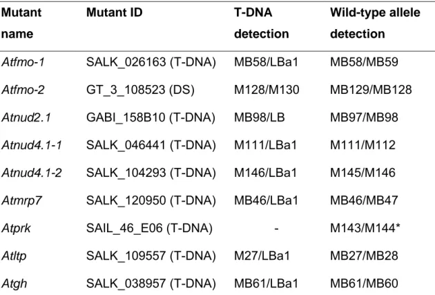
2.2.18 Standard DNA sequence analysis 29 2.2.19 Staining for beta-glucuronidase (GUS) activity 29 2.2.20 Determination of total salicylic acid levels in leaves 29 2.2.21 Identification of Arabidopsis insertion mutants 30
2.2.22 Sequence analysis 31
2.3 Microarray analysis 33
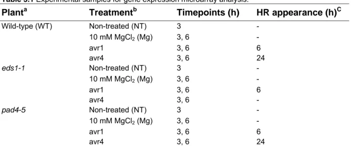
2.3.1 Sample preparation 33
2.3.2 Data analysis 34
3. RESULTS 37
3.1 Microarray analysis 37
3.1.1 Experimental design 37
3.1.2 Sample preparation and quality control 39 3.1.3 Global transcriptional changes upon inoculation with avirulent P. syringae in wild-type plants 40 3.1.4 Effect of the eds1 and pad4 mutations on avr1- and avr4-triggered transcriptional changes 44 3.1.5 Identification of genes controlled by EDS1 and PAD4 48
3.2 Screen for altered resistance phenotypes to the oomycete Peronospora parasitica isolate Cala2 53 3.2.1 Aberrant defence responses in Atfmo and Atnud4.1 T-DNA insertional mutants 53
3.3 Defining the role of AtFMO in plant defence 55 3.3.1 AtFMO is required for EDS1/PAD4-controlled defence responses 55 3.3.2 Analysis of AtFMO expression and SA accumulation in wild-type and mutants 59 3.3.3 Double mutant analysis supports an SA-independent function of AtFMO 63 3.3.4 AtFMO has motifs characteristic for flavin-dependent monooxygenases but does not have close homologues in Arabidopsis 65
3.4 Functional characterisation of AtNUD2.1 and AtNUD4.1 68 3.4.1 Sequence and transcriptional analysis for AtNUD2.1 and AtNUD4.1 68 3.4.2 T-DNA mutants of two homologous NUDIX hydrolases display constitutive defence responses and
enhanced basal resistance 70
4. DISCUSSION 77
4.1 A quantitative model to explain defence-related global transcriptional reprogramming and corresponding mutant effects 77
4.2 EDS1/PAD4-regulated genes might function either downstream of EDS1/PAD4 or as part of a
positive feed back loop 81
4.3 EDS1/PAD4-dependent genes – More to discover? 83
4.4 AtFMO - a positive regulator of plant defence responses 84 4.4.1 AtFMO transcript accumulation and defence function is partially independent of SA 84 4.4.2 AtFMO – possible biochemical activity and substrates 85
4.5 NUDIX hydrolases as negative regulators of plant defence responses 90 4.5.1 Constitutive defence symptoms and enhanced resistance in NUDIX hydrolase knock-out mutants 90 4.5.2 Potential NUDIX hydrolase function in regulating plant stress responses 92 4.5.3 Control of NUDIX hydrolases by EDS1 and PAD4 94
OUTLOOK 96
REFERENCES 97
SUPPLEMENTARY DATA 111
DANKSAGUNG 112
PUBLIKATION 113
ERKLÄRUNG 114
LEBENSLAUF 115
1. Introduction
Plants are the ultimate source of food for humans and animals. The concentration on a few plant species in agriculture and forestry and their cultivation in monoculture has favoured the out break of plant diseases, sometimes with devastating consequences.
A better understanding of plant-pathogen interactions will enable us to develop means that will help to make plant production more predictable.
1.1 Arabidopsis thaliana as a model host of plant pathogens
Despite the lack of any important commercial value of this member of the mustard family, Arabidopsis thaliana has several traits which make it an ideal model organism for plant genetic research. Its small plant size, the short life cycle (about 6 weeks) and its large seed production make it possible to grow it on limited space and in rapid manner. Because of the named advantages and its relatively small genome (125 Mb), Arabidopsis was the first plant whose genome has been fully sequenced in a multinational effort (Initiative, 2000). The availability of the genome sequence was the starting point for the development of genomic tools, making it possible to study transcriptional changes at a whole genome-wide scale (Redman et al., 2004).
Arabidopsis is host to different classes of pathogens including oomycetes, fungi, viruses and bacteria (Mauch-Mani and Slusarenko, 1993). Depending on the mode of the infection, pathogens are classified as necrotrophic (host cell is killed), biotrophic (cell remains alive) or hemibiotrophic (cell is killed later in the infection process).
Knowledge about the existence of pathogens that attack Arabidopsis was subsequently used to study plant-microbe interactions in this model organism. The obligate biotrophic pathogen oomycete Peronospora parasitica (Crute et al., 1992;
Parker et al., 1993; McDowell et al., 2000) and biotrophic bacterial strains of
Pseudomonas syringae (Whalen et al., 1991; Volko et al., 1998) were especially
useful to unravel mechanisms of plant disease and host resistance.
1.2 Plant defence against biotrophic pathogens
The observation that most plants appear healthy in an environment full of potential disease causing agents lead to the conclusion that plants have developed extremely effective defence systems that normally protect them from disease.
1.2.1 Non-host resistance
The most prevalent form of disease resistance in the field is called “non-host resistance”. It is defined as resistance expressed by an entire plant species to a pathogen that normally infects another plant species (Heath, 2000). Although non- host resistance is not well understood at the molecular level, it involves preformed defence barriers (e.g. cuticle on the leaf surface) and inhibitory plant metabolites (e.g. alkaloids, cyanogenic glycosides, phenols) as well as inducible defence mechanisms. Induction of non-host defence responses can be triggered by recognition of pathogen-associated molecular patterns (PAMPs) (reviewed in Medzhitov and Janeway, 2002). PAMPs are molecules which are characteristic for an entire pathogen class, e.g. flagellin of bacterial pathogens and are recognized in flies, mammals and plants by Toll-like receptors (TLR) (Lemaitre et al., 1997; Felix et al., 1999; Hayashi et al., 2001; Zipfel et al., 2004). Although broadly structurally similar molecular components mediate PAMP-triggered signal transduction pathways in plants and animals, it is not clear if these similarities are due to convergent evolution or common ancestral origin (Nurnberger et al., 2004).
1.2.2 R gene-mediated resistance
Although non-host resistance provides plants with a relatively robust protection from disease, pathogens have developed virulence factors (effectors) that help them to overcome defence mechanisms of certain plant species. As a countermeasure against virulent pathogens, plants have evolved a race-specific resistance that is effective against specific pathogen isolates, thus turning a normally compatible interaction (host develops disease) into an incompatible interaction (host is resistant).
The genetic basis for this so-called race-specific resistance is embodied in the gene-
for-gene hypothesis (Flor, 1971) which states that pathogen recognition is conferred
by products of plant resistance (R) and corresponding pathogen avirulence (Avr)
genes. The recognition event triggers a rapid defence response that often includes a localised programmed cell death of plant cells at the site of attempted invasion, a phenomenon termed hypersensitive response (HR). The accumulation of the phenolic defence molecule salicylic acid (SA) can contribute to HR but is not essential in all cases (Mur, 1997; Kachroo et al., 2000; Mur et al., 2000; Shapiro and Zhang, 2001; Chandra-Shekara et al., 2004). The function of SA in plant defence was extensively studied on SA depleted plants expressing a bacterial salicylate hydroxylase (NahG) which removes SA by conversion to catechol and on the Arabidopsis SA synthesis mutant sid2 (Salicylic Acid Induction Deficient2) (Wildermuth et al., 2001). SID2 encodes an isochorismate synthase, suggesting that SA accumulated during pathogen infection is derived from chorismate. Substantial SA accumulation and transcriptional activation of the SA marker gene PR1 (Pathogenesis-Related1) were detected in Arabidopsis treated with avirulent strains of P. syringae as early as 4 and 10 hours post inoculation (h), respectively (Feys et al., 2001; Zhang et al., 2004).
Several R and corresponding Avr genes have been cloned. The largest class of R genes encode intracellular proteins with a central nucleotide-binding site (NBS) and C-terminal leucine rich repeats (LRRs). Based on the domain at the N-terminus, NBS-LRR proteins are divided into two sub-classes. One class is defined by a domain that has homology to Drosophila Toll and mammalian interleukin (IL)-1 receptors (TIR) and the other class by a coiled-coil domain (CC). Examples of Avr-R protein pairs, identified in genetic studies of the Arabidopsis-Pseudomonas syringae system, are avrRpm1 recognised by RPM1 (CC-NBS-LRR type) and avrRps4 recognised by RPS4 (TIR-NBS-LRR type) (Dangl et al., 1992; Hinsch and Staskawicz, 1996). By introducing avrRpm1 or avRps4 into the virulent strain Pseudomonas syringae pathovar tomato DC3000 (Pst), both resulting strains (Pst- avr1 and Pst-avr4) are converted into avirulent pathogens in Arabidopsis plants that express the corresponding R protein.
Avr genes are defined by their ability to induce disease resistance in host plants but
subsequent studies demonstrated that Avr genes in the absence of corresponding R
genes confer a selective advantage to the pathogen as they act as virulence factors
(Kearney and Staskawicz, 1990; Ritter and Dangl, 1995). Bacterial pathogens of
animals and plants utilise both a type III secretion pathway to deliver Avr gene
products into the host cell (Hueck, 1998).
It was assumed that the recognition process between the products of R and Avr genes is based on a direct interaction. However, molecular characterisation of corresponding R and Avr protein pairs indicate that this event is rather an exception than the rule. Based on these observations the guard hypothesis postulated that Avr proteins (as their function as virulence factors) bind to plant virulence targets (Dangl and Jones, 2001). The role of R proteins is in detection of this Avr protein-virulence target complexes rather than the perception of the Avr protein alone. Thus, R proteins may monitor the binding status or stability of plant virulence targets (“guardees”). The guard hypothesis is supported by various recent findings. In the Arabidopsis-Pseudomonas interaction, RIN4 (RPM1-interactor protein4) was identified as guardee targeted by the two sequence unrelated virulence factors avrRpm1 and avrB (Mackey et al., 2002). RIN4 was shown to interact with avrB and avrRpm1 but also with the CC-NBS-LRR protein RPM1 conferring resistance against Pseudomonas syringae expressing avrRpm1 or avrB. Reduction of RIN4 protein levels hampers resistance to both bacterial strains, indicating that RIN4 is essential for RPM1 mediated resistance. The guard hypothesis made it plausible how a limited number of approximately 128 NBS-LRR type genes in the Arabidopsis genome (Initiative, 2000; Dangl and Jones, 2001) can confer resistance to numerous pathogen races. As R proteins may be guarding a limited number of plant components that are the preferred targets of multiple virulence factors, R proteins should be able to detect invasion of multiple races of pathogens.
1.2.3 Basal resistance
Even when plants are attacked by virulent pathogen (not detected by R proteins),
pathogen growth is to some extent restricted. This phenomenon, called basal
resistance, became apparent with the identification of “enhanced disease
susceptibility” mutants that allowed an even stronger development of disease than
wild-type susceptible hosts (Glazebrook et al., 1996). Although, virulent pathogens
escape R gene-mediated recognition, they trigger a delayed and weak defence
response with similarity to R-mediated defence (e.g. SA accumulation and host
transcriptional reprogramming but no HR). Thus R protein action ensures that
defence responses are triggered in a rapid and strong manner, thus preventing host
colonisation.
1.2.4 Systemic acquired resistance and the role of salicylic acid
A successful local defence response in R-mediated resistance not only leads to local resistance but mediates enhanced resistance to subsequent infections in previously unchallenged parts of the plant. This type of resistance, often referred to as systemic acquired resistance (SAR), is effective against a broad range of biotrophic pathogens (Ryals et al., 1996). The defence signalling molecule SA accumulates locally in R- mediated resistance and is then allocated to systemic leaves via the phloem (Metraux et al., 1990; Shulaev et al., 1995). The coincidence of SA accumulation in systemic leaves and the establishment of SAR led many researchers to believe that SA is the SAR mediating signal. Results from grafting experiments between wild-type and transgenic tobacco expressing the bacterial SA degrading enzyme, NahG, led to the conclusion that SA is not the SAR signal as it was found that the NahG rootstock (SA deficient) was still able to produce and translocate the SAR signal to the wild- type rootstock scion (Vernooij et al., 1994). The reciprocal grafting experiment demonstrated that the NahG systemic scion was unable to perceive the SAR signal emitted from the wild-type scion, indicating that SA accumulation in the systemic tissue is essential for the establishment of SAR.
Consistent with the requirement for SA in SAR establishment, spray application of SA or its synthetic analogue BTH (benzo(1,2,3)thiadiazole-7-carbothioic acid S-methyl ester) induces SAR and transcriptional activation of a typical set of PR genes. Recent publications revealed that SA-mediated redox changes activate the key SA-response regulator, NPR1 (Non-expresser of Pathogenesis-Related genes1), by shifting NPR1 from its inactive oligomeric to its active monomeric form (Mou et al., 2003). Upon its activation, NPR1 binds to TGA transcription factors and stimulates the DNA-binding activity of these transcription factors to promoter elements of SA-responsive genes, resulting in PR gene up-regulation (Despres et al., 2003).
1.3 SA-independent signalling
The findings that SA deficiency in plants only partially compromises local resistance,
the relatively late accumulation of SA and the SA-independent nature of the SAR
signal demonstrate that SA-independent signalling pathways exist.
1.3.1 Early signalling events
Early cellular re-programming events, preceding SA signalling, are induced upon pathogen recognition. Changes in the ion permeability of the plasma membrane resulting in influxes of calcium (Ca
2+), protons (H
+) and an efflux of potassium (K+) and chloride (Cl
-) ions are one of the earliest signalling events after pathogen exposure. Increased intracellular Ca
2+levels are upstream of the production of reactive oxygen species (ROS) (Ligterink et al., 1997; Grant et al., 2000b), a process known as oxidative burst. ROS are produced during the oxidative burst by plasma- membrane-bound NADPH oxidases (Torres et al., 2002), cell wall attached peroxidases (Kawano, 2003) and apoplast-located amine oxidases (Allan and Fluhr, 1997). There are several roles discussed for ROS, including direct pathogen toxicity (Bussink and Oliver, 2001) and the reinforcement of plant cell walls by cross-linking of cell wall polymers (Bradley et al., 1992). The oxidative burst also triggers a change of the cellular redox status, thus connecting it to the SA signalling cascade. There is also evidence for ROS as SA-independent signal that controls early changes in gene expression via a mitogen-activated protein kinase (MAPK) (Grant et al., 2000a). Also, plant protein tyrosine phosphates (PTPs) are discussed to detect redox changes and subsequently regulate MAPKs which then might activate transcription factors (Gupta and Luan, 2003; Laloi et al., 2004). Another early signalling event is mediated by Ca
2+-dependent protein kinases (CDPKs). CDPKs are thought to sense the increase of the Ca
2+concentration triggered in response to different abiotic and biotic stresses and transduce this information via protein kinase activity to downstream signalling events (Ludwig et al., 2004). The biological relevance of CDPK-signalling was reinforced by the finding that CDPK-silenced Nicotiana benthamiana plants showed a weaker and delayed hypersensitive response upon race-specific elicitation in a R- mediated resistance response (Romeis et al., 2001).
1.3.2 Jasmonic acid and ethylene signalling
Plants defective in SA signalling are not more susceptible to the necrotrophic fungus
Botrytis cinerea. Whereas Arabidopsis mutant plants with defects in jasmonic acid
(JA) signalling (coi1, coronatine insensitive1) and ethylene (ET) perception (ein2,
ethylene insensitive2) display an impaired resistance to Botrytis cinerea. In contrast
to hyper-susceptible SA-deficient mutants, coi1 plants are more resistant to virulent
stains of Pseudomonas syringae. The contrasting actions of SA and JA signalling were also reflected by gene expression analysis in which coi1 and SA signalling mutants had mainly opposite effects on global gene expression (Glazebrook et al., 2003). Notably, comparative Arabidopsis gene expression profiling experiments after JA and SA application found that these signalling molecules also induce a common set of genes (Schenk et al., 2000). These results suggest a complex interplay of SA and JA/ET in modulating gene expression and resistance to plant pathogens.
The observation that in Arabidopsis JA/ET-controlled gene expression was induced by non-host but not by host biotrophic powdery mildew pathogens and that ectopic activation of JA/ET signalling conferred resistance to two biotrophic host pathogens suggests that host biotrophic pathogens either fail or actively repress the JA/ET signalling cascade (Zimmerli et al., 2004).
1.3.3 Evidence for lipid-derived signals in regulating plant defences
Besides JA that is derived from linolenic acid, other fatty acid-derived molecules have been implicated as modulators of plant defence signalling. Changes in abundance and composition of oxylipins that are derived from oxidation of fatty acids occurred upon pathogen attack or after wounding (Weber et al., 1997). Alméras et al. (2003) demonstrated that the electrophilic character of many oxylipins makes them potent transcriptional activators of certain marker genes for abiotic and biotic stress.
The identification of several Arabidopsis mutants deficient in aspects of lipid metabolism also points to an important role of lipid signalling in plant defence. For example, Arabidopsis mutant ssi2 (suppressor of SA-insensitivity2) is deficient in oleate caused by a mutated gene encoding a stearoyl-acyl-carrier-protein desaturase and displays constitutive high levels of SA and a enhanced resistance to various biotrophic pathogens (Kachroo et al., 2001; Shah et al., 2001). Analysis of ssi2NahG plants revealed that elevated levels of SA were not essential for the ssi2 phenotype.
Deficiencies in the synthesis of polyunsaturated glycerol lipids in the double mutant
of fatty acid desaturase7 (fad7) and fad8 resulted in a partially defective oxidative
burst, reduced cell death and impaired resistance to avirulent strains of P. syringae
(Yaeno et al., 2004). Another protein, ACD11 (accelerated cell death11) with in vitro
sphingolipid transfer activity was shown to be a negative regulator of programmed
cell death in Arabidopsis (Brodersen et al., 2002). The lethal recessive acd11
mutation triggered spontaneous cell death and constitutive up-regulation of a subset of defence genes including genes encoding the lipase-like proteins EDS1 and PAD4 (EDS1/PAD4 regulatory role in plant defence is discussed in sections 1.5 and 1.6).
Spontaneous cell death was abolished in acd11NahG but can be restored by BTH treatment. Notably, BTH induced cell death did not occur in acd11eds1 and only partially in acd11pad4.
Evidence for the role of lipid-derived molecules in SAR signalling comes from the finding that a mutation in DIR1 (Defective in induced resistance1) encoding a lipid transfer-like protein prevents the emission of a yet unidentified SAR signal after an otherwise intact local defence response. Similarly, mutations in the SFD1 (Suppressor of fatty acid desaturase deficiency1) gene, which affects plastidic glycerolipid composition, compromises the SAR response but not basal resistance to P. syringae (Nandi et al., 2004).
1.4 Transcriptional reprogramming during plant defence responses
The complexity of signalling events following pathogen recognition with multiple signalling molecules and regulators is so immense that a global view on changes at the level of the metabolome or proteome is not yet technically feasible. However, development of large scale gene expression profiling technologies, in particular the emergence of oligonucleotide arrays, allows monitoring of transcriptional reprogramming during plant defence on a genome-wide scale (Redman et al., 2004).
In the following overview I will focus on studies performed on Arabidopsis as high quality, large scale and comparable gene expression profiling data sets are most advanced for this plant species. Examining the transcriptional changes in Arabidopsis upon challenge with different pathogens revealed that up to 23% of the total genes had altered transcript levels (Scheideler et al., 2002; Tao et al., 2003).
The earliest transcriptional changes are thought to be triggered by recognition of
PAMPs. Zipfel et al. (2004) reports that treatment with the bacteria derived flagellin
led to an up-regulation of 966 of approximately 23000 monitored Arabidopsis genes
within 30 minutes (min) of application. Consistent with the idea that the first
transcriptional changes are triggered by PAMP recognition is the finding that the
transcriptional profiles of plants treated with Pst-hrpA
-(mutant strain unable to deliver
type III effectors), Pst or Pst-avr1 do not differ considerably within first 2 h but at later time points (de Torres et al., 2003). Pst-avr1 triggered changes in gene expression were not observed before 3 h.
Induced non-host, basal and R gene-mediated resistance share common signalling events (e.g. Ca
2+-fluxes, ROS burst, SA induction). These similarities are reflected by the observation that all three defence systems induce and repress common sets of genes (Maleck et al., 2000; Tao et al., 2003; Zipfel et al., 2004).
Large differences in the host transcriptional profiles after infiltration of virulent and avirulent P. syringae strains were only observable at early time points (3, 6 and 9 h) but the profile of the compatible interaction at later time points (30 h) were similar to profile of the incompatible interaction at earlier time points (9 h) (Tao et al., 2003).
Thus, differences in the transcriptional profiles between R-meditated and basal resistance appear to be quantitative rather than qualitative. The action of R proteins seems to accelerate and amplify transcriptional reprogramming of basal defence responses.
Although R proteins of the TIR- and CC-NBS-LRR class differ in their dependency on some signalling components, their action induces and represses a common set of genes. This was recently demonstrated by comparing large-scale gene expression profiles of RPP4- (Recognition of Peronospora parasitica4; TIR-NBS-LRR type), RPP7- and RPP8- (both CC-NBS-LRR type) mediated resistance responses to Peronospora parasitica (Eulgem et al., 2004).
1.5 EDS1 and PAD4 are positive regulators in plant defence signalling
Genetic screens in Arabidopsis for mutants with altered defences led to the identification of several important resistance signalling components. EDS1 was first identified as a mutant compromised in R-mediated resistance to Peronospora parasitica (Parker et al., 1996). PAD4 was discovered in a screen for mutants with defects in basal resistance to virulent P. syringae pathovar maculicola (Psm) (Glazebrook et al., 1996). Since their discovery nearly a decade ago, a great amount of knowledge accumulated about their important role in defence signalling.
EDS1 and PAD4 are both required for resistance to various classes of pathogens
(oomycetes, bacteria and viruses) recognised by TIR-NBS-LRR type R proteins
(Parker et al., 2000; Peart et al., 2002). Although, CC-NBS-LRR-mediated resistance signalling is normally dependent on NDR1 (Non-race specific Disease Resistance1) and independent of EDS1 and PAD4 at least one exceptions was found with CC- NBS-LRR type R protein HRT mediating viral resistance in an EDS1/PAD4- dependent manner (Chandra-Shekara et al., 2004). Also two CC-domain containing proteins with predicted trans-membrane domain, RPW8.1 and RPW8.2, conferring resistance to powdery mildew pathogens, depend on EDS1 and PAD4 (Xiao et al., 2003; Xiao et al., 2005).
Characteristically, TIR-NBS-LRR type R protein-mediated resistance is totally abolished in eds1 null mutants but still partially functional in pad4 mutant lines. This was illustrated well in the different phenotypes of eds1 and pad4 to avirulent P.
parasitica strains. In contrast to strictly delimited HR in wild-type, pathogen growth is unimpeded in eds1 whereas in pad4 a delayed HR response allows hyphal growth leading to trailing plant cell necrosis (Feys et al., 2001). Further, SA accumulation in TIR-NBS-LRR-mediated resistance was totally abolished in eds1 but only partially disabled in pad4 (Feys et al., 2001). Similarly, the ROS burst was found to be still intact in pad4 but not in eds1 plants (Rusterucci et al., 2001).
Constitutive resistance triggered by deregulated TIR-NBS-LRR type R proteins was found to be dependent on EDS1 and PAD4 (Shirano et al., 2002; Zhang et al., 2003), suggesting a signalling role for EDS1/PAD4 genetically down-stream of R protein action. Further evidence for an EDS1/PAD4 function downstream of R protein action comes from analysis of BONZAI1 (BON1) encoding a calcium-dependent phospholipid-binding protein. BON1 is a negative regulator of the R gene SNC1. The bon1 mutation results in SNC1-mediated constitutive defence responses and growth defects which require EDS1, PAD4 and SA accumulation (Yang and Hua, 2004).
Studies on Arabidopsis genes that negatively effect the EDS1/PAD4 pathway
were valuable to elucidate the role of EDS1/PAD4 in transducing ROS and SA
defence signals. The lesion-mimic mutant lsd1 (lesion simulating disease resistance
response1) displays a deregulated cell death response (run away cell death) upon
various abiotic and biotic stresses (Dietrich et al., 1994). The deregulated cell death
in lsd1 is caused by its inability to restrict ROS-derived signals. Epistatic analysis
revealed that both EDS1 and PAD4 are necessary for lsd1 conditioned run away cell
death even in response to an artificial provision of ROS or an SA analog (Rusterucci
et al., 2001). Cooperation of ROS and SA is known to be important in triggering
resistance to pathogens (Shirasu et al., 1997). Thus, it was proposed that EDS1/PAD4 regulate an ROS/SA signal amplification loop under negative control of LSD1 (Rusterucci et al., 2001).
MPK4 (MAP kinase4) was recently found to negatively regulate SA accumulation (Petersen et al., 2000) and positively regulate JA/ET signalling both in an EDS1- and PAD4-dependent manner (P. Brodersen and colleagues, personal communication).
Thus, EDS1 and PAD4 might modulate the previously discussed antagonism between SA and JA/ET signalling.
While expression of local resistance and plant cell death triggered by CC-NBS-LRR R proteins upon pathogen recognition is the same as wild-type in eds1 and pad4, these mutants fail to establish SAR. Experiments with phloem exudates indicate that eds1 is defective in emitting SAR signals from local tissue but also in its perception in systemic tissue (L. Jorda, unpublished). Although eds1 compared to pad4 is more defective in TIR-mediated resistance, their deficiency in basal resistance seems to be equivalent (Feys et al., 2001; Rusterucci et al., 2001).
Jirage et al. (1999) found that PAD4 function is redundant in the defence response to Psm-avrRpt2 (recognised by CC-NBS-LRR R protein RPS2) but required in response to virulent Psm. Thus, it was proposed that PAD4 is required for amplification of weak signals that occur by infection of virulent pathogens. In contrast, RPS2-derived signals are strong enough to trigger defence responses and thus do not require amplification by PAD4 (Jirage et al., 1999).
Disabled SA accumulation can only partially explain the eds1 and pad4 mutant phenotypes as R gene-mediated resistance in eds1 and pad4 is more severely compromised than in the SA-deficient sid2 or NahG plants (Feys et al., 2001 and this study).
EDS1 and PAD4 have pockets of homology to eukaryotic lipases (Falk et al., 1999;
Jirage et al., 1999). It was therefore suggested that they might play a role in lipid
based signalling by hydrolysing a lipid substrate. Lipase activity has indeed been
reported for the EDS1/PAD4-related Arabidopsis protein associated with senescence
control, SAG101 (Senescence Associated Gene101) (He and Gan, 2002). Despite
trying various potential substrates under different reaction conditions, S. Rietz and
colleagues (MPIZ, Cologne) did not observe lipase activity for EDS1, PAD4 and
SAG101. Thus, the biochemical nature of EDS1/PAD4 derived signal remains
elusive. Recent findings demonstrate that EDS1, PAD4 and SAG101 work in concert
to regulate basal and TIR-NBS-LRR resistance (Feys et al., submitted). A common signalling function of the three lipase-like proteins is further supported by the finding that EDS1 forms dimeric and potentially multimeric complexes with PAD4 and SAG101 inside the plant cell (Feys et al., 2001 and Feys et al., submitted).
1.6 EDS1 and PAD4-controlled gene expression
As observed for other signalling components involved in plant defence responses, mRNA and protein levels of EDS1 and PAD4 are induced upon pathogen challenge (Feys et al., 2001). Notably, EDS1 and PAD4 proteins exist in the cell prior to pathogen attack, potentially transducing the early defence promoting signals.
Furthermore, it was shown that EDS1 and PAD4 positively influence mutually their mRNA accumulation upon pathogen challenge (Jirage et al., 1999; Feys et al., 2001;
Eulgem et al., 2004). Consistent with their signalling role closely downstream of TIR- NBS-LRR function and in basal resistance, EDS1 and PAD4 are required for pathogen-triggered gene induction from early time points on (Zhou et al., 1998; de Torres et al., 2003).
Glazebrook et al. (2003) applied the microarray technology to study transcriptional changes in Arabidopsis wild-type and mutant plants upon inoculation with virulent P.
syringae. Their study revealed that mutations in SID2 and PAD4 effected common but also different sets of genes. The common set of SID2 and PAD4-controlled genes includes PR1 and most likely represents genes that are induced by SA. The set of genes which is effected by pad4 but not by sid2 was predicted to function in a yet unknown signalling pathway.
By monitoring transcriptional changes in 8000 genes during RPP4-signalling, Eulgem et al. (2004) recently identified seven PAD4 co-regulated genes (including EDS1) with no requirement for NDR1, NPR1 or SA (NahG) but with suppressed mRNA levels in pad4-1. The authors predict that these genes are involved in the EDS1/PAD4 signalling process but they did not demonstrate the biological relevance of these PAD4-coregulated genes in pathogen resistance.
I was interested in identifying essential components of this EDS1/PAD4-regulated
SA-independent defence pathway.
1.7 Thesis aims
As described, several lines of evidence point to the existence of an EDS1/PAD4- controlled signalling pathway in Arabidopsis that functions independently of SA. This mainly uncharacterised EDS1/PAD4 pathway is important for full expression of local R gene-triggered and basal resistance as well as for systemic defence responses.
However, the nature of this signalling pathway or the genetic components involved
are largely unknown. I intended to characterise this important EDS1/PAD4-
conditioned pathway by means of comparative transcriptional profiling of defence
responses in wild-type, eds1 and pad4. In particular, I aimed to combine data derived
from the transcriptional profiling experiment with the use of Arabidopsis insertion
mutant resources to identify, in a targeted approach, novel essential regulators in the
EDS1/PAD4-controlled defence signalling pathway.
2. Materials and Methods
The Materials and Methods section is subdivided into three parts. In the first part (2.1) materials used throughout this study, including plant lines, pathogens, bacterial strains, chemicals, enzymes, media, buffers and solutions are listed, whereas methods applied in this work are described in the second part (2.2) and microarray- related methods are addressed in the third part (2.3).
2.1 Materials
2.1.1 Plant materials
Arabidopsis thaliana wild-type and mutant lines used in this study are listed in the following two tables.
Table 2.1 Wild-type Arabidopsis accessions used in this study.
Accession Abbreviation Original source
Columbia-0 Col-0 J. Dangla
Landsberg-erecta-0 Ler-0 Nottingham Arabidopsis Stock Centreb Wassilewskija-0 Ws-0 K. Feldmannc
aUniversity of North Carolina, Chapel Hill, USA
bNottingham, UK
cUniversity of Arizona, Tucson, USA
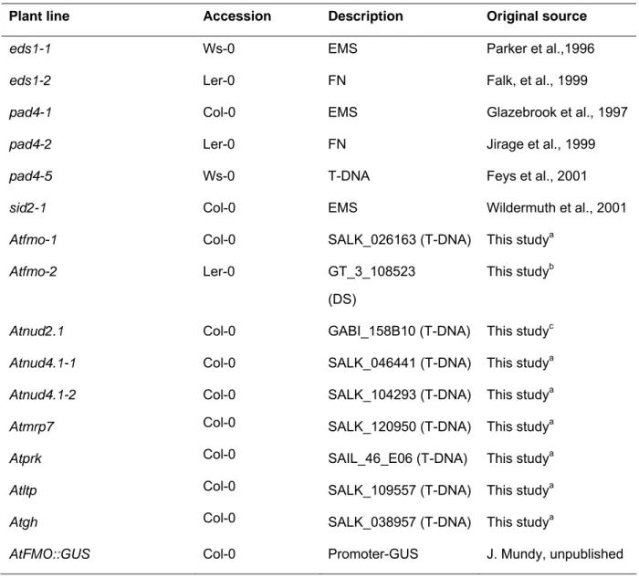
Table 2.2 Mutant and transgenic Arabidopsis lines used in this study.
Plant line Accession Description Original source
eds1-1 Ws-0 EMS Parker et al.,1996
eds1-2 Ler-0 FN Falk, et al., 1999
pad4-1 Col-0 EMS Glazebrook et al., 1997
pad4-2 Ler-0 FN Jirage et al., 1999
pad4-5 Ws-0 T-DNA Feys et al., 2001
sid2-1 Col-0 EMS Wildermuth et al., 2001
Atfmo-1 Col-0 SALK_026163 (T-DNA) This studya
Atfmo-2 Ler-0 GT_3_108523
(DS)
This studyb
Atnud2.1 Col-0 GABI_158B10 (T-DNA) This studyc
Atnud4.1-1 Col-0 SALK_046441 (T-DNA) This studya
Atnud4.1-2 Col-0 SALK_104293 (T-DNA) This studya
Atmrp7 Col-0 SALK_120950 (T-DNA) This studya
Atprk Col-0 SAIL_46_E06 (T-DNA) This studya
Atltp Col-0 SALK_109557 (T-DNA) This studya
Atgh Col-0 SALK_038957 (T-DNA) This studya
AtFMO::GUS Col-0 Promoter-GUS J. Mundy, unpublished
a SALK collection (Alonso et al., 2003) distributed by Nottingham Arabidopsis Stock Centre.
b Ds-insertion line (Sundaresan et al., 1995) distributed by Nottingham Arabidopsis Stock Centre.
c GABI-Kat, Max-Planck-Institute for Plant Breeding Research (Rosso et al., 2003).
2.1.2 Pathogens
2.1.2.1 Peronospora parasitica
Table 2.3 Peronospora parasitica isolates used in this study.
Isolate Original source Reference
Cala2 Oospore infection of a single seedling (Holub et al., 1994a) Emco5 Oospore infection of a single seedling (Holub et al., 1994a) Noco2 Conidia isolated from a single seedling (Parker et al., 1993)
Peronospora parasitica isolates and their interaction with Arabidopsis ecotypes Arabidopsis ecotype Peronospora parasitica isolate
Cala2 Emco5 Noco2
Col-0 incompatible (RPP2)
intermediate (sporulation on cotyledons)
compatible
Ler-0 compatible incompatible
(RPP8)
incompatible (RPP5) Ws-0 incompatible
(RPP1A)
compatible incompatible (RPP1)
2.1.2.2 Pseudomonas syringae pv. tomato
Pseudomonas syringae pv. tomato strain DC3000 expressing the avirulence determinants avrRps4 (Hinsch and Staskawicz, 1996) or avrRpm1 (Grant et al., 1995) from the broad host range plasmid pVSP61 (Innes et al., 1993) or DC3000 containing empty pVSP61 were used throughout this study. The P. syringae pv.
tomato isolates were originally obtained from R. Innes (Indiana University, Bloomington Indiana, USA).
2.1.2.3 Golovinomyces orontii
Inoculum of Golovinomyces orontii was kindly provided by the group of R. Panstruga (Max-Planck-Institute for Plant Breeding Research).
2.1.3 Oligonucleotides
Listed below are primers used in this study which were synthesised by Operon or
Metabion. Lyophilised primers were resuspended in nuclease-free water to a final
concentration of 100 pmol/µl (= 100 µM), working stocks were diluted to 10 pmol/µl
(=10 µM).
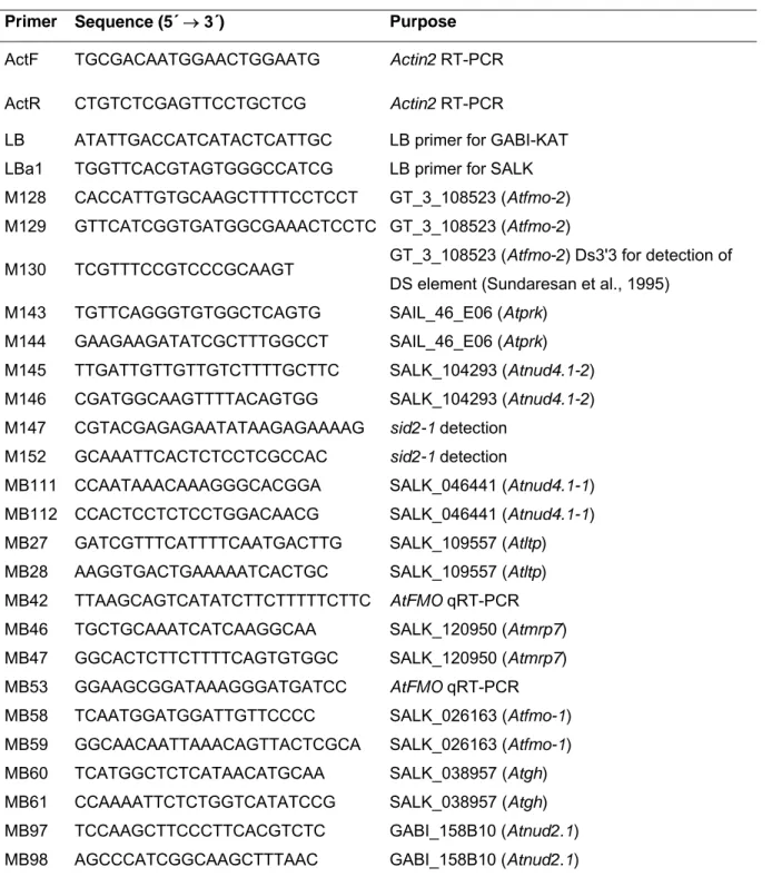
Table 2.4 List of primers used in this study.
Primer Sequence (5´ → 3´) Purpose
ActF TGCGACAATGGAACTGGAATG Actin2 RT-PCR ActR CTGTCTCGAGTTCCTGCTCG Actin2 RT-PCR
LB ATATTGACCATCATACTCATTGC LB primer for GABI-KAT LBa1 TGGTTCACGTAGTGGGCCATCG LB primer for SALK M128 CACCATTGTGCAAGCTTTTCCTCCT GT_3_108523 (Atfmo-2) M129 GTTCATCGGTGATGGCGAAACTCCTC GT_3_108523 (Atfmo-2)
M130 TCGTTTCCGTCCCGCAAGT GT_3_108523 (Atfmo-2) Ds3'3 for detection of DS element (Sundaresan et al., 1995)
M143 TGTTCAGGGTGTGGCTCAGTG SAIL_46_E06 (Atprk) M144 GAAGAAGATATCGCTTTGGCCT SAIL_46_E06 (Atprk) M145 TTGATTGTTGTTGTCTTTTGCTTC SALK_104293 (Atnud4.1-2) M146 CGATGGCAAGTTTTACAGTGG SALK_104293 (Atnud4.1-2) M147 CGTACGAGAGAATATAAGAGAAAAG sid2-1 detection
M152 GCAAATTCACTCTCCTCGCCAC sid2-1 detection
MB111 CCAATAAACAAAGGGCACGGA SALK_046441 (Atnud4.1-1) MB112 CCACTCCTCTCCTGGACAACG SALK_046441 (Atnud4.1-1) MB27 GATCGTTTCATTTTCAATGACTTG SALK_109557 (Atltp) MB28 AAGGTGACTGAAAAATCACTGC SALK_109557 (Atltp) MB42 TTAAGCAGTCATATCTTCTTTTTCTTC AtFMO qRT-PCR MB46 TGCTGCAAATCATCAAGGCAA SALK_120950 (Atmrp7) MB47 GGCACTCTTCTTTTCAGTGTGGC SALK_120950 (Atmrp7) MB53 GGAAGCGGATAAAGGGATGATCC AtFMO qRT-PCR MB58 TCAATGGATGGATTGTTCCCC SALK_026163 (Atfmo-1) MB59 GGCAACAATTAAACAGTTACTCGCA SALK_026163 (Atfmo-1) MB60 TCATGGCTCTCATAACATGCAA SALK_038957 (Atgh) MB61 CCAAAATTCTCTGGTCATATCCG SALK_038957 (Atgh) MB97 TCCAAGCTTCCCTTCACGTCTC GABI_158B10 (Atnud2.1) MB98 AGCCCATCGGCAAGCTTTAAC GABI_158B10 (Atnud2.1)
2.1.4 Enzymes
2.1.4.1 Restriction Endonucleases
Restriction enzymes were purchased from New England Biolabs (Frankfurt,
Germany) unless otherwise stated. Enzymes were supplied with 10 x reaction buffer.
2.1.4.2 Nucleic acid modifying enzymes
Standard PCR reactions were performed using home made Taq DNA polymerase.
To achieve highest accuracy, Pfu polymerase was used when PCR products were generated for later cloning reactions. Modifying enzymes and their suppliers are listed below:
Taq DNA polymerase (home made)
PfuTurbo
®DNA polymerase (Stratagene
®, Heidelberg, Germany) T4 DNA ligase (Roche, Mannheim, Germany)
DNaseI (Roche, Mannheim, Germany)
SuperScript™ II RNase H
-Reverse Transcriptase (Invitrogen™ , Karlsruhe,Germany) Gateway™-Technology
LR Clonase™ Enzyme mix (Invitrogen™, Karlsruhe, Germany)
2.1.5 Chemicals
Laboratory grade chemicals and reagents were purchased from Sigma-Aldrich (Deisenhofen, Germany), Roth (Karlsruhe, Germany), Merck (Darmstadt, Germany), Invitrogen™ (Karlsruhe, Germany), Serva (Heidelberg, Germany), and Gibco™ BRL
®(Neu Isenburg, Germany) unless otherwise stated.
2.1.6 Antibiotics
Ampicillin (Amp) 100 mg/ml in H
2O Carbenicillin (Carb) 50 mg/ml in H
2O Gentamycin (Gent) 15 mg/ml in H
2O Kanamycin (Kan) 50 mg/ml in H
2O Rifampicin (Rif) 100 mg/ml in DMSO
Tetracycline (Tet) 12.5 mg/ml in 70 % ethanol
Stock solutions (1000x) stored at -20° C. Aqueous solutions were sterile filtrated.
2.1.7 Buffers and solutions
General buffers and solutions are displayed in the following listing. All buffers and
solutions were prepared with Milli-Q
®water. Buffers and solutions for molecular
biological experiments were autoclaved and sterilised using filter sterilisation units, respectively. Buffers and solutions not displayed in this listing are denoted with the corresponding methods.
DEPC-H
2O Diethylpyrocarbonate 0.1 % in H
2O
Well mixed, left O/N and autoclaved for 30 min.
DNA extraction buffer (Quick prep) Tris 200 mM
NaCl 250 mM
EDTA 25 mM
SDS 0.5 %
pH 7.5 (HCl)
DNA gel loading dye (6 x) Sucrose 4 g EDTA (0.5 M) 2 ml
Bromphenol blue 25 mg
H
2O to 10 ml
Ethidium bromide stock solution Ethidium bromide 10 mg/ml in H
2O Diluted 1:40000 in agarose solution
GUS staining solution Na
2HPO
4(1M) 11.54 ml
NaH
2PO
4(1M) 8.46 ml
K
3Fe(CN)
6(0.05 M) 2 ml K
4Fe(CN)
6(0.05 M) 2 ml EDTA (0.05 M) 4 ml Triton X-100 (10 %) 2 ml
H
2O 90 ml
pH 7.0
Prior to use add 5 ml methanol and 550 µl X-Gluc
stock solution (50 mg/ml DMF) to 50 ml staining
solution.
Lactophenol trypan blue Lactic acid 10 ml
Glycerol 10 ml
H
2O 10 ml
Phenol 10 g
Trypan blue 10 mg
Before use dilute 1:1 in ethanol.
PCR reaction buffer (10 x) Tris 100 mM
KCl 500 mM
MgCl
215 mM
Triton X-100 1 %
pH 9.0
Stock solution was sterilised by autoclaving and used with Taq DNA polymerase.
BTH solution BTH (commercial product BION
®, Syngenta) was resuspended in dH
20 to the desired concentration prior use.
2.2 Methods
2.2.1 Maintenance and cultivation of Arabidopsis plant material
Arabidopsis seeds were germinated by sowing directly onto moist compost (Stender AG, Schermbeck, Germany) containing insecticide (10 mg l
-1Confidor WG 70 (Bayer, Germany)). Seeds were cold treated by placing sawn pots on a tray with a lid and incubating them in the dark at 4° C for three days. Pots were subsequently transferred to a controlled environment growth chamber, covered with a propagator lid and maintained under short day conditions (10 hour photoperiod, light intensity of approximately 200 µEinsteins m
-2sec
-1, 23° C day, 22° C night, and 65 % humidity).
Propagator lids were removed when seeds had germinated. If required for setting
seed, plants were transferred to long day conditions (16 hour photoperiod) to allow
early bolting and setting of seed. To collect seed, aerial tissue was enveloped with a paper bag and sealed with tape at its base until siliques shattered.
2.2.2 Generation of Arabidopsis F
1and F
2progeny
Fine tweezers and a magnifying-glass were used to emasculate an individual flower.
To prevent self-pollination, only flowers that had a well-developed stigma but immature stamen were used for crossing purpose. Fresh pollen from three to four independent donor stamens was dabbed onto each single stigma. Mature siliques containing F
1seed were harvested and allowed to dry. Approximately five F
1seeds per cross were grown as described above and allowed to self pollinate. Produced F
2seeds were collected and stored.
2.2.3 Inoculation and maintenance of P. parasitica
P. parasitica isolates were maintained as mass conidiosporangia cultures on leaves of their genetically susceptible Arabidopsis ecotypes over a 7 day cycle. Leaf tissue from infected seedlings was harvested into a 50 ml Falcon tube 7 days after inoculation. Conidiospores were collected by vigorously vortexing harvested leaf material in dH
2O for 15 seconds and after the leaf material was removed by filtering through miracloth (Calbiochem) the spore suspension was adjusted to the desired concentration using a Neubauer counting cell chamber. Plants to be inoculated were grown under short day conditions as described above. P. parasitica conidiospores were applied onto 2-week-old seedlings by spraying until imminent run-off using an aerosol-spray-gun. Inoculated seedlings were kept under a propagator lid to create a high humidity atmosphere and incubated in a growth chamber at 18°C and a 10 hour light period. For long term storage P. parasitica isolate stocks were kept as mass conidiosporangia cultures on plant leaves at -80° C.
2.2.4 Quantification of P. parasitica sporulation
To determine sporulation levels, seedlings were harvested 5-7 d after inoculation in a
50 ml Falcon tube and vortexed vigorously in 5 – 10 ml water for 15 seconds. Whilst
the conidiospores were still in suspension 10 µl were removed twice and spores were
counted under a light microscope using a Neubauer counting cell chamber. For each
tested Arabidopsis genotype, two pots containing approximately 30 seedlings were
infected per experiment and harvested spores from all seedlings of each pot were counted twice with sporulation levels expressed as the number of conidiospores per gram fresh weight.
2.2.5 Lactophenol trypan blue staining
Lactophenol trypan blue staining was used to visualise necrotic plant tissue and P.
parasitica mycelium (Koch and Slusarenko, 1990a). Leaf material was placed in a 15 ml Sarstedt tube (Nümbrecht, Germany) and immersed in lactophenol trypan blue.
The tube was placed into a boiling water bath for 2 minutes followed by destaining in 5 ml chloral hydrate solution (2.5 g/ml water) for 2 h and a second time overnight on an orbital shaker. After leaf material was left for several hours in 70 % glycerol, samples were mounted onto glass microscope slides in 70 % glycerol and examined using a light microscope (Axiovert 135 TV, Zeiss, Germany) connected to a Nikon DXM1200 Digital Camera. For Figure 3.13 infected leaves were examined under UV- light to exhibit cell death-associated fluorescence.
2.2.6 Maintenance of P. syringae pv. tomato cultures
Pseudomonas syringae pv. tomato strains described in 2.1.2.2 were streaked onto selective NYG agar plates containing rifampicin (100 µg/ml) and kanamycin (50 µg/ml) from -80° C DMSO stocks. Streaked plates were incubated at 28° C for 48 hours before storing at 4° C and refreshed weekly.
2.2.7 P. syringae pv. tomato inoculations and growth assay
P. syringae pv. tomato cultures were started from a small amount of bacteria grown
on NYG agar plates containing rifampicin (100 µg/ml) and kanamycin (50 µg/ml) in 20
ml NYG broth containing rifampicin (100 µg/ml) and kanamycin (50 µg/ml). The 20 ml
cultures were incubated overnight at 28° C and 200 rpm in a rotary shaker. For hand
infiltrations applied for the microarray samples see section 2.3.1. For growth assays,
2.5 ml of the overnight cultures were used to inoculate 50 ml of NYG broth in 300 ml
Erlenmeyer flasks supplemented with antibiotics. The flasks were incubated at 28° C
and 200 rpm in a rotary shaker for 3 hours. The required OD
600reading at this time
point was 0.2. Cultures were transferred to sterile 50 ml Falcon tubes and pelleted at
4500 rpm for 10 minutes at 20° C. Bacteria were washed by resuspending the pellet
in 40 ml of 10 mM sterile MgCl
2and subsequent centrifugation at 4500 rpm for 10 minutes at 20° C. The supernatant was promptly removed and each pellet resuspended in 50 ml of sterile 10 mM MgCl
2. For vacuum-infiltration the concentration of bacteria was adjusted to 5 x 10
5cfu/ml in 600 ml of 10 mM MgCl
2containing 0.002 % Silwet L-77 (Lehle seeds, USA).
Single pots of nine 4- to 5-week old plants, grown under short day conditions were routinely used for bacterial growth assays. Two hours before vacuum-infiltration, plants were watered and kept under a dH
2O-humidified lid. Plants were vacuum- infiltrated with bacteria by inverting the pots and carefully submerging all leaf material in 600 ml of diluted bacterial suspension contained within a plastic exsiccator.
Vacuum was applied and maintained within the exsiccator for 3 minutes before being gradually released. Periodic swirling and tapping of the exsiccator helped to dislodge any air bubbles that accumulated at the surface of the leaves. Any non-infiltrated leaves remaining at this stage were removed by hand. Excess of bacterial solution was removed by inverting the pots and gently dipping the plants in water.
Day zero (T0) samples were taken one hour after infiltration by using a cork borer (∅
0.55 cm) to excise and transfer four leaf discs from four independent plants to a 1.5 ml centrifuge tube, resulting in a total excised area of 1 cm
2. This was repeated with a second batch of four leaf discs from four independent plants. The discs were then macerated with a plastic pestle in 100 µl of sterile 10 mM MgCl
2. Subsequently, 900 µl of sterile 10 mM MgCl
2were added (10
-1dilution) and 100 ml of each sample were plated onto NYG agar (Rif
100, Kan
50). Day three (T3) samples were taken in an identical manner to that of T0 except that four leaf discs from four independent plants per genotype were taken in triplicates. For each sample a dilution series ranging between 10
-1and 10
-7was made and 15 µl aliquots from each dilution were spotted sequentially onto a single NYG agar plate (Rif
100, Kan
50). All bacteria plates were incubated at 28° C for two days before colony numbers were counted.
2.2.8 A. thaliana powdery mildew Golovinomyces orontii
Powdery mildew G. orontii was propagated on A. thaliana ecotype Col-0 plants
cultivated at 20° C and 16 h light/ 8 h darkness, 80 % humidity in a growth chamber.
2.2.9 Isolation of Arabidopsis genomic DNA (Quick prep for PCR)
This procedure yields a small quantity of poor quality DNA. However, the DNA is of sufficient quality for PCR amplification. The aliquots were stored at -20° C.
The cap of a 1.5 ml microcentrifuge tube was closed onto a leaf to cut out a section of tissue and 400 µl of DNA extraction buffer were added. A micropestle was used to grind the tissue in the tube until the tissue was well mashed. The solution was centrifuged at maximum speed for 5 minutes in a bench top microcentrifuge and 300 µl supernatant were transferred to a fresh tube. One volume of isopropanol was added to precipitate DNA and centrifuged at maximum speed for 5 minutes in a bench top microcentrifuge. The supernatant was discarded carefully. The pellet was washed with 70 % ethanol and dried. Finally the pellet was dissolved in 100 µl 10 mM Tris-HCl pH 8.0 and 1 µl of the DNA solution was used for a 20 µl PCR reaction mixture.
2.2.10 Isolation of total RNA from Arabidopsis
Total RNA was prepared from 4-week old plant materials. Liquid nitrogen frozen leaf samples (approximately 80-100 mg) were homogenized 15 seconds to a fine powder using a Mini-Bead-Beater-8
TM(Biospec Products) and 1.2 mm stainless steel beads (Roth) in 2 ml centrifuge tubes. After homogenisation samples were kept frozen in liquid nitrogen until the next step of the extraction procedure. 1 ml of RNAwiz
®Reagent (Ambion) was added and samples were homogenised by vortexing for 1 minute. For dissociation of nucleoprotein complexes the homogenised samples were incubated for 5 minutes at room temperature. 0.2 ml of chloroform was added and samples were shaken vigorously for 20 seconds. After incubation for 10 minutes at room temperature samples were centrifuged for 15 minutes at 12000 g and 4° C. The upper aqueous, RNA containing phase was transferred to a fresh 2 ml microcentrifuge tube containing 0.5 ml DEPC-water. The RNA was precipitated by adding 1 ml isopropanol, subsequent mixing and incubation for 10 minutes at room temperature. Samples were centrifuged for 15 minutes at 12000 g and 4° C. The supernatant was removed and the pellet was washed by vortexing in 1 ml of ice cold 75 % ethanol. Samples were again centrifuged for 5 minutes at 12000 g and 4° C.
Supernatant was discarded and pellets were allowed to air-dry for 10 minutes and
dissolved in 25 µl DEPC-water. Samples were immediately transferred to and stored at -80° C.
2.2.11 Polymerase chain reaction (PCR)
Standard PCR reactions were performed using Taq DNA polymerase while for cloning of PCR products Pfu or Pfx polymerases were used according to the manufacturer instructions. All PCRs were carried out using a PTC-225 Peltier thermal cycler (MJ Research). A typical PCR reaction mix and thermal profile is shown below.
Reaction mix (20 µl total volume):
Component
aVolume
Template DNA (genomic or plasmid) 0.1 - 20 ng
10 x PCR reaction buffer 2 µl
dNTP mix (2.5 mM each: dATP, dCTP, dGTP,
dTTP) 2 µl
Forward primer (10 µM) 1 µl
Reverse primer (10 µM) 1 µl
Taq DNA polymerase (4U/µl) 0.5 µl
Nuclease free water to 20 µl total volume
Thermal profile
Stage Temperature (°C) Time period No. of cycle
Initial denaturation 94 3 min 1 x
Denaturation 94 30 sec
Annealing 50 - 60 30 sec 25 - 40
Extension 72 1 min per kb
Final extension 72 3 min 1 x
2.2.12 Reverse transcription-polymerase chain reaction (RT-PCR)
RT-PCR was carried out in two steps. SuperScript™ II RNase H
-Reverse Transcriptase (Invitrogen) was used for first strand cDNA synthesis by combining 1 µg template total RNA, 1 µl primer dT
18V (0.5 µg/µl, V standing for an variable nucleotide), 5 µl dNTP mix in a volume of 13.5 µl (deficit made up with DEPC-water).
Samples were incubated at 65° C for 10 minutes. Subsequently, the reactions were filled up to a total volume of 20 µl with 4 µl of 5 x reaction buffer, 2 µl of 0.1 M DTT and 0.5 µl reverse transcriptase. The reactions were incubated at 42° C for 60 minutes before the enzyme was heat inactivated at 70° C for 10 minutes. For subsequent normal PCR, 1 µl of the above RT-reaction was used as cDNA template.
As template total RNA for the reverse transcription reaction was not DNase treated, a control reaction for each RNA preparation was performed in which the reverse transcription reaction was incubated without reverse transcriptase enzyme (enzyme replaced by equal volume of DEPC-water) to check in the following PCR for contamination by genomic DNA.
2.2.13 Quantitative real-time polymerase chain reaction (qRT-PCR)
A quantitative real-time PCR kit (Brilliant SYBR Green QPCR Core Kit, Stratagene) was used to determine the amount of transcript accumulation of a gene of interest.
Reactions were carried out according to the manufacturer’s protocol. Primer combinations that specifically amplify the investigated gene and a gene serving as an internal standard were used in independent reactions performed on an ABI PRISM 7700 Sequence Detection System (Applied Biosystems, Foster City, California, USA). Data were analysed by the comparative ∆∆C
T