XIAP Confers Immunity Against Intracellular Bacteria
Inaugural-Dissertation zur
!Erlangung des Doktorgrades
!der Mathematisch-Naturwissenschaftlichen Fakultät der Universität zu Köln
vorgelegt von
Maria Andree
aus Nordhorn
Berichterstatter: Prof. Dr. Elena Rugarli Prof. Dr. Rudolf Wiesner
Tag der mündlichen Prüfung: 25. Juni 2013
TABLE OF CONTENTS
I
Table of contents
ABBREVIATIONS III
ABSTRACT 1
ZUSAMMENFASSUNG 2
INTRODUCTION 4
Inhibitor of Apoptosis Proteins (IAPs) - regulators of cell death 4
The cellular apoptotic machinery 5
Regulators of mitochondrial outer membrane permeabilisation – the BCL2 proteins 7
Regulation of IAPs by IBM-containing proteins 9
IAPs in inflammatory signalling 10
Shigella flexneri as a physiological stimulus of immune responses 12
Aim of the study 13
MATERIALS AND METHODS 14
Cell culture 14
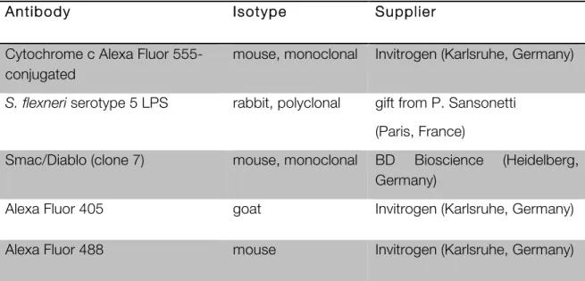
Antibodies and reagents 14
DNA constructs 16
Bacterial infection 16
Gentamycin protection assay 17
Cell viability and cell death 17
NF-κB activity 17
Cell fractionation, sample preparation and Western blotting 18
Immunoprecipitation of active BAX 19
RNAi interference 19
Caspase-3 activity 20
Mitochondrial membrane potential 20
Microscopy 20
Mouse strains 21
Infection of mice with S. flexneri 22
Tissue immunohistochemistry (IHC) 22
Statistical analysis 22
Software 22
TABLE OF CONTENTS
II
RESULTS 23
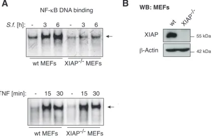
XIAP is required for S. flexneri-induced NF-κB activation 23 S. flexneri-induced release of mitochondrial SMAC interrupts NOD1/XIAP-
mediated inflammatory signalling 25
Intracellular S. flexneri induces the release of mitochondrial SMAC without
inducing collateral cellular damage 29
Calpain-cleaved BID induces the mitochondrial release of SMAC in S. flexneri–
infected cells 33
XIAP confers immunity against S. flexneri infection in vivo 36
DISCUSSION 40
XIAP is essential for the immune response against intracellular S. flexneri 40
SMAC and OMI are selectively released from mitochondria 42
Calpain-cleaved BID induces the selective release of SMAC 43
Therapeutic implications 45
Conclusion 46
REFERENCES 47
APPENDIX 54
Human BID sequence 54
Vector map 54
ERKLÄRUNG 55
ABBREVIATIONS
III
Abbreviations
BAD BCL2 antagonist of cell death
BAX BCL2 associated x protein
BAK BCL2 antagonist killer 1 BCL2 B-cell lymphoma 2
BH BCL2 homology
BID BH3-interacting domain death antagonist
BIM BCL2 interacting mediator of cell death
BIR Baculovirus IAP Repeat
bp Base pairs
BSA Bovine serum albumin
C- Carboxy-
Ca2+ Calcium
CFU Colony forming units cIAP Cellular IAP
Da, kDa Dalton, Kilodalton
DIABLO Direct IAP binding protein with low pI
DMSO Dimethylsulfoxid DTT Dithiothreitol EDTA Ethylenediamine-
tetraacetic acid et al. et alteri /-a /-um (and
others)
HtrA2 Hight temperature requirement A2 IAP Inhibitor of apoptosis IBM IAP-binding motif
IP Immunoprecipitation i.v. intra veneous
M molar, mol/l
MTS mitochondrial targeting sequence
N- Amino-
NF-κB Nuclear Factor κB
MAPK Mitogen activated protein Kinase
MOMP Mitochondrial outer
membrane permeabilization NF-κB Nuclear factor kappa B PAGE Polyacrylamid gel electro-
phoresis
PBS Phosphate buffered saline PCR Polymerase chain reaction p.i. Post infection
pmol Picomol
PMSF Phenylmethylsulfonyl fluoride rpm Revolutions per minute
RT Room temperature
SDS Sodium dodecylsulfate S.f. Shigella flexneri
SMAC Second mitochondrial
derived activator of caspases STS Staurosporine
UV Ultra violet
WB Western blot
XIAP X-linked inhibitor of apoptosis
ABSTRACT
1
Abstract
The so-called X-linked inhibitor of apoptosis protein (XIAP) is the most intensively studied member of the inhibitor of apoptosis proteins (IAPs) and is the only cellular protein that directly binds to and inhibits caspases thereby blocking apoptosis. XIAP is frequently overexpressed in malignant cells and has therefore been investigated as a promising therapeutic target in cancer.
Recently, IAPs have also been shown to influence pathways that modulate immune signalling via activation of NF-κB. One such pathway is the NOD signalling cascade that is responsible for the detection and defence against intracellular bacteria. XIAP has previously been shown to be an essential component of the NOD signalling cascade required for the activation of NF-κB mediated inflammatory responses.
In order to study the role of XIAP in NOD signalling in response to bacterial infection, we used Shigella flexneri as a physiological stimulus for the NOD signalling cascade. Our data demonstrate that XIAP is essential for NF-κB activation after S. flexneri infection, but S. flexneri is able to efficiently down-regulate the XIAP-mediated inflammatory response.
In particular, we show that S. flexneri infection co-opts the host protease calpain to cleave the BCL2 protein BID. Calpain-cleaved BID then translocates to the mitochondria where it mediates the selective release of the XIAP-antagonists SMAC and OMI from the mitochondrial intermembrane space. In the cytosol, SMAC and OMI inhibit XIAP and therefore interrupt the NOD-mediated inflammatory response. Critically, calpain-cleaved BID induces the release of SMAC and OMI, but not of Cyt c from the mitochondria. This selective permeability differentiates this process from the mitochondrial permeabilisation induced by apoptotic stimuli and permits the infected host cell not only to survive but also ensures bacterial propagation.
The physiological importance of XIAP in controlling bacterial infection was confirmed in vivo in XIAP whole-body and liver-specific knockout mice, which show more bacterially induced necrotic liver lesions in response to S. flexneri infection than wildtype littermates. In contrast, SMAC/OMI and BID knockout mice, with unopposed XIAP activity, survived S. flexneri infection for much longer than wild type animals.
Our findings demonstrate how the non-apoptotic antagonisation of XIAP by mitochondrial SMAC and OMI can control the delicate interaction between intracellular microbial pathogens and their hosts.
ZUSAMMENFASSUNG
2
Zusammenfassung
Das sogenannte X-linked inhibitor of apoptosis protein (XIAP) zählt zu den am besten untersuchten Mitgliedern der Familie der Inhibitoren der Apoptose (kurz IAPs). Bislang ist es das einzig bekannte Protein, das in der Lage ist, Caspasen direkt zu binden und zu inhibieren und somit Apoptose zu blockieren. XIAP wird in malignen Zellen häufig überexprimiert und stellt daher ein attraktives therapeutisches Ziel in der Krebstherapie dar.
In den letzten Jahren wurden IAPs zudem als Bestandteil von immunregulatorischen Signalwegen identifiziert, die meist die Aktivierung von NF-κB beinhalten. In der NOD- Signalkaskade, die für die Erkennung und Abwehr von intrazellulären Bakterien verantwortlich ist, wurde XIAP als entscheidender Faktor für die Aktivierung von NF-κB identifiziert.
Um die Funktion von XIAP im Laufe einer bakteriellen Infektion zu untersuchen, wurde das Bakterium Shigella flexneri als physiologischer Stimulus der NOD Signalkaskade eingesetzt.
Unsere Untersuchungen zeigen, dass S. flexneri in der Lage ist, die XIAP-vermittelte NOD- Aktivierung zu unterdrücken. Insbesondere wurde gezeigt, dass S. flexneri die selektive Freisetzung der XIAP-Antagonisten SMAC und OMI aus dem mitochondrialen Intermembranraum induziert und somit XIAP inhibiert. S. flexneri vermittelt die Freisetzung von SMAC und OMI durch die Protease Calpain, die eine proteolytische Spaltung von BID katalysiert. Das gespaltene BID-Fragment transloziert an die Mitochondrien und induziert dort die Freisetzung von SMAC und OMI in das Cytosol. BID, prozessiert durch Calpain, permeabilisiert die Mitochondrien zwar für SMAC und OMI, jedoch ohne dabei auch Cytochrom c freizusetzen und gewährleistet somit das Überleben der infizierten Wirtszelle, um ungehinderte Vermehrung der Bakterien zu ermöglichen.
Die Bedeutung von XIAP in der Kontrolle bakterieller Infektion konnte in „XIAP knockout“
Mäusen bestätigt werden. Ganzkörper XIAP knockout- sowie leberspezifische XIAP- knockout Mäuse weisen deutlich mehr bakteriell-induzierte nekrotische Areale in der Leber auf, im Vergleich zu Wildtyp Mäusen. Im Gegensatz dazu überlebten SMAC/OMI und BID knockout Mäuse - mit ungehemmter XIAP Funktion - die S. flexneri Infektion deutlich länger als die Wildtyp Kontrollen. Grund hierfür ist die uneingeschränkte Funktion von XIAP, die eine bessere Bekämpfung der Infektion ermöglicht.
ZUSAMMENFASSUNG
3
Zusammenfassend zeigt die vorliegende Arbeit, wie nicht-apoptotische Antagonisierung von XIAP durch mitochondriales SMAC und OMI die delikate Interaktion zwischen intrazellulären Bakterien und ihren Wirten beeinflusst.
INTRODUCTION
4
Introduction
Inhibitor of Apoptosis Proteins (IAPs) - regulators of cell death
The inhibitor of apoptosis proteins (IAPs) were originally identified in baculoviruses when homologs for p35, a protein that prevents apoptosis after viral infection, were investigated.
The viral CpGV gene product was found to functionally complement p35 in preventing apoptosis, caused by the Autographa californica nuclear polyhedrosis virus, which was thus called the inhibitor of apoptosis (Crook et al., 1993). One year later Clem and Miller demonstrated that IAP homologs block apoptosis independently of viral infection, indicating that IAPs interact directly with the cellular apoptotic machinery (Clem and Miller, 1994). In the following years, IAPs were found to be an evolutionary conserved protein family in various eukaryotic species including yeast, fly and mammals (Deveraux and Reed, 1999). Their defining feature is the presence of one or several zinc-finger motifs, known as baculoviral IAP repeat (BIR) domains (Birnbaum et al., 1994), some of which are required for their function to inhibit caspases - the main executioners of apoptosis, which is a form of programmed cell death.
The human genome encodes eight different IAPs, but in spite of their name as inhibitor of apoptosis, several members of the family are known to have distinct functions that are unrelated to apoptosis. Cellular processes such as cell cycle regulation, protein degradation and other signal transduction cascades are also subject to regulation by IAPs (Salvesen and Duckett, 2002). Nevertheless, the cellular IAPs (cIAP1 and cIAP2) efficiently impede apoptosis, triggered by multiple stimuli, when introduced into mammalian cells (Deveraux and Reed, 1999). However, cIAPs are weak caspase inhibitors, presumably lacking structural features present in other IAPs. Therefore they represent protein scaffolds suitable for direct caspase inhibition but have lost or never acquired specific caspase inhibitory interaction sites (Eckelman and Salvesen, 2006). Currently, they are widely recognised for their regulatory function in the TNF-receptor pathway (Vandenabeele and Bertrand, 2012).
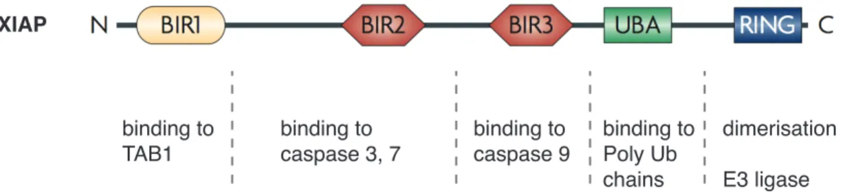
The X-linked inhibitor of apoptosis protein (XIAP, encoded by BIRC4) is the best-studied IAP and the only family member that can directly bind to and inhibit caspases, thereby blocking apoptosis (Deveraux et al., 1997) (Figure 1).
INTRODUCTION
5
Figure 1. Domain structure of XIAP. XIAP comprises three BIR domains, each exerting different functions, a ubiquitin-associated (UBA) domain, which allows binding to poly-ubiquitin (Ub) chains and a RING finger domain, which is responsible for dimerisation and E3-ligase function. (modified from Gyrd-Hansen and Meier, 2010)
On the one hand, the BIR3 domain of XIAP can bind to the dimerisation surface of caspase- 9, maintaining it in an inactive monomeric form. On the other hand, the linker region between the BIR1 and BIR2 domains binds to caspase-3 or -7 and blocks their activity by steric occlusion of caspase-substrates (Salvesen and Duckett, 2002). The BIR1 domain itself is not involved in caspase inactivation and interacts with a different set of proteins, implicating XIAP in other cellular functions, which will be described below.
Based on their ability to inhibit apoptosis and the frequently elevated expression of IAPs in tumour cells, IAPs have been mainly studied in mammalian cancer. Specifically, elevated expression of XIAP is often found in malignant cells and is thought to be responsible for resistance to anti-cancer therapeutics (Schimmer et al., 2006; Tamm et al., 2004a, 2004b;
Mizutani et al., 2007). Congruently, ectopic overexpression of XIAP in tumour cell lines confers resistance to apoptosis (Deveraux et al., 1997) and vice versa XIAP-targeting by its specific knock-down sensitises tumour cells to apoptosis (Kashkar et al., 2007).
Nevertheless, XIAP-targeting has thus far failed to completely restore the apoptotic machinery under physiological conditions. Furthermore, studies assessing the prognostic importance of XIAP in tumour patients or its predictive role for chemotherapy have been inconsistent between different malignancies (reviewed in Kashkar, 2010). Additional factors such as a defective mitochondrial apoptotic pathway might be responsible for this discrepancy.
The cellular apoptotic machinery
The regulation of tissue homeostasis is critical for all multicellular organisms. The number of cells is tightly controlled by apoptosis - a form of programmed cell death (Kerr et al., 1972).
Apoptosis ultimately involves the activation of caspases, members of a highly conserved binding to
TAB1
binding to caspase 3, 7 SMAC / OMI
binding to caspase 9
binding to Poly Ub chains
dimerisation E3 ligase XIAP
INTRODUCTION
6
family of cystein proteases, which are responsible for the majority of proteolytic events that result in the destruction of the cell (Fuentes-Prior and Salvesen, 2004). Caspases can be activated through two major pathways: the “extrinsic” and the “intrinsic” (also called mitochondrial) pathway of apoptosis.
The extrinsic pathway of apoptosis can be initiated externally by ligands of the TNF-receptor family (e.g. TNF, FAS-ligand) that induce receptor oligomerisation and recruitment of the adaptor molecule FAS-associated death domain protein (FADD) to form the death inducing signalling complex (DISC) together with pro-caspase-8. Activated caspase-8 can then directly cleave and activate executioner caspases 3 and 7.
The intrinsic or “mitochondrial pathway of apoptosis” can be initiated autonomously by the cell and is caused by overwhelming cellular stress such as DNA damage, extensive protein aggregation or growth factor withdrawal (Strasser et al., 2000). The mitochondrial pathway of apoptosis is characterised by mitochondrial outer membrane permeabilisation (MOMP), which results in the release of Cytochrome c (Cyt c) from the mitochondrial intermembrane space (IMS). The release of Cyt c into the cytosol promotes the ATP-dependent formation of the apoptosome complex – comprising of Cyt c, apoptotic protease-activating factor 1 (APAF-1) and pro-caspase-9 - which catalyses auto-activation of the initiator caspase-9 (Li et al., 1997; Zou et al., 1999) followed by activation of executioner caspases.
In addition, both pathways of apoptosis can intersect. Beside direct activation of executioner caspases, caspase-8-dependent cleavage of the BH3-only protein BID allows amplification of the caspase cascade to ensure efficient cell demolition (Kaufmann et al., 2012). The caspase-8-cleaved C-terminal fragment of BID (tBID) translocates to the mitochondria and leads to MOMP, followed by the release of Cyt c and downstream caspase activation.
Nevertheless this step is not critical to all cell types, which are accordingly categorised into type I (BID-independent) and type II (BID-dependent) cells. Experiments with diverse cell types from BID-deficient mice have revealed an important role for BID in Fas-induced apoptosis in type II cells, which include hepatocytes and pancreatic β-cells (Yin et al., 1999;
McKenzie et al., 2008). Of note, while BIDknockout mice are resistant to Fas-ligand-induced hepatocyte apoptosis, additional loss of XIAP can rescue this effect. Accordingly, XIAP is one important discriminator between type I and type II Fas-induced apoptosis (Jost et al., 2009).
But whether a cell behaves like type I or type II does not depend on a single determinant, but on the ratio between processed effector caspases, DISC formation, caspase-8 activation and on the levels of free XIAP, respectively (Kaufmann et al., 2012).
INTRODUCTION
7
Apoptosis is a tightly regulated process and its dysregulation culminates in multiple human diseases. Whereas the failure to execute apoptosis in regenerating tissues contributes to cancer, excessive apoptosis in post-mitotic tissues can cause degenerative disorders.
Caspases – the executioner of both apoptotic pathways - are subject to inhibition by the IAPs. IAPs therefore confer protection from death-inducing stimuli, providing an important level of control of the apoptotic pathway.
Regulators of mitochondrial outer membrane permeabilisation – the BCL2 proteins
IAP antagonisation is dependent on the liberation of mitochondrial IAP-binding motif (IBM)- containing proteins by MOMP. MOMP is often regarded as a point of no return and therefore must be tightly regulated. Members of the B-cell lymphoma 2 (BCL2) family are crucial regulators of MOMP. The pro-apoptotic BCL2 members can sense cellular damage and directly induce MOMP by forming pores in the outer mitochondrial membrane (OMM) in contrast to the anti-apoptotic members that can block this event. The precise regulation of MOMP therefore depends on the fine-tuned interactions among pro- and anti-apoptotic BCL2 family members.
The common structural feature shared by the members of this protein family is the BCL2 homology (BH) domain. Functionally, BCL2 family proteins can be divided into three groups:
i) anti-apoptotic members, ii) pro-apoptotic effectors and iii) pro-apoptotic BH3 only proteins (Cory and Adams, 2002) (Figure 2).
Anti-apoptotic BCL2 proteins
BCL2, BCL-W, BCL-XL, A1, MCL-1
Pro-apoptotic BCL2 proteins Effectors
BAK, BAX, BOK
BH3-only proteins
BID, BIM, BAD, BIK, BMF, BNIP3, HRK, NOXA, PUMA
Nature Reviews | Molecular Cell Biology
BH4 BH3 BH1 BH2 TM
BH3 BH1 BH2 TM
BH3 Effectors
Anti-apoptotic
BCL-2 protein BAX or BAK Pro-apoptotic BCL-2 proteins
BH3-only proteins
BH3-only protein
Anti-apoptotic BCL-2 protein
BH3-only protein BH3-only protein Sensitizer Direct activator BCL-2, BCL-W, BCL-XL, A1 and MCL-1
BAK, BAX and BOK BID, BIM, BAD, BIK, BMF, BNIP3, HRK, NOXA and PUMA
Direct activator–derepressor model
Active
BAX or BAK Active
a Anti-apoptotic BCL-2 proteins
b Indirect activator model
Patch clamping An electrophysiological technique used for measuring ion channel activity over membranes. Typically, a small diameter (1μm) micropipette serves as the electrode and is applied to a small area of membrane (the ‘patch’), allowing the activity of one or a few ion channels to be measured.
Micelle
An aggregate (typically spherical) of varying size comprised of lipids. In aqueous environments, the hydrophobic lipid tails orientate to the centre of the micelle and the hydrophilic head groups are on the surface.
analysis of BAX-permeabilized liposomes revealed openings of varying size (25–100 nm) that appeared concurrently with permeabilization in a manner that was BCL-XL inhibitable49. The diameter of these open- ings is consistent with the ability of BAX to induce the size-independent release of dextrans. Supporting a lipidic pore model, the edges of these BAX-induced pores are smooth and devoid of proteinaceous material. In contrast, protein pores formed by the toxin pneumolysin, as ana- lysed by cryo-EM, are uniform in nature and decorated around the edges with toxin molecules50. However, similar pore-like structures have yet to be found on mitochondria during MOMP.
Post-MOMP regulation of IMS proteins
Although MOMP itself provides little specificity as to which IMS proteins are released, studies suggest that release of different IMS proteins can be selective. This may be through the regulation of IMS protein inter action with mitochondrial membranes or by mitochondrial inner membrane remodelling.
IMS protein membrane attachment. Apoptosis induc- ing factor (AIF) is an IMS protein that is anchored on the mitochondrial inner membrane and displays much slower release kinetics than cytochrome c following MOMP31. This protracted release may be owing to the requirement for AIF cleavage post-MOMP by cytosolic proteases to liberate AIF from the mitochondrial inner membrane. One candidate protease is cytosolic calpain I, which can cleave AIF in vitro to promote its release from permeabilized mitochondria51.
Electrostatic interactions between cytochrome c and the mitochondrial lipid cardiolipin have been proposed to regulate its release52. However, it might be expected that the ionic strength of the cytosol should suffice to disrupt these interactions53.
Post-MOMP mitochondrial inner membrane remodelling.
Other mechanisms may control IMS protein release fol- lowing MOMP. Mitochondrial cristae are involutions of the mitochondrial inner membrane that greatly increase the mitochondrial surface area available for oxidative phosphorylation and ATP synthesis. Cristae are dynamic structures and their accessibility to the IMS is largely dictated through regulation of cristae junction size. As most cytochrome c resides in mitochondrial cristae, sev- eral studies have addressed whether cristae remodelling provides an additional means of regulating cytochrome c release following MOMP. Various BH3-only proteins, including BID, BIM, BNIP3 (BCL-2 and adenovirus E1B 19 kDa protein-interacting protein 3) and BCL-2- interacting killer (BIK) have been found to promote mitochondrial cristae remodelling54–57. Treatment of mitochondria in vitro with the BH3 protein tBID induced dramatic inner membrane remodelling, leading to inter- connected cristae with widened junctions and cyto- chrome c mobilization into the IMS55. Two IMS proteins, optic atrophy protein 1 (OPA1; a dynamin-like GTPase) and presenilins-associated rhomboid-like protein (PARL;
a rhomboid protease) have been found to regulate cristae remodelling during apoptosis58,59. Following MOMP, dis- assembly of OPA1 hetero-oligomers is required for the widening of cristae junctions, whereas PARL cleavage of OPA1 generates an OPA1 cleavage product that maintains tight cristae junctions that prevent cytochrome c release.
Functionally, PARL loss renders cells more susceptible to apoptosis induced by intrinsic stimuli, whereas OPA1 overexpression is protective58,59.
Although cristae remodelling requires activated BAX or BAK, it can occur in the absence of MOMP because pharmacological inhibitors of MOMP still allow remod- elling to occur54. Remodelling was associated with the mobilization of cytochrome c to the IMS and, like pre vious studies, disassembly of OPA1 was required for remodel- ling to occur. In this study, however, gross changes in mitochondrial morphology were not apparent. Instead, a subtle narrowing, rather than widening, of cristae junc- tions occurred. Similarly, correlative light microscopy and EM of apoptotic cells revealed that gross alterations in mitochondrial structure were detected only after MOMP and caspase activation had occurred, arguing against a causal role for large mitochondrial structural changes Box 3 | Regulation of MOMP by the BCL-2 family
The B cell lymphoma 2 (BCL-2) family of proteins is divided into three groups based on their BCL-2 homology (BH) domain organization (see the figure, part a). Pro-apoptotic BCL-2 proteins can be sub-divided into effectors (the proteins that actually cause
mitochondrial outer membrane permeabilization (MOMP)) or BH3 only (the proteins that relay the apoptotic signal to the effectors). Although BCL-2-related ovarian killer protein (BOK) displays similar domain architecture to BCL-2-associated X protein (BAX) and BCL-2 antagonist or killer (BAK), there is little evidence that it is a functional effector. Two
prominent models of BAX and BAK activation have been proposed, termed the indirect activator (or neutralization) and direct activator–derepressor models of activation150,151 (see the figure, part b). The indirect activator model asserts that BAX and BAK are bound in a constitutively active state by anti-apoptotic BCL-2 proteins and that competitive interactions of BH3-only proteins with anti-apoptotic BCL-2 family members is sufficient to release activated BAX and BAK. In the direct activator–derepressor model, BAX and BAK are activated following interaction with a subset of BH3-only proteins known as direct activators, and anti-apoptotic BCL-2 proteins prevent MOMP either by sequestering the activating BH3-only proteins or by inhibiting activated BAX and BAK. A second subset of BH3-only proteins, termed sensitizers, cannot directly activate BAX and BAK but neutralize anti-apoptotic BCL-2 proteins. Definitive proof for either model has proved challenging; it is likely that aspects of both models are correct. BAD, BCL-2 antagonist of cell death; BID, BH3-interacting domain death agonist; BIK, BCL-2-interacting killer; BIM, BCL-2-interacting mediator of cell death; BMF, BCL-2-modifying factor; BNIP3, BCL-2 and adenovirus E1B 19 kDa protein-interacting protein 3; HRK, harakiri; PUMA, p53
upregulated modulator of apoptosis; TM, transmembrane.
R E V I E W S
NATURE REVIEWS |MOLECULAR CELL BIOLOGY VOLUME 11 | SEPTEMBER 2010 | 625
© 20 Macmillan Publishers Limited. All rights reserved10 Nature Reviews | Molecular Cell Biology
BH4 BH3 BH1 BH2 TM
BH3 BH1 BH2 TM
BH3 Effectors
Anti-apoptotic
BCL-2 protein BAX or BAK Pro-apoptotic BCL-2 proteins
BH3-only proteins
BH3-only protein
Anti-apoptotic BCL-2 protein
BH3-only protein BH3-only protein Sensitizer Direct activator BCL-2, BCL-W, BCL-XL, A1 and MCL-1
BAK, BAX and BOK BID, BIM, BAD, BIK, BMF, BNIP3, HRK, NOXA and PUMA
Direct activator–derepressor model
Active
BAX or BAK Active
a Anti-apoptotic BCL-2 proteins
b Indirect activator model
Patch clamping An electrophysiological technique used for measuring ion channel activity over membranes. Typically, a small diameter (1μm) micropipette serves as the electrode and is applied to a small area of membrane (the ‘patch’), allowing the activity of one or a few ion channels to be measured.
Micelle
An aggregate (typically spherical) of varying size comprised of lipids. In aqueous environments, the hydrophobic lipid tails orientate to the centre of the micelle and the hydrophilic head groups are on the surface.
analysis of BAX-permeabilized liposomes revealed openings of varying size (25–100 nm) that appeared concurrently with permeabilization in a manner that was BCL-XL inhibitable49. The diameter of these open- ings is consistent with the ability of BAX to induce the size-independent release of dextrans. Supporting a lipidic pore model, the edges of these BAX-induced pores are smooth and devoid of proteinaceous material. In contrast, protein pores formed by the toxin pneumolysin, as ana- lysed by cryo-EM, are uniform in nature and decorated around the edges with toxin molecules50. However, similar pore-like structures have yet to be found on mitochondria during MOMP.
Post-MOMP regulation of IMS proteins
Although MOMP itself provides little specificity as to which IMS proteins are released, studies suggest that release of different IMS proteins can be selective. This may be through the regulation of IMS protein inter action with mitochondrial membranes or by mitochondrial inner membrane remodelling.
IMS protein membrane attachment. Apoptosis induc- ing factor (AIF) is an IMS protein that is anchored on the mitochondrial inner membrane and displays much slower release kinetics than cytochrome c following MOMP31. This protracted release may be owing to the requirement for AIF cleavage post-MOMP by cytosolic proteases to liberate AIF from the mitochondrial inner membrane. One candidate protease is cytosolic calpain I, which can cleave AIF in vitro to promote its release from permeabilized mitochondria51.
Electrostatic interactions between cytochrome c and the mitochondrial lipid cardiolipin have been proposed to regulate its release52. However, it might be expected that the ionic strength of the cytosol should suffice to disrupt these interactions53.
Post-MOMP mitochondrial inner membrane remodelling.
Other mechanisms may control IMS protein release fol- lowing MOMP. Mitochondrial cristae are involutions of the mitochondrial inner membrane that greatly increase the mitochondrial surface area available for oxidative phosphorylation and ATP synthesis. Cristae are dynamic structures and their accessibility to the IMS is largely dictated through regulation of cristae junction size. As most cytochrome c resides in mitochondrial cristae, sev- eral studies have addressed whether cristae remodelling provides an additional means of regulating cytochrome c release following MOMP. Various BH3-only proteins, including BID, BIM, BNIP3 (BCL-2 and adenovirus E1B 19 kDa protein-interacting protein 3) and BCL-2- interacting killer (BIK) have been found to promote mitochondrial cristae remodelling54–57. Treatment of mitochondria in vitro with the BH3 protein tBID induced dramatic inner membrane remodelling, leading to inter- connected cristae with widened junctions and cyto- chrome c mobilization into the IMS55. Two IMS proteins, optic atrophy protein 1 (OPA1; a dynamin-like GTPase) and presenilins-associated rhomboid-like protein (PARL;
a rhomboid protease) have been found to regulate cristae remodelling during apoptosis58,59. Following MOMP, dis- assembly of OPA1 hetero-oligomers is required for the widening of cristae junctions, whereas PARL cleavage of OPA1 generates an OPA1 cleavage product that maintains tight cristae junctions that prevent cytochrome c release.
Functionally, PARL loss renders cells more susceptible to apoptosis induced by intrinsic stimuli, whereas OPA1 overexpression is protective58,59.
Although cristae remodelling requires activated BAX or BAK, it can occur in the absence of MOMP because pharmacological inhibitors of MOMP still allow remod- elling to occur54. Remodelling was associated with the mobilization of cytochrome c to the IMS and, like pre vious studies, disassembly of OPA1 was required for remodel- ling to occur. In this study, however, gross changes in mitochondrial morphology were not apparent. Instead, a subtle narrowing, rather than widening, of cristae junc- tions occurred. Similarly, correlative light microscopy and EM of apoptotic cells revealed that gross alterations in mitochondrial structure were detected only after MOMP and caspase activation had occurred, arguing against a causal role for large mitochondrial structural changes Box 3 | Regulation of MOMP by the BCL-2 family
The B cell lymphoma 2 (BCL-2) family of proteins is divided into three groups based on their BCL-2 homology (BH) domain organization (see the figure, part a). Pro-apoptotic BCL-2 proteins can be sub-divided into effectors (the proteins that actually cause
mitochondrial outer membrane permeabilization (MOMP)) or BH3 only (the proteins that relay the apoptotic signal to the effectors). Although BCL-2-related ovarian killer protein (BOK) displays similar domain architecture to BCL-2-associated X protein (BAX) and BCL-2 antagonist or killer (BAK), there is little evidence that it is a functional effector. Two
prominent models of BAX and BAK activation have been proposed, termed the indirect activator (or neutralization) and direct activator–derepressor models of activation150,151 (see the figure, part b). The indirect activator model asserts that BAX and BAK are bound in a constitutively active state by anti-apoptotic BCL-2 proteins and that competitive interactions of BH3-only proteins with anti-apoptotic BCL-2 family members is sufficient to release activated BAX and BAK. In the direct activator–derepressor model, BAX and BAK are activated following interaction with a subset of BH3-only proteins known as direct activators, and anti-apoptotic BCL-2 proteins prevent MOMP either by sequestering the activating BH3-only proteins or by inhibiting activated BAX and BAK. A second subset of BH3-only proteins, termed sensitizers, cannot directly activate BAX and BAK but neutralize anti-apoptotic BCL-2 proteins. Definitive proof for either model has proved challenging; it is likely that aspects of both models are correct. BAD, BCL-2 antagonist of cell death; BID, BH3-interacting domain death agonist; BIK, BCL-2-interacting killer; BIM, BCL-2-interacting mediator of cell death; BMF, BCL-2-modifying factor; BNIP3, BCL-2 and adenovirus E1B 19 kDa protein-interacting protein 3; HRK, harakiri; PUMA, p53
upregulated modulator of apoptosis; TM, transmembrane.
R E V I E W S
NATURE REVIEWS |MOLECULAR CELL BIOLOGY VOLUME 11 | SEPTEMBER 2010 |625
© 20 Macmillan Publishers Limited. All rights reserved10
INTRODUCTION
8
Figure 2. The BCL2 protein family. The BCL2 protein family comprises three subgroups based on their different domain structure. Anti-apoptotic members contain four conserved BH domains and one transmembrane (TM) domain. Pro-apoptotic members can be further subdivided into effectors (multi-domain proteins that cause MOMP) with three BH domains and a TM domain, while BH3-only proteins (regulators of the effectors) contain only one BH domain (modified from Tait and Green, 2010).
Anti-apoptotic BCL2 proteins, such as BCL2 itself or BCL-XL are not exclusively expressed on the OMM, but also on other cellular membranes. Nevertheless, their function has mostly been studied in the context of mitochondria where they potently inhibit apoptosis induced by cytotoxic stress. With a hydrophobic groove on their surface, anti-apoptotic BCL2 proteins can bind to pro-apoptotic family members and thus retain them in an inactive state (Sattler et al., 1997).
BAX and BAK are members of the pro-apoptotic BCL2 family and indispensable for MOMP.
While individual null mutations of BAX or BAK in mice have no altered apoptotic response, BAX/BAK double null mutants show dramatically impaired apoptosis during development (Lindsten et al., 2000). Whereas BAK resides on the OMM where it is bound to anti- apoptotic MCL1 and BCL-XL (Willis et al., 2005), BAX is a soluble cytosolic monomer that undergoes conformational change upon apoptotic stimuli followed by translocation to the OMM. Once in the OMM the exposed BH3 domain of BAX binds to a hydrophobic groove of another active BAX molecule, allowing homo-oligomerisation (Dewson and Kluck, 2009). The currently held view is that during MOMP, BAX and/or BAK oligomers form pores in the OMM, that facilitate the release of Cyt c, SMAC, OMI and other soluble proteins from the IMS through a mitochondrial pore (Munoz-Pinedo et al., 2006; Youle and Strasser, 2008).
However, despite decades of intensive research, the exact biochemical nature of pore formation (i.e. the number of BAX/BAK molecules required; further interaction partners) remains controversial and different models have been proposed (reviewed in Tait and Green, 2010).
The third group of BCL2 proteins, known as the “BH3-only proteins” consists of a large and diverse collection of proteins whose only common feature is the conserved BH3-domain (e.g. BID, BAD, NOXA, PUMA). They are sentinels for cellular damage and promote apoptosis either by neutralising their anti-apoptotic relatives („sensitiser“) or by directly activating BAX and BAK („direct activators“) (Chipuk et al., 2010).
INTRODUCTION
9
Regulation of IAPs by IBM-containing proteins
Regulation of the IAPs is executed by the mitochondrial IBM-containing proteins that are released from the mitochondria following MOMP. All mammalian IBM-containing proteins that have been discovered so far, reside mainly in the IMS and need to undergo proteolytic processing to expose their IBM (Salvesen and Duckett, 2002). Second mitochondrial derived activator of caspases (SMAC) and OMI (also called HtrA2) are the most important IBM- containing proteins involved in apoptosis (Verhagen et al., 2000; Martins et al., 2002). SMAC and OMI are synthesised as precursor proteins in the cytosol comprising an N-terminal mitochondrial targeting sequence (MTS) that is cleaved off following mitochondrial import, exposing the conserved N-terminal IBM of the mature protein (“AVPI” in the case of SMAC,
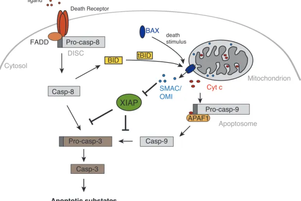
“AVPS” in the case of OMI). When apoptosis is initiated and MOMP occurs, SMAC and OMI are, simultaneously with Cyt c, released into the cytosol to relieve caspases from IAP inhibition (Figure 3).
Figure 3. Extrinsic and intrinsic (mitochondrial) apoptotic pathways. Apoptosis can be induced by stimulation of death receptors with their respective ligands (TNF, FAS, TRAIL). The death receptor oligomerises and induces formation of the DISC, which activates pro-caspase-8. Active initiator caspase-8 activates executioner caspase-3 followed by cleavage of apoptotic substrates. The mitochondrial apoptotic pathway is activated upon death stimuli that lead to oligomerisation of cytosolic BAX, which inserts into the OMM and induces MOMP. SMAC, OMI and Cyt c are released into the cytosol where the latter triggers the formation of the apoptosome (together with pro-caspase- 9 and APAF1), results in activation of initiator caspase-9. Caspase-9 can cleave and activate executioner caspase-3 leading to apoptotic cell death. Furthermore, SMAC and OMI antagonise XIAP,
XIAP
Cytosol
SMAC/
OMI
Mitochondrion BAX death
stimulus
Cyt c
Apoptotic substates
Pro-casp-9 APAF1 Apoptosome Casp-9
Casp-3 Pro-casp-3
Death Receptor
Pro-casp-8 FADD
DISC
Casp-8
ligand
BID tBID
INTRODUCTION
10
preventing it from inactivating caspases, thus supporting the execution step of apoptosis. Caspase-8 can also activate the mitochondrial pathways of apoptosis by cleaving BID into a tBID fragment, which translocates to the mitochondria and activates BAX.
Significant efforts to develop pharmacological IAP antagonists as anti-cancer drugs have been focussing on the synthesis of the purified IAP binding motif (IBM) – that is capable of inhibiting IAPs. In recent years, several novel types of these IAP antagonists, frequently referred to as “SMAC mimetics” have entered clinical trials (LaCasse et al., 2008).
IAPs in inflammatory signalling
In addition to their well-known function in apoptosis, IAPs are increasingly being associated with diverse non-apoptotic pathways such as inflammation and innate immunity. A remarkably conserved RING finger domain equips some IAPs with E3-ligase function (Galbán and Duckett, 2010) and implicates them in ubiquitin-dependent signalling events that regulate the activation of the transcription factor nuclear factor κB (NF-κB) and mitogen activated protein (MAP) kinase pathways that drive the expression of genes involved in inflammation and cell survival (Gyrd-Hansen and Meier, 2010).
Evidence for the involvement of IAPs in innate immunity first emerged in 2005, when Gesellchen and colleagues showed that the Drosophila IAP homologue DIAP2 is indispensable for NF-κB activation in response to Gram-negative bacterial infection (Gesellchen et al., 2005).
XIAP, in particular, has been implicated in inflammatory signalling in response to intracellular bacterial infection. Specifically, XIAP knockout mice failed to clear bacterial infection with Listeria monocytogenes (Bauler et al., 2008) or Chlamydophila pneumonia (Prakash et al., 2010). It is only recently that the molecular mechanisms of XIAP in immune signalling are beginning to be understood in more detail. XIAP - and especially its E3-ligase function - was reported to be important in the NOD signalling cascade (Krieg et al., 2009; Damgaard et al., 2012). Nucleotide-binding oligomerisation domain (NOD) receptors are members of the nucleotide-binding domain and leucine-rich repeat (NLR) containing proteins that function as cytosolic pattern-recognition receptors and trigger inflammatory responses against intracellular bacteria through activation of NF-κB (Chamaillard et al., 2003). The NOD signalling pathway culminates in expression of pro-inflammatory cytokines and chemokines, necessary for a robust anti-microbial environment and proper activation of an immune response. In 2009, Krieg and colleagues proposed XIAP as a new and indispensable
INTRODUCTION
11
component in the NOD signalling pathway in which XIAP was shown to interact with the receptor interacting protein kinase 2 (RIPK2) via its BIR2 domain (Krieg et al., 2009).
According to this view, XIAP serves as a link between the NOD signalling complex (NOD, RIPK2, XIAP) and components of the IKK-activating complex, since an interaction between XIAP and TAB1, an upstream adaptor for activation of the kinase TAK1, has been described (Lu et al., 2007). TAK1 in turn phosphorylates the catalytic subunits of the IKK complex (IKKα and IKKβ) leading to phosphorylation and proteasomal degradation of IκBs (IκBα and IκBβ).
After IκB degradation, NF-κB subunits can translocate to the nucleus and activate target genes involved in the inflammatory response.
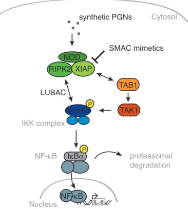
Further insight into the detailed molecular signalling pathway was recently provided by Damgaard and colleagues, who investigated the role of XIAP in NOD signalling. They provide evidence that XIAP ubiquitylates RIPK2 in response to NOD2 stimulation and thereby recruits the linear ubiquitin chain assembly complex (LUBAC) to the NOD signalling complex. LUBAC in turn serves as an ubiquitin ligase downstream of XIAP and is required for efficient activation of NF-κB and cytokine secretion (Damgaard et al., 2012) (Figure 4). Since NOD1 and NOD2 differ only in ligand specificity and share the majority of their signalling pathway, this finding might be relevant for NOD1 activation and needs further investigation.
Figure 4. Intracellular NOD signalling cascade. Synthetic minimal peptidoglycan (PGN) fragments stimulate NOD, leading to complex formation with XIAP and RIPK2. Recruitment of TAB1 to poly-ubiquitylated RIPK2 activates TAK1, which in turn activates the IKK complex finally leading to proteasomal degradation of IκBα and liberation of the NF-κB subunits for nuclear translocation where
TAK1 RIPK2 XIAP
Nucleus IL-8
NF-κB NOD
synthetic PGNs Cytosol
NF-κB IκBα IKK complex
TAB1 NEMOP
proteasomal degradation
P
LUBAC
SMAC mimetics
INTRODUCTION
12
they induce expression of target genes, such as IL-8. LUBAC is recruited to polyubiquitylated RIPK2 and stabilises interaction with NEMO.
Shigella flexneri as a physiological stimulus of immune responses
The Gram-negative enteropathogenic bacterium Shigella flexneri represents a well- established model for intracellular infections. In patients, invasion and inflammatory destruction of the colonic epithelium by S. flexneri causes bacillary dysentery (Shigellosis) a severe form of bloody diarrhea, predominantly occurring in developing countries due to poor sanitary conditions.
S. flexneri is known to stimulate an innate immune response through the activation of NOD1 (Girardin et al., 2001, 2003) and is a well-suited model to analyse the complex host- pathogen interactions that shape the immune response of intestinal epithelial cells. The pathogenesis of S. flexneri infection is closely linked to itsability to invade and replicate in the cytosol of epithelial cells thus avoiding exposure to the extracellular host immune defence (Philpott et al., 2000). In epithelial cells an inflammatory response is produced by activation of NF-κB, followed by production of cytokines such as IL-8, leading to recruitment of immune cells to contain the bacterial infection (Phalipon and Sansonetti, 2007) (Figure 5).
Figure 5: Infection of the colonic epithelium by S. flexneri. Schematic model illustrating S. flexneri infection of the colonic epithelium. Multiplication of S. flexneri in epithelial cells leads to a severe form of inflammatory colitis (Shigellosis) (modified from Ashida et al., 2011).
INTRODUCTION
13
Aim of the study
In addition to its well-recognised function as an inhibitor of apoptosis, XIAP has recently been shown to play an important role in NOD signalling during innate immune responses (Gyrd- Hansen and Meier, 2010). Specifically, NOD signalling in response to stimulation by minimal peptidic fragments induced an inflammatory response involving NF-κB activation and cytokine release that was dependent on XIAP (Krieg et al., 2009; Damgaard et al., 2012).
However, minimal peptidic fragments are insufficient in mimicking the complex circumstances of a physiological infection and the relevance of these findings to physiological stimulation of the NOD-mediated immune responses against bacterial infection remained unresolved and is the subject of this thesis. As a natural antagonist of XIAP function, the mitochondrial intermembrane space protein SMAC is capable of disrupting XIAPs function as an inhibitor of apoptosis (Verhagen et al., 2000). On this basis we investigated whether XIAPs function in an innate immune response to bacterial infection by S. flexneri was similarly regulated by SMAC.