SMAC dependency of XIAP mediated chemoresistance
I n a u g u r a l – D i s s e r t a t i o n
zur
Erlangung des Doktorgrades
der Mathematisch-Naturwissenschaftlichen Fakultät der Universität zu Köln
vorgelegt von Jens Michael Seeger
aus Leverkusen
Köln 2009
Berichterstatter: Prof. Dr. Thomas Langer Prof. Dr. Martin Krönke
Tag der mündlichen Prüfung: 23.10.2008
Abstract ... 4
Zusammenfassung ... 6
Aberrations ... 9
Introduction... 10
Apoptosis ... 10
The two pathways of apoptosis... 12
The role of mitochondria in apoptosis ... 13
Bcl2-family proteins with opposing apoptotic activities... 14
IAPs ... 15
XIAP antagonists ... 17
Apoptosis and Cancer... 18
Resistance to chemotherapeutic treatments and apoptosis... 20
XIAP and Chemoresistance ... 21
Aim of the study ... 22
Methods... 24
Cell lines and cell culture ... 24
siRNA and lentiviral gene transfer... 24
Sample preparation... 25
Immunoblotting ... 25
Immunoprecipitation of active Bax ... 26
Cell viability ... 26
Caspase activation... 27
Immunofluorescence and fluorescence microscopy ... 27
Results... 29
XIAP overexpression in HeLa cells does not confer resistance against cytostatic agents ... 29
Caspase-independent mitochondrial SMAC release in response to cytostatic agents regulates the anti-apoptotic potential of XIAP ... 30
The Role of XIAP in chemoresistant Hodgkin Lymphoma (HL) derived B-cell lines ... 37
XIAP targeting completely restored granzyme B-mediated apoptosis in HL ... 42
Discussion ... 46
References ... 52
Abstract
An important hallmark of tumor cells is their resistance to apoptosis. Apoptosis is a tightly regulated cellular response that ultimately results in the elimination and disposal of unwanted or damaged cells. Apoptosis is brought about by a family of proteases known as the caspases, the activity of which is responsible for the organized destruction of the cell. Each step of the apoptotic signaling cascade is under stringent control. Apoptotic signaling can be regulated at the apical point of the apoptotic cascade by controlling the translation of death-inducing signals into proteolytic activity or more critically by direct modulation of proteolytic activity of caspases. The later is modulated by direct interaction of caspases with members of the inhibitor of apoptosis protein (IAP) family, the most studied one, X-linked IAP (XIAP), has evolved to potently inhibit the enzymatic activity of mammalian caspases.
By efficiently inhibiting caspases XIAP has been shown to block apoptosis and described as a factor conferring resistance against different chemotherapeutic drugs (chemoresistant factor) in a variety of tumor cells. Furthermore, elevated XIAP expression has been frequently observed in several tumor tissues and XIAP targeting sensitizes diverse tumor cell lines for chemotherapeutic agents underlining the role of XIAP in tumor chemoresistance. However, by generating stable cell lines overexpressing XIAP the data provided show that XIAP overexpression alone does not generate a chemoresistant phenotype. Experiments evaluating both XIAP overexpression and stable knock-down of SMAC, a critical regulator of XIAP, show thatXIAP action as a chemoresistant factor is tightly controlled by SMAC. In contrast to Bcl2 that acts as a mitochondrial gatekeeper, XIAP does not alter mitochondrial functions. Cytostatic drugs readily induce release of SMAC in cells with functionally intact mitochondria independent of caspase action, thereby completely neutralizing the anti-apoptotic action of even overexpressed XIAP. Although increased cytotoxic activity by different cytostatic drugs was observed, XIAP targeting failed to restore chemosensitivity in chemoresistant Hodgkin Lymphoma-derived cell lines indicating limited involvement of XIAP in chemoresistance. Unlike chemotherapeutic agents, XIAP targeting resulted in complete reactivation of the apoptotic machinery in response to grzB treatment regardless of mitochondrial functional state. These data demonstrated for the first time that it is essential to assess the mitochondrial capacity
to release SMAC as well as the expression levels of both XIAP and SMAC in order to predict the chemosensitivity of particular tumours, a relationship that has not previously been recognised.
Zusammenfassung
Ein wichtiges Merkmal von Krebszellen ist ihre Apoptoseresistenz, welche wesentlich zu der neoplastischen Proliferation sowie der Resistenz gegenüber dem Immunsystem und der Chemotherapie beiträgt. Bei der Apoptose handelt es sich um eine streng regulierte zelluläre Reaktion, deren charakteristische Merkmale unter anderem die Veränderungen der Plasmamembran (membrane blebbing) und die Kondensierung des Zellkernes (den Abbau der genomischen DNA) beinhalten. Dabei ist die Aktivität einer Familie von Proteasen, die als Caspasen bekannt sind, essentiell für die systematische Eliminierung der Zelle. Die Initiation der Apoptose erfolgt über die Signaltransduktion eines apoptotischen Stimulus zur proteolytischen Prozessierung/Aktivierung der Initiatorcaspasen (Caspase-8 und -9), die ihrerseits die Effektorcaspasen (Caspase-3, -6 und -7) proteolytisch aktivieren. Die Aktivierung von Caspasen kann über mindestens zwei Apoptosesignalwege erfolgen: Der erste Signalweg wird durch die Aktivierung der Todesrezeptoren auf der Zelloberfläche durch die Bindung ihrer spezifischen Liganden eingeleitet, was zur Rekrutierung und Aktivierung von Caspase-8 führt. Im zweiten Signalweg spielen die Mitochondrien eine zentrale Rolle. Die Aktivierung des mitochondrialen Apoptosesignalweges führt zur Permeabilisierung der äußeren mitochondrialen Membran (mitochondrial outer membrane permeabilization; MOMP) und dadurch zur Freisetzung von proapoptotischen Proteinen wie Cytochrome c aus dem Intermembranraum (intermembrane space; IMS) ins Zytosol. Zusammen mit den zytosolischen Komponenten Apaf-1 und Caspase-9 initiiert es die Formierung des Apoptosoms wodurch die Aktivierung von Caspase-9 in Abhängigkeit von ATP-Verbrauch erfolgt.
Aufgrund der immensen Bedeutung der Apoptose und der damit verbundenen Konsequenzen für die Zelle wird jeder Schritt in der apoptotischen Signalkaskade strengstens kontrolliert. Die Funktion von Caspasen als Haupteffektoren der Apoptose kann mittels verschiedener (Adapter-) Proteine reguliert werden, die entweder direkt oder indirekt die proteolytische Aktivierung und Aktivität der Caspasen beeinflussen und somit die Apoptose modulieren können. Die Mitglieder der Inhibitor of Apoptosis Protein (IAP) Familie können die Apoptose durch die direkte Interaktion mit den Caspasen regulieren. Das am besten erforschte Protein
dieser Familie, das X-chromosomale IAP (XIAP), ist in der Lage die enzymatische Aktivität von Caspasen effizient zu hemmen und gilt als der potenteste zelluläre Caspaseinhibitor. Weiterhin wird die Funktion von IAPs durch verschiedene zelluläre Proteine moduliert, die IAPs binden und damit deren inhibitorische Funktion aufheben können. Der wohl bekannteste zelluläre Inhibitor von den IAPs ist der second mitochondrial derived activator of caspases (SMAC), der als inaktives, zytosolisches Protein translatiert wird. Die Reifung von SMAC erfolgt im Intermembranraum der Mitochondrien, wohin es mittels seiner NH2-terminalen mitochondrialen Zielsequenz (MTS) transloziert wird. Dort wird die MTS proteolytisch entfernt und dadurch das NH2-terminale AVPI-Motiv freigelegt, das für die Bindung und die Inhibition von IAPs essentiell ist. Während der Apoptose wird neben Cytochrome c unter anderem auch das maturierte SMAC ins Zytosol freigesetzt, wo es dann die IAPs inhibieren kann.
Durch die direkte Interaktion und effiziente Inhibition von Caspasen wurde XIAP als ein anti-apoptotisches Protein identifiziert. Weiterhin wurde eine erhöhte Expression von XIAP in verschiedenen Tumorzellen beobachtet, die mit einem geringen Therapieerfolg assoziiert war. Darüber hinaus führte die Verminderung der Expression von XIAP mittels RNAi oder die Blockade seiner inhibitorischen Funktion, z. B. durch SMAC, zu einer erhöhten Suszeptibilität gegenüber Chemotherapeutika in einer Vielzahl von Tumorzellen. Somit wurde XIAP in den vergangenen Jahren als ein viel versprechendes Therapieziel in malignen Erkrankungen identifiziert.
In der vorliegenden Arbeit wurde die Bedeutung von XIAP in der Chemoresistenz von Tumoren untersucht. Dabei wurden stabile Zelllinien mit Modifikation der Expression von XIAP und SMAC etabliert und mit verschiedenen Zytostatika behandelt. Im Gegensatz zum aktuellen Modell über die Rolle von XIAP bei der Resistenz gegenüber chemotherapeutischen Substanzen zeigen diese Daten, dass eine Überexpression von XIAP allein keine Chemoresistenz erzeugen kann. Alle hier getesteten konventionellen Zytostatika induzieren unabhängig von der Caspaseaktivität oder von XIAP die Freisetzung von SMAC aus den Mitochondrien und neutralisieren damit vollständig die antiapoptotische Wirkung von sogar überexpremiertem XIAP. Dahingegen führte ein stabiler knock-down von SMAC in der Kombination mit einer Überexpression von XIAP zu einer deutlichen
Chemoresistenz, was folglich die Funktion von XIAP als Chemoresistenzfaktor in der Abhängigkeit der Expression und der mitochondrialen Freisetzung von SMAC stellt.
Die Analyse der Chemosensitivität von malignen Hodgkin Lymphom Zelllinien zeigt, dass der knock-down von XIAP in diesen Zellen zwar zu einer erhöhten Chemosuszeptibilität führt aber nicht vollständig den Apoptosesignalweg reaktivieren kann. Diese Beobachtungen lassen auf eine eher geringfügige Bedeutung der erhöhten XIAP Expression für die Prognose über die Tumor-Chemosuszeptibilität schließen.
Diese Daten zeigen zum ersten Mal, dass es unerlässlich ist die mitochondriale Fähigkeit zur Freisetzung SMAC sowie die Expressionslevel von XIAP und SMAC zu berücksichtigen, um eine Vorhersage über die Chemosensitivität von Tumoren zu treffen - eine Beziehung zueinander, die bisher nicht anerkannt wurde.
Aberrations
MOMP Mitochondrial Outer Membrane Permeabilization SMAC Second Mitochondrial derived Activator of Caspases
IAP Inhibitor of Apoptosis Protein
XIAP X-linked IAP
Caspase Cysteinyl-Aspartate Specific ProteASE DISC Death Inducing Signaling Complex TNF-R Tumor Necrosis Factor Receptor
FADD Fas Associated protein with Death Domain FLICE FADD-Like Interleukin-1β-Converting Enzyme Apaf1 Apoptosis Protease-Activating Factor 1
AIF Apoptosis Inducing Factor
BIR Baculovirus IAP Repeat
HL Hodgkin Lymphoma
H-RS cells Hodgkin-Reed/Sternberg cells STS Staurosporine ETO Etoposide DOX Doxorubicin VBL Vinblastine VCR Vincristine MTX Mitoxanthrone CIS cis-Platin
SDS-PAGE Sodium Dodecyl Sulfate- PolyAcrylamide Gel Electrophoresis CVS Cristal Violet Staining
XTT Cell proliferation kit II (Roche)
IB Immune Blot
IP Immune Precipitation
PARP Poly-(ADP-Ribose) Polymerase
grzB Granzyme B
Ub Ubiquitin
Pfu proliferating units
Introduction
Apoptosis
Life means death – homeostasis in complex organisms is maintained by the finely tuned balance between cellular regeneration and death. About 10 billion of our cells will die each day just to counter the numbers of new cells that arise through mitosis.
A number of modes of cell death have been described so far, and it seems quite possible that others await discovery. Two major modes of cell death have been intensively investigated: Necrosis and apoptosis. Whereas necrosis describes the process of pathological cell death, where amongst other things the release of cellular material into the extracellular space occurs, eliciting an immune response and inflammation (Gallucci et al., 1999; Shi et al., 2000), apoptosis describes the physiological cell death, that is an orchestrated collapse of cells, which manifests in membrane blebbing, cell shrinkage, protein processing, chromatin condensation and DNA degradation (Kerr et al., 1972; Wyllie et al., 1980) followed by rapid engulfment of corpses by neighboring cells thus avoiding an inflammatory response. This type of programmed cell death has been discovered and rediscovered several times by various developmental biologists and cytologists, and has been named differently over the past two centuries (Lockshin and Zakeri, 2001). The term finally adopted is apoptosis, coined by Wyllie and colleagues in 1972 (Kerr et al., 1972). The authors noticed that apoptotic cells shared many morphological features, which were distinct from those observed in cells undergoing necrotic cell death, and they suggested that these shared morphological features might be the result of an underlying common, conserved, endogenous cell death program (reviewed by Goodlett and Horn, 2001;
see also figure 1).
For any multicellular organism, apoptosis plays an essential part in many processes of life, ranging from organ development to the purging of pathogen-infected cells from the body and the elimination of activated or auto-aggressive immune cells. Naturally, apoptosis has to be tightly regulated because uncontrolled cell death may result in developmental defects, autoimmune diseases, neurodegeneration or cancer.
The ‘decision’ of a cell to die depends on the activity of many molecules that determine a cell's likelihood of activating its self-destruction program. Most of the
specific morphological changes that were observed in apoptotic cells upon apoptotic stimuli are mediated by a set of cysteine proteases. These death inducing proteases are homologous to each other, and are part of a large protein family known as caspases (Cysteinyl-Aspartate Specific ProteASE) (Alnemri et al., 1996). Caspases have diverse biological functions, including apoptosis, necrosis and inflammation.
Thus far twelve caspases have been identified in humans, of which seven are key regulators of apoptosis. There are two types of apoptotic caspases: initiator (apical) caspases and effector (executioner) caspases (Kumar, 2007). Initiator caspases (-2, -8, -9, -10) proteolytically activate inactive pro-forms (zymogens) of effector caspases (-3, -6, -7). In turn, effector caspases cleave other proteins within the cell resulting in the apoptotic process. These include approximately 400 substrates (Luthi and Martin, 2007), such as lamins, kinases, DNA repair enzymes, and proteins involved in mRNA splicing, DNA replication, and cell survival, and this is presumed to trigger the morphological process of cell death defined as apoptosis (Kerr et al., 1972). The caspases themselves are activated by intrinsic and extrinsic apoptotic stimuli which will be discussed in the following.
Figure 1: Structural changes of cells undergoing necrosis or apoptosis (taken from Goodlett et al., 2001)
The two pathways of apoptosis
Despite the cellular diversity of our body, all cells appear to activate the same basic elements of the death-inducing program. Two major apoptotic pathways have been identified, one responding to immune-mediated signals, dubbed the ‘‘extrinsic’’
pathway, and the other, the ‘‘intrinsic’’ pathway, is engaged following developmental cues or cellular stresses (Fig. 2). The extrinsic pathway is triggered through ligation of specific cell-surface death receptors, whereas the intrinsic pathway depends on Mitochondrial Outer Membrane Permeabilization (MOMP), which causes the release of apoptogenic factors from the intermembrane space into the cytoplasm. The eventual consequences of each pathway are similar, as they both result in the activation of members of the caspase cascade (Meier and Vousden, 2007).
The extrinsic apoptotic pathway is initiated by the oligomerization, most probably the trimerization, of Tumor Necrosis Factor Receptor (TNF-R) family members, such as CD95, induced by ligation of their specific ligand. Receptor oligomerization results in formation of a complex of proteins associated with activated receptors. The so-called Death-Inducing Signaling Complex (DISC) is the first initiator complex after apoptotic induction. The death signal is propagated by a caspase cascade initiated by the activation of caspase-8, also called FLICE (FADD-Like Interleukin-1β-Converting Enzyme), at the DISC followed by a rapid cleavage of executioner caspases, like caspase-3 and other caspases, which eventually cleave vital substrates in the cell (Ashkenazi and Dixit, 1998; Kischkel et al., 1995; Krammer, 2000).
The intrinsic apoptotic pathway involves mitochondria and results in the release of cytochrome c, which subsequently initiates the caspase activation. The released cytochrome c together with the Apoptosis Protease-Activating Factor 1 (Apaf-1) and procaspase-9 in the cytoplasm forms the apoptosome, the other initiator complex of apoptosis (Li et al., 1997; Saleh et al., 1999). This apoptosome complex in turn activates downstream executioner caspases, like caspase-3, -6 and -7 (Green and Reed, 1998; Li et al., 1997; Reed et al., 1998).
The role of mitochondria in apoptosis
Morphological, biochemical, and molecular genetic studies have shown that mitochondria are the convergence point for diverse signaling cascades initiated by cellular damage. In addition to being crucial for energy production and essential metabolic pathways, mitochondria also play key roles in executing the apoptotic program. The mitochondrial check point permits cells to arrest in the cell cycle in order to repair/restore cellular function after chromosomal damage or may allow cells to undergo apoptosis upon overwhelming, persistent, or severe damage (Green and
Figure 2: The two pathways of apoptosis. The extrinsic pathway of apoptosis is initialized by ligand binding to the death receptors (DR; i. e. CD95L to CD95) resulting in activation of the DR. To the active DR several proteins are recruited forming together the death inducing signaling complex (DISC). This platform recruits and activated the initiator caspase-8, following activation of the executioner caspase-3 which leads to apoptosis. The application of cytostatic drugs or UV treatment leads to cellular stress which induces the intrinsic apoptotic pathway. Herein the mitochondria become activated resulting in mitochondrial outer membrane permeabilization (MOMP). One of the proteins released from mitochondria after MOMP is cytochrome c which is necessary for forming the so called apoptosome together with other factors such as the initiator caspase-9, Apaf-1 and ATP, leading to the activation of caspase-9 which in turn activates caspase-3. As a further regulation point, the inhibitor of apoptosis protein (IAP) XIAP is able to bind both caspase-3 and caspase-9 and inhibit apoptosis. To brake this blockade another protein, the second derived mitochondrial activator of caspases (SMAC), is also released from mitochondria while MOMP. SMAC binds XIAP and inhibits it in its function, resulting in ongoing apoptosis.
Kroemer, 2004). The mitochondria act as pivotal decision centers, because they respond to apoptotic stimuli with Mitochondrial Outer Membrane Permeabilization (MOMP), releasing death-promoting factors from their intermembrane space into the cytosol. The release of cytochrome c from mitochondria couples these organelles to a pathway for caspase activation and apoptosis (Matsuyama and Reed, 2000; Reed and Kroemer, 2000). In addition to cytochrome c other mitochondrial pro-apoptotic factors are released, such as SMAC/DIABLO, which antagonizes the inhibitory effect of the Inhibitors Of Apoptosis (IAP) (Du et al., 2000; Verhagen et al., 2000), Apoptosis Inducing Factor (AIF), a flavoprotein with potent but relatively mysterious apoptotic activity (Lorenzo et al., 1999) and Omi/HtrA2, a serine protease which interacts and inhibits the X-linked Inhibitor Of Apoptosis Protein (XIAP) and enhances caspase activity (Hegde et al., 2002).
The apoptotic activity of mitochondria is known to be regulated by the family of Bcl2 proteins. The Bcl2 proteins, which have either pro- or anti-apoptotic activity, have been studied intensively over the past decade owing to their importance in the regulation of apoptosis, tumorigenesis and cellular responses to anti-cancer therapy (Youle and Strasser, 2008).
Bcl2-family proteins with opposing apoptotic activities
This protein family was named after the founding member of the family, which was isolated as a gene involved in follicular B cell lymphoma (Tsujimoto et al., 1985). The Bcl-2 family is comprised of well over a dozen proteins, which have been classified into three functional groups (Youle and Strasser, 2008). The key feature of the members of group I, such as BCL-2, BCL-XL, BCL-W, MCL1, BCL-B (also known as BCL-2L10) and A1 (also known as BCL-2A1) is that they all possess anti-apoptotic activity and protect cells from death. In contrast, group II consists of Bcl-2 family members with pro-apoptotic activity. Members of this group, including Bax, Bak and BOK (also known as MTD), have a similar overall structure to group I proteins, but promote apoptosis. It appears that the pro-apoptotic family members like Bax and Bak are crucial for initiating MOMP and their function is tightly controlled by anti- apoptotic Bcl2 members. In apoptotic cells, Bax and Bak undergo a conformational change, oligomerize and gain their pro-apoptotic activity by triggering the release of cytochrome c/SMAC. Group III consists of a large and diverse collection of proteins
including BAD, BIK (also known as BLK or NBK), BID, HRK (also known as Death Protein-5 (DP5)), BIM (also known as BOD), BMF, NOXA and PUMA (also known as BBC3) whose only common feature is the presence of the 12–16-amino-acid BH3 domain (Adams and Cory, 1998). Through their conserved BH3 domain these proteins can bind and regulate the activity of BCL-2 proteins. Recent evidence indicates that BH3-only proteins de-repress BAX and BAK by direct binding and inhibition of BCL-2 and other anti-apoptotic family members. By contrast, an opposing model postulates direct activation of BAX and BAK by some BH3-only proteins (specifically BIM, tBID and PUMA) (Youle, 2007).
IAPs
The cytosolic Inhibitor of Apoptosis Proteins (IAPs) are the key regulators of caspase activity in the cell. Both, the initiator as well as the effector caspases, can be regulated by IAP binding, inhibiting the caspase activity.
The first members of the IAP family identified in 1993, were the baculoviral IAPs Cp- IAP and Op-IAP, that had a cytoprotective effect by inhibiting virus induced apoptotic pathways in their target cells (Clem and Miller, 1994; Crook et al., 1993). The common structural feature of these IAPs is a motif termed the Baculovirus IAP Repeat (BIR) that is required for the cytoprotective function of IAPs. Subsequently, host proteins containing BIR domains have been identified in a wide range of lower and higher eukaryotic species, from yeast to mammals (Deveraux and Reed, 1999).
The first human IAP identified was the Neuronal Apoptosis Inhibitory Protein (NAIP), involved in Spinal Muscular Atrophy (SMA), a neurodegenerative disorder (Liston et al., 1996; Roy et al., 1995). Further human IAPs including c-IAP-1/HIAP-2/hMIHB, c- IAP-2/HIAP-1/hMIHC, X-IAP/hILP/MIHA, survivin, BRUCE and ML-IAP were subsequently identified by sequence homology and examined for their anti-apoptotic capacity (Ambrosini et al., 1997; Duckett et al., 1996; Hauser et al., 1998; Hawkins et al., 1996; Hay et al., 1995; Rothe et al., 1995; Roy et al., 1995; Uren et al., 1996;
Vucic et al., 2000). The fact that the BIR motif is shared by all IAP members, suggests a central role for this domain in interacting with conserved components of the apoptotic machinery (Fig. 3).
The most extensively studied family member, XIAP, is a particularly potent inhibitor of caspase activity. Biochemical characterization of XIAP revealed that the second BIR domain (BIR2) inhibits caspase-3 and caspase-7, whereas the third BIR domain (BIR3) inhibits caspase-9 (Deveraux et al., 1999; Deveraux et al., 1998; Deveraux et al., 1997). Further biochemical and structural studies have precisely mapped the XIAP elements required for caspase inhibition, showing that some of these elements are specific to XIAP and not conserved amongst the IAP family (Eckelman and Salvesen, 2006; Eckelman et al., 2006). Indeed XIAP is the only member of this family able to directly inhibit both the initiation and execution phase of the caspase cascade and has garnered the most attention (Fig. 4).
In addition to caspase binding capability, it has been shown that some IAPs have a ubiquitin-E3-ligase activity and are able to catalyze their own ubiquitination in vitro (Yang and Li, 2000) and not surprisingly are able to catalyze the ubiquitination of
Figure 3: Mammalian IAPs/BIRPs. Alternative designations are shown in parentheses. The X-linked IAP (XIAP) is the best-characterized member of this family. BIR, Baculovirus IAP Repeat; CARD, Caspase-Recruitment Domain; ILP, IAP-Like Protein; MIHA, Mammalian IAP Homologue A; NAIP, Neuronal Apoptosis Inhibitory Protein (taken from Salvesen and Duckett, 2002).
Figure 4: Interactions of caspase-3 and caspase-9 with XIAP. (A) The XIAP (in red) consists of three BIR domains; two of them (BIR2 & BIR3) harbors a binding groove. Additionally a RING domain is localized at the COOH-terminus. Caspase-3 and caspase-9 are shown as monomers in blue. (B) Inhibition of caspase 3 occurs after binding of the linker NH2-terminal of BIR2 from XIAP to caspase- 3 through sterical blockage of the catalytically center of caspase-3. (C) Caspase-9 is inhibited by binding to the binding groove of BIR3, resulting in avoidance of dimerization of caspase-9.
their substrates. XIAP in particular, can ubiquitinate caspase 3 (Suzuki et al., 2001b) and SMAC (MacFarlane et al., 2002) amongst other proteins. The ubiquitin-E3-ligase activity is associated with a RING domain that has been found in XIAP as well as other IAP family members (cIAP1 and cIAP2) that are subject to proteasomal degradation (Yang and Li, 2000).
Furthermore, accumulating evidence suggest that XIAP has important roles in other cellular processes including morphogenesis (Olayioye et al., 2005), heavy metal homeostasis (Burstein et al., 2004; Mufti et al., 2006), NF-κB activation and TNF signalling (Gaither et al., 2007; Hofer-Warbinek et al., 2000), MAP kinase signaling (Lu et al., 2007), and neuronal differentiation (Yamaguchi et al., 1999), which may in fact prove to be more important in normal physiological processes.
XIAP antagonists
Several factors play critical roles in controlling the XIAP function by specifically interacting with and relieving its caspase-inhibitory effects. One of them is the XIAP Associated Factor 1 (XAF1), a cytosolic protein that upon binding to XIAP results in increased caspase 3 activity and sensitization to etoposide induced cell death even in XIAP overexpressing cells, suggesting an antagonizing effect of XAF1 in chemoresistance mediated by XIAP overexpression (Liston et al., 2001).
Another XIAP binding protein, is the evolutionary conserved serine proteases HtrA2, (Hegde et al., 2002; Martins et al., 2002; Suzuki et al., 2001a; Verhagen et al., 2002) that in humans binds XIAP in a similar manner to SMAC, thereby facilitating caspase inhibition (Suzuki et al., 2001a). HtrA2 contains an NH2-terminal Mitochondrial Targeting Sequence (MTS), which is removed by proteolytic cleavage following translocation into the intermembrane space of mitochondria in the maturation process that exposes an NH2-terminal motif required for XIAP binding. In contrast to SMAC, HtrA2 can also promote cell death without membrane blebbing or apoptotic body formation that requires the serine protease activity (Suzuki et al., 2001a) and is not affected by caspase inhibitors (Hegde et al., 2002), suggesting two different mechanisms for cell death induction: Similar to SMAC, one mechanism depends on binding to and inhibition of XIAP and therefore results in increased caspase activity.
The second mechanism relies on the serine protease activity of HtrA2, independently of caspase activity (Hegde et al., 2002; Suzuki et al., 2001a).
The ‘Second derived Mitochondrial Activator of Caspases’ (SMAC), also known as DIABLO, is the most popular and best investigated XIAP antagonist, promoting caspase activation by antagonizing IAPs (Du et al., 2000; Verhagen et al., 2000).
SMAC is a nuclear-encoded, cytosolically translated protein which harbors an NH2- terminal MTS similar to HtrA2, targeting it to the mitochondrion for translocation into the intermembranespace, where its MTS is proteolytically removed by the Inner Membrane Peptidase (IMP), resulting in the maturation of SMAC (Burri et al., 2005).
After apoptotic activation of mitochondria, that results in MOMP, mature SMAC is released into the cytosol (Du et al., 2000; Verhagen et al., 2000). In its mature form SMAC reveals its NH2-terminal AVPI-sequence, which is an IAP Binding Motive (IBM), required for XIAP binding (Chai et al., 2000; Wu et al., 2000). With this AVPI- sequence SMAC disrupt XIAPs caspase inhibitory activity (Liu et al., 2000) by binding to both the BIR2 and the BIR3 domain (Fig. 5), resulting in ongoing apoptosis.
Apoptosis and Cancer
The ability of tumor cell populations to expand in number is determined not only by the rate of cell proliferation but also by the rate of cell attrition. Apoptosis represents a major source of this attrition. The evidence is mounting, principally from studies in mouse models and cultured cells, as well as from descriptive analyses of biopsied stages in human carcinogenesis, that acquired resistance toward apoptosis is a hallmark of most and perhaps all types of cancer (Hanahan and Weinberg, 2000).
The possibility that apoptosis serves as a barrier to cancer was first raised in 1972, when Kerr, Wyllie, and Currie described massive apoptosis in the cells populating rapidly growing, hormone-dependent tumors following hormone withdrawal. The discovery of the bcl-2 oncogene by its upregulation via chromosomal translocation in follicular lymphoma (reviewed by Korsmeyer, 1992 and Strasser et al., 1990) and its recognition as having antiapoptotic activity (Vaux et al., 1988) opened up the
Figure 5: Binding of SMAC (green) to XIAP (red).
(A) Mature SMAC bears its NH2-terminal AVPI- sequence like a horn on the surface.
(B) Dimerized SMAC binds both the Bir2 and Bir3 domain of XIAP, resulting in inhibition of XIAP function.
investigation of apoptosis in cancer at the molecular level. Other examples including inactivation of the p53 tumor suppressor protein, a component of the apoptotic signaling circuitry, which led to rapidly growing tumors containing low numbers of apoptotic cells (Symonds et al., 1994) strengthen the consensus that apoptosis is a major barrier to cancer that is broken during tumorigenesis. Collectively, these observations argue that altering components of the apoptotic machinery can dramatically affect the dynamics of tumor progression, providing a rationale for the inactivation of this machinery during tumor development.
The most common malignant tumors of the lymphatic system are the Hodgkin Lymphomas (HL), identified by Sir Thomas Hodgkin in 1832. Typical for the Hodgkin lymphoma is the presence of giant abnormal cells, the Hodgkin-Reed/Sternberg cells (H-RS cells) in affected tissues (reviewed by Kuppers, 2003). In classical HL (cHL), malignant H-RS cells represent about 1% of the cell population in the affected lymph nodes (reviewed by Kuppers, 2003). Genetic and immunologic characterization of the H-RS revealed that they have rearranged B-cell receptor genes but no functional B- cell receptors (Kanzler et al., 1996). In only minor cases (about 2% of the HL) T-cell receptor rearrangement has been observed (Muschen et al., 2000), suggesting a B- cell origin for most classical HL cases. Nevertheless neither mature B-cell receptors nor T-cell receptors are presented on the surface of H-RS cells which would normally result in induction of apoptosis (Lam et al., 1997). The fact that H-RS cells survive without functional immune receptors suggests that H-RS cells are resistant to apoptosis. Indeed, a blockade of both the extrinsic and intrinsic apoptotic pathway is seen in H-RS cells. The extrinsic pathway has been shown to be blocked by the overexpression of cFlip, a caspase-8 inhibitor (Thomas et al., 2002), while the intrinsic pathway is blocked by deficient Bax activation and failure of the mitochondrial cytochrome c release (Kashkar et al., 2002; Kashkar et al., 2006). In addition, elevated expression of XIAP has been identified as a hallmark of HL cell lines and H-RS cells of primary tumor tissues (Kashkar et al., 2003) (Fig. 6). Classical HL is a fatal disease with 90% of untreated patients dying within 2 to 3 years. With modern polychemotherapy,more than 80% of patients suffering from cHL are cured.
Despitethis treatment success rate, the pathogenesis of cHL is stilllargely unknown.
Dissolving the mechanisms by which H-RS cells escape apoptosis help to identify new strategies for anti cancer therapy.
Resistance to chemotherapeutic treatments and apoptosis
Resistance to chemotherapy represents a major obstacle to cancer therapy.
Although different forms of chemotherapy are aimed at a variety of biochemical targets, it is generally believed that chemotherapy kills cancer cells by induction of a final common pathway that leads to apoptosis (Houghton, 1999; Martin et al., 1997).
The agents that are commonly used act by damaging cellular components including DNA (crosslinking DNA by cisplatin), and microtubules (vinblastine) or by inhibiting protein kinases to such an extent that apoptosis is induced. Thus, defects in the apoptotic machinery may result in chemoresistance (Lowe et al., 1994).
Accumulating evidence suggest that the pathway involved in chemotherapeutic agent-induced apoptosis seems to be a consequence of damage to the mitochondria (Houghton, 1999), although the mechanisms that might induce these changes are not well understood. Correspondingly, elevated expression levels of Bcl2 have been identified as a chemoresistance factor (Reed, 1995). Activation of the transcription
Figure 6: Expression of caspase activators and inhibitors in HL-derived B cell lines. (A) Equal amounts of proteins from total cell lysate of L1236, L591, L428, and KMH2 cells and of control B cells L1309 were subjected to SDS-PAGE and Western blot analysis. Proteins were detected using antibodies against Apaf-1 and XIAP. (B) XIAP expression in primary Hodgkin's lymphoma (HL) tissues. XIAP positivity in H-RS cells: + = very weak; ++ = weak; +++ = moderate; ++++ = strong staining. (C) Paraffin section immunohistochemistry of cases 3, 5, 6, 7, 10, and 11 of classical HL cases listed in Fig. 2 B. XIAP was stained using anti-XIAP specific mAb. There are strong (Nos. 5, 6, 7, and 11) or moderate (Nos. 3 and 10) granular intracytoplasmic staining for XIAP in essentially all morphologically recognizable Hodgkin or Reed-Sternberg cells. Background lymphocytes are negative for XIAP staining (taken from Kashkar et al., 2003).
factor p53 is frequently associated with chemotherapy-induced apoptosis (Li et al., 1998) and mutations in TP53 (the gene that encodes p53) are common in some cancers and lead to resistance to DNA-damaging agents (reviewed in Hersey and Zhang, 2001). Furthermore, the blockade of death receptor signaling (CD95 and CD95L) leads to apoptosis resistance in drug sensitive neuroblastoma cells (Fulda et al., 1997) and IAPs as potent caspase inhibitors, have also been suggested to play a role in drug resistance (reviewed by Schimmer et al., 2006). A range of diverse strategies are currently investigated in order to counter chemoresistance in cancer therapy. Targeting Bcl2 by antisense oligonucleotides (G3139 (Genasense), a Bcl2 antisense phosphothioate oligonucleotide, is in phase I clinical trials) or by BH3 mimetics approaches has been shown to suppress the oncogenic effect of anti- apoptotic Bcl2 members and promises to be beneficial in patients suffering from malignancies (reviewed by Labi et al., 2008, Cotter et al., 1994 and Waters et al., 2000). Targeting IAP by RNAi or SMAC mimetics have also been shown to sensitize tumor cells to apoptosis (reviewed by Schimmer et al., 2006, Li et al., 2004 and Nikolovska-Coleska et al., 2004).
XIAP and Chemoresistance
Given its role in apoptosis, there has been much interest in understanding the role of XIAP in cancer and evaluating XIAP as a therapeutic target. XIAP is overexpressed in malignant cells and, in certain patients, is associated with poor clinical outcome and has been viewed as the resistance factor for multiagent chemotherapy (Schimmer et al., 2006). From the discovery of XIAP in the second half of the 1990s, research on this unique IAP has been exponential giving us a detailed structural and mechanistic view of its activity in addition to abundant cell biological data. As a result, the development of potential drugs targeting XIAP has become possible offering targeted therapies to counteract cancer and overcome drug resistance. Several lines of evidence suggest that XIAP represents a promising target in cancer therapy: First, in vitro, XIAP overexpression confers resistance to different apoptotic stimuli (Schimmer et al., 2006).
Second, elevated XIAP expression was reported in a variety of human cancers (Jaffer et al., 2007; Kashkar et al., 2003; Kluger et al., 2007; Krajewska et al., 2003;
Lopes et al., 2007; Mizutani et al., 2007; Tamm et al., 2000; Tamm et al., 2004b) and
was associated with more aggressive tumor histology and decreased survival in patients (Mizutani et al., 2007; Tamm et al., 2004a; Tamm et al., 2004b).
Finally, XIAP targeting by small molecules primarily designed to relieve XIAP- mediated caspase-binding/inhibition or specific down-regulation of XIAP expression by RNA interference or anti-sense oligonucleotides induced cell death directly or synergistically with chemotherapeutic agents in a broad range of tumor cell lines in vitro and in tumor xenograft models (Arnt et al., 2002; Chawla-Sarkar et al., 2004;
Fulda et al., 2002; Kashkar et al., 2007; Lima et al., 2004; McManus et al., 2004;
Sasaki et al., 2000; Schimmer et al., 2004; Yang et al., 2003).
However, recent data suggests that elevated XIAP expression in different tumor tissues is not an applicable prognostic marker for chemoresistant tumors and even associated with a more favorable clinical outcome (Ferreira et al., 2001; Hwang et al., 2008; Seligson et al., 2007). Our knowledge about XIAP as a chemoresistance factor has been initially derived from caspase activity studies mainly using recombinant proteins in cell free systems (Deveraux et al., 1997) or from transient-overexpression studies of XIAP in intact cells (Duckett et al., 1998). Accordingly, the results obtained are limited in describing the capacity of XIAP to inhibit cell death in a living cell and fail to properly examine the potency of XIAP in the context of regulatory circuits. The crucial event in chemotherapeutic drugs-induced apoptosis is the mitochondrial outer membrane permeabilization with the subsequent release of multiple pro-apoptotic factors including cytochrome c and SMAC, initiating caspase proteolytic activity and regulating XIAP function, respectively. Thus, the physiological impact of XIAP on caspase activity is further determined by mitochondria and mitochondrial SMAC, a phenomenon which is completely disregarded in the initial previous works.
Aim of the study
From the discovery of XIAP in the second half of the 1990s, research on this unique IAP has been exponential giving us a detailed structural and mechanistic view of its activity in addition to abundant cell biology data (Eckelman et al., 2006). The aim of this study is to investigate the role of XIAP in tumor chemoresistance by stably modifying the expression level of XIAP and XIAP-antagonists in HeLa cell lines. The
impact of these findings will be further investigated in Hodgkin-Lymphoma tumor cell lines with elevated XIAP expression and defective apoptotic machinery.
Methods
Cell lines and cell culture
HeLa, HeLa-Bcl2 and HeLa-mycXIAP (Kashkar et al., 2007; Kashkar et al., 2005) cell lines were cultured in DMEM and Jurkat, Jurkat-Bcl2, L591 and L428 cell lines (Kashkar et al., 2002) were cultured in VLE RPMI 1640 supplemented with 10% fetal calf serum, 2 mM L-glutamine, 100 µg/ml streptomycin, and 100 units/ml penicillin (Biochrom, Berlin, Germany). All chemicals were purchased from Sigma (Hamburg, Germany) unless indicated otherwise. Cells were treated with cytostatic agents at the indicated concentrations including Staurosporine (STS) (Alexis, Lausen, Switzerland), Etoposide (ETO), Doxorubicin (DOX), Mitoxanthrone (MTO), Vinblastine (VBL), Vincristine (VCR) and cis-Platin (CIS) incubated as indicated.
Caspase activity was blocked by co-treatment of cells with 20 µM Z-VAD(Ome)-FMK (Alexis, Grünberg, Germany).
GrzB/Ad (adenovirus)-mediated cell death was carried out as described previously by Goping et al. (Goping et al., 2003). In brief, 106 cells/ml were treated with 600 ng isolated human grzB (Alexis, Lausen, Switzerland) and 100 pfu Ad (a gift from Dr U.
Protzer, University of Cologne/Germany) in serum-free media supplemented with 0.1% (w/v) BSA. Cells were incubated for 4 to 5 hours at 37°C, then harvested and assessed for cell death by trypan blue staining.
siRNA and lentiviral gene transfer
To silence XIAP and SMAC expression, pENTR constructs were generated with a pair of oligonucleotides derived from XIAP and SMAC mRNAs (the sequence was obtained from Ambion Europe, Huntingdon, United Kingdom), which includes the unique N-19 target as described in pSUPER RNAi System Manual (OligoEngine, Seattle, WA). Three shRNA sequences were designed to down-regulate each gene.
All shRNAs were examined in HeLa and HEK293 cells and the most potent shRNA was identified (data not shown) and stably expressed in cell lines using lentiviral gene transfer. The vector uses the polymerase-III H1-RNA gene promoter. After generating a pENTR clone, the pLenti6/V5DEST XIAPshRNA-, SMACshRNA- or scrshRNA-expressing vectors were created using LR recombination. The viral particles were produced according to the instructions of the manufacturers
(ViraPower Lentiviral expression system; Invitrogen). Cells were transiently transfected with pENTR vectors or transduced by the recombinant lentiviral constructs, and stable cell lines were generated by blasticidin (Invitrogen, Karlsruhe, Germany) selection.
Sample preparation
To extract whole cell lysates, cells were washed twice in cold (4°C) phosphate- buffered saline (PBS), pelleted at 1.200xg for 3 min at 4°C and resuspended in 1 volume of CHAPS lysis buffer (10 mM HEPES [pH 7.4], 150 mM NaCl, 1% CHAPS, complete protease inhibitor cocktail) on ice for 30 minutes. Samples were centrifuged at 14.000xg for 20 min at 4°C and supernatants (= whole cell lysates) as well as the resulting pellets (= nuclear extracts) were recovered.
Poly(ADP-ribose) polymerase (PARP) cleavage was assessed after incubation of pellets in urea extraction buffer (50 mM Tris [pH 6.8], 6 M urea, 3% SDS, 10%
glycerol, 0.00125% bromophenol blue, 5% 2-mercaptoethanol) denatured at 100°C for 10 minutes.
For the preparation of cytosolic extracts about 107 cells were washed twice in cold (4°C) PBS and centrifuged at 1.200xg for 3 min at 4°C. The cells were then resuspended in 1 volume Buffer A (20 mM PIPES [pH 7.0], 50 mM KCl, 2 mM MgCl2, 5 mM EGTA, 1 mM dithiothreitol, 10µM cytochalasin B) and incubated for 20 min on ice allowing to swell, following plasma membrane disruption by homogenization through a 27-gauge needle. The cell breakage was verified microscopically by trypan blue staining. After centrifugation at 14.000xg for 30 min at 4°C the supernatants (=
cytosolic extract) was recovered and stored at -20°C.
Protein concentration was determined by the bicinchoninic acid assay method (BCA- Assay, Pierce) using BSA as a standard using an anthos ht2 (anthos Mikrosysteme GmbH, Krefeld, Germany)
Immunoblotting
Equal amounts of protein were separated by Sodium Dodecyl Sulfate–
PolyAcrylamide Gel Electrophoresis (SDS-PAGE) and transferred to nitrocellulose membrane (Protran 0.2 µm; Schleicher and Schuell) as previously described (Kashkar et al., 2002).
Rabbit polyclonal antiserum specific for Bax, mouse polyclonal antiserum specific for PARP and mouse monoclonal antibodies specific for XIAP, Bcl2, Bax clone 6A7 and cytochrome c were obtained from BD Laboratories (Heidelberg, Germany). Rabbit anti–caspase-3 and mouse anti-SMAC antibodies were obtained from Cell Signaling Technology (Beverly, MA). The mouse anti-GFP antibody was provided from Invitrogen (Karlsruhe, Germany). Affinity purified rabbit anti-cIAP1 and anti-cIAP2 antibodies were obtained from R&D Systems (Wiesbaden-Nordenstadt, Germany).
The mouse anti-actin antibody and the HorseRadish Peroxidase (HRP) conjugates of anti-rabbit and anti-mouse IgG were purchased from Sigma-Aldrich (Munich, Germany). The HRP-conjugated antibodies were used as secondary antibodies and chemiluminescent signals were detected by ECL (Pierce) on films (Amersham, GE Healthcare, Buckinghamshire, UK).
Immunoprecipitation of active Bax
Equal volumes of whole cell lysates (107 cells/ml) were used for immunoprecipitation.
The KCl concentration of the cell lysates was adjusted to 150 mM, and all samples were brought to a final volume of 500 ml with CHAPS lysis buffer. Samples were rotated for 12 h at 4°C with 2 µg of monoclonal anti-Bax 6A7 antibody. Antigen- antibody complexes were immobilized by rotation for 2 h at 4°C with GammaBind G Sepharose (Pharmacia Biotech). The complexes were pelleted (1 min, 14 000xg) and the supernatant removed. The complexes were then washed three times with the same buffer used for the immunoprecipitation and subjected to SDS - PAGE and immunoblotted as described above.
Cell viability
Cells (104 per well for HeLa, 105 per well for Jurkat/HL cells) were incubated in 96- well plastic plates at 37°C in full medium and treated with the indicated concentrations of cytostatic agents and durations as indicated. Cell viability was assessed either by the XTT test (Cell Proliferation Kit II [XTT]; Roche Applied Sciences, Manheim, Germany) or Crystal Violet assay System (CVS). In XTT assay after treatments cells were incubated with the XTT reagents at 37°C for 4 hours according to the instructions of the manufacturer. The absorbance of the samples was measured with an enzyme-linked immunosorbent assay (ELISA) reader
(wavelength, 450 nm; reference wavelength, 620 nm). For CVS after treatment cells were washed twice with PBS, stained with crystal violet (0.2% w/v in 2% EtOH) and resolved by a mixture of 0,2 M sodium citrate and 100% EtOH (1:1 v/v). The colorimetric measurement was carried out at 595nm using a Tecan GENious Pro (Tecan, Crailsheim, Germany). All data are mean values from at least 3 different experiments in triplicate. Blank absorbance from wells that lacked cells was subtracted from that of the samples, and the difference between the absorbance is referred to as "% cell viability" (100% in untreated cells).
Caspase activation
For initiating caspase activation, 20 µg cell extracts were treated with either increasing amounts of horse heart cytochrome c as indicated and additional 1 mM dATP or 20 ng recombinant murine grzB with or without SMAC N-terminal peptide, H-AVPIAQK-OH (10 µM; Calbiochem) for 1 hour at 30°C. Alternative, whole cells where treated with increasing amounts of STS as indicated and cell extracts were prepared. For the following Caspase-3 activity measurement were 5 µg cell extracts in a total volume of 100 µl in caspase activation buffer (20 mM Pipes, 100 mM NaCl, 1 mM EDTA, 0.1% CHAPS, 10 % sucrose, 10 mM dithiothreitol) used and the reaction was initiated by the addition of 100 µM Ac-DEVD–7-amino-4- trifluoromethylcomarin (Ac-DEVD-AFC), following fluorescent readout at 400/505 nm using a Tecan GENious Pro (Tecan, Crailsheim, Germany).
Immunofluorescence and fluorescence microscopy
Cells were treated with 0.5 µM staurosporine or 5 µM doxorubicin for 8h. Cells were then fixed with 3% paraformaldehyde for 20 min, permeabilized with 0.1% saponin in PBS for 10 min, and blocked with 3% BSA and 0.1% saponin in PBS for 30 min. For immunostaining, cells were incubated with primary mouse anti-SMAC or anti Cytochrome c antibody for 1 h, washed with 0.1% saponin in PBS and then incubated with goat anti-mouse antibody conjugated with Alexaflour 568 (Molecular Probes) for 30 min. Nuclei were counterstained with Hoechst 33258 (10 mg/ml PBS) and mounted on glass slides and analyzed by a motorized inverted microscope (Olympus Ix81; Tokyo, Japan) using a 63x/1.40 numerical aperture Planapo oil
objective. Images were acquired using analy-SIS software (Soft Imaging System, Münster, Germany).
Results
XIAP overexpression in HeLa cells does not confer resistance against cytostatic agents
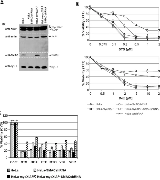
In order to characterize the role of XIAP as a chemoresistance factor, a HeLa cell line stably expressing myc-tagged XIAP (mycXIAP) was established (Fig. 7A and Fig.
10). The cytoprotective potency of XIAP was analyzed in the HeLa-mycXIAP cell line after treatment with different cytostatic agents including staurosporine (STS),
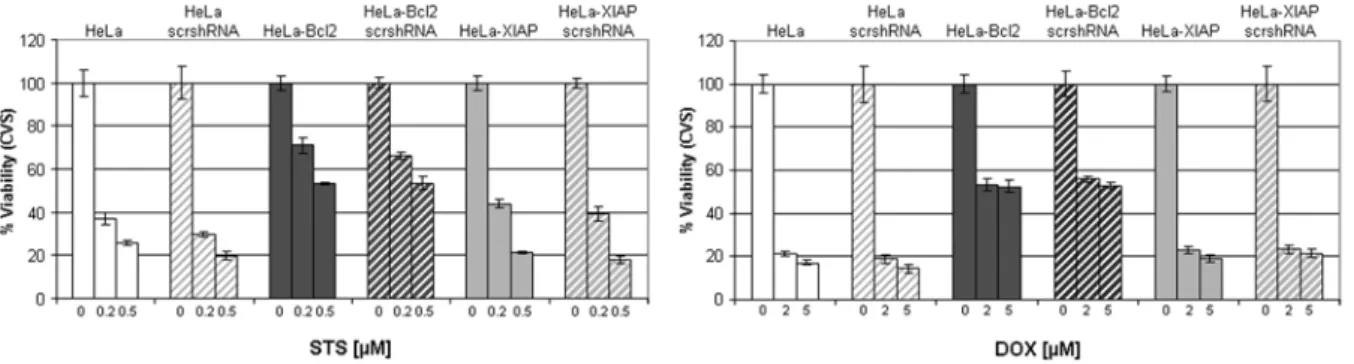
doxorubicine (DOX), etoposide (ETO), mitoxantrone (MTO), vinblastin (VBL), and vincristin (VCR) and compared to HeLa and HeLa cell line overexpressing Bcl2 (HeLa-Bcl2) as an established chemoresistance factor (Kashkar et al., 2005; Letai, 2008; Reed, 1995) (Fig. 7A). Strikingly, overexpression of XIAP does not generate any cytoprotective effect nor does it confer an anti-apoptotic effect against cytostatic agents (Fig. 7B-C). All cytostatic agents exerted equal cytotoxic effects in HeLa and HeLa-mycXIAP cell lines as analyzed by two separate toxicity assays, XTT (Fig. 7B) and crystal violet staining (CVS) (Fig. 7C). Analysis of PARP cleavage to monitor the ongoing apoptotic process demonstrated the involvement of a functional apoptotic cascade in HeLa and HeLa-mycXIAP cell lines generating cleaved PARP upon cytostatic treatments (Fig. 7D). In contrast, Bcl2 overexpression markedly reduced cytotoxicity and completely abrogated the apoptotic capability of cells after different cytostatic drug treatments (Fig. 7B-D).
Caspase-independent mitochondrial SMAC release in response to cytostatic agents regulates the anti-apoptotic potential of XIAP
In order to investigate the caspase inhibitory potency of overexpressed XIAP in the HeLa-mycXIAP cell line, cytosolic extracts of intact HeLa cell lines were prepared and caspase activity was initiated by exogenously added cytochrome c and dATP (Kashkar et al., 2003). In contrast to HeLa cells cytochrome c-induced caspase-3 activity was significantly impaired in cytosolic extracts derived from HeLa-mycXIAP cells demonstrating the caspase inhibitory potency of overexpressed mycXIAP. As the sensory centers of cytotoxic stresses, mitochondria promote caspase activity by releasing pro-apoptotic factors including cytochrome c and SMAC. Once released into the cytosol, SMAC interacts with XIAP to release XIAP-mediated inhibition of caspase-3. Accordingly, in cytosolic extracts of HeLa and HeLa-mycXIAP cells, addition of the synthetic SMAC N7 peptide enhanced caspase-3 activity initiated by cytochrome c/dATP and restored the caspase-activity blocked by overexpressed
were detected in total cell extracts of HeLa, HeLa-mycXIAP and HeLa-Bcl2 by Western blot analysis using specific antibodies. (B-D) HeLa, HeLa-mycXIAP and HeLa-Bcl2 cell lines were left untreated or treated for 24 hours with staurosporine (STS, 0.5 µM), doxorubicin (DOX, 5 µM), etoposide (ETO, 100 µM), mitoxantrone (MTO, 5 µM), vinblastine (VBL, 50 nM), and vincristin (VCR, 50 nM). Viable cell number was determined using the XTT (B) or crystal violet staining assay (CVS). PARP cleavage was detected in nuclear extracts using mouse anti-PARP antibody detecting cleaved PARP (D).
Asterisk indicates nonspecific bands recognized by anti-XIAP antibody.
mycXIAP (Fig. 8A). Detailed analyses of the mitochondrial apoptotic pathway in HeLa, HeLa-mycXIAP and HeLa-Bcl2 cell lines showed that all tested cytostatic agents initiated the mitochondrial release of cytochrome c and SMAC in HeLa and HeLa-mycXIAP cells but not in the HeLa-Bcl2 cells with blocked mitochondria (Kashkar et al., 2005) (Fig. 8B). The engagement of the mitochondrial apoptotic pathway and MOMP by cytostatic drug treatment was predominantly a caspase- independent process as demonstrated by pre-treatment with the universal caspase inhibitor z-VAD (Fig. 8B). In contrast to Bcl2, XIAP was not able to prevent cytostatic agent-induced mitochondrial release of SMAC (Fig. 8B) that exerts neutralizing activity toward XIAP.
Figure 8: Cytostatic agents induce caspase-independent SMAC release. (A) Cytosolic extracts of HeLa and HeLa-mycXIAP cells were prepared and equal amounts of protein were incubated with increasing amount of cytochrome c (0, 0.25, 0.5, and 1 µM) and dATP with or without SMAC N7 peptide for 15 min at 30°C. Relative caspase-3 activity was measured by using 100 µM Ac-DEVD- AFC and presented as arbitrary fluorescence units. (B) HeLa, HeLa-mycXIAP and HeLa-Bcl2 cells (all 106) were treated with STS (0, 0.25, and 0.5 µM), DOX (0, 2, and 5 µM), ETO (0, 10 and 50 µM), MTO (0, 1 and 5 µM), VBL (0, 5 and 20 nM), and VCR (0, 5 and 20 nM) for 12 h. SMAC and cytochrome c were detected in cytosolic extracts by specific antibodies. Reprobing for actin ensured equal loading of cytosolic extracts. The involvement of caspase activity in cytostatic drugs-induced cytchrome c/SMAC release was examined in HeLa cells by z-VAD-FMK (20 µM) co-treatment.
If mitochondrial release of SMAC caused neutralization of XIAP, down-regulation of SMAC expression should restore XIAPs caspase inhibitory function and confer resistance against cytostatic agents. In order to address this issue we specifically down-regulated SMAC expression by using small hairpin RNA (shRNA) targeting SMAC mRNA (Kashkar et al., 2006). Only HeLa and HeLa-mycXIAP cells expressing
Figure 9: SMAC knock-down promotes resistance against cytostatic agents and facilitates XIAP anti- apoptotic function. (A) XIAP, actin, SMAC and cytocherome c were detected in total cell extracts of HeLa, HeLa-SMACshRNA, HeLa-mycXIAP and HeLa-mycXIAP-SMACshRNA cells by Western blot analysis using specific antibodies against XIAP, cytochrome c, actin (reprobed and merged) and SMAC (reprobed and merged). (B) Cells were left untreated or treated for 24 hours with increasing concentrations of STS or DOX. The percentage viable cell number was determined using an XTT assay. (C) Cells were left untreated or treated for 24 hours with STS (0.5 µM), DOX (5 µM), ETO (100 µM), MTO (5 µM), VBL (50 nM), and VCR (50 nM). The percentage viable cell number was determined using a CVS assay. Asterisk indicates nonspecific bands recognized by anti-XIAP antibody.
shRNA against SMAC (HeLa-SMACshRNA and HeLa-mycXIAP-SMACshRNA) displayed down-regulated SMAC expression, while scrambled (scr) shRNA (HeLa- scrshRNA and HeLa-mycXIAP-scrshRNA) remained ineffective (Fig. 9a and 10). The specificity of the SMAC knockdown was revealed by unaltered cytochrome c, Bcl2, XIAP, and actin expression (Fig. 9A and Fig. 10). We next examined whether SMAC down-regulation results in decreased susceptibility of cells to cytostatic agents.
HeLa-SMACshRNA and HeLa-mycXIAP-SMACshRNA but not HeLa-scrshRNA showed markedly reduced susceptibility to STS and DOX treatment compared to their parental counterparts HeLa and HeLa-mycXIAP, respectively (Fig. 9B). This is additionally confirmed by a separate cytotoxicity assay and holds true for other cytostatic drugs demonstrating the key regulatory role of SMAC in cytostatic agent- induced cell death (Fig. 9C and Fig. 11).
Fig. 10: Western blot analysis of cell lines used in this work. XIAP, actin, Bcl2, SMAC and Cytochrome c were detected in total cell extracts of HeLa, HeLa-scrshRNA, HeLa-SMACshRNA, HeLa-XIAPshRNA, HeLa-Bcl2, HeLa-Bcl2- scrshRNA, HeLa-Bcl2-XIAPshRNA, HeLa- mycXIAP, HeLa-mycXIAP-scrshRNA, and HeLa- myXIAP-SMACshRNA cell lines by Western blot analysis using specific antibodies. Asterisk indicates nonspecific bands recognized by anti- XIAP antibody.
Fig. 11: Expression of scrshRNA does not influence cytotoxic activity of cytostatic agents. Cells were left untreated or treated for 24 hours with indicated concentrations of STS and DOX. The percentage viable cell number was determined using a CVS assay.
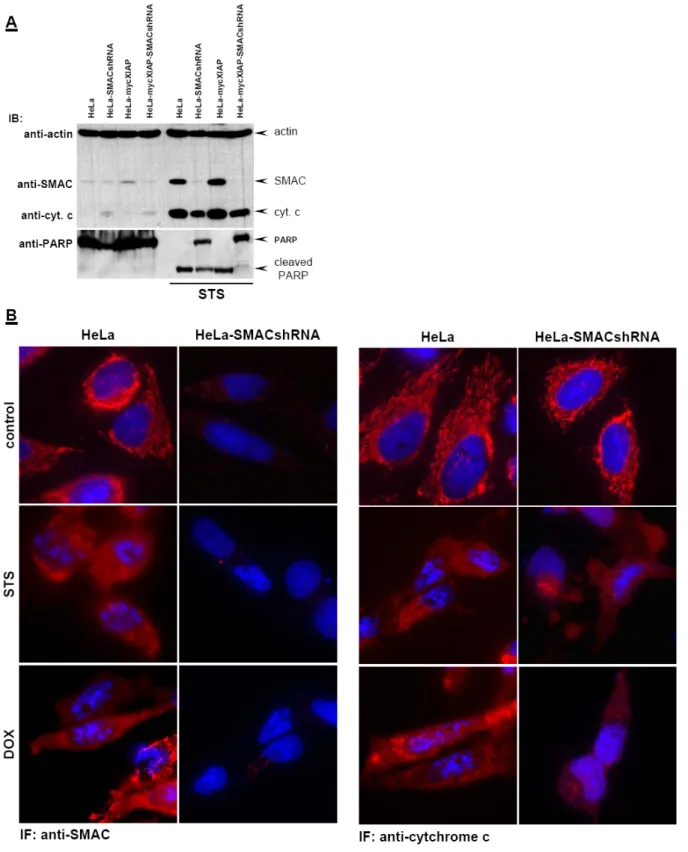
It is important to note that SMAC knock-down did not cause any alteration in cytochrome c release after cytostatic drug treatment as shown in Fig. 12. The lack of
Figure 12: SMAC knock-down does not influence mitochondrial release of cytochrome c. (A) Cells were treated with STS (0.5 µM) for 12 h. SMAC and cytochrome c were detected in cytosolic extracts by specific antibodies. Reprobing for actin ensured equal loading of cytosolic extracts. PARP cleavage was detected in nuclear extracts using mouse anti-PARP antibody detecting cleaved PARP. (B) IF analysis of SMAC and cytochrome c release after STS and DOX treatment in HeLa and HeLa-SMACshRNA cell lines. Cells were left untreated or treated with STS (0.5 µM) or DOX (5 µM) for 8h. SMAC and cytochrome c were detected by specific primary and secondary Alexa Fluor 568- conjugated antibodies (red). Nuclei were co-stained with Hoechst 33258.
SMAC in HeLa-SMACshRNA resulted in blockade of caspase activity by XIAP as demonstrated by incomplete PARP cleavage (Fig. 12A) and failure of nuclear fragmentation (Fig. 12B). This effect was significantly enhanced in HeLa-mycXIAP- SMACshRNA lacking SMAC expression and overexpressing XIAP as shown by complete blockade of PARP cleavage (Fig. 12A).
Correspondingly, XIAP knock-down in HeLa-SMACshRNA cells restored their apoptotic capability and enhanced the cytotoxicity mediated by STS (Fig. 13). HeLa- SMACshRNA cells were transiently transfected with DNA constructs containing XIAPshRNA or scrshRNA expression cassettes which in addition co-expressed EGFP to visualize the transfected cells. As shown in Fig. 13A, XIAP expression was specifically down-regulated in the HeLa-SMACshRNA cells transfected with EGFP- XIAPshRNA but not with EGFP-scr-shRNA. Immunofluorescence (IF) analysis revealed nuclear fragmentation as a sign of the ongoing apoptotic process after STS treatment in HeLa-SMACshRNA cells transiently transfected with EGFP-XIAPshRNA but not in untransfected cells or cells transfected with EGFP-scr-RNA (Fig. 13B).
Correspondingly, HeLa-SMACshRNA cells depleted of XIAP displayed increased susceptibility to STS (Fig. 13C). IF analysis of cytochrome c showed that XIAP down- regulation did not facilitate cytochrome c release nor did it promote any nuclear fragmentation in untreated cells (Fig. 13B) underscoring the interplay between XIAP expression and mitochondrial SMAC release in modulating caspase activity initiated by cytosolic cytochrome c. Taken together, these data suggest that the XIAP- mediated chemoresistance is SMAC dependent.
Figure 13: XIAP knock-down restores inefficient apoptosis in HeLa-SMACshRNA cells. HeLa- SMACshRNA cells were transiently transfected with DNA constructs encoding EGFP together with XIAPshRNA or scrshRNA expression cassettes. (A) After 48 hours cell extracts were prepared and XIAP, actin and GFP were detected in Western blot analysis using specific antibodies. (B) Cells were left untreated or treated with STS (0.5 µM) for 8h. Cytochrome c was detected by specific primary and secondary Alexa Fluor 568-conjugated antibodies (red). Nuclei were co-stained with Hoechst 33258. Transfected cells appeared green by co-expression of EGFP. (C) Viability was microscopically determined by evaluating >300 EGFP expressing cells. The percentage of viable cells was calculated relative to the total cell number. Asterisk indicates nonspecific bands recognized by anti-XIAP antibody.
The Role of XIAP in chemoresistant Hodgkin Lymphoma (HL) derived B-cell lines
One of the central piece of evidence validating XIAP as a chemoresistance factor is derived from observations that XIAP targeting markedly enhanced the cytotoxic activity of different cytostatic drugs in different tumor types (Schimmer et al., 2006).
The malignant Hodgkin and Reed-Sternberg (H-RS)cells of Hodgkin lymphoma (HL) are germinal center B cellswith a dysfunctional apoptotic machinery (Marafioti et al., 2000). Previous results demonstrated an elevated expression of XIAP in H-RS cell lines and primary tumor tissues as a hallmark of HL (Akyurek et al., 2006; Kashkar et al., 2003).
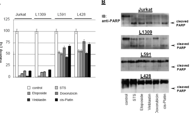
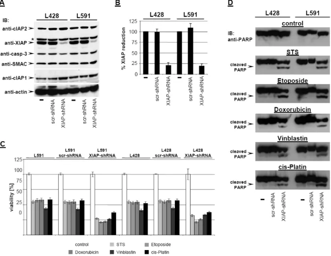
As shown in Figure 14, HL B-cell lines L428 and L591 were resistant to staurosporine (STS), etoposide, doxorubicin, vinblastine, and cisplatin. In contrast, treatment of Jurkat T cells or the control B-cell line L1309 with all cytotoxic agents resulted in cell death (Figure 14A) associated with PARP cleavage (Figure 14B) indicating an apoptotic cell death. If XIAP is the key mediator of chemoresistance in HL B-cells, knock-down of XIAP by RNAi should sensitize HL cells to cytotoxic drugs. XIAP expression was specifically down-regulated by generating HL cell lines stably
Figure 14: Chemoresistance of HL B-cell lines against cytostatic agents. Control B-cell line L1309, control Jurkat T-cell line, and HL B-cell lines L428 and L591 were treated with staurosporine (0.5 µM), etoposide (50 µM), doxorubicin (1 µM), vinblastine (0.2 µM), or cisplatin (200 µM) and incubated for 24 hours. (A) Viability was assayed by XTT test. The means ± SD are from 3 individual experiments performed in triplicate. (B) PARP cleavage was detected in nuclear extracts by mouse anti-PARP antibody.
Figure 15: Knock-down of XIAP by RNAi sensitizes HL B-cell lines for cytostatic agents. (A) cIAP2, XIAP, caspase-3, SMAC, cIAP1, and actin were detected in total cell extracts of L428, L428-scr- shRNA, L428-XIAP-shRNA, L591, L591-scr-shRNA, and L591-XIAP-shRNA by Western blot analysis using specific antibodies. (B) Quantitative analysis of panel A. Graphs of mean band intensity of XIAP from Western blot images were acquired on an Alpha Innotech documentation station. All values were normalized to actin expression levels and are presented as percentage of the mean levels in untreated cells (100%). The SD values were calculated from 3 individual experiments. (C-D) HL B-cell lines L428, L428-scr-shRNA, L428-XIAP-shRNA, L591, L591-scr-shRNA, and L591-XIAP- shRNA were left untreated or treated for 24 hours with STS (0.5 µM), etoposide (50 µM), doxorubicin (1 µM), vinblastine (0.2 µM), or cisplatin (200 µM). Viable cell number was determined using an XTT assay (C). PARP cleavage was detected in nuclear extracts by mouse anti-PARP antibody (D).
expressing small hairpin RNA (shRNA) targeting XIAP mRNA using lentiviral gene transfer.
As shown in Figure 15A, only HL cells expressing shRNA against XIAP (L428- XIAPshRNA and L591-XIAPshRNA) displayed down-regulated XIAP expression (up to 82% reduction; Figure 15B), while scrambled (scr) shRNA (L428-scrshRNA and L591-scrshRNA) remained ineffective. The specificity of XIAP knockdown was revealed by unaltered expression of caspase 3, SMAC, cIAP1, cIAP2, and actin (Figure 15A). We finally examined whether XIAP down-regulation results in increased susceptibility to cytotoxic drugs by analyzing cell viability and PARP cleavage. As shown in Figure 15C-D, cytotoxic agents induced significantly increased cell death
and promoted PARP cleavage in L428-XIAPshRNA and L591-XIAPshRNA cell lines lacking XIAP expression. As already shown in Figure14, parental L428 and L591 or derivatives expressing scrambled shRNA (Figure 15D) were only partially affected and did not show any PARP cleavage. Apparently, selective downregulation of XIAP enhances chemosensitivity in HL cells and promotes caspase activity.
The question remaining and raised now is whether XIAP knock-down completely reactivated the dysfunctional apoptotic machinery of HL cells which might strongly indicate the central role of XIAP as a chemoresistance factor in these tumor cells. As shown in Fig. 16A, although potentiating some caspase activity and significantly upregulating cytotoxicity mediated by high-dose STS (compare L591-scrshRNA and L428-scrshRNA with L591-XIAPshRNA and L428-XIAPshRNA, respectively), XIAP knock-down in HL cell lines failed to completely restore the cytotoxicity initiated by STS (compare Jurkat control cell line with L591-XIAPshRNA and L428-XIAPshRNA treated with 0.5 µM STS) (Fig. 16A). Mitochondria as the central molecular mechanism determining the chemosusceptibility (Reed, 1995) have been previously shown to be blocked in HL cells through an as yet unknown mechanism (Kashkar et al., 2002). Correspondingly, analysis of the STS-induced mitochondrial apoptotic pathway including Bax activation and cytochrome c release revealed that XIAP knock-down did not restore the defective mitochondria in HL cells (Fig. 16B) and thus could not completely restore the cytotoxic effect of STS. In contrast to control cells, only small amounts of cytochrome c were detected in the cytosolic fractions of HL cells after high-dose STS treatment which might be a result of intracellular organelle injury without involving Bax action (Fig. 16C). Analysis of caspase-3 activity in a cell- free system using exogenous cytochrome c revealed that the lack of XIAP in L591- XIAPshRNA and L428-XIAPshRNA significantly facilitated caspase activity (Fig.
16D). Thus, small amounts of cytosolic cytochrome c in HL cells depleted of XIAP might be capable to initiate some caspase activation which underlies the enhanced STS-induced cytotoxicity (Fig. 15A&C). The results obtained revealed that XIAP knock-down reproduced the effect of cytosolic SMAC (Fig. 16D) and thus restored, only in part, the mitochondrial apoptotic function. Moreover, these data suggest that the chemoresistant phenotype in HL cells is not entirely a result of elevated XIAP expression and hence might not be completely restored by XIAP targeting.
This issue was additionally addressed by XIAP knock-down in chemosensitive HeLa and chemoresistant HeLa-Bcl2 cell lines (HeLa-XIAPshRNA and HeLa-Bcl2-
Figure 16: XIAP down-regulation does not restore the mitochondrial apoptotic pathway in H-RS cell lines. (A-C) Jurkat, Jurkat-Bcl2, L591-scrshRNA, L428-scrshRNA, L591-XIAPshRNA and L428- XIAPshRNA cells were left untreated or treated with indicated increasing concentrations of STS. (A) After 12 hours, cytosolic extracts were isolated and caspase-3 activity was measured using DEVD- AFC as substrate. Cell death was assessed by trypan blue exclusion after 24 hours. Each time point represents the average of triplicates. (B) Activated Bax was immunoprecipitated in CHAPS total cell lysates after 12 hours using the conformation-specific anti-Bax antibody 6A7, followed by Western blotting using anti-human Bax antiserum. (C) Cytosolic cytochrome c was detected in cytosolic fractions of cells after 12 hours STS treatment using anti-cytochrome c antibody. Reprobing for actin ensured equal loading of cytosolic extracts. (D) Cytosolic extracts of L591-scrshRNA, L428- scrshRNA, L591-XIAPshRNA and L428-XIAPshRNA cells were prepared and equal amounts of protein were incubated with increasing amount of cytochrome c (0, 0.25, 0.5, and 1 µM) and dATP with or without SMAC N7 peptide for 15 min at 30°C. Relative caspase-3 activity was measured by using 100 µM Ac-DEVD-AFC and presented as arbitrary fluorescence units. The experimental values represent mean ± SD values from at least 3 individual experiments performed in triplicate.