• Keine Ergebnisse gefunden

Magnetic Resonance Imaging of the Axial Skeleton in Patients With Spondyloarthritis: Distribution Pattern of Inflammatory and Structural Lesions

N/A
N/A
Protected

Academic year: 2021

Aktie "Magnetic Resonance Imaging of the Axial Skeleton in Patients With Spondyloarthritis: Distribution Pattern of Inflammatory and Structural Lesions"

Copied!
7
0
0

Wird geladen.... (Jetzt Volltext ansehen)

Volltext

(1)

https://doi.org/10.1177/1179544117728081

Creative Commons Non Commercial CC BY-NC: This article is distributed under the terms of the Creative Commons Attribution-NonCommercial 4.0 License (http://www.creativecommons.org/licenses/by-nc/4.0/) which permits non-commercial use, reproduction and distribution of the work without further permission provided the original work is attributed as specified on the SAGE and Open Access pages (https://us.sagepub.com/en-us/nam/open-access-at-sage).

Clinical Medicine Insights: Arthritis and Musculoskeletal Disorders

Volume 10: 1–7

© The Author(s) 2017 Reprints and permissions:

sagepub.co.uk/journalsPermissions.nav DOI: 10.1177/1179544117728081

Introduction

The spondyloarthritis (SpA) represents a heterogeneous group of chronic inflammatory disorders of the musculoskeletal system that commonly affect the axial skeleton. The individual sub- groups have numerous clinical, genetic, and immunological sim- ilarities. The underlying pathophysiology is believed to be driven by the enthesitis. The most common subgroup of SpA repre- sents the ankylosing spondylitis (AS). Further subgroups are the psoriatic arthritis (PsA), the reactive SpA, and the enteropathic SpA. The prevalence of SpA is believed to be around 2%. 1,2

Magnetic resonance imaging (MRI) is established as the imaging method of choice for the diagnosis and the management of SpA. 3 Although a positive imaging detection of the axial skel- eton is no longer compulsory based on the new Assessment of Spondyloarthritis International Society (ASAS) classification criteria, MRI imaging still plays an important role, especially in

the early diagnosis of SpA. 4 The strength of the method lies in the highly sensitive detection of acute inflammatory processes as well as the high-resolution visualization of anatomical changes.

The lack of radiation exposure makes MRI ideal for monitoring response to specific anti-inflammatory therapy.

The guidelines of the ASAS and the Outcome Measures in Rheumatology Clinical Trials (OMERACT) differentiate the MRI morphologic changes observed in SpA in acute inflamma- tory from chronic structural changes. This classification applies to both, the spine and the sacroiliac joints (SIJs). 5,6 In particular, chronic SpA changes are more commonly seen at the SIJ. This also applies to conventional radiography, which is expressed in the modified New York criteria for the classification of radiographic SpA. 7 Some authors claim that the changes primarily occur at the SIJ and only secondarily and to a lesser extent involve the spine. 8,9

Magnetic Resonance Imaging of the Axial Skeleton in Patients With Spondyloarthritis: Distribution Pattern of Inflammatory and Structural Lesions

Patrick Hoffstetter 1,2 , Mohammed H Al Suwaidi 3 , Alexander Joist 4 , Achim Benditz 5 , Martin Fleck 3,6 , Christian Stroszczynski 1

and Christian Dornia 1,2

1 Department of Radiology, University Medical Center, Regensburg, Germany. 2 Department of Radiology, Asklepios Medical Center, Bad Abbach, Germany. 3 Department of Rheumatology and Clinical Immunology, Asklepios Medical Center, Bad Abbach, Germany. 4 Department of

Orthopedics II, Asklepios Medical Center, Bad Abbach, Germany. 5 Department of Orthopedics, University Medical Center, Regensburg, Germany. 6 Department of Internal Medicine I, University Medical Center, Regensburg, Germany.

ABSTRACT

PuRPoSe: Spondyloarthritis is a chronic inflammatory disorder of the musculoskeletal system driven by systemic enthesitis and typically involving the axial skeleton, ie, the spine and the sacroiliac joints. The purpose of this study was to assess the distribution pattern of inflam- matory and structural magnetic resonance imaging (MRI) findings in spondyloarthritis.

MeThoDS: Retrospective study of 193 patients with axial spondyloarthritis who received MRI of the spine and the sacroiliac joints. We quantitatively assessed inflammatory and structural lesions using established MRI-based scoring methods. The significance of the differ- ences between gender, HLA-B27 status, and spine and sacroiliac involvement was determined.

ReSuLTS: In total, 174 patients (90.2%) showed a sacroiliac involvement and 120 patients (62.2%) a combined involvement of the sacroiliac joints and the spine. An isolated sacroiliac involvement was found in 54 patients (28.0%) and an isolated spine involvement in 19 patients (9.8%). The sacroiliac joint was significantly more involved in men than in women (P < .01), and men had significantly higher scores for struc- tural lesions (P < .001). The subgroup of HLA-B27–positive patients showed a significantly higher percentage of sacroiliac involvement com- pared with HLA-B27–negative patients (P < .05).

ConCLuSIonS: Spondyloarthritis is a systemic disorder predominantly involving the sacroiliac joints. However, the entire axial skeleton may be affected. In particular, HLA-B27–negative women show atypical manifestations without sacroiliac involvement. Magnetic resonance imaging in spondyloarthritis should cover the entire axial skeleton, ie, sacroiliac joints and the spine to meet the pathophysiology of this dis- order and capture the true extent of inflammatory and structural lesions.

KeyWoRDS: Spondyloarthritis (SpA), magnetic resonance imaging (MRI), axial skeleton

ReCeIVeD: May 10, 2017. ACCePTeD: July 27, 2017.

PeeR ReVIeW: Three peer reviewers contributed to the peer review report. Reviewers’

reports totaled 176 words, excluding any confidential comments to the academic editor.

TyPe: Original Research

FunDIng: The author(s) received no financial support for the research, authorship, and/or publication of this article.

DeCLARATIon oF ConFLICTIng InTeReSTS: The author(s) declared no potential conflicts of interest with respect to the research, authorship, and/or publication of this article.

CoRReSPonDIng AuThoR: Christian Dornia, Department of Radiology, University Medical Center, Franz-Josef-Strauss-Allee 11, 93053 Regensburg, Germany.

Email: christian.dornia@ukr.de

(2)

However, the increasing use of MRI for early diagnosis and mon- itoring of SpA led to the realization that the inflammatory pro- cesses of SpA can be present simultaneously at the spine and the SIJ or else primarily at the spine. 10 This is again contrary to recent data showing that combination studies of the spine and the SIJ hardly supply any additional diagnostic information compared with sole investigations of the SIJ. 11

Investigations of the spine and the SIJ are among the most frequent MRI examinations, besides being part of primary rheumatologic workup. The axial SpA is an important differ- ential diagnosis in patients with back pain. Radiologists should be familiar with the pattern of involvement of SpA in addition to the morphology to increase the diagnostic accuracy of their medical reports.

The aim of this study is to evaluate the distribution pattern of inflammatory and structural MRI findings in a large patient group with confirmed axial SpA.

Material and Methods

A retrospective analysis of 193 patients who received MRI scans of both, the spine and the SIJ, between the period February 2012 and July 2015 was conducted. All patients were referred from the Department of Rheumatology in our hospital. All patients had a confirmed SpA. Out of 193 patients, 62 underwent a combined examination of the entire spine and the SIJ and the remaining 131 patients underwent a combined examination of the thoracic spine, the lumbar spine, and the SIJ, the same examination, however, except for the cervical spine area. All patients were examined using the following uniform examination protocol on a 1.5-T MRI scanner (Philips Ingenia, Amsterdam, The Netherlands):

SIJ:

1. Paracoronal. Short tau inversion recovery sequence (STIR)

TR 2857 ms, TE 40 ms, matrix 400 × 400

2. Axial. Nonenhanced T1-weighted turbo spin echo sequence (T1 TSE)

TR 683 ms, TE 10 ms, matrix 432 × 434

3. Axial. Nonenhanced T2*-weighted steady-state gradient echo sequence (T2* GRE)

TR 671 ms, TE 9 ms, matrix 384 × 384, flip angle 35°

4. Paracoronal. Contrast-enhanced fat-saturated T1-weighted turbo spin echo sequence (T1 TSE fs)

TR 532 ms, TE 8 ms, matrix 400 × 400.

Spine:

1. Sagittal. Short tau inversion recovery sequence (STIR) TR 3200 ms, TE 65 ms, matrix 512 × 512

2. Sagittal. Nonenhanced T1-weighted turbo spin echo sequence (T1 TSE)

TR 464 ms, TE 5 ms, matrix 864 × 864

3. Sagittal. T2-weighted turbo spin echo sequence (T2 TR 2800 ms, TE 100 ms, matrix 880 × 880 TSE)

4. Sagittal. Contrast-enhanced fat-saturated T1-weighted gradient echo sequence (DIXON)

TR 6.5 ms, TE1 2.1 ms, TE2 4.6 ms, matrix 200 × 377, flip angle 10°.

The examinations of the SIJ and the entire spine were performed on 2 separate occasions, whereas the SIJ and the thoracic/lumbar spine in one go. The paracoronal plane was parallel to the longitudinal axis of the SIJ and the axial plane perpendicular to the longitudinal axis. All sequences were prepared with a slice thickness of 4 mm. The MRI scans were performed in the supine position using a digital surface coil.

The intravenous contrast agent used was gadoteric acid in a concentration of 0.5 mmol/mL (Dotarem; Guerbet GmbH, Sulzbach/Taunus, Germany) at a dose of 0.1 mL/kg of body weight.

Need for informed consent and study approval was waived by the local Ethical Committee, as all devices are approved for clinical use; only routine laboratory parameters and no person- alized data were used. The study has been performed in accord- ance with the ethical standards laid down in the 1964 Declaration of Helsinki and its later amendments.

In the retrospective analysis, the MRI scans were blinded to and interpreted by 2 readers from the Department of Radiology and 2 readers from the Department of Orthopedics. The evalu- ation regarding SpA-typical changes was conducted on a digi- tal workstation (IMPAX EE; Agfa HealthCare, Bonn, Germany) with 2 high-resolution medical monitors (Barco GmbH, Karlsruhe, Germany).

In the initial analysis, the investigations of the SIJ and the spine were evaluated for the presence of typical SpA manifes- tations according to the ASAS classification criteria. To diag- nose axial SpA, a detection of a characteristic bone marrow edema (BME) at not less than 3 vertebral end plates or at least 2 different locations of the SIJ was required. In case of unilocu- lar BME of the SIJ, this finding had to be present on at least 2 consecutive slices to fulfill the diagnostic criteria of axial SpA.

After 4 weeks, a second analysis was conducted, and the

MRI examinations were evaluated quantitatively using differ-

ent scores. For the inflammatory lesions, the scoring system of

the Spondyloarthritis Research Consortium of Canada

(SPARCC) was applied. 12,13 The structural MRI findings of

the SIJ were assessed using the chronicity score by Hermann

et  al. 14,15 For the structural lesions of the spine, the Stoke

Ankylosing Spondylitis Spine Score (SASSS) was used. 16

(3)

SPARCC score for the SIJ

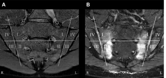
The score quantifies periarticular edema-like signal altera- tions. 12 In the paracoronal plane on the STIR sequence, both SIJs are divided into 4 quadrants (Figure 1) and scored for the presence or absence of BME to 0 or 1; the maximum value thus corresponds initially to 8. Continuous edema-like signal of depth ≥ 1 cm from the articular surface as well as a very high signal intensity will assign each an additional point per joint, corresponding to a maximum score increased by 4. The maxi- mum possible score for a paracoronal slice is thus 12. There is a total of 6 paracoronal slices evaluated, resulting in a maximum possible value of 72.

SPARCC score for the spine

The score quantifies edema-like signal alterations of the verte- bral bodies on a sagittal STIR sequence. 13 Two vertebrae form a discovertebral unit (DVU). This is divided into 4 quadrants (Figure 2). Each quadrant is scored for the presence or absence of BME to 0 or 1. On each slice, the presence of a lesion with a very high signal intensity in any quadrant is given an addi- tional score of 1. Similarly, the presence of a lesion exhibiting depth ≥ 1 cm in any quadrant is given an additional score of 1, leading to a maximum score of 6 for each DVU. A total of 3 sagittal slices will be assessed, and the maximum score of each DVU corresponds therefore to 18. A maximum of 6 DVUs are evaluated, and the maximum value for the SPARCC score of the spine is therefore 108. If more than 6 positive DVUs are present, then the highest single score is used.

Chronicity score of the SIJ

The score includes 4 degrees of severity regarding sclerosis, erosions, and ankylosis of the SIJ and assigns corresponding values of 0 to 4. 14,15 The right and left SIJs were rated

separately from one another and then formed a total score with a maximum value of 8.

SASSS for structural spine lesions

All corners of each vertebra between the lower border of T12 and the upper border of S1, ie, a total of 24 corners are assessed looking for structural changes. 16 Findings were scored as fol- lows: normal result (0), erosion (1), syndesmophyte formation (2), and total bony bridging (3) giving a maximum possible score of 24 × 3 = 72.

HLA-B27 status was recorded in all patients. The C-reactive protein (CRP) levels and the Bath Ankylosing

Figure 1. SPARCC score for inflammatory lesions in the sacroiliac joints. (A) Division of the sacroiliac joints into 4 quadrants. (B) SPARCC score = 10 for the shown slice (3 positive quadrants on both sides plus 2 points for signal intensity and depth on both sides). Total SPARCC score = 45 for all 6 slices (not shown). SPARCC indicates Spondyloarthritis Research Consortium of Canada.

Figure 2. SPARCC score for inflammatory lesions in the spine. (A) Division of a DVU in 4 quadrants. (B) SPARCC score = 15 for the shown slice (DVU 1: 4 positive quadrants plus 1 point for depth, DVU 2: 2 positive quadrants, DVU 3: 3 positive quadrants, DVU 4: 3 positive quadrants, DVU 5: 1 positive quadrant, DVU 6: 1 positive quadrant). Total SPARCC score = 32 for all 3 slices (not shown). DVU indicates discovertebral unit;

SPARCC, Spondyloarthritis Research Consortium of Canada.

(4)

Spondylitis Disease Activity Index (BASDAI) of all patients were determined for the evaluation of disease activity at the time of the MRI scan.

The significance between sex of the patient, the HLA-B27 status, and the percentage of spine and SIJ involvement was determined by the t test for independent samples. In addition, an investigation of the distribution pattern of the individual subgroups of SpA was also conducted using t test. Possible differences in the imaging-based scores were determined using the Wilcoxon test. The scores of the imaging, the BASDAI, and the CRP levels were also tested by the Spearman rank correlation coefficient regarding a possible correlation.

The statistical analysis was performed using WinSTAT for Microsoft Excel (R. Fitch Software; Bad Krozingen, Germany).

Statistical significance was considered if P ≤ .05.

Results

A total of 193 patients were evaluated, of whom 111 (57.5%) were men and 82 (42.5%) were women. The median age was 46 (range: 17-72) years. In all, 137 patients (71.0%) were HLA- B27 positive. The average CRP level of all patients was 11.3 mg/L (minimum: 0.1; maximum: 92.0; SD: 15.9), and the average BASDAI was 5.4 (minimum: 0.2; maximum: 10.0;

median: 5.8; SD: 2.0). The patients had the following disease subgroups: 127 (65.8%) AS, 30 (15.5%) PsA, 24 (12.4%) undif- ferentiated SpA, and 12 (6.2%) enteropathic SpA.

All 193 patients had manifestations of axial SpA. In all, 174 patients (90.2%) showed a sacroiliac involvement, 120 patients (62.2%) a combined involvement of the SIJs and the spine. An isolated sacroiliac involvement was found in 54 patients (28.0%), an isolated spine involvement in 19 patients (9.8%).

Between male and female patients, significant differ- ences in the distribution pattern were observed. In male patients, a significant higher proportion of SIJ involvement was observed (P < .01). Detailed gender-related results are given in Table 1. Regarding the subgroups, HLA-B27–

positive patients had a significantly higher proportion of SIJ involvement than the subgroup without this genetic trait (P < .05; Table 2). Between the individual entities of SpA, no relevant difference in the pattern of involvement of the axial skeleton was seen.

The scores for inflammatory lesions are given in Table 3 and showed gender-related differences. The scores for the spine were significantly higher among men than women (P < .05).

However, women had a higher score for the SIJ, but the differ- ence was not statistically significant.

The scores for structural lesions are shown in Table 4 and were significantly higher in men than in women, for the spine as well as for the SIJ (P < .001).

Between the scores for inflammatory and structural lesions and the CRP levels and BASDAI, no significant correlations

Table 1. Gender-related distribution.

AxiAl SPOnDylOARTHRiTiS (n = 193)

MAlE (n = 111) FEMAlE (n = 82)

Sole SiJ

involvement SiJ and spine

involvement Sole spine

involvement Sole SiJ

involvement SiJ and spine

involvement Sole spine

involvement n = 24 (21.6%) n = 82 (73.9%) n = 5 (4.5%) n = 30 (36.6%) n = 39 (47.6%) n = 13 (15.8%)

Spine involvement n = 87 (78.4%)* Spine involvement n = 52 (63.4%)*

SiJ involvement n = 106 (95.5%)** SiJ involvement n = 69 (84.1%)**

Abbreviation: SiJ, sacroiliac joint.

*P < .05; **P < .01.

Table 2. HlA-B27–related distribution.

AxiAl SPOnDylOARTHRiTiS (n = 193)

HlA-B27 POSiTiVE (n = 137) HlA-B27 nEGATiVE (n = 56)

Sole SiJ

involvement SiJ and spine

involvement Sole spine

involvement Sole SiJ

involvement SiJ and spine

involvement Sole spine

involvement n = 40 (29.2%) n = 89 (65.0%) n = 8 (5.8%) n = 15 (26.8%) n = 32 (57.1%) n = 9 (16.1%)

Spine involvement n = 97 (70.8%) Spine involvement n = 41 (73.2%)

SiJ involvement n = 129 (94.2%)* SiJ involvement n = 47 (83.9%)*

Abbreviation: SiJ, sacroiliac joint.

*P < .05.

(5)

were detected. Between CRP levels and BASDAI, however, there was a borderline significant correlation (P = .07, r = .17).

Discussion

The results of our study indicate that morphologically detect- able changes in MRI, although primarily seen at the SIJ level, can basically affect any part of the axial skeleton. The patient population of our analysis corresponds to that of other studies that have looked at axial SpA. Most of the patients were men and middle-aged, and about 60% to 85% were HLA-B27 posi- tive. 17,18 The most common subgroup of SpA was the AS and therefore referred to as the prototype of this group of diseases.

All patients fulfilled the ASAS criteria of the imaging arm;

they all had a positive MRI and at least one further clinical feature of SpA. The low values for CRP and BASDAI in our patients indicate a low disease activity, and this is reflected in the relatively low SPARCC scores. The average score for spine and SIJ was 9 so that accounting for only 5% of the maximum value. The scores for the structural and therefore chronic changes, however, had higher values. The average SASSS was 6.3 and the chronicity score of the SIJ was 4.2 corresponding to 9% and 52%, respectively, of the maximum value. It should be noted that the SASSS only rated the segments T12 to S1; the remaining vertebral bodies were not considered. Due to the fact that not all patients of the collective received a full investi- gation of the spine, ie, including the cervical spine, the modi- fied SASSS could not be applied in our evaluation. In relation, therefore, the structural changes of the spine compared with the SIJ are determined too low. However, the results of the

structural scores are generally associated with a longer disease course. However, data on the duration of symptoms of patients were not available.

The scores for the inflammatory as well as for the structural changes were higher for the SIJ than for the spine. One expla- nation could be that the inflammatory changes first occur at the SIJ and at a later stage involve the spine. A recently pub- lished study by Weber et al 11 concludes that combined studies of spine and SIJ provide only little extra diagnostic information over sole investigations of the SIJ. These results are contrary to our data, as in our collective, 62% of the patients had SpA man- ifestations on both, the SIJ and the spine, and in approximately 10% of the patients, manifestations were demonstrated even only on the spine. This circumstance is corroborated by a study by van der Heijde et al. 10 The authors of this study came to the conclusion that up to 50% of the patients with negative find- ings at the SIJ exhibit inflammatory changes in the spine. In a study by Weber et al 19 on patients with AS, MRI had a sensi- tivity of 69% and a specificity of 94% for the detection of acute inflammatory changes of the spine.

Particularly, in the interest of monitoring of the disease, it appears to us that combined MRI protocols are yielding and justified, so as to ensure early detection of all inflammatory lesions of the axial skeleton. According to the guidelines of the ASAS/OMERACT, the diagnosis of axial SpA requires the presence of a characteristic BME of the vertebral bodies or the SIJ, in the spine at not less than 3 different vertebral end plates, at the SIJ in at least 2 different locations or on 2 consecutive slices. 5,6 However, edema-like bone marrow changes are among

Table 3. SPARCC score for inflammatory lesions of the spine and the SiJ.

SCORES FOR inFlAMMATORy lESiOnS (n = 193)

Spine (SPARCC: 0-108) SiJ (SPARCC; 0-72)

4.1 (0-65; median: 4; SD: 7.5) 5.04 (0-54; median: 2; SD: 9.1)

MAlE (n = 111) FEMAlE (n = 82) MAlE (n = 111) FEMAlE (n = 82)

4.9 (0-65; median: 2; SD: 8.8) 2.9 (0-24; median: 0; SD: 4.9) 4.4 (0-48; median: 0; SD: 8.8) 5.5 (0-54; median: 3; SD: 9.5)

P < .05 nonsignificant

Abbreviations: SiJ, sacroiliac joint; SPARCC, Spondyloarthritis Research Consortium of Canada.

Table 4. Scores for structural lesions of the spine and the SiJ.

SCORES FOR STRUCTURAl lESiOnS (n = 193)

Spine (SASSS: 0-72) SiJ (Hermann et al: 0-8)

6.3 (0-68; median: 2; SD: 12.1) 4.2 (0-8; median: 4; SD: 2.1)

MAlE (n = 111) FEMAlE (n = 82) MAlE (n = 111) FEMAlE (n = 82)

12 (0-68; median: 4; SD: 15.1) 2.2 (0-22; median: 2; SD: 3.3) 4.8 (0-8; median: 4; SD: 2.0) 3 (0-54; median: 3; SD: 1.8)

P < .001 P < .001

Abbreviations: SASSS, Stoke Ankylosing Spondylitis Spine Score; SiJ, sacroiliac joint.

(6)

the most frequent and nonspecific musculoskeletal MRI find- ings. Numerous and sometimes difficult differential diagnoses must be weighed when interpreting edema-like signal changes. 20,21 Through the integrative assessment of all mor- phological changes of both, the SIJ and the spine, the specific- ity of inflammatory lesions can potentially increase. Besides the typical signal changes of the vertebral bodies and the SIJ, the ASAS/OMERACT defines other inflammatory lesions such as arthritis of the facet, the costovertebral, and the costotrans- versal joints. However, these are not enough to diagnose an axial SpA manifestation. These findings are therefore not included in the SPARCC score and therefore also not in our study. According to a study by Althoff et  al, 22 fat-saturated T2-STIR sequences alone are sufficient to diagnose certain edema-like bone marrow changes in patients with sacroiliitis.

The authors see that the additional use of contrast-enhanced T1-weighted images can increase diagnostic accuracy in the initial stage of inflammation. In our view, T2-STIR sequences are slightly more sensitive than contrast-enhanced T1-weighted sequences. By evaluating the contrast-enhanced sequences, however, the specificity of the study may be increased by the additional evidence of further inflammatory lesions. These findings were not considered quantitatively in our analysis, as there are no established scores.

Underlining the differences in the distribution pattern of inflammatory and structural changes between male and female patients or the HLA-B27 status reflects the heterogeneity of axial SpA disorders. Typical SpA patterns are observed in HLA-B27 positive and male patients presenting with a pre- dominant involvement of the SIJ. Atypical patterns without SIJ manifestation are more commonly observed in HLA-B27 negative and female patients. Significant differences in the pat- tern of involvement between the individual subgroups of SpA disorders could not be shown in our study probably due to the sample size.

The SIJ seems to be the focus of inflammatory changes of the axial skeleton. Another explanation could be that the size of the enthesial surface of the SIJ results in an increased sensi- tivity in detecting the inflammatory processes via imaging as opposed to the significantly smaller enthesial size of the end plates of the vertebral bodies.

When interpreting the results above, the limitations of our study should be considered, such as the relatively small sample size and the heterogeneous composition of the patient popula- tion. Only 62 patients received an examination of the entire spine, whereas only the thoracic and lumbar spines were exam- ined in the remaining patients. It is therefore possible that the scores of the spine for inflammatory and structural lesions that were determined were too low. In this context, one should also remember that MRI compared with radiography has a lower spatial resolution, with a consequent reduction in sensitivity for erosive and osteoproliferative skeletal changes. Through our use of gradient echo sequences with SIJ studies, however, the

sensitivity of MRI could be increased. 15,23 Another limitation may lie in the fact that the patient population has a rather low systemic disease activity.

The strengths of our study are the uniform and consistent MRI examination protocol and the use of well-defined classi- fication criteria and established scoring methods for spine and SIJ changes.

In conclusion, SpA is a systemic disorder predominantly involving the SIJs. However, the entire axial skeleton may be affected. In particular, HLA-B27–negative female patients show atypical manifestations without sacroiliac involvement.

MRI in SpA should cover the entire axial skeleton, ie, SIJs and the spine to meet the pathophysiology of this disorder and cap- ture the true extent of inflammatory and structural lesions.

Main points

• Spondyloarthritis is regarded as a systemic disorder that can manifest in the entire axial skeleton.

• Spondyloarthritis is almost always accompanied by an involvement of the SIJs, but an isolated involvement of the SIJs is rather rare.

• Magnetic resonance imaging of patients with suspected SpA should cover the entire axial skeleton to capture the true extent of inflammatory and structural changes.

Author Contributions

PH and CD designed the study. PH, AJ, AB and CD carried out data analysis and interpretation. PH performed the statisti- cal analysis. PH, MHS and CD drafted the manuscript. MF and CS critically revised and edited the manuscript. All authors read and approved the final version of the manuscript.

RefeRenCes

1. Braun J, Sieper J. Ankylosing spondylitis. Lancet. 2007;369:1379–1390.

2. Braun J, Bollow M, Remlinger G, et al. Prevalence of spondylarthropathies in HLA-B27 positive and negative blood donors. Arthritis Rheum. 1998;41:58–67.

3. Poddubnyy D, Vahldiek J, Spiller I, et al. Evaluation of 2 screening strategies for early identification of patients with axial spondyloarthritis in primary care. J Rheumatol. 2011;38:2452–2460.

4. Rudwaleit M, van der Heijde D, Landewé R, et al. The Assessment of SpondyloArthritis International Society classification criteria for peripheral spondyloarthritis and for spondyloarthritis in general. Ann Rheum Dis.

2011;70:25–31.

5. Rudwaleit M, Jurik AG, Hermann KG, et al. Defining active sacroiliitis on mag- netic resonance imaging (MRI) for classification of axial spondyloarthritis: a consensual approach by the ASAS/OMERACT MRI group. Ann Rheum Dis.

2009;68:1520–1527.

6. Hermann KG, Baraliakos X, van der Heijde D, et al. Descriptions of spinal MRI lesions and definition of a positive MRI of the spine in axial spondyloarthritis: a consensual approach by the ASAS/OMERACT MRI study group. Ann Rheum Dis. 2012;71:1278–1288.

7. van der Linden S, Valkenburg HA, Cats A. Evaluation of diagnostic criteria for ankylosing spondylitis. A proposal for modification of the New York criteria.

Arthritis Rheum. 1984;27:361–368.

8. Braun J, Sieper J. The sacroiliac joint in the spondyloarthropathies. Curr Opin Rheumatol. 1996;8:275–287.

9. Baraliakos X, Listing J, Rudwaleit M, Sieper J, Braun J. Development of a radio- graphic scoring tool for ankylosing spondylitis only based on bone formation:

addition of the thoracic spine improves sensitivity to change. Arthritis Rheum.

2009;61:764–771.

(7)

10. van der Heijde D, Sieper J, Maksymowych WP, et al. Spinal inflammation in the absence of sacroiliac joint inflammation on magnetic resonance imaging in pa- tients with active nonradiographic axial spondyloarthritis. Arthritis Rheum.

2014;66:667–673.

11. Weber U, Zubler V, Zhao Z, et al. Does spinal MRI add incremental diagnostic value to MRI of the sacroiliac joints alone in patients with non-radiographic axial spondyloarthritis? Ann Rheum Dis. 2015;74:985–992.

12. Maksymowych WP, Inman RD, Salonen D, et al. Spondyloarthritis research Consortium of Canada magnetic resonance imaging index for assessment of sacroiliac joint inflammation in ankylosing spondylitis. Arthritis Rheum.

2005;53:703–709.

13. Maksymowych WP, Inman RD, Salonen D, et al. Spondyloarthritis Research Consortium of Canada magnetic resonance imaging index for as- sessment of spinal inflammation in ankylosing spondylitis. Arthritis Rheum.

2005;53:502–509.

14. Hermann KG, Braun J, Fischer T, Reisshauer H, Bollow M. Magnetic resonance tomography of sacroiliitis: anatomy, histological pathology, MR-morphology, and grading. Radiologe. 2004;44:217–228.

15. Dornia C, Fleck M, Hartung W, Niessen C, Stroszczynski C, Hoffstetter P.

Magnetic resonance imaging of the sacroiliac joints in patients with suspect- ed spondyloarthritis—comparison of turbo spin-echo and gradient- echo sequences for the detection of structural alterations. Rofo. 2015;187:

109–114.

16. Spoorenberg A, de Vlam K, van der Linden S, et al. Radiological scoring meth- ods in ankylosing spondylitis. Reliability and change over 1 and 2 years. J Rheumatol. 2004;31:125–132.

17. Rudwaleit M, Haibel H, Baraliakos X, et al. The early disease stage in axial spondylarthritis: results from the German Spondyloarthritis Inception Cohort.

Arthritis Rheum. 2009;60:717–727.

18. Sampaio-Barros PD, Bertolo MB, Kraemer MH, Marques-Neto JF, Samara AM. Undifferentiated spondyloarthropathies: a 2-year follow-up study. Clin Rheumatol. 2001;20:201–206.

19. Weber U, Hodler J, Kubik RA, et al. Sensitivity and specificity of spinal inflam- matory lesions assessed by whole-body magnetic resonance imaging in patients with ankylosing spondylitis or recent-onset inflammatory back pain. Arthritis Rheum. 2009;61:900–908.

20. Prakash D, Prabhu SM, Irodi A. Seronegative spondyloarthropathy-related sacroili- itis: CT, MRI features and differentials. Indian J Radiol Imaging. 2014;24:271–278.

21. Arnbak B, Jensen TS, Manniche C, Zejden A, Egund N, Jurik AG.

Spondyloarthritis-related and degenerative MRI changes in the axial skeleton—an inter- and intra-observer agreement study. BMC Musculoskelet Disord. 2013;14:274.

22. Althoff CE, Feist E, Burova E, et al. Magnetic resonance imaging of active sac- roiliitis: do we really need gadolinium? Eur J Radiol. 2009;71:232–236.

23. Krohn M, Braum LS, Sieper J, et al. Erosions and fatty lesions of sacroiliac joints

in patients with axial spondyloarthritis: evaluation of different MRI techniques

and two scoring methods. J Rheumatol. 2014;41:473–480.

Abbildung

Figure 1.  SPARCC score for inflammatory lesions in the sacroiliac joints. (A) Division of the sacroiliac joints into 4 quadrants
Table 1.  Gender-related distribution.
Table 3.  SPARCC score for inflammatory lesions of the spine and the SiJ.

Referenzen

ÄHNLICHE DOKUMENTE

Percentage of current year earlywood vessel-elements connected to previous year latewood vessel-elements (A) and mean (±SD) flow (B) expressed as the proportion of axial flow in

Woodhams R, Matsunaga K, Iwabuchi K, Kan S, Hata H, Kuranami M, Watanabe M, Hayakawa K (2005) Diffusion-weighted imaging of malig- nant breast tumors: the usefulness of

Pearce RH (1991) Magnetic resonance imaging reflects the chemical changes of aging degeneration in the human inter- vertebral disc. Tertti M, Paajanen H, Laato

Pearce RH (1991) Magnetic resonance imaging reflects the chemical changes of aging degeneration in the human inter- vertebral disc. Tertti M, Paajanen H, Laato

chapter 4: How does the complex consistency of brain tissue, consisting of coupled pools of protons (e.g., myelin water – free water), affect the outcome of the

However, it is worth noting that the improvement in lesion visualization seen on combined pre- plus post-contrast images relative to pre-contrast images alone bears excellent

To investigate the response of the volume elements to the excitation detached from the signal of the falling motion, the relative phase ∆ϕ r with respect to a reference point in

This subset comprises volumetric MRI scans of the speaker’s vocal tract during sustained production of vowels and consonants, as well as dynamic mid- sagittal scans of