AUS DEM LEHRSTUHL FÜR INNERE MEDIZIN II Prof. Dr. Lars S. Maier DER FAKULTÄT FÜR MEDIZIN DER UNIVERSITÄT REGENSBURG
SR CA2+-LEAK AND DISORDERED EXCITATION-CONTRACTION COUPLING AS THE BASIS FOR ARRHYTHMOGENIC AND NEGATIVE INOTROPIC EFFECTS OF
ACUTE ETHANOL EXPOSURE
Inaugural – Dissertation zur Erlangung des Doktorgrades
der Medizin der
Fakultät für Medizin der Universität Regensburg
vorgelegt von Julian Mustroph
2018
Dekan: Prof. Dr. Dr. Torsten E. Reichert 1. Berichterstatter: Prof. Dr. Lars S. Maier
2. Berichterstatter: Prof. Dr. Christof Schmid
Tag der mündlichen Prüfung: 10.04.2018
Deutschsprachige Zusammenfassung der wissenschaftlichen Originalarbeit
SR Ca 2+ -leak and disordered excitation-contraction coupling as the basis for arrhythmogenic and negative inotropic effects of acute ethanol exposure
von
Julian Mustroph et al.
publiziert im
Journal of Molecular and Cellular Cardiology 2018
vorgelegt von
Julian Mustroph
Einleitung und Fragestellung des Projektes
In der vorliegenden Arbeit wurden Effekte von Ethanol auf isolierte Kardiomyozyten, insbesondere die elektromechanische Kopplung und das diastolische Kalziumleck untersucht. Diese Zusammenfassung beleuchtet die wichtigsten Ergebnisse dieser Studie, zudem soll ein Ausblick auf mögliche Anschlussarbeiten und klinische Implikationen geliefert werden, der in der Originalarbeit aus editorialen Gründen kürzer gehalten war. Da in dieser Zusammenfassung auch eine Interpretation und Wichtung der beobachteten Effekte stattfinden soll, werden Daten teilweise in anderer Reihenfolge als im Manuskript der Publikation diskutiert.
Der Konsum von Alkohol als Genussmittel ist in vielen Kulturen weit verbreitet.
Während der schädliche Einfluss von Ethanol auf Organe wie Leber und Gehirn auch populärwissenschaftlich weithin bekannt ist, existieren nur wenige Daten zur toxischen Ethanolwirkung auf das Herz - insbesondere die molekularen Mechanismen sind nur unzureichend verstanden (1,2).
Zudem muss bei der Untersuchung der Ethanolwirkung auch zwischen akuten und chronischen Effekten auf das Myokard differenziert werden, da chronischer Stress am Herzen oftmals mit einem kardialen Remodelling einhergeht (3–6).
Ausgangspunkt der vorliegenden Arbeit war die klinische Beobachtung, dass nach einem akutem Alkoholexzess vermehrt Vorhofflimmern auftritt (7–9), das so genannte holiday-heart-Syndrom, und dass interessanterweise bei bis zu 62 % der Patienten bei Erstdiagnose eines Vorhofflimmerns ein unmittelbar zurückliegender Alkoholkonsum stattgefunden hat (1). Zudem ist bekannt, dass Alkoholexzesse jenseits der chronischen Schädigung des Herzenz eine akut negative inotrope Wirkung haben können und auch ventrikuläre Arrhythmien auslösen können (7,8,10–
12).
Auf Ebene des einzelnen Kardiomyozyten ist für das Vorhofflimmern eine Dysregulation der zellulären Kalziumhomöostase bekannt (13).
Ein zentraler Regulator des kardiomyozytären Kalziumhaushalts ist hierbei die
Kalzium/Calmodulin-abhängige Kinase II (CaMKII), eine Serin-Threonin-
Proteinkinase. Es sind gegenwärtig vier Isoformen und über 30 verschiedene
Spliceformen der CaMKII bekannt (14–16). Der generelle Aufbau der CaMKII ist
zwischen den Isoformen vergleichbar. N-Terminal ist eine katalytische Kinase-
Domäne mit ATP-Bindungsstelle und Proteinsubstratbindungsstellen angesiedelt.
Nachfolgend befindet sich eine autoregulatorische Domäne mit Calmodulin- Bindungsstelle und der für die Enzymaktivität wichtigen Autophosphorylierungstelle Threonin 287 (15,16). C-Terminal befindet sich die Assoziationsdomäne, welche wichtig für die Faltung und Ausbildung des Nativzustands des Holoenzyms bzw. von Holoenzymkomplexen ist.
Das Enzym ist im Nativzustand durch die autoregulatorische Domäne inhibiert, die als so genanntes Pseudosubstrat die katalytische Domäne blockiert. Calmodulin (CaM) weist vier Kalziumbindungstellen auf – die Bindung von Kalzium erhöht die Affinität zu Bindungszielen, z.B. der CaMKII. Zunächst kommt es durch Ca 2+ - Calmodulin-Anlagerung an die CaM-Bindungsstelle der CaMKII (Threonin 206 und Threonin 207) zu einem Konformationswechsel des Enzyms, infolge dessen die katalytische Domäne disinhibiert und die autoregulatorische Domäne freigelegt wird.
Die CaM-Bindung an ein CaMKII-Monomer erhöht die Affinität anderer Kinasemonomere für CaM (auch Kooperativität genannt (17)). CaM fungiert nach Enzymbindung als allosterischer Aktivator, der die inhibitorische Potenz der autoregulatorischen Domäne als Pseudosubstrat senkt. Ein Substratzugang zur katalytischen Untereinheit wird ermöglicht. Sofern ATP und Mg 2+ vorhanden sind, ist bedingt durch die multimere Struktur des Enzyms und die Konformationsänderung ("Streckung") der CaMKII eine so genannte Intersubunit-Phosphorylierung möglich:
hierbei wird der Threoninrest 287 der autoregulatorischen Domäne durch die Kinase- Domänen anderer CaMKII-Moleküle phosphoryliert (16). Die Phosphorylierung des Threoninrests bewirkt zum einen eine Erhöhung der Affinität für CaM um den Faktor 1000, zum anderen verhindert diese Autophosphorylierung ein Wiederanlagerung von katalytischer und autoinhibitorischer Domäne auch nach Dissoziation des CaM.
Damit wird das Enzym kalziumunabhängig i.e. autonom aktiv, zumindest so lange es nicht zu einer Abspaltung des Phosphatrests kommt.
Auch die Amplitude des systolischen Kalziumtransienten scheint eine Rolle für die Dauer der autonomen Aktivität zu spielen (19). Neben der "klassischen", Ca 2+ - Calmodulin-vermittelten Aktivierung sind auch weitere Aktivierungswege der CaMKII bekannt: das Enzym kann auch durch Oxidation (20) oder Glykosylierung (21) aktiviert werden und dann Zielproteine phosphorylieren.
Insbesondere bewirkt eine CaMKII-abhängige (Hyper-)Phosphorylierung des
Kalziumkanals des sarkoplasmatischen Retikulums, des Ryanodinrezeptor Typ 2
(RYR2), ein verstärktes diastolisches Kalziumleck, welches einen typischen Befund
für das Vorhofflimmern, aber auch für ventrikuläre Arrhythmien und die Herzinsuffizienz darstellt (13,22–25). Eine oxidative Aktivierung der CaMKII kann im Herzen durch reaktive Sauerstoffspezies (ROS) vermittelt sein, die im Herzen vor allem durch die NADPH-Oxidase 2 (NOX2) gebildet werden (20,26).
Interessanterweise ist eine Aktivierung der NOX2 durch Ethanol mit gesteigerter ROS-Produktion in der Leber vorbeschrieben (27,28). Zudem konnte in einem Modell der chronischen alkoholischen Kardiomyopathie eine vermehrte Aktivität der NOX2 gezeigt werden (29).
Folgende Hypothesen wurden in meiner Publikation untersucht:
1) Ethanol bewirkt in isolierten Kardiomyozyten eine akute Aktivierung der NOX2 mit gesteigerter Produktion von ROS.
2) Ethanol bewirkt durch eine gesteigerte ROS-Bildung eine oxidative Aktivierung der CaMKII.
3) Ethanol induziert CaMKII-vermittelt ein gesteigertes diastolisches Kalziumleck aus dem sarkoplasmatischen Retikulum.
4) Eine Inhibition der CaMKII, die Verhinderung der CaMKII-Oxidation, sowie ein transgener knock-out der NOX2 können das durch Ethanol pathologisch erhöhte diastolische Kalziumleck signifikant absenken und stellen somit mögliche Therapieansätze dar.
Methoden
Für die vorliegende Arbeit wurden humane rechtsatriale Gewebeproben von
Patienten verwendet, welche aus operationstechnischen Gründen bei Bypass-
Operationen entnommen wurden. Eine Einwillung der Patienten war im Vorfeld der
Operation eingeholt worden (nach positivem Ethikvotum). Aus den Biopsien wurden
Herzmuskelzellen nach etablierten Verfahren mittels enzymatischem Verdau des
Gewebes isoliert und für zelluläre Untersuchungen eingesetzt. Zudem wurden
Mauskardiomyozyten zur weiteren mechanistischen Untersuchung der
Ethanoleffekte aus C57BL6-Wildtypmäusen und zwei transgenen Modellen (s.u.)
isoliert. Da Ethanol wie bereits ausgeführt sowohl im Vorhof (Vorhofflimmern), als
auch im Ventrikel (negative Inotropie, ventrikuläre Arrhythmien) Effekte hat,
untersuchten wir neben atrialen auch ventrikuläre Kardiomyozyten. In den unten
genannten Experimenten wurde immer, wenn nicht anders beschrieben, unvergällter
Ethanol (Reinheit > 99%, Firma Roth) mit Kontrolllösung
(Normaltyrode/physiologischem Medium) verglichen. Die Konzentrationen von 1, 3
und 6 ‰ Ethanol wurden hierbei von mir nach klinischen aber auch mechanistischen
Gesichtspunkten ausgewählt. Bei der Literaturrecherche hatte ich festgestellt, dass
eine akute Intoxikation vor allem im Bereich von ca. 1-6 ‰ Ethanol klinisch apparent
wird, bzw. zur stationären Aufnahme führt. Auch präklinisch werden, soweit hierzu
Daten verfügbar sind, vor allem Konzentrationen > 1 ‰ erfasst (30–34). Zudem wird
der akute Alkoholexzess ("binge drinking") als eine akute Intoxikation >0.8 ‰
definiert (1), welcher nach aktuellen Daten regelmäßig von bis zu 24% der
amerikanischen Bevölkerung betrieben wird (1). Die Wahrscheinlichkeit eines
Vorhofflimmerns nach Alkoholkonsum, also einem "holiday heart syndrome", nimmt
interessanterweise mit jedem zusätzlich aufgenommenen alkoholischen (Standard-)
Getränk um ca. 8 % zu (35). Somit sollte im Konzentrationsbereich von 1-6 ‰,
sowohl ein klinisch relevantes Kollektiv erfasst sein, als auch eine mechanistische
Untersuchung mit ausgeprägter Aktivierung pathophysiologischer Signalkaskaden
möglich sein. Eine Schlüsselmethode für meine Untersuchungen war die
Bestimmung des diastolischen SR Kalziumlecks mittels konfokaler
Laserscanmikroskopie im Line-scan-Verfahren. Hierfür wurden isolierte
Kardiomyozyten mit 10 µmol/L des fluoreszierenden Kalziumsensors Fluo-4-AM
((36), Firma Invitrogen) beladen, der nach endogener Deesterifikation im Zytosol
Ca 2+ -Ionen bindet und durch Laserimpulse (488 nm Anregungswellenlänge) angeregt
werden kann. Durch Messung eines bestimmten, örtlich eng begrenzten Zellareals
über eine bestimmte Messdauer kann eine Erfassung diastolischer
Kalziumfreisetzungsereignisse (Kalzium-"Sparks") aus dem sarkoplasmatischen
Retikulum erfolgen. Diese Sparks wurden gezählt und normiert auf Zellbreite und
Messdauer, um die Sparkfrequenz zu erhalten (unter zu Hilfenahme des
Sparkmaster-Programms (37)). Zudem berechnete ich die Sparkgröße als
Sparkamplitude * halb-maximale-Sparkbreite * halb-maximale-Sparkdauer, woraus
sich in Kombination mit der Sparkfrequenz das diastolische Kalziumleck berechnen
lässt. Aus technischen Gründen wurden nur Zellen mit Sparkereignissen in die
statistische Analyse mit aufgenommen. Weiterhin setzte ich die
Epifluoreszenzmikroskopie mit Fluo-4-AM und zusätzlich dem ratiometrischen
Kalziumsensor Fura-2-AM (ebenfalls 10 µmol/L, Firma Invitrogen) ein, um die
systolische Kalziumfreisetzung und die diastolische Kalziumwiederaufnahme in
isolierten Kardiomyozyten zu erfassen. Die Abfallskinetik von Fluo-4- Kalziumtransienten wurde dabei als Maß der SERCA2a-Funktion untersucht (38). Da in Mauskardiomyozyten allerdings auch die Transportfunktion des NCX ca. 10% der Relaxation des Kalziumtransienten ausmacht (38), berechnete ich zusätzlich aus dem Kalziumtransientenabfall in Kombination mit der Kinetik des zu dieser Zelle gehörenden Koffein-induzierten Kalziumtransienten (Surrogatparameter für die NCX- Funktion), das so genannte tau SERCA2A (39) als
!"#$%&'(2! =
!!
!"# !"#$%&'()"*+%,*(
!
!"# !"##$%&'()&*%$&'!.
Zudem führte ich Western Blot Experimente durch, des Weiteren erfolgten durch uns Muskelstreifen-Kontraktiliätsmessungen und Patch-Clamp-Untersuchungen.
Ergebnisse und Diskussion
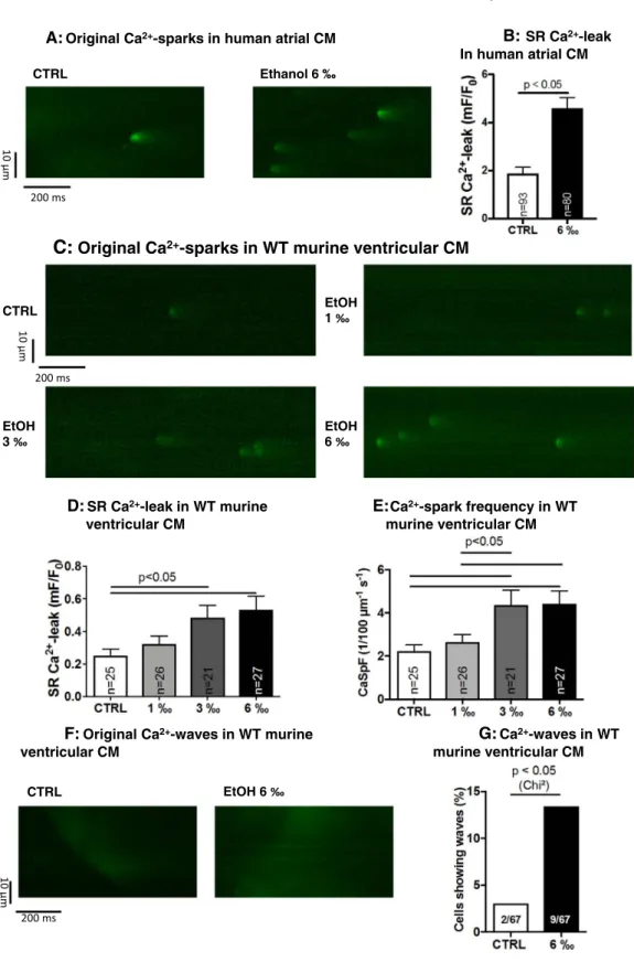
Ein Hauptergebnis meiner Publikation war der Befund eines erhöhten diastolischen SR Kalziumlecks in humanen atrialen Kardiomyozyten nach Behandlung mit 6 ‰ Ethanol im Vergleich zu ethanolfreier Kontrolllösung (Fig. 1A und 1B), welches ich mittels konfokaler Ca 2+ -Sparkmessung erheben konnte.
Da humane Kardiomyozyten nur eingeschränkt zur Verfügung stehen, verifizierten wir diesen Befund zunächst in murinen ventrikulären und atrialen Kardiomyozyten (Fig. 1C-E bzw. Suppl. Fig. 2 respektive) und konnten hierbei zudem eine positiv- korrelierte Abhängigkeit von der Ethanolkonzentration (0, 1, 3 und 6 ‰ Ethanol) nachweisen. Zur weiteren Verifikation des erhöhten diastolischen Kalziumlecks unter Ethanol wendeten wir in der Publikation zudem die etablierte Tetracaine-shift- Methode an (40), die mittels Epifluoreszenzmikroskopie auch Anteile des Kalziumlecks erfassen kann, die durch konfokale Sparkmessungen nicht nachweisbar sind (41). Wie erwartet zeigte auch dieses Verfahren unter 6 ‰ Ethanol ein signifikant erhöhtes diastolisches Kalziumleck (Suppl. Fig. 1B). Ich konnte zeigen, dass das erhöhte diastolische Kalziumleck unter Ethanol in murinen Kardiomyozyten auch mit einer gesteigerten Inzidenz von so genannten Kalziumwaves assoziiert war (Fig. 1F), die ebenfalls diastolische SR Kalziumfreisetzungen sind und ein proarrhythmogenes Substrat darstellen (41,42).
Wir konnten dabei zeigen, dass Ethanol zur vermehrten Aktivierung der CaMKII
(Autophosphorylierung an Thr-287), exemplarisch untersucht für die Konzentration
von 3 ‰, führt. Das diastolische Kalziumlecks kann über eine elektrogene Aktivierung des membranständigen Natrium-Kalziumaustauschers (NCX) und konsekutiven elektrogenen Ionenaustausch eine atypische Zelldepolarisation (und somit Arrhythmien) bewirken. Weiterhin kann das diastolische Kalziumleck zu einer kontraktilen Dysfunktion, bedingt durch Depletion der systolischen Kontraktionsamplitude, führen (22,41). Insofern ist der Befund eines erhöhten diastolischen Kalziumlecks mit vermehrten Kalziumwaves unter Ethanol von hoher pathophysiologischer Relevanz, da hierdurch insbesondere der o.g. Mechanismus der NCX-Aktivierung mit nachfolgenden proarrhythmogenen späten Nachdepolarisationen (delayed afterdepolarizations, DADs) in der Zelle ausgelöst werden kann. Späte Nachdepolarisationen sind ein etablierter Surrogatparameter für eine verstärkte Arrhythmieneigung von Kardiomyozyten (43) und wir konnten zeigen, dass diese in der Tat unter Ethanol signifikant vermehrt auftraten (Suppl. Fig. 3A).
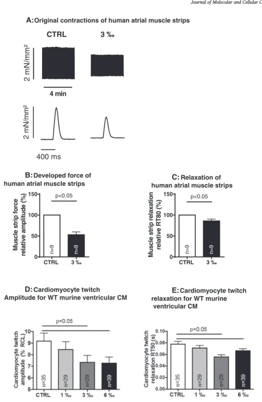
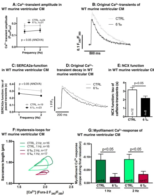
Interessanterweise ist die systolische Kalziumfreisetzung aus dem sarkoplasmatischen Retikulum durch Ethanol vermindert: die Amplitude des systolischen Kalziumtransienten, welche ich mittels Epifluoreszenzmikroskopie ermittelt habe, wurde durch 6 ‰ Ethanol in murinen Kardiomyozyten signifikant niedriger (Fig. 3A). Dies stellt eine mögliche Erklärung für die klinisch beobachtete negative Inotropie von Alkohol im Tiermodell und im Menschen dar (10–12,44,45).
Spannenderweise war jedoch die Kalziumkonzentration im Kalziumspeicher der Kardiomyozyten, dem sarkoplasmatische Retikulum, in zunächst scheinbarer Diskrepanz zum Befund der erniedrigten Kalziumtransienten, unter Ethanolexposition nicht signifikant niedriger im Vergleich zur Kontrolllösung (siehe Textabschnitt 3.2 im Manuskript). Ein möglicher Erklärungsansatz ist hierbei eine gesteigerte Kalziumwiederaufnahme unter Ethanolbehandlung über die sarkoplasmatische endoplasmatische Kalzium-ATPase (SERCA2a), die tatsächlich mittels Analyse der Kalziumtransientenrelaxation nachgewiesen werden konnte (Fig. 3C). Um einen Einfluss des NCX auf Abfallskinetik des Kalziumtransienten zu exkludieren, berechnete ich zudem tau SERCA2A (39), welches ebenfalls eine signifikant stärkere SERCA2a-Funktion (i.e. ein kleineres tau SERCA2a ) ergab (Suppl. Fig. 5).
Nebenbefundlich ergab sich hierbei eine signifikant gesteigerte Funktion des NCX unter Ethanol (Fig. 3E).
Passend zur gesteigerten Kalziumtransientenrelaxation war auch die CaMKII-
abhängige Phosphorylierung des endogenen SERCA2a-Inhibitors Phospholamban
an Threonin-17 signifikant erhöht unter Ethanol (Suppl. Fig. 7). Phosphoryliertes Phospholamban dissoziiert von der SERCA2a, sodass es zu einer Desinhibition der SERCA2a kommt. Zudem bestätigt die vermehrte Phospholamban-Phosphorylierung an der CaMKII-spezifischen Thr-17-Stelle die Beobachtung einer gesteigerten CaMKII-Aktivität unter Ethanol.
Wir bestimmten als Parameter der SERCA2a-Funktion die Abfallskinetik des Kalziumtransienten und maßen nicht direkt die Transporterfunktion, z.B. mittels
45 Ca 2+ -Oxalat-Wiederaufnahme-Assay. Für derartige direkte Transporterassays
müssten Kardiomyozyten membranpermeabilisiert werden, was die Interpretation der
Wirkung von Ethanol auf die Zelle verkomplizieren würde, daher entschieden wir uns
für diese Studie gegen diese Verfahren. Die in unserer Studie unter Ethanol
gesteigerte NCX-Funktion sollte allerdings zukünftig auch mittels Patch-Clamp-
Messungen evaluiert werden, um Einflüsse des (in unseren Experimenten
möglicherweise variablen) Membranpotentials auf die NCX-Messungen zu
minimieren. Des Weiteren würde dies eine Evaluation der zytosolischen
Natriumkonzentration ermöglichen, die ebenfalls durch eine verstärkte CaMKII-
Aktivierung unter Ethanol beeinflusst werden könnte (46,47) (erste eigene
präliminäre Untersuchungen der zytosolischen Natriumkonzentration unter Ethanol
mittels Fluoreszenzmikroskopie stützen diese Vermutung, siehe Zusammenfassung
und Ausblick). Negativ inotrope Effekte akuter Ethanol-Exposition waren in vivo
beobachtet worden (11,12,45). Wir konnten dies auf zellulärer Ebene nachvollziehen
und konnte zeigen, dass Ethanol dosisabhängig die Kontraktionsamplitude von
Kardiomyozyten erniedrigt (Fig 2D). Auch in multizellulären Präparaten (humane
atriale Muskelstreifen) war dieser negativ inotrope Effekt feststellbar (Abnahme der
Kontraktionskraft, Fig. 2B). Dieser deutliche negativ inotrope Effekt ist durch die nur
etwas reduzierte Amplitude der Kalziumtransienten nur teilweise zu erklären. Die
Untersuchung von Hysterese-Schleifen für Kalziumkonzentration und resultierende
Sarkomerlängen während des Kontraktionszyklus ermöglichte es die
Kalziumsensitivität in Ethanol-exponierten Zellen zu untersuchen. Hier stellte ich fest,
dass die Empfindlichkeit der Myofilamente für Kalzium durch Ethanol abgesenkt wird
(Hystereseschleifen in Fig. 3F und Quantifizierung (48) in 3G). Dies ist in
Zusammenschau mit der o.g. erhöhten CaMKII-Aktivität unter Ethanol erklärbar - in
anderen Studien konnte bereits eine Absenkung der Myofilament-Kalziumsensitivität
nach CaMKII-Aktivierung, unter anderem durch Phosphorylierung des regulatorischen Myofilamentproteins Titin, gezeigt werden (49,50).
Es verblieb zu klären, über welchen Mechanismus Ethanol diese Effekte auf die CaMKII und die elektromechanische Kopplung herbeiführt. Wie oben bereits beschrieben werden in Kardiomyozyten ROS klassischerweise vor allem durch die NOX2 gebildet (51) und Ethanol führt in anderen Geweben zu einer NOX2- Aktivierung (27). Zur Evaluation der ROS-Produktion nach Ethanolexposition setzten wir den allgemeinen ROS-Fluoreszenzsensor CMH 2 -DCFDA in murinen Kardiomyozyten ein und konnten eine vermehrte ROS-Produktion (i.e. CMH 2 - DCFDA-Fluoreszenzsteigerung) unter 3 ‰ Ethanol nachweisen (Fig. 4B).
Um die NOX2-Abhängigkeit des Ethanol-induzierten diastolischen Kalziumlecks zu evaluieren, bediente ich mich eines transgenen Mausmodells, in welchem die NOX2- Untereinheit gp91phox ausgeknockt ist (NOX2 KO, (26,52,53)), wodurch die NOX2 funktionsunfähig wird.
In isolierten ventrikulären und atrialen Kardiomyozyten aus diesem Modell konnten selbst 6 ‰ Ethanol keine Erhöhung des diastolischen Kalziumlecks bewirken (Fig.
4D und Suppl. Fig. 8B respektive). Ich konnte zeigen, dass bei Verlust der NOX2 Ethanol nicht mehr zur Aktivierung der CaMKII führt, i.e. die CaMKII- Autophosphorylierung in Ethanol-behandelten NOX2 KO Kardiomyozyten nicht durch Ethanol erhöht wurde.
Zudem stellte ich fest, dass Kalziumtransienten und SERCA2a-Funktion (Kalziumtransientenrelaxation) in NOX2 KO Kardiomyozyten nicht durch Ethanol verändert waren (Fig. 8C und 8D respektive). Somit erscheinen die von mir beobachteten Effekte von Ethanol durch NOX2-generierte ROS vermittelt zu sein.
Spannenderweise konnte ich nachweisen, dass auch die Myofilament- Kalziumsensitivität im NOX2 KO unter Ethanol nicht mehr nicht mehr signifikant beeinträchtigt wird.
Um die Bedeutung von ROS für die kontraktile Dysfunktion unter Ethanol noch
stärker zu beleuchten, führten wir Versuche mit dem ROS-Fänger (Antioxidanz) N-
Acetylcystein (NAC) durch. Dieser verhinderte eine Verringerung der
Kontraktionskraft unter 3 ‰ Ethanol im Vergleich zur alleinigen Ethanolapplikation
(Fig. 2B).
Um nachzuweisen, dass das diastolische Kalziumleck durch Ethanol tatsächlich durch die CaMKII vermittelt wird, setzte ich überdies den CaMKII-Inhibitor AIP (autocamtide-related inhibitory peptide 2) ein. Dieser reduzierte in Wildtypkardiomyozyten das durch Ethanol (6 ‰) hervorgerufene diastolische Kalziumleck tatsächlich deutlich (Fig. 4G).
Zu klären galt noch der Mechanismus der CaMKII-Aktivierung. Es existiert ein Antikörper zum direkten Nachweis der Oxidation der CaMKII an den Konsensusstellen Methionin 281 und 282 (20). Dieser Antikörper ist unserer Erfahrung nach insgesamt eher geeignet für den qualitativen Nachweis einer Oxidation, als deren exakte Quantifizierung, sodass letztlich die Autophosphorylierung der CaMKII der entscheidende Readout für ihre Aktivierung ist. Diesen qualitativen Nachweis einer gesteigerten CaMKII-Oxidation konnte ich in der Tat erbringen (siehe Abbildung Z1 am Ende dieser Zusammenfassung, ANOVA p<0.05, n=3 Mäuse). Um weiter zu bestätigen, dass die oxidative Aktivierung der CaMKII eine zentrale Rolle für die Ethanol-Effekte spielt, untersuchte ich ein transgenes Mausmodell, in welchem die CaMKII nicht mehr durch Oxidation aktiviert werden kann (Met281/282Val-Mutation, "MMVV"-Modell (20,54)). In ventrikulären und auch atrialen Kardiomyozyten aus diesen Mäusen führt Ethanol nicht mehr zu pathologischem diastolischen Kalziumleck (Fig. 4I und Suppl. Fig. 9A respektive).
Auch stellte ich fest, dass die systolische Kalziumtransientenamplitude und Kalziumtransientenrelaxation unter Ethanol in MMVV-Kardiomyozyten nicht beeinträchtigt war (Suppl. Fig 9B und 9C respektive).
Wie auch im NOX2 KO konnte ich zudem für die MMVV-transgenen Kardiomyozyten eine unveränderte Myofilament-Kalziumsensitivität zeigen (Supp. Fig. 8E und 8F).
Zusammenfassung und Ausblick
In meiner Publikation wird erstmals ein erhöhtes diastolisches Kalziumleck aus dem
sarkoplasmatischen Retikulum als Folge einer akuten Ethanolexposition von
isolierten Kardiomyozyten gezeigt. Zudem wird dieses u.a. durch Nutzung zweier
transgener Mausmodelle als Folge einer ROS-vermittelten oxidativen CaMKII-
Aktivierung mechanistisch aufgeklärt. Den Schlüsselbefund eines erhöhten
diastolischen Kalziumlecks konnte ich zudem in humanen atrialen Kardiomyozyten
validieren, was die Relevanz der Befunde für die klinisch hochrelevanten akuten
Ethanoleffekten unterstreicht (1).
Hierbei ist jedoch zunächst zu berücksichtigen, dass meine Experimente auf zellulärer Ebene valide, jedoch eine direkte klinische Übertragbarkeit in die in vivo Situation nicht zwingend gegeben sein muss. Neben den direkten Effekten von Ethanol auf die Herzmuskelzellen könnten in vivo auch Effekte wie z.B. eine Volumenüberladung oder Störungen des Elektrolythaushaltes und vegetative Effekte einen wichtigen Beitrag zur toxischen Ethanolwirkung leisten. Auch am Herzen ist nicht auszuschließen, dass andere Mechanismen (z.B. Mitochondriopathien) zu den chronischen Effekte von Ethanol beitragen.
Dennoch wäre in Zukunft für Patienten mit Vorhofflimmern nach Ethanolintoxikation die Entwicklung einer Therapie mit neuartigen, auch oral verfügbaren CaMKII- Inhibitoren ein auf diese Arbeit aufbauender möglicher neuer pharmakologischer Ansatz. (25,39).
Eine Wirksamkeit vieler dieser Inhibitoren ist für Modelle des Vorhofflimmerns (humane atriale Kardiomyozyten und in vivo Tiermodelle) bereits gezeigt (25) und ich konnte in der aktuellen Publikation die Wirksamkeit eines dieser Inhibitoren, AIP, auch für die Ethanol-vermittelte CaMKII Aktivierung validieren: Behandlung von Ethanol-exponierten Kardiomyozyten mit AIP führte zu einer signifikanten Reduktion des diastolischen Kalziumlecks (unpublizierte Daten aus unserer Arbeitsgruppe zeigen dies auch für einen weiteren CaMKII-Inhibitor [SMP-114]).
Eine Therapie mit Antioxidanzien (z.B. NAC) wäre ebenfalls denkbar, um die gesteigerte Ethanol-induzierte ROS-Produktion zu antagonisieren. Jedoch hat sich in den letzten Jahren gezeigt, dass ein unkritischer Einsatz dieser Substanzen auch mit unerwünschten Ereignisse, wie z.B. einer beschleunigten Progression und Entstehung von Tumorerkrankungen assoziiert ist (55). Möglicherweise wäre die direkte Inhibition der NOX2 eine Therapiealternative: erste vielversprechende NOX2- Inhibitoren befinden sich in der Entwicklung (56,57).
Für die Zukunft sind weitere Untersuchungen der kardialen Ethanolwirkung im Lichte der hohen Prävalenz ethanolinduzierter Herzerkrankungen (1) von großer Relevanz.
Eine CaMKII-Aktivierung kann auch Alterationen der zellulären Natriumhomöostase bewirken z.B. über eine Verstärkung des späten Natriumstroms (lateI Na ; (46,47)).
Dies kann zudem eine Verstärkung des diastolischen Kalziumlecks im Sinne eines
circulus vitiosus (25,47) bewirken.
Dieses Feld möchte ich in Anschlussarbeiten an meine Dissertation weiter untersuchen. In ersten Folgearbeiten konnte ich tatsächlich bereits einen signifikanten Anstieg der zytosolischen Natriumkonzentration nach Ethanolexposition zeigen (Abbildung Z2, Fluoreszenz des Natriumfarbstoffs SBFI, Kontrolle/CTRL versus 3 ‰ Ethanol, 2-Wege-ANOVA p<0.05). Ich werde daran mit Untersuchungen wichtiger Natriumtransporter unter Ethanol mittels Patch-clamp anschließen, um weitere therapeutische Optionen, wie z.B. eine Inhibition des späten Natriumstrom oder eine NCX-Blockade zu evaluieren. Langfristig soll aufbauend auf diese Arbeit eine Translation in ein in vivo Modell erfolgen und die antiarrhythmische Wirksamkeit der CaMKII-Inhibition mittels transjugulärer Rechtsherzkatheter mit Arrhythmieinduktion in Mäusen getestet werden.
Abbildungen der Zusammenfassung Z1:
Z2:
0.5 Hz 1.0 Hz 2.0 Hz
0.45 0.50 0.55 0.60 0.65
Zytosolische Natriumkonzentration (SBFI F 340 /F 380 ) CTRL, n=32 Zellen
3 ‰ Ethanol, n=30 Zellen
*
0.0 0.5 1.0 1.5
ox-CaMKII/CaMKII
*
CTRL 1 ‰ WT ventricular CM
3 ‰
n=3 n=3 n=3
Literaturverzeichnis der Zusammenfassung
1. Voskoboinik A, Prabhu S, Ling L, Kalman JM, Kistler PM. Alcohol and Atrial Fibrillation. J Am Coll Cardiol. 2016 Dec;68(23):2567–76.
2. Whitman IR, Agarwal V, Nah G, Dukes JW, Vittinghoff E, Dewland TA, et al.
Alcohol Abuse and Cardiac Disease. J Am Coll Cardiol. 2017 Jan 2;69(1):13.
3. Knollmann BC, Knollmann-Ritschel BE, Weissman NJ, Jones LR, Morad M.
Remodelling of ionic currents in hypertrophied and failing hearts of transgenic mice overexpressing calsequestrin. J Physiol. 2000;525(2):483–98.
4. Backs J, Backs T, Neef S, Kreusser MM, Lehmann LH, Patrick DM, et al. The δ isoform of CaM kinase II is required for pathological cardiac hypertrophy and remodeling after pressure overload. Proc Natl Acad Sci. 2009;106(7):2342–7.
5. Dobrev D, Ravens U. Remodeling of cardiomyocyte ion channels in human atrial fibrillation. Basic Res Cardiol. 2003 May 1;98(3):137–48.
6. Kreusser MM, Backs J. Integrated mechanisms of CaMKII-dependent ventricular remodeling. Front Pharmacol. 2014;5:36.
7. Ettinger PO, Wu CF, Cruz CDL Jr, Weisse AB, Sultan Ahmed S, Regan TJ.
Arrhythmias and the “Holiday Heart”: Alcoholassociated cardiac rhythm disorders. Am Heart J. 95(5):555–62.
8. Buckingham TA, Kennedy HL, Goenjian AK, Vasilomanolakis EC, Shriver KK, Sprague MK, et al. Cardiac arrhythmias in a population admitted to an acute alcoholic detoxification center. Am Heart J. 1985 Nov;110(5):961–5.
9. Thornton JR. ATRIAL FIBRILLATION IN HEALTHY NON-ALCOHOLIC PEOPLE AFTER AN ALCOHOLIC BINGE. Orig Publ Vol 2 Issue 8410. 1984 Nov 3;324(8410):1013–5.
10. Ibrahim BM, Fan M, Abdel-Rahman AA. Oxidative stress and autonomic dysregulation contribute to the acute time-dependent myocardial depressant effect of ethanol in conscious female rats. Alcohol Clin Exp Res. 2014 May;38(5):1205–15.
11. Kelbaek H, Gjørup T, Brynjolf I, Christensen NJ, Godtfredsen J. Acute effects of alcohol on left ventricular function in healthy subjects at rest and during upright exercise. Am J Cardiol. 1985 Jan 1;55(1):164–7.
12. Cheng CP, Shihabi Z, Little WC. Acute effects of mildly intoxicating levels of alcohol on left ventricular function in conscious dogs. J Clin Invest. 1990 Jun 1;85(6):1858–65.
13. Chelu MG, Sarma S, Sood S, Wang S, van Oort RJ, Skapura DG, et al.
Calmodulin kinase II–mediated sarcoplasmic reticulum Ca2+ leak promotes atrial fibrillation in mice. J Clin Invest. 2009 Jun 15;(119(7)):1940–51.
14. Salas MA, Valverde CA, Sánchez G, Said M, Rodriguez JS, Portiansky EL, et al.
THE SIGNALLING PATHWAY OF CAMKII-MEDIATED APOPTOSIS AND
NECROSIS IN THE ISCHEMIA/REPERFUSION INJURY. J Mol Cell Cardiol.
2010 Jun;48(6):1298–306.
15. Hudmon A, Schulman H. Structure-function of the multifunctional Ca2+/calmodulin-dependent protein kinase II. Biochem J. 2002;364(3):593–611.
16. Rellos P, Pike ACW, Niesen FH, Salah E, Lee WH, Von Delft F, et al. Structure of the CaMKII delta calmodulin complex reveals the molecular mechanism of CaMKII kinase activation. PLoS Biol. 2010;8(7):e1000426.
17. Johnson DE, Meng J, Hudmon A. Mechanisms Underlying Cooperativity in CaMKII Autophosphorylation and Substrate Phosphorylation. Biophys J.
106(2):528a.
18. Eshete F, Fields RD. Spike frequency decoding and autonomous activation of Ca2+-calmodulin-dependent protein kinase II in dorsal root ganglion neurons. J Neurosci Off J Soc Neurosci. 2001 Sep 1;21(17):6694–705.
19. De Koninck P, Schulman H. Sensitivity of CaM kinase II to the frequency of Ca2+ oscillations. Science. 1998 Jan 9;279(5348):227–30.
20. Erickson JR, Joiner MA, Guan X, Kutschke W, Yang J, Oddis CV, et al. A Dynamic Pathway for Calcium-Independent Activation of CaMKII by Methionine Oxidation. Cell. 2008 May 2;133(3):462–74.
21. Erickson JR, Pereira L, Wang L, Han G, Ferguson A, Dao K, et al. Diabetic hyperglycaemia activates CaMKII and arrhythmias by O-linked glycosylation.
Nature. 2013 Oct 17;502(7471):372–6.
22. Neef S, Dybkova N, Sossalla S, Ort KR, Fluschnik N, Neumann K, et al. CaMKII- dependent diastolic SR Ca2+ leak and elevated diastolic Ca2+ levels in right atrial myocardium of patients with atrial fibrillation. Circ Res. 2010 Apr 2;106(6):1134–44.
23. Fischer TH, Herting J, Mason FE, Hartmann N, Watanabe S, Nikolaev VO, et al.
Late INa increases diastolic SR-Ca2+-leak in atrial myocardium by activating PKA and CaMKII. Cardiovasc Res. 2015 Jul 1;107(1):184–96.
24. Anderson ME, Brown JH, Bers DM. CaMKII in myocardial hypertrophy and heart failure. J Mol Cell Cardiol. 2011 Oct;51(4):468–73.
25. Mustroph J, Neef S, Maier LS. CaMKII as a target for arrhythmia suppression.
Pharmacol Ther. 2017 Aug;(176):22–31.
26. Bendall JK, Cave AC, Heymes C, Gall N, Shah AM. Pivotal Role of a gp91phox- Containing NADPH Oxidase in Angiotensin II-Induced Cardiac Hypertrophy in Mice. Circulation. 2002 Jan 22;105(3):293.
27. Paik Y-H, Kim J, Aoyama T, De Minicis S, Bataller R, Brenner DA. Role of
NADPH Oxidases in Liver Fibrosis. Antioxidants Redox Signal. 2014 Jun
10;20(17):2854–72.
28. McDonough KH. Antioxidant nutrients and alcohol. Environ Nutr Interactions Antioxid Nutr Environ Heal Part C. 2003 Jul 15;189(1–2):89–97.
29. Brandt M, Garlapati V, Oelze M, Sotiriou E, Knorr M, Kröller-Schön S, et al.
NOX2 amplifies acetaldehyde-mediated cardiomyocyte mitochondrial dysfunction in alcoholic cardiomyopathy. Sci Reports. 2016 Sep 14;6:32554.
30. Pelissier F, Lauque D, Charpentier S, Franchitto N. Blood alcohol concentration in intoxicated patients seen in the emergency department: does it influence discharge decisions? J Stud Alcohol Drugs. 2014;75(6):937–44.
31. Wundersitz L, Raftery S. Understanding the context of alcohol impaired driving for fatal crash–involved drivers: A descriptive case analysis. Traffic Inj Prev.
2017 May 2;1–7.
32. Mitra B, Charters KE, Spencer JC, Fitzgerald MC, Cameron PA. Alcohol intoxication in non-motorised road trauma: ALCOHOL INTOXICATION IN NON- MOTORISED ROAD TRAUMA. Emerg Med Australas. 2017 Feb;29(1):96–100.
33. Grüne B, Piontek D, Pogarell O, Grübl A, Groß C, Reis O, et al. Acute alcohol intoxication among adolescents—the role of the context of drinking. Eur J Pediatr. 2017 Jan;176(1):31–9.
34. Pajunen T, Vuori E, Vincenzi FF, Lillsunde P, Smith G, Lunetta P. Unintentional drowning: Role of medicinal drugs and alcohol. BMC Public Health [Internet].
2017 Dec [cited 2017 Sep 12];17(1). Available from:
http://bmcpublichealth.biomedcentral.com/articles/10.1186/s12889-017-4306-8 35. Larsson SC, Drca N, Wolk A. Alcohol Consumption and Risk of Atrial Fibrillation.
J Am Coll Cardiol. 2014 Jul 22;64(3):281.
36. Gee KR, Brown KA, Chen W-NU, Bishop-Stewart J, Gray D, Johnson I.
Chemical and physiological characterization of fluo-4 Ca2+-indicator dyes. Cell Calcium. 2000 Feb;27(2):97–106.
37. Picht E, Zima AV, Blatter LA, Bers DM. SparkMaster: automated calcium spark analysis with ImageJ. Am J Physiol - Cell Physiol. 2007 Sep 11;293(3):C1073.
38. Bers DM. Excitation-Contraction Coupling and Cardiac Contractile Force [Internet]. Springer Netherlands; 2001. (Developments in Cardiovascular Medicine). Available from: http://books.google.de/books?id=0p8AqZP7D5UC 39. Neef S, Steffens A, Pellicena P, Mustroph J, Lebek S, Ort KR, et al.
Improvement of cardiomyocyte function by a novel pyrimidine-based CaMKII- inhibitor. J Mol Cell Cardiol. 2017 Dec 30;(115):73–81.
40. Shannon TR. Quantitative Assessment of the SR Ca2+ Leak-Load Relationship.
Circ Res. 2002 Oct 4;91(7):594–600.
41. Bers DM. Cardiac Sarcoplasmic Reticulum Calcium Leak: Basis and Roles in
Cardiac Dysfunction. Annu Rev Physiol. 2014 Feb 10;76(1):107–27.
42. Aronsen JM, Skogestad J, Lewalle A, Louch WE, Hougen K, Stokke MK, et al.
Hypokalaemia induces Ca(2+) overload and Ca(2+) waves in ventricular myocytes by reducing Na(+),K(+)-ATPase α(2) activity. J Physiol. 2015 Mar 15;593(Pt 6):1509–21.
43. Fozzard HA. Afterdepolarizations and triggered activity. Basic Res Cardiol.
1992;87 Suppl 2:105–13.
44. Li Z, Guo X, Bai Y, Sun G, Guan Y, Sun Y, et al. The Association Between Alcohol Consumption and Left Ventricular Ejection Fraction: An Observational Study on a General Population. Gilardi. L, editor. Medicine (Baltimore). 2016 May;95(21):e3763.
45. Delgado CE, Gortuin NJ, Ross RS. Acute effects of low doses of alcohol on left ventricular function by echocardiography. Circulation. 1975 Mar;51(3):535–40.
46. Wagner et al. Ca/Calmodulin-dependent protein kinase II regulates cardiac Na channels. J Clin Invest. 2006
47. Sag CM, Mallwitz A, Wagner S, Hartmann N, Schotola H, Fischer TH, et al.
Enhanced late INa induces proarrhythmogenic SR Ca leak in a CaMKII- dependent manner. J Mol Cell Cardiol. 2014 Nov;76:94–105.
48. Howarth FC, Qureshi MA. Myofilament sensitivity to Ca2+ in ventricular myocytes from the Goto–Kakizaki diabetic rat. Mol Cell Biochem.
2008;315(1):69–74.
49. Guilbert A, Lim HJ, Cheng J, Wang Y. CaMKII-dependent myofilament Ca(2+) desensitization contributes to the frequency-dependent acceleration of relaxation. Cell Calcium. 2015 Nov;58(5):489–99.
50. Hamdani N, Krysiak J, Kreusser MM, Neef S, dos Remedios CG, Maier LS, et al. Crucial Role for Ca2+/Calmodulin-Dependent Protein Kinase-II in Regulating Diastolic Stress of Normal and Failing Hearts via Titin Phosphorylation. Circ Res. 2013 Feb 14;112(4):664.
51. Santos CXC, Anilkumar N, Zhang M, Brewer AC, Shah AM. Redox signaling in cardiac myocytes. Free Radic Biol Med. 2011 Apr 1;50(7):777–93.
52. Wagner S, Dantz C, Flebbe H, Azizian A, Sag CM, Engels S, et al. NADPH oxidase 2 mediates angiotensin II-dependent cellular arrhythmias via PKA and CaMKII. J Mol Cell Cardiol. 2014;75:206–15.
53. Dworakowski R, Alom-Ruiz SP, Shah AM. NADPH oxidase-derived reactive oxygen species in the regulation of endothelial phenotype. Pharmacol Reports PR. 2008 Feb;60(1):21–8.
54. Anderson ME. Oxidant stress promotes disease by activating CaMKII. J Mol Cell Cardiol. 2015 Dec;89(Pt B):160–7.
55. Sayin VI, Ibrahim MX, Larsson E, Nilsson JA, Lindahl P, Bergo MO. Antioxidants
Accelerate Lung Cancer Progression in Mice. Sci Transl Med. 2014 Jan
29;6(221):221ra15.
56. Hirano K, Chen WS, Chueng AL, Dunne AA, Seredenina T, Filippova A, et al.
Discovery of GSK2795039, a Novel Small Molecule NADPH Oxidase 2 Inhibitor.
Antioxidants Redox Signal. 2015 Aug 10;23(5):358–74.
57. Joseph LC, Kokkinaki D, Valenti M-C, Kim GJ, Barca E, Tomar D, et al.
Inhibition of NADPH oxidase 2 (NOX2) prevents sepsis-induced cardiomyopathy
by improving calcium handling and mitochondrial function. JCI Insight. 2017 Sep
7;2(17).
Contents lists available at ScienceDirect
Journal of Molecular and Cellular Cardiology
journal homepage: www.elsevier.com/locate/yjmcc
Original article
SR Ca 2+ -leak and disordered excitation-contraction coupling as the basis for arrhythmogenic and negative inotropic e ff ects of acute ethanol exposure
Julian Mustroph a , Olivia Wagemann a , Simon Lebek a , Daniel Tarnowski a , Jasmin Ackermann a , Marzena Drzymalski a , Ste ff en Pabel a , Christof Schmid b , Stefan Wagner a , Samuel Sossalla a , Lars S. Maier a , Stefan Neef a,⁎
aDept. of Internal Medicine II, University Medical Center Regensburg, Regensburg, Germany
bDept. of Cardiothoracic Surgery, University Medical Center Regensburg, Regensburg, Germany
A R T I C L E I N F O
Keywords:
Ethanol SR Ca
2+-leak CaMKII
Holiday heart syndrome NOX2
A B S T R A C T
Aims: Ethanol has acute negative inotropic and arrhythmogenic effects. The underlying mechanisms, however, are largely unknown. Sarcoplasmic reticulum Ca
2+-leak is an important mechanism for reduced contractility and arrhythmias. Ca
2+-leak can be induced by oxidative stress and Ca
2+/Calmodulin-dependent protein kinase II (CaMKII). Therefore, we investigated the influence of acute ethanol exposure on excitation-contraction cou- pling in atrial and ventricular cardiomyocytes.
Methods and results: Isolated human atrial and murine atrial or ventricular cardiomyocytes were preincubated for 30 min and then superfused with control solution or solution containing ethanol. Ethanol had acute negative inotropic and positive lusitropic e ff ects in human atrial muscle strips and murine ventricular cardiomyocytes.
Accordingly, Ca
2+-imaging indicated lower Ca
2+-transient amplitudes and increased SERCA2a activity, while myofilament Ca
2+-sensitivity was reduced. SR Ca
2+-leak was assessed by measuring Ca
2+-sparks. Ethanol in- duced severe SR Ca
2+-leak in human atrial cardiomyocytes (calculated leak: 4.60 ± 0.45 mF/F
0vs 1.86 ± 0.26 in control, n ≥ 80). This effect was dose-dependent, while spontaneous arrhythmogenic Ca
2+- waves increased ~5-fold, as investigated in murine cardiomyocytes. Delayed afterdepolarizations, which can result from increased SR Ca
2+-leak, were significantly increased by ethanol. Measurements using the reactive oxygen species (ROS) sensor CM-H
2DCFDA showed increased ROS-stress in ethanol treated cells. ROS-scaven- ging with N-acetylcysteine prevented negative inotropic and positive lusitropic effects in human muscle strips.
Ethanol-induced Ca
2+-leak was abolished in mice with knockout of NOX2 (the main source for ROS in cardi- omyocytes). Importantly, mice with oxidation-resistant CaMKII (Met281/282Val mutation) were protected from ethanol-induced Ca
2+-leak.
Conclusion: We show for the fi rst time that ethanol acutely induces strong SR Ca
2+-leak, also altering excitation- contraction coupling. Acute negative inotropic e ff ects of ethanol can be explained by reduced systolic Ca
2+- release. Mechanistically, ROS-production via NOX2 and oxidative activation of CaMKII appear to play central roles. This provides a mechanism for the arrhythmogenic and negative inotropic effects of ethanol and suggests a druggable target (CaMKII).
Non-standard abbreviations
BAC Blood alcohol content
CaMKII Ca
2+-/Calmodulin-dependent protein kinase II NCX Na
+/Ca
2+-exchanger
NOX2 NADPH oxidase 2
PLN Phospholamban
ROS Reactive oxygen species
RyR2 Ryanodine receptor type 2
SERCA2a Sarcoplasmic reticulum Ca
2+-ATPase type 2a
SR Sarcoplasmic reticulum
1. Introduction
Recent research [1] highlights the importance of ethanol abuse for heart failure and atrial fibrillation, the incidence of both is more than
https://doi.org/10.1016/j.yjmcc.2018.02.002
Received 9 June 2017; Received in revised form 23 January 2018; Accepted 1 February 2018
⁎
Corresponding author at: Dept. of Internal Medicine II, University Medical Center Regensburg, Franz-Josef-Strauß-Allee 11, 93053 Regensburg, Germany.
E-mail address:
Stefan.Neef@ukr.de (S. Neef).
-RXUQDORI0ROHFXODUDQG&HOOXODU&DUGLRORJ\²
$YDLODEOHRQOLQH)HEUXDU\
7
doubled independent of other risk factors. In fact, chronic ethanol consumption, especially of large quantities, is a leading cause of dilated cardiomyopathy (DCM) [2] and chronic consumption of only moderate amounts of alcohol already impairs left ventricular ejection fraction [3]. Importantly, recent data shows that occasions of binge drinking might be more harmful for the heart than the underlying chronic con- sumption [4], indicating the relevance of acute e ff ects of high ethanol doses. Interestingly, of the 53% of Americans who consume alcohol regularly, 44% partake in frequent binge drinking [5]. Thus, ~24% of all Americans likely frequently experience blood alcohol contents (BACs) of 0.8‰ or higher. Strikingly, 20–30% of people admitted to cardiology departments (and general hospital) are reported to be al- cohol abusers [6,7]. In a study investigating admissions for acute ethanol intoxication, the mean blood alcohol content was 2.5‰, while BACs of up to 6‰ were measured [8]. Other studies investigating clinical and pre-clinical settings similarly found BAC-ranges of 1 – 6 ‰ common for acute intoxication [9–12].
Malignant ventricular arrhythmias occur more frequently in alco- holic cardiomyopathy than in idiopathic dilated cardiomyopathy [13], suggesting a specific arrhythmogenic substrate. Indeed, the risk for ventricular fi brillation in the context of ST-elevation myocardial in- farction is considerably higher in persons who consume alcohol com- pared to non-drinkers [14] and 19% of non-ischemic sudden cardiac deaths in Finland have been attributed to alcoholic cardiomyopathy [15].
Aside from chronic e ff ects, ethanol consumption can acutely induce atrial fibrillation [16,17] also in people who do not habitually drink [18], the so-called “holiday heart syndrome”. Strikingly, 35–62% of admissions with a first diagnosis of atrial fibrillation had consumed ethanol before the onset of symptoms [5]. Furthermore, recent ethanol consumption has been associated with sudden cardiac death [19] and higher acute doses of ethanol have been found to favor the inducibility of experimental ventricular tachycardia and fibrillation [20]. Besides arrhythmias, ethanol acutely has negative inotropic effects [21–24]
even in healthy human subjects independent of the autonomic nervous system [25] and also beginning at relatively low concentrations of
~1.3‰ ethanol [24,26]. This is also true in isolated myocardium in the absence of any in vivo systemic regulators [27]. Even mildly in- toxicating levels of ethanol acutely impair cardiac function [22 – 24].
While the overall systemic effect of acute ethanol consumption in healthy persons is an increase in cardiac output (potentially due to vasodilation and increased heart rate [28]), alcohol acutely impairs cardiac index and stroke index in patients with heart failure [29]. Thus, acute ethanol consumption impairs cardiac function and provides an arrhythmogenic substrate.
In chronic ethanol consumption, mechanisms such as disrupted myofibrillar architecture, apoptosis, mitochondrial alterations, and imbalances in fatty acid metabolism are emerging as important pa- thomechanisms [30]. While indications of ethanol e ff ects on Ca
2+- homeostasis have been reported [31], the mechanisms by which ethanol acutely impacts cardiac function and promotes ar- rhythmogenesis are so far still not well understood.
Cardiac contraction is strongly dependent on cardiomyocyte Ca
2+- homeostasis. Central regulators of cardiac excitation-contraction cou- pling are protein kinase A (PKA) and Ca
2+/Calmodulin-dependent protein kinase II (CaMKII). Besides activation by the Ca
2+/Calmodulin complex, CaMKII can be activated by oxidation (at Met281/282).
CaMKII overactivity in heart failure has been show to induce sarco- plasmic reticulum (SR) Ca
2+-leak through the ryanodine receptor (RyR2) and is believed to cause SR Ca
2+-depletion and to be centrally involved in impaired contractility [32]. CaMKII-induced SR Ca
2+-leak has also been implicated in cardiac arrhythmias [33], which appears to be especially true for atrial fi brillation [34 – 36]. Thus, we speculated that alterations in Ca
2+-handling, including SR Ca
2+-leak, might be involved in acute effects of ethanol on cardiomyocyte function. Speci-
reactive oxygen species (ROS) [37], we set out to investigate whether ROS might be involved in e ff ects of ethanol also in the heart and whether CaMKII might be a mediator of these effects.
2. Methods
An extended methods section can be found in the supplement.
2.1. Isolation of murine and human cardiomyocytes
Murine atrial and ventricular cardiomyocytes were isolated as pre- viously described [38,39]. Explanted hearts were retrogradely Lan- gendorff-perfused with solution containing 7.5 mg/mL liberase™
(Roche) and trypsin 0.6% and hearts were then mechanically dis- sociated. NOX2 KO mice (“gp91
phox−/−“) [40] were kindly provided by Prof. Ajay M. Shah. Mice with the CaMKII Met281/282Val mutation [41,42] were kindly provided by Prof. Mark E. Anderson. All mice were on the C57BL/6 background. All investigations conformed to Directive 2010/63/EU of the European Parliament and to local institutional guidelines.
Human atrial cardiomyocytes were isolated from human right atrial appendages using the chunk-isolation technique in a spinner flask with 0.775 mg/mL collagenase type I (370 U/mL; Worthington) and 0.4 mg/
mL protease type XXIV (Sigma-Aldrich), as described before [43]. Right atrial appendages from human patients were obtained during coronary artery bypass or cardiac valve surgery. Written consent had been given by the donors and approval had been granted by the ethics committee to this investigation that conforms to the principles outlined in the Declaration of Helsinki. Patient data can be found in Suppl. Table 1.
2.2. Experimental solutions & (pre)-treatment of isolated cardiomyocytes Normal Tyrode's solution (NT) was used, consisting of (in mmol/L) 140 NaCl, 4 KCl, 5 HEPES, 1 MgCl
2, 10 glucose, 2 CaCl
2(pH 7.4 at 37 °C with NaOH). Loading bu ff ers for Ca
2+- fl uorescent dyes Fluo-4 and Fura-2 loading consisted of NT with Pluronic F-127 0.2 mg/mL in ad- dition to the dye.
As preliminary experiments had indicated that a certain duration of exposure to ethanol was required before the onset of experiments in order to attain effects, isolated cells were pre-treated with ethanol or with control solution for 30 min before starting experiments. Ethanol exposure was then continued with superfusion. 2 μ mol/L myristoylated AIP (myrAIP) was used to specifically inhibit CaMKII.
2.3. Epifluorescence and sarcomere length measurements
Ca
2+-epifluorescence and sarcomere length were simultaneously measured using an epi fl uorescence setup (IonOptix Corp) [44]. Myo- cytes were loaded with either Fluo-4 AM or Fura-2 AM (10 μ mol/L, Molecular Probes). All experiments were performed at 37 °C. Fura-2 fluorescence-ratio was calculated using alternating (240 Hz) excitation at 340 nm and 380 nm. For better evaluation of transient decay kinetics, experiments using Fluo-4 (which is not as strong a Ca
2+-bu ff er as Fura- 2) were used, and F/F
0of the fluorescence signal was calculated to assess amplitudes. Myocytes were field-stimulated at a basal frequency of 1 Hz (increased to 2 Hz to investigate frequency-dependence). SR Ca
2+-content was estimated by caffeine pulse (10 mmol/L). The monoexponential time-constant τ ( “ tau ” ) of the ca ff eine-induced tran- sient was calculated to estimate NCX function. To investigate ROS production, cells were loaded with 10 μ mol/L CM-H
2DCFDA and the (linear) percental change of the fluorescence signal over 30 s was used as a measure of ROS production [45].
2.4. Ca
2+-spark measurements
J. Mustroph et al. -RXUQDORI0ROHFXODUDQG&HOOXODU&DUGLRORJ\²
experiments were performed at 37 °C. Ca
2+-sparks were recorded using an LSM 5 (for Fig. 4D) and LSM 7 (for all other experiments) with laser- scan excitation at 488 nm. Ca
2+-sparks were detected and quantified using Sparkmaster [46] with visual confirmation of sparks detected.
Ca
2+-spark frequency (CaSpF) was calculated from this and normalized to scanned myocyte width and scanning interval. Ca
2+-spark size (CaSpS, calculated as: amplitude * half-maximum-width * half-max- imum-duration) was added for all sparks within a cell to calculate the SR Ca
2+-leak for this cell. Only cells displaying Ca
2+-sparks were in- cluded in the statistics.
2.5. Muscle strip experiments
Muscle strip experiments were performed as previously reported [47]. Thin trabeculae were isolated from human right atrial appendages and connected to a force transducer (World Precision Instruments) for isometric force recordings. Contractions were induced by electrical field stimulation (1 Hz, 5 mA). After 30 min of equilibration, the trabeculae were stretched until reaching the maximum steady-state twitch force to improve comparability. After recording steady state basal contractions, the superfusion was switched to solution containing 3 ‰ ethanol.
Ethanol effects were recorded in steady state after 30 min. To explore the role of ROS for ethanol effects, experiments utilizing 10 mmol/L of the ROS-scavenger N-acetylcysteine in the superfusate were performed.
2.6. Blinding & statistics
For all experiments, investigators were blinded with respect to the intervention substance used and/or genotype. All data are presented as mean ± SEM. Statistical analyses were performed using Student's t-test for unpaired or paired values, one-way ANOVA, two-way repeated measures ANOVA or Fisher's exact test, as appropriate. Values p < .05 were considered as statistically significant.
3. Results
3.1. Ethanol effects on SR Ca
2+-leak and Ca
2+-waves
SR Ca
2+-leak was investigated by measuring Ca
2+-sparks. Ethanol acutely induced SR Ca
2+-leak in human atrial cardiomyocytes (original registrations in Fig. 1A, mean data in 1B). Dose-dependence of SR Ca
2+- leak was investigated in wildtype murine ventricular cardiomyocytes, in which ethanol dose-dependently induced SR Ca
2+-leak at con- centrations of 1, 3, and 6 ‰ (original registrations in Fig. 1C, mean data in 1D), with the highest leak measured at 6‰. This was driven by an increase in Ca
2+-spark frequency (Fig. 1E), while Ca
2+-spark size was unaltered (Suppl. Fig. 1A). To verify the central result of increased SR Ca
2+-leak upon ethanol, we also employed tetracaine-measurements as another approach to determine leak [48,49] upon 6 ‰ of ethanol, which could confirm the increased Ca
2+-leak (Suppl. Fig. 1B).
Furthermore, we found that acute alcohol exposure increased the incidence of spontaneous arrhythmogenic Ca
2+-waves, which was 4.5- fold higher upon 6 ‰ ethanol with 13.4% of cells showing spontaneous waves (original registrations for CTRL and 6‰ in Fig. 1F, mean data in Fig. 1G). We could confirm increased SR Ca
2+-leak upon 1, 3, and 6‰
of ethanol also in WT murine atrial cardiomyocytes (Suppl. Fig. 2).
Increased SR Ca
2+-leak can induce pro-arrhythmogenic delayed afterdepolarizations via NCX-dependent membrane depolarization [35]. Indeed, we could verify this in WT murine atrial cardiomyocytes, where 3‰ of ethanol induced delayed afterdepolarizations in 87.5% of investigated cells versus 0% in CTRL cells (7/8 vs. 0/6 cells, original registrations in Suppl. Fig. 3A, mean data in B).
3.2. Ethanol effects on Ca
2+-handling
Investigation of healthy human atrial intact muscle strip
preparations showed clear negative inotropic (impaired contraction force amplitude, Fig. 2B) as well as positive lusitropic (i.e. faster re- laxation time RT80, Fig. 2C) effects of ethanol, as illustrated in the original recording in Fig. 2A. (Absolute values were also significantly different, Suppl. Fig. 4A&B).
Ethanol effects on ventricular isolated cardiomyocyte contractility were investigated using murine cells in the absence of Ca
2+-sensitive dyes (as these would buffer Ca
2+). Here, we found that ethanol acutely impaired myocyte contractility by trend at a concentration of 1‰ (8%
reduction in contractility), and clearly and significantly so at con- centrations of 3‰ (19% reduction) and 6‰ (21% reduction; Fig. 2D).
Positive lusitropic e ff ects of ethanol were also present in the isolated cardiomyocytes (again by trend at 1‰, but significantly at 3‰ and 6‰, Fig. 2E).
Ethanol effects on systolic Ca
2+-release (Ca
2+-transient amplitudes) were investigated using the ratiometric Ca
2+-sensitive dye Fura-2 in the murine ventricular cells. Ethanol clearly reduced systolic Ca
2+- transient amplitudes, as shown in Fig. 3A and depicted in Ca
2+-traces in Fig. 3B. Diastolic Ca
2+-elimination is 90% due to SERCA2a in murine cardiomyocytes [50], so that SERCA2a function can be measured by analyzing Ca
2+-transient decay kinetics. Thus, we performed experi- ments using the non-ratiometric dye Fluo-4 (which is a less strong Ca
2+-buffer and has a much better temporal resolution than Fura-2) to investigate SR Ca
2+-reuptake by SERCA2a. As shown in Fig. 3C and illustrated by original registrations in Fig. 3D, we found much faster transient decay upon ethanol, suggesting higher SERCA2a activity and thus higher SR Ca
2+-reuptake. NCX activity was investigated by ana- lyzing the decay of caffeine-induced transients, which revealed higher NCX activity upon ethanol (Fig. 3E). Increased NCX activity could in- fl uence the measurement of SERCA2a-function from the decay of electrically stimulated Ca
2+-transients. To exclude the effect of NCX, we therefore calculated tau
SERCA2a[49] from cells for which both Ca
2+- transient relaxation and caffeine transient relaxation was available.
Tau
SERCA2awas significantly shorter in ethanol-exposed cells (Suppl.
Fig. 5). Also, SR Ca
2+-leak could in fl uence transient decay. To exclude this confounder as well, we determined in paired experiments tau after rapid addition of tetracaine (to block Ca
2+-leak through the ryanodine- receptor [48,49]) as well as under control conditions. These experi- ments again showed signi fi cantly lower tau in ethanol-treated cells (Suppl. Fig. 6). Thus, our findings indicate increased SERCA2a-function upon ethanol exposure.
SR Ca
2+-content was also measured using caffeine-induced SR Ca
2+-release, but using Fura-2 (instead of Fluo-4) to quantify transient amplitudes. These experiments revealed unaltered SR Ca
2+-content in the ethanol treated cells (Fura-2 ration ethanol 6‰ 0.385 ± 0.039 vs.
control 0.408 ± 0.031 n = 20 vs. n = 12, n.s.).
Myofilament Ca
2+-response (“Ca
2+-sensitivity”) was explored in murine WT ventricular cardiomyocytes by investigating hysteresis loops for Ca
2+-concentration (i.e. Fura-2 ratio) and corresponding sarcomere length, as shown in the loops averaged from all experiments in Fig. 3F. These indicate a clear shift towards longer sarcomere lengths (i.e. reduced myofilament response to Ca
2+) upon ethanol treatment.
To substantiate this, we employed the method of Howarth and Qureshi [51] and analyzed the final phase of the relaxation (200–400 ms after electrical stimulation), during which the myofilaments come into quasi- equilibrium with cytosolic Ca
2+. This analysis confirmed significantly less myofilament Ca
2+-response upon ethanol exposure (Fig. 3G).
3.3. Role of oxidative CaMKII-activation by reactive oxygen species (ROS) for ethanol effects
We had speculated that ethanol could induce activation of CaMKII via increased generation of ROS. Using the fluorescent ROS-sensor CM- H
2DCFDA, we could demonstrate higher ROS generation in ethanol exposed cells (original registration in Fig. 4A, mean data in 4B).
Accordingly, we found a significant increase in Thr287-
J. Mustroph et al. -RXUQDORI0ROHFXODUDQG&HOOXODU&DUGLRORJ\²
autophosphorylated (and thus active) CaMKII already at a relatively low concentration of 3‰ ethanol (Suppl. Fig. 7) in isolated WT murine ventricular cardiomyocytes. Furthermore, phosphorylation of the most
specific Thr17 site was also significantly increased upon ethanol (pT17/
PLN-ratio, Suppl. Fig. 7), confirming increased CaMKII-activity and supporting the increased lusitropy observed in Fig. 3C.
Ethanol 6 ‰ CTRL
A: Original Ca
2+-sparks in human atrial CM
F: Original Ca
2+-waves in WT murine ventricular CM
G: Ca
2+-waves in WT murine ventricular CM D: SR Ca
2+-leak in WT murine
ventricular CM E: Ca
2+-spark frequency in WT murine ventricular CM
B: SR Ca
2+-leak In human atrial CM
EtOH CTRL 1
EtOH
3 EtOH
6
C: Original Ca 2+ -sparks in WT murine ventricular CM
EtOH 6 CTRL
Fig. 1.
SR Ca
2+-leak and Ca
2+-waves: Original recordings (A) and mean data (B) show induction of SR Ca
2+-leak by ethanol in human atrial cardiomyocytes (CTRL n = 93, 6‰ n = 80 cells, n = 4 patients). Effects of different concentrations of ethanol were tested in murine ventricular cardiomyocytes, demonstrating that SR Ca
2+-leak was induced at concentrations of 1, 3, and 6‰ ethanol (original recordings in C, mean data for Ca
2+-leak in D, CTRL n = 25 cells, 1‰ n = 26 cells, 3‰ n = 21 cells, 6‰ n = 27 cells, n = 5 mice), which was mainly driven by an increase in Ca
2+-spark frequency (E). Also, arrhythmogenic Ca
2+-waves were potently induced by ethanol (original recording in F, mean data in G, CTRL n = 67 cells, 6‰
n = 69 cells, n = 5 mice).
J. Mustroph et al. -RXUQDORI0ROHFXODUDQG&HOOXODU&DUGLRORJ\²
cardiomyocytes [52]. We used NOX2-knockout (KO) mice to investigate the role of ROS for ethanol e ff ects and found that KO of NOX2 pre- vented CaMKII-activation and CaMKII-dependent phospholamban- phosphorylation upon ethanol (Suppl. Fig. 8A), suggesting a critical role of NOX2 for CaMKII-activation due to ethanol. We furthermore found that NOX2-KO consistently prevented induction of SR Ca
2+-leak even at 6‰ both in ventricular cardiomyocytes (original registrations in Fig. 4C, mean data in 4D) as well as atrial cardiomyocytes (Suppl.
Fig. 8B). Also, Ca
2+-transient amplitude (Suppl. Fig. 8C), and SERCA2a-function (Ca
2+-transient decay as RT 80%, Suppl. Fig. 8D) were not altered in murine NOX2-KO ventricular cardiomyocytes even
by 6‰ ethanol. Thus, ethanol-induced alterations of Ca
2+-handling seem to depend on ROS-generation via NOX2. Interestingly, myo fi la- ment Ca
2+-response was also not altered upon 6‰ ethanol in NOX2-KO ventricular cardiomyocytes (averaged hysteresis-loops in Suppl. Fig. 8E, myofilament Ca
2+-response in F), suggesting a role of ROS also for this ethanol e ff ect.
To further investigate the role of ROS and explore the potential of antioxidative strategies to treat ethanol effects, we performed muscle strip experiments with healthy human atrial tissue using the ROS scavenging compound N-acetylcysteine (NAC), which completely pre- vented the negative inotropic (Fig. 4E) and positive lusitropic (Fig. 4F)
M us cl e stri p re la xati on re lat iv e R T8 0 (% )
CTRL 3 ‰ 0
50 100 150
n= 9 n= 9
p<0.05
Mus cl e str ip for ce re lat iv e amp lit ude (% )
CTRL 3 ‰ 0
50 100
150 p<0.05
n=9 n= 9
B: Developed force of
human atrial muscle strips C: Relaxation of
human atrial muscle strips
D: Cardiomyocyte twitch
Amplitude for WT murine ventricular CM
A: Original contractions of human atrial muscle strips
400 ms
2m N /m m ²
4 min
2m N /m m ²
CTRL 3 ‰
E: Cardiomyocyte twitch relaxation for WT murine
ventricular CM
Fig. 2.
Contractile function and relaxation: In paired experiments using human atrial muscle strips (n = 9 muscle strips, n = 9 patients, original recordings in Fig. A), ethanol clearly impaired contractile force (B), but accelerated relaxation (C). These negative inotropic (D) and positive lusitropic (E) effects were also observed in isolated single murine ventricular cardiomyocytes at doses ranging from 1 to 6‰ ethanol (CTRL n = 39 cells, 1‰ n = 29 cells, 3‰ n = 29 cells, 6‰ n = 39 cells, n = 8 mice).
J. Mustroph et al. -RXUQDORI0ROHFXODUDQG&HOOXODU&DUGLRORJ\²