Role of host-derived ADAM-9 in tumor invasion and metastasis of malignant melanoma
A dissertation
Submitted in partial fulfillment of the requirements for the award of Doctor of Philosophy Degree
in the Faculty of Natural Science University of Cologne
Submitted by
Ngum Anna Abety from Bafou-Djuttitsa, Cameroon
Cologne 2012
Reviewer: PD Dr. Roswitha Nischt
Prof. Dr. Matthias Hammerschmidt Prof. Dr. Siegfried Roth
Date of Oral Examination: 08.10.2012
For my Father
"I thank God every time I remember you. In all my prayers for you, I always pray with joy."
Philippians 1:3-4
Table of content
Abstract ... I Zusammenfassung ... II
1 Introduction ... 1
1.1 Malignant melanoma ... 1
1.1.1 Origin of melanoma ... 1
1.1.2 Melanoma invasion ... 2
1.2 ADAM proteases ... 4
1.2.1 ADAM domain structure and function ... 4
1.2.2 Regulation of ADAM expression and activity ... 6
1.3 ADAM-9... 8
1.3.1 ADAM-9 molecular chracteristics and function ... 8
1.3.2 ADAM-9 in cancer ... 11
1.4 Animal models for malignant melanoma ... 12
1.4.1 Mouse models ... 12
1.4.2 Medaka model ... 13
1.5 Aims of the thesis ... 14
2 Materials and methods ... 16
2.1 Materials... 16
2.1.1 Chemicals ... 16
2.1.2 Cell culture material ... 16
2.1.3 Bacteria, vectors and plasmids ... 16
2.1.4 Antibodies ... 17
2.1.5 Buffers ... 18
2.2 Methods ... 18
2.2.1 Cell biology ... 18
2.2.1.1 Cells and cell culture ... 18
2.2.1.2 Isolation of primary fibroblasts ... 19
2.2.1.3 Preparation of fibroblast conditioned media, cell monolayer and matrix...19
2.2.1.4 Bromodeoxyuridine (BrdU) incorporation assay ... 20
2.2.1.5 Apoptosis assay ... 20
2.2.2 Protein analysis ... 21
2.2.2.1 Preparation of lysates ... 21
2.2.2.2 SDS-PAGE and immunodetection ... 22
2.2.2.3 Zymographic analysis ... 22
2.2.2.4 Mouse cytokine antibody array ... 22
2.2.2.5 Murine TNF-alpha ELISA... 23
2.2.2.6 Immunohistochemistry and immunofluorescence ... 23
2.2.2.7 Tissue histology ... 24
2.2.2.8 Image analysis of stainings ... 24
2.2.3 Analysis of nucleic acids ... 24
2.2.3.1 RNA isolation ... 24
2.2.3.2 Reverse transcriptase and polymerase chain reaction (RT-PCR) ... 25
2.2.3.3 Genomic DNA preparation from mouse tails... 25
2.2.4 Animal experiment ... 27
2.2.4.1 Ethics and animal care ... 27
2.2.4.2 Melanoma grafting ... 27
2.2.5 Production of recombinant soluble ADAM-9 protein ... 28
2.2.5.1 Cloning and site directed mutagenesis ... 28
2.2.5.2 Production and purification of recombinant soluble ADAM-9 ... 31
2.2.5.3 In vitro enzymatic analysis ... 31
2.2.6 Statistical analysis ... 32
3 Results ... 33
3.1 ADAM-9 in melanoma ... 33
3.1.1 ADAM-9 in human melanoma in vivo ... 33
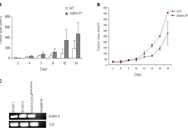
3.1.2 Analysis of Adam-9 in murine melanoma grafts ... 33
3.2 Role of host-derived ADAM-9 in growth of B16F1 melanoma in vivo ... 34
3.2.1 ADAM-9 expression in melanoma from Adam-9-/- in comparism to WT mice...34
3.2.2 B16F1 tumor growth kinetics ... 35
3.2.3 Melanoma metastasis in Adam-9-/- mice ... 37
3.3 Processes involved in melanoma growth ... 39
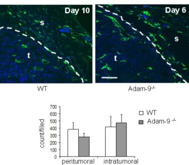
3.3.1 Vascularization of tumors ... 39
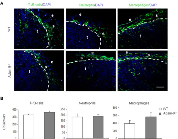
3.3.2 Inflammatory response in tumors ... 41
3.3.3 Proliferation and apoptosis of B16F1 melanoma cells in vivo ... 42
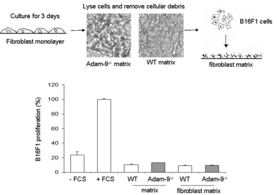
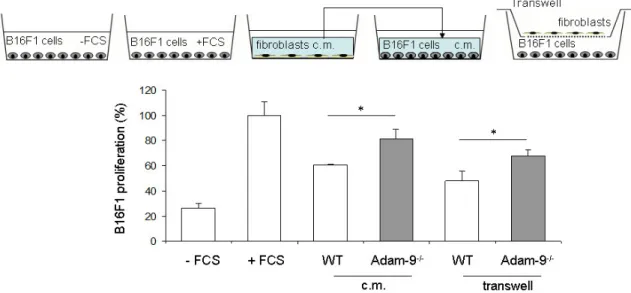
3.4 Role of fibroblast-derived Adam-9 on melanoma cell growth ... 43
3.4.1 Influence of cellular contacts and matrix synthesis/modification on melanoma cell proliferation ... 43
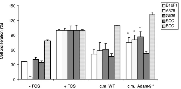
3.4.2 Soluble factors as mediators of melanoma-stroma communication leading to cell proliferation ... 46
3.5 The role of soluble factors in the regulation of melanoma cell proliferation . 49 3.5.1 Identification of soluble factors in Adam-9-/- fibroblasts ... 49
3.5.2 TIMP-1 expression in fibroblasts and melanoma cells ... 50
3.5.3 Effect of TIMP-1 on melanoma cell proliferation ... 51
3.5.4 Expression of TNF-α and TNFRI ... 54
3.5.5 Modulation of apoptosis induced by TNF-α and TNFRI ... 56
3.5.6 Differential fibroblast gene expression in the absence of Adam-9 ... 58
3.6 ADAM-9 and its role in the modification of the peritumoral matrix ... 60
3.6.1 Extracellular matrix protein expression at the tumor-stroma border... 60
3.6.2 Expression of ECM proteins by fibroblasts ... 63
3.7 Cleavage of ECM proteins by ADAM-9 ... 66
3.7.1 Production of recombinant soluble ADAM-9 ... 66
3.7.2 Degradation of proteins by ADAM-9 ... 68
3.7.3 Significance of collagen type I accumulation for melanoma progression 70 3.8 ADAMs and MMPs in melanoma models ... 71
4 Discussion ... 75
4.1 Regulation of tumor development by ADAM-9 ... 76
4.2 Regulation of tumor metastasis by ADAM-9... 78
4.3 Melanoma cell proliferation and apoptosis in the absence of host-derived ADAM-9...79
4.4 Extracellular matrix proteins in the absence of ADAM-9 ... 83
4.5 Melanoma fish model ... 86
5 Conclusion and perspectives ... 88
6 References ... 89
Abbreviation ... 104
Acknowledgement ... 105
Erklärung ... 106
Curriculum vitae ... 107
Abstract
A Disintegrin And Metalloproteases (ADAMs) represents a family of transmembrane proteins with a distinct multidomain structure including a metalloprotease, disintegrin, and cysteine-rich domain. These proteins function in adhesion, regulation of cell signaling by proteolysis of cell surface proteins and their receptors. ADAM-9 is a protease that cleaves membrane-bound proteins such as VCAM-1, TNF-α, as well as extracellular matrix proteins like fibronectin and laminin-111. ADAM-9 also contains adhesive domains that are involved in cell adhesion and migration. In human and murine melanoma, ADAM-9 is expressed by both tumor and stroma cells at the tumor-stroma border. The significance of this expression is however not clear. To explore the functional role of ADAM-9 produced by the host in melanoma progression, B16F1 murine melanoma cells were injected in the flank of Adam-9-/- and wild type animals. Tumors developed in Adam-9-/- mice were significantly larger compared to wild type animals and displayed significant increase in tumor cell proliferation accompanied by decrease in apoptosis. Using co-culture systems of primary fibroblasts and B16F1 melanoma cells, we could detect increased melanoma cell proliferation when cultured in the presence of supernatants from Adam-9-/- but not wild type fibroblasts. Among the proteins secreted in strongly enhanced amounts from Adam-9-/- fibroblasts, was tissue inhibitor of metalloproteinases-1 (TIMP-1).
Neutralization of TIMP-1 in Adam-9-/- fibroblasts supernatant using specific antibodies abolished the induced B16F1 proliferation. Besides TIMP-1, soluble TNF-α and TNFR1 were also up-regulated in Adam-9-/- fibroblasts supernatants. Using various in vitro approaches, we could demonstrate that neutralization of TNF-α in Adam-9-/- fibroblast resulted in reduced B16F1 cell apoptosis. In addition, by RNA and proteomic analysis, we found altered expression of extracellular matrix proteins at the tumor-stroma border of tumors from Adam-9-/- animals compared to wild type, which likely contributes to the melanoma phenotype observed in Adam-9-/- animals.
Interestingly, we identified collagen type I as a new substrate of ADAM-9. In summary, our results indicate that loss of ADAM-9 in stromal fibroblasts results in altered release of soluble factors, which in turn affect melanoma cell proliferation and apoptosis.
Zusammenfassung
Die A Disintegrin and Metalloproteases (ADAMs) sind Transmembranproteine und weisen eine Multidomänen-Struktur mit einer Metalloprotease-, einer Disintegrin- und einer Cystein-reichen Domäne auf. ADAMs vermitteln Zelladhäsion und sind an zellulärer Signaltransduktion durch die Proteolyse von Oberflächenproteinen und deren Rezeptoren beteiligt. ADAM-9 ist eine aktive Protease, welche membran- gebundene Proteine, wie z.B. VCAM-1 und TNF-α sowie Proteine der extrazelllulären Matrix wie Fibronectin und Laminin-111 prozessiert. ADAM-9 vermittelt ebenfalls Zelladhäsion und -migration. In humanen und murinen Melanomen wurde gezeigt, dass ADAM-9 an der Tumor-Stroma-Grenze sowohl in stromalen als auch in Tumorzellen lokalisiert ist. Die Bedeutung der ADAM-9 Expression für das Tumorwachstum ist unklar. Um die Funktion der Expression von ADAM-9 in Stromazellen für die Melanomentwicklung zu analysieren, wurden B16F1 Melanomzellen in die Flanke von Adam-9-/- und Wildtyp Tieren injiziert. Tumore die in Adam-9-/- Mäusen entstanden, waren signifikant größer als die in Wildtyp-Tieren und wiesen eine signifikant erhöhte Tumorzellproliferation und eine erniedrigte Apoptose auf. In Kokultur-Systemen mit primären Fibroblasten und B16F1 Melanomzellen konnten wir eine erhöhte Proliferation der Melanomzellen durch Inkubation mit Überständen von Adam-9-/- Fibroblasten, aber nicht von Wildtyp-Zellen, feststellen.
Wir konnten zeigen, dass Adam-9-/- Fibroblasten unter anderem erhöhte Menge des TIMP-1 (tissue inhibitor of metalloproteinases) sekretieren. Die Neutralisation von TIMP-1 in Überständen von Adam-9-/- Fibroblasten durch spezifische Antikörper führte zur aufhebung der erhöhten Proliferation der Melanom Zellen. Neben TIMP-1 waren auch lösliches TNF-α (tumornekrosefaktor-α) und TNFR1 erhöht in Überständen von Adam-9-/- Fibroblasten nachzuweisen. Durch verschiedene in vitro Experimente konnten wir zeigen, dass die Neutralisation von TNF-α in Adam-9-/- Fibroblasten zu einer reduzierten Apoptose der B16F1 Zellen führt. Zusätzlich konnten wir mit Hilfe von RNA- und Proteomarrays eine veränderte Expression von Proteinen der extrazellluären Matrix in der Tumor-Stroma-Grenze von Adam-9-/- im Vergleich zu Wildtyp-Tieren feststellen. Diese Veränderungen könnten zu erhöhte Tumorwachstum in Adam-9-/- Tieren beitragen. Interessanterweise konnten wir Kollagen Typ I als neues Substrat für ADAM-9 identifizieren. Zusammenfassend haben unsere Ergebnisse gezeigt, dass der Verlust von ADAM-9 in stromalen
Fibroblasten zu einer veränderten Freisetzung löslicher Faktoren führt, welche die Melanomzellproliferation und- apoptose beeinflussen.
1 Introduction
1.1 Malignant melanoma 1.1.1 Origin of melanoma
Melanoma is the most serious form of skin cancer that originates from melanocytes (Bandarchi et al., 2010). During embryonic development, melanocytes from the neural-crest migrate into the skin where their survival, proliferation and differentiation are under control of several tissue factors (Herlyn et al., 2000). Gene mutations, altered production of growth factors and loss of adhesion molecules, contribute to the disruption of pathways involved in the regulation of melanocytes homeostasis and lead to malignant transformation (Schopfer et al., 2007). One of the initial events of transformation is the loss of intercellular contacts with keratinocytes, which release melanocytes from the regulatory control of these cells; this is followed by increased melanocyte proliferation and formation of nevus. Nevi are regarded as benign formations. Malignant transformation of melanocytes or nevi leads to exponential growth within the epidermis, also known as radial-growth-phase (RGP) melanoma.
From the RGP, melanoma can invade the underlying dermal tissue thus initiating the vertical-growth-phase (VGP) ultimately leading to metastatic spread (figure 1) (Schopfer et al., 2007). According to histological and clinical features, human melanoma can be classified into four main subtypes. 1) Superficial spreading melanoma (SSM), which is the most common type and is located at the intraepidermal compartment. 2) Nodular melanoma (NMM), the second most common type forms raised nodules and have no radial phase. 3) Lentigo maligna melanoma (LMM) is flat in appearance and occurs on sun-exposed skin. 4) Acral lentiginous melanoma (ALM) is common on the palms of the hand, the soles of the feet and nail bed (Clark et al., 1984; Bandarchi et al., 2010).
Transformation of a nevus into a melanoma has been hypothesized to be as a result of accumulation of genetic alterations, some of which are caused by exposure to UV light, this includes mutations in molecules of signaling pathways regulating cell survival and proliferation such as Ras/Raf/MEK/ERK and PI3K/AKT (Cohen et al., 2002; Satyamoorthy et al., 2003). The most common mutation in the Ras/Raf/MEK/ERK pathway is a mutation in BRAF, which is found in about 50% of melanomas (Davies et al., 2002). The substitution of glutamic acid with valine at position 600 (V600E) is the most common BRAF mutation and leads to constitutive ERK signaling. RAS is also a target for gain-of-function mutations in melanoma.
NRAS, one of the three human Ras genes, is mutated in approximately 30% of melanomas with the substitution of glutamine with leucine at position 61 being the most common (Davies et al., 2002).
1.1.2 Melanoma invasion
Upon transformation, tumor progression into the tissue greatly depends on the cross talk with neighboring cells and matrix components. During malignant transformation, down-regulation of E-cadherin in transformed melanonocytes allows their escape from keratinocytes control. Following this event, N-cadherin is up-regulated (Hsu et al., 1996) allowing the melanoma cells to switch their communication partners from keratinocytes to fibroblasts. In addition to cell-cell communication, soluble factors produced by the melanoma cells can induce fibroblasts to secrete growth factors, cytokines, matrix proteins, and proteases to generate a tumor-promoting microenvironment (Tang et al., 1994; Hsu et al., 2000). Melanoma cells produce several factors with stroma-inducing potential, for example basic fibroblast growth factor (bFGF) and vascular endothelial growth factor (VEGF) (Mueller and Fusenig 2004). Stimulated fibroblasts are also a rich source of soluble factors such as stromal cell-derived factor 1 (SDF-1) which stimulates tumor cell proliferation (Kalluri and Zeisberg 2006). Several cell biological processes are activated in this microenvironment, which leads to the growth and progression of melanoma cells into the host tissue. The activity of several proteolytic enzymes in the tumor microenvironment has been shown to be pivotal in promoting melanoma cell invasion by degradation of matrix components or shedding of cellular receptors (Hofmann et al., 2000). Several protease families are involved in melanoma invasion, these include serine proteases, matrix metalloproeases (MMPs) and more recently also the family of A disintegrin and metalloproteases (ADAMs) (Egebald and Werb 2002;
Mochizuli and Okada 2007).
Matrix metalloproteinases (MMPs) are zinc-dependent endopeptidases which are capable of degrading extracellular matrix proteins, but also process bioactive molecules (Visse and Nagase 2003). MMPs produced by tumor or stromal cells participate in melanoma progression by degrading basement membrane, remodeling the ECM and releasing ECM- and membrane-bound growth factors (McGuire et al., 2003). These processes in turn influence cell proliferation, migration, invasion, and metastasis (reviewed by Zigrino and Mauch 2011). MMPs also affect inflammation
and angiogenesis during melanoma progression. For example, the early stage of inflammation during melanoma growth is delayed in Mmp-12 knockout mice (Balbin et al., 2003). Furthermore, endothelial cells secrete MMPs to degrade blood vessel basement membrane and ECM components but also generate endogenous angiogenic inhibitors like endostatin from collagen XVIII (Stetler-Stevenson et al., 1999).
Figure 1 Transformation of melanocytes to melanoma. Benign transformation of melanocytes leads to formation of melanocytic nevi which progress into dysplastic nevi. The progression into a malignant radial or vertical growth melanoma is characterized by the activity of several proteolytic enzymes. These proteases contribute to cellular functions leading to malignant cell progression through the tissue and intravasation of melanoma cells into blood and lymph vessels. (From Zigrino and Mauch, Melanoma Development: Proteases in Melanoma 2011, Springer).
Another example of proteolytic enzymes implicated in melanoma progression are the serine proteases. The serine proteases plasmin and urokinase plasminogen activator (uPA) are involved in cleaving ECM proteins and activate MMPs in the extracellular space (Nagase 1997). It is thought that through their role in activating MMP-2 and - 14, serine proteases may play a role in the vertical growth phase transition of melanoma (Antonicelli et al., 2007). In recent years, another group of proteases has become interesting in melanoma progression, namely the ADAMs.
ADAMs are membrane-bound proteins involved in proteolysis and cell adhesion (White 2003). ADAM-9 and -10 were shown to be up-regulated in melanoma while ADAM-9 was shown to mediate cellular contact between fibroblasts and melanoma cells while ADAM-10 was found to increase melanoma cell migration in vitro (Zigrino et al., 2011, Lee et al., 2010). Another member, ADAM-15, was shown to be down- regulated in melanoma metastasis as compared to primary melanoma (Ungerer et al., 2010). The specific role of ADAMs in melanoma however, is presently not yet clear.
1.2 ADAM proteases
1.2.1 ADAM domain structure and function
The ADAMs are multidomain, type I transmembrane proteins that together with the ADAMs containing thrombospondin motifs (ADAMTS), form the adamalysin subfamily under the metzincin family of metalloproteases. Members of the ADAM family are characterized by a conserved domain structure including, a signal peptide, prodomain, metalloprotease, disintegrin, cysteine-rich, epidermal growth factor-like domain, transmembrane domains and a cytoplasmic tail (figure 2) (Seals and Courtneidge 2003). ADAMs may exist in two forms, membrane-bound and soluble, which derive from alternative splicing.
The signal peptide at the N-terminus is important in directing ADAM proteins to the secretory pathway (Vitale and Denecke 1999). ADAMs may contain a zinc-binding motif coordinated by three histidine residues (HExxHxxGxxHD) within their metalloproteinase domain. However, only 13 of the circa 22 identified mammalian ADAMs contain the catalytic consensus sequence and are thought to be catalytically active (Duffy et al., 2011). The prodomain chaperones the proper folding of ADAM proteins (Milla et al., 1999) and keeps the metalloprotease site inactive by forming a supramolecular complex, the so-called “cysteine-switch”. The “cysteine-switch” is formed between a conserved cysteine residue in the prodomain and the active zinc ion in the metalloprotease domain. This keeps the protein inactive until the prodomain is removed by proprotein convertases or organomercurials, cleaving at a conserved Rx(R/K)R site (Roghani et al. 1999; Howard et al. 2000). The removal of the prodomain occurs in the trans-Golgi network (Van Wart and Birkedal-Hansen 1990).
Cytoplasmic tail Transmembrane EGF-like Cystein-rich Disintegrin Metalloprotease Prodomain
Signal peptide
Cytoplasmic tail Transmembrane EGF-like Cystein-rich Disintegrin Metalloprotease Prodomain
Signal peptide
Figure 2. Schematic representation of the general domain structure of membrane-bound ADAM protein. ADAMs are activated intracellularly by removal of the prodomain. ADAMs appear on the cell surface in the activated form anchored to the plasma membrane by the transmembrane domain.
ADAMs proteolytic activity has been associated with ectodomain shedding. One of the best studied examples of shedding is that of TNF-α originally described by Black et al., (1997). Using genetic mouse models Horiuchi et al., (2007) showed that shedding of TNF-α in vivo is primarily mediated by ADAM-17. Another example is the involvement of ADAM proteases in regulation of notch signaling. ADAM-10 (kuzbanian, in Drosophila) and ADAM-17 have been implicated in the ectodomain shedding of the notch receptor leading to intramembrane proteolysis and release of the cytoplasmic stub of the receptor (Pan et al. 1997; Brou et al., 2000).
Translocation of the intracellular domain into the nucleus results in the activation of transcription and regulation of developmental processes (Pan et al. 1997; Brou et al., 2000). ADAMs have also been implicated in the generation of APPα. The accumulation of β-amyloid peptide in the brain leads to the development of Alzheimer´s disease (Gandy and Petanceska 2000). β-amyloid peptide is produced by processing of amyloid precursor protein (APP) by β- and γ-secretases, but a soluble form of APPα is produced by the action of α-secretases such as ADAM-9, - 10 and -17 which opposes the adverse effects of the β-amyloid peptide (Amour et al., 2000; Slack et al., 2001; Hotoda et al., 2002). Moreover, ADAMs also cleave components of the extracellular matrix as has been shown for ADAM-10 and -15 cleaving collagen type IV in vitro (Millichip et al. 1998; Martin et al. 2002).
Apart from the proteolytic activity, ADAMs are involved, through their disintegrin- cysteine domains, in receptor binding and mediate cellular interactions. It is unclear which sequence in these domains is responsible for adhesive activity as only ADAM- 15 contains the known integrin-binding sequence RGD (arginine-glycine-aspartic acid). However ADAM-15 has been shown to bind to αvβ3 and α5β1 in an RGD- dependent and independent manner (Nath et al., 1999; Eto et al., 2000). Most of the ADAMs were thought to interact with integrins through an aspartic acid-containing sequence (ECD; glutamic acid-cysteine-aspartic acid) located in the disintegrin loop.
Mutation of ECD in ADAM-2 to ECA leads to inhibition of sperm-egg binding (Bigler et al. 2000; Zhu et al. 2000). However, resolution of the crystal structure of vascular- apoptosis-inducing protein-1 (VAP-1), a snake venom homologue of mammalian ADAMs, has revealed that the disintegrin loop, which contains the ECD motif, is masked and inaccessible for protein binding (Takeda et al., 2006). In this study, the high variable region in the cysteine-rich domain was shown to be responsible for substrate interaction. The function of the EGF-like domain is not clear but it is thought that, together with the cysteine-rich domain, it is involved in protein-protein interaction leading to cell fusion (Huovila et al., 1996). This has been shown for ADAM-12 during the fusion of myoblasts cells in vitro (Yagami-Hiromasa et al., 1995).
The cytoplasmic tails of the ADAMs contain proline-rich motifs that can bind to SH3 domain containing-proteins (Howard et al. 1999; Poghosyan et al., 2001). Several ADAMs have potential phosphorylation sites for serine-threonine and/or tyrosine kinases. Phosphorylation of the cytoplasmic tail and interaction with cytoplasmic proteins may influence cell signaling, control maturation, sub cellular localization and metalloprotease activity of ADAMs (Izumi et al., 1998; Roghani et al., 1999).
1.2.2 Regulation of ADAM expression and activity
Potential regulatory mechanisms at the transcriptional and post-translational levels have been discussed for the regulation of ADAMs, however lack of knowledge on their promoter regions have limited these studies. For ADAM-17, it was shown that under hypoxia, activation of the specificity protein 1 (Sp1) a transcription factor that binds to the promoter region of ADAM-17 leads to up-regulation of this protein (Szalad et al., 2009). ADAM-8 and -9 are also up-regulated under reduced oxygen conditions (Valkovskaya 2008; Guaiquil et al., 2009). However, the mechanism that governs this regulation, whether it involves Sp1 or another member of this family is
unclear. Sequencing of the proximal promoter regions of Adam-9 has identified three putative binding sites for transcriptional regulatory elements, namely sites for human muscle-specific Mt, barbiturate-inducible element box and CAS interacting (Cong and Jia 2009). Binding of these proteins to the Adam-9 promoter and how they affect its expression has not yet been analyzed. ADAMs are also regulated at by extracellular stimuli for example; ADAM-9 expression is down-regulated by IL-1α at transcript and protein level via a JNK mediated mechanism (Zigrino et al., 2005 and A. Schönefuss, doctoral thesis) while ADAM-17 expression is induced by IL-1β and TNF-α (Cesaro et al., 2009; Turner et al., 2010).
Protein trafficking and sub cellular localization also regulate the expression of ADAMs. ADAMs are trafficked through the golgi compartment and need to be transported to the cell surface where their activity is implemented (Mochizuki and Okada 2007). Their cytoplasmic tail regulates the sub cellular localization and catalytic activity of ADAMs through binding to other cytoplasmic proteins (Izumi et al., 1998; Roghani et al., 1999). For example, binding of protein kinase C to ADAM-9 and -17 regulates shedding of HB-EGF when stimulated with phorbol esters (Izumi et al., 1998; Doedens and Black 2000). Intracellular binding of PASCIN3 to the cytoplasmic tail of ADAM-12 regulates shedding of HB-EGF. Therefore, it was suggested that PACSIN3 is involved in the correct transportation of ADAM-12 to the plasma membrane (Mori et al., 2003).
The activity of ADAMs is also regulated by their prodomain which maintains the protein in the latent state (Deuss et al., 2008), or by endogenous inhibitors, e.g.
TIMPs with relative specificity. TIMP-1 was shown to inhibit ADAM-10 while TIMP-3 inhibits ADAM-10, -12 and -17. However, not all ADAMs are inhibited by TIMPs, for instance ADAM-8 and -9 are not inhibited by any member of the TIMPs (Amour et al., 2002 Amour et al., 1998 and 2000; Loechel et al., 2000). A number of synthetic inhibitors with a broad inhibitory spectrum towards ADAMs and MMPs have been described, most of these inhibitors function by binding to the catalytic zinc ion. One of the most investigated inhibitors is INCB3619, which inhibits ADAM-8, -9, -10, -17 and -33 and blocks the release of EFGR ligands (Fridman et al., 2007). Other synthetic inhibitors include hydroxamate-based inhibitorsCGS 27023 for ADAM-9 (Roghani et al, 1999), GW280264 and GI254023 for ADAM-10 and -17 (Ludwig et al., 2005).
Hence, the regulation of ADAMs will depend on their expression profile, stimulus, inhibitor and possibly substrate specificity as it has been suggested that ADAMs
have intrinsic substrate specificity which limits their proteolytic activity (Hattori et al., 2000).
1.3 ADAM-9
1.3.1 ADAM-9 molecular chracteristics and function
Like all other ADAMs, ADAM-9 is characterized by a general conserved domain structure (Weskamp et al., 1996). ADAM-9 is ubiquitously expressed and exists in two forms; membrane-bound and soluble (figure 3). Two soluble variants of the ADAM-9 protein have been described. The first was described by Hotoda et al., (2002) which lacks exon 18 thus resulting in a frame shift and an early stop codon (figure 3; ADAM-9s L). Consequently, it lacks part of the EGF-like domain, the transmembrane domain and the cytoplasmic tail and is released in a soluble form.
The second variant is generated by insertion of an extra exon, designated exon 12 that produces an early stop codon resulting in the deletion of part of the cysteine-rich domain, the EGF-like domain, the transmembrane and the cytoplasmic domain (figure 3; ADAM-9s S) (Mazzocca et al., 2005). The first variant was cloned from A- 172 human glioblastoma cells and was identified in various tissues such as heart, liver, kidneys and lungs (Hotoda et al., 2002). The second variant has been described in activated hepatic stellate cells (Mazzocca et al., 2005) and in breast cancer tissue and cell lines (Fry and Toker 2010).
Insertion of an extra exon with a TGA stop codon prodomain metalloprotease disintegrin Cyseine-rich EGF-like cytoplasmic
tail
ADAM-9 Full length
ADAM-9s L
Lack of exon 18 results in frameshift and an early stop codon.
TM
RRRR ADAM-9s S
Insertion of an extra exon with a TGA stop codon prodomain metalloprotease disintegrin Cyseine-rich EGF-like cytoplasmic
tail
ADAM-9 Full length
ADAM-9s L
Lack of exon 18 results in frameshift and an early stop codon.
TM
RRRR ADAM-9s S
Figure 3. Schematic representation of membrane-bound and soluble forms of ADAM-9. The two soluble variants of ADAM-9 are shown. The furin cleavage site (RRRR) is indicated by the arrow.
ADAM-9 is activated intracellularly in the golgi apparatus by removal of the prodomain by a pro-protein convertase such as furin (Roghani et al., 1999), thus it is exposed on the cell surface or released in the extracellular environment as active
enzyme. Proteolytic activity of ADAM-9 has been shown towards molecules with important roles in growth and angiogenesis such as EGF, FGFR2iiib (Hotoda et al., 2002; Izumi et al. 1998; Peduto et al. 2005), VCAM-1 and EphB4 (Guaiquil et al., 2009). However, ADAM-9 can also directly cleave ECM proteins such as laminin 111 (Mazzocca et al., 2005) and fibronectin (Schwettman and Tschesche 2001). More recently, ADAM-9 was involved in the shedding of ADAM-10, a mechanism that may regulate ADAM-10 levels on the cell membrane (Parkin and Harris 2009).
Apart from its role as a sheddase, ADAM-9 can mediate binding to integrin receptors.
Interaction of ADAM-9 has been shown with α6β1 integrin on fibrosarcoma cells where it enhances cell migration on laminin-coated plates (Nath et al., 2000). Using recombinant disintegrin-cysteine rich domain of ADAM-9, ADAM-9 was shown to interact with α3β1 integrins on the surface of keratinocytes (Zigrino et al., 2007).
Further, on fibroblasts interaction of the disintegrin-cysteine rich domain of ADAM-9 with β1 integrins led to increased secretion of proteolytic enzymes (Zigrino et al., 2011). Interestingly, the adhesion of these cells to ADAM-9 was shown to be independent of the ECD motif, which is in agreement with lack of exposure of this sequence (Takeda et al., 2006; see section 1.2.1). Soluble ADAM-9 has also been shown to promote colon carcinoma cell invasion in vitro by binding to α2β1 and α6β4 integrins (Mazzocca et al., 2005). More recently, the same group has demonstrated that when express as membrane-bound or secreted as soluble form, ADAM-9 may differentially modulate migration of breast cancer cells (Fry and Toker 2010).
The cytoplasmic tail of ADAM-9 can bind to endophilin and SH3PX1 (Howard et al., 1999). Given the potential role of endophilin and SH3PX1 in vesicle sorting (Wang et al., 2008), this interaction may play a role in regulating maturation and sub cellular localization of ADAM-9. Using yeast two-hybrid analysis, MAD2β was identified as a cytoplasmic binding partner for ADAM-9. MAD2β shows 23 % similarity to MAD2, which is a component of the spindle assembly checkpoint (Nelson et al., 1999).
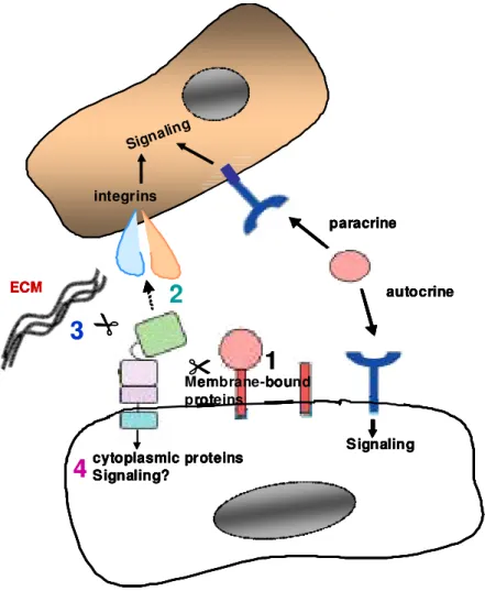
Perhaps these interactions with MAD2β are involved in coordinating ADAM-9 function with the cell cycle. In addition, ADAM-9 cytoplasmic tail can also bind to PKCdelta and regulates phorbol ester induced shedding of heparin-binding EGF and amyloid precursor protein (Izumi et al., 1998; Roghani et al., 1999). The different characterized functions of ADAM-9 are summarized in figure 4 below.
autocrine paracrine
cytoplasmic proteins Signaling?
Signaling Signaling
Membrane-bound proteins
ECM
integrins
autocrine paracrine
cytoplasmic proteins Signaling?
Signaling Signaling
Membrane-bound proteins
ECM
integrins
1 2
3
4
autocrine paracrine
cytoplasmic proteins Signaling?
Signaling Signaling
Membrane-bound proteins
ECM
integrins
autocrine paracrine
cytoplasmic proteins Signaling?
Signaling Signaling
Membrane-bound proteins
ECM
integrins
1 2
3
4
Figure 4. Overview of ADAM-9 functions. 1, ectodomain shedding of membrane-bound growth factors/cytokines and their receptors lead to release of soluble factors which can act in an autocrine or paracrine manner. 2, interaction with integrins can influence adhesion/migration and signaling. 3, processing of the ECM may affect signaling through integrins and cell migration. 4, localization and activity of ADAM-9 can be influenced by its cytoplasmic tail.
As to the control of Adam-9 transcriptional activity, it has been suggested that a single nucleotide substitution at -1314 influences the transcriptional activity of ADAM- 9 with a gain/loss binding of three putative transcriptional regulatory elements. These include the human muscle-specific Mt, the barbiturate-inducible element box and CAS interacting (Cong and Jia 2009). Estrogen is possibly an activator of Adam-9 promoter activity as the transcriptional activity of Adam-9 is increased in human neuroblastoma (SH-SY5Y) and human prostate adenocarcinoma (LNCaP) cell lines after treatment with estrogen (Cong and Jia 2009). Estrogen functions as a transcription factor by binding to the estrogen response element (ERE), altogether eight possible ERE sites have been found in the Adam-9 promoter region (Gruber et al., 2004; Cong and Jia 2009).
1.3.2 ADAM-9 in cancer
ADAM-9 is up-regulated in various human cancers such as renal, prostate, breast cancer and melanoma (O´Shea et al., 2003; Peduto et al., 2005; Zigrino et al., 2005;
Fritzsche et al. 2008). ADAM-9 is thought to participate in the escape of hepatocellular carcinoma (HCC) cells from NK cell-mediated immunosurveillance by shedding major histocompatibility complex (MHC) class I–related chain A (MICA), a ligand for NKG2D receptor (Kogha et al., 2010). Treatment of HCC cells with Sorafenib, a multikinase inhibitor (Keating and Santoro 2009), reduces MICA shedding by down-regulating ADAM-9 (Kogha et al., 2010). In a mouse model for prostate cancer, ADAM-9 was shown to be highly expressed in well-differentiated carcinomas but not in advanced poorly-differentiated tumors (Peduto et al. 2005).
Ablation of ADAM-9 in W10 animals, which spontaneously develop prostate tumors, resulted in the development of only well-differentiated tumors as compared to wild type mice, which develop poorly-differentiated tumors suggesting that ADAM-9 is involved in the progression of prostate tumors to advanced grades (Peduto et al., 2005). Using cell-based assays, the authors identified altered FGF and EFG signaling because of reduced FGFR2iiib and EGFR cleavage, as possible molecular mechanism for the observed effect. However, no in vivo evidence was provided on the alteration of these two signaling pathways. Soluble ADAM-9 has been found in increased amounts in the peritumoral stroma of liver metastases (Mazzocca et al., 2005). Interestingly, the soluble and membrane-bound forms of ADAM-9 have opposing roles in the migration of breast cancer cells. The soluble form enhances cell migration in a protease-dependent manner, while the membrane-bound form inhibits cell migration by altering integrin-mediated signaling (Fry and Toker 2010). In vitro primary keratinocytes lacking ADAM-9 show enhanced migration, this is reflected in vivo during wound healing (Mauch et al., 2010). Accelerated wound re- epithelialization is observed in Adam-9-/- animals because of altered shedding of collagen type XVII (Mauch et al., 2010). Shed collagen type XVII has been described as to have inhibitory effects on cell migration (Franzke et al., 2002).
In human melanoma, ADAM-9 is highly expressed at the tumor-stroma border where it is localized in both tumor and stromal cells (Zigrino et al., 2005). ADAM-9 expression in melanoma cells was confirmed in vitro. However, no correlation was observed between Adam-9 transcript levels and the invasive capacity of the cells (Zigrino et al., 2005). One functional activity of ADAM-9 in melanoma cells, which
might be important in progression of melanoma, is the mediation of heterotypic cell interaction between dermal fibroblasts and melanoma cells (Zigrino et al., 2011).
Despite these observations, the exact role of ADAM-9 in melanoma growth has not been determined.
1.4 Animal models for malignant melanoma 1.4.1 Mouse models
The increase incidence of melanoma has led to the need for better understanding of the molecular mechanisms involved in the commencement and progression of melanoma. Relevant animal models for addressing experimental questions would benefit melanoma research. The studies of melanoma in vivo have employed mouse xenograft models and genetically modified animals with spontaneous development of melanoma. In the xenograph model, melanoma cells propagated in vitro are grafted under the mouse skin and allowed to grow (Overwijk and Restifo 2001). Although melanoma does not spontaneously develop in these models, it is efficient in evaluating the contribution of specific host proteins in tumor progression. Tumor development is followed in a short period of 10 to 14 days, tumor cells with different genetic abnormalities or manipulations can be used (Richmond and Su 2008).
Mouse melanoma models, which spontaneously develop melanoma, have been generated to epitomize the histopathology of human melanoma. These models have the advantage that distinct genetic alterations leading to the development of melanoma in humans can be reproduced (Refaeli et al., 2009). Important differences in melanoma development between human and mouse is the localization of the melanocytes. In humans, the melanocytes are located in the basal layer of the epidermis while in mouse they are confined to the hair follicle in the dermis (Herlyn et al., 2000); hence, the histopathologies are not exactly the same (Ha et al., 2005).
Tormo et al., (2006), have described an interesting murine melanoma model HGF/CDK4R24C/R24C. This mice over-express hepatocyte growth factor/scatter factor (HGF) and carries a knock-in gene of the mutated cyclin-dependent kinase 4 (CDK4), a mutation which renders CDK4 insensitive to inhibition by p16/INK4a. This mutation has been observed in hereditary and sporadic human melanoma (Chin 2003). The over-expression of HGF leads to continuous signaling through its receptor tyrosine kinase c-Met and subsequent activation of the ras signal transduction pathway, which promotes melanomagenesis (Otsuka et al., 1998). These mice
develop melanoma spontaneously or upon carcinogen treatment in the epidermo- dermal junction of the skin. Importantly, these tumors are invasive and metastatic (Tormo et al., 2006). Another mouse model of melanoma is the Tg(Grm1)EPv(E) mice, that over-express the metabotropic glutamate receptor 1 (mGrm1) in melanocytes under control of dopachrome tautomerase promoter (Pollock et al., 2003). This mouse strain develops melanoma at about 6 months of age with major location in the tail (Pollock et al., 2003). Ectopic expression of mGrm1 transforms melanocytes and causes malignant melanoma in vivo (Oritz et al., 2007). The Tg(Grm1)EPv(E) model may recapitulate human uveal melanoma as activation of this pathway has been observed in human uveal melanoma (Van Raamsdonk et al., 2009). The Tyr::CreER; BRAFCA; Ptenlox/lox mouse melanoma model (Dankort et al., 2009) initiates the substitution of glutamic acid with valine at position 600, a mutation observed in human melanoma leading to constitutive ERK signaling (Davies et al., 2002). All mice develop melanoma with metastasis to lymph nodes and lungs (Dankort et al., 2009).
1.4.2 Medaka model
Non-mammalian animals like fish also develop melanoma. Fish animal models are increasingly being use to study melanoma (Craig et al., 2008). Fish models for melanoma were first established in Xiphophorus, followed by transgenic melanoma models in zebrafish and medaka (Patton et al., 2011). The medaka melanoma model was generated by over-expression of the oncogene, Xiphophorus medaka receptor kinase (Xmrk) an orthologue of the mammalian epidermal growth factor receptor gene set under the control of the pigment cell-specific promoter, microphthalmia- associated transcription factor (mitf) (Schartl et al., 2009; Meierjohann et al., 2004).
Fish homozygous for Xmrk all develop melanoma between 2 to 6 weeks of age, which are highly invasive and metastatic (Schartl et al., 2009). Uveal melanoma and tumor formation in the red and yellow pigment cells called xanthophoroma and erythrophoroma respectively have been described (Schartl et al., 2009). This model has many similarities with human melanoma for one thing, Xrmk receptor induces signaling pathways, which mediate proliferation, resistance to apoptosis and initiation of dedifferentiation, events that are similar to those found downstream of mammalian Egfr (Meierjohan and Schartl 2006). Xmrk induces the Ras-Raf-MAPK signaling pathway, which mediates cell proliferation and differentiation. PI3-K can also bind to
the c-terminal of Xmrk leading to activation of Akt and induction of anti-apoptotic signaling. Signal transducer and activator of transcription (STAT5) is also activated promoting anti-apoptosis by increasing the expression of Bcl-x. These two pathways are well described in human melanoma (Satyamoorthy et al., 2003).
Apart from these indicated similarities, no data on the expression and putative role of molecules such as proteolytic enzymes in this model. Whether tumoral and peritumoral proteolysis plays a role for disease progression in this model, as shown for human and murine melanoma, needs to be investigated. Technical advantages of this model is among others, their easy breeding and large number of offsprings produced, thus offering a model with screening possibilities for pharmacological analysis of potential inhibitors for therapeutic use.
1.5 Aims of the thesis
After formation of a primary tumor within the epidermal layer, melanoma cells progress into the tissue and metastasize to distant organs. Invasion into the dermal compartment and distant organs requiresamong others, cancer cell adhesion to/and degradation of the ECM. In addition to several proteolytic enzymes, ADAM-9 was found expressed by tumor and stromal cells in malignant melanoma. This protein can exert dual functions. On one hand, it is proteolytically active, and on the other hand, it functions as a cell adhesion receptor making it a molecule with pivotal roles in melanoma development and metastasis. The main aim of this study was to investigate at a molecular level how ADAM-9 produced by the host affects growth and metastasis of malignant melanoma. To achieve this well established in vivo melanoma cell grafting model was used to analyze melanoma growth in mice lacking ADAM-9 in the host. Tumor progression was monitored as a function of time and all processes important for tumor progression such as inflammation, tumor-induced neoangiogenesis and proliferation were analyzed in detail to investigate whether ablation of ADAM-9 had an impact on these processes. In addition, the protein modifications in the peritumoral environment in tumors from WT or Adam-9-/- animals were further investigated in vivo by proteomics. In vitro co-culture systems using fibroblasts isolated from WT and Adam-9-/- animals and murine melanoma cells was employed to analyze at a molecular level how the expression of ADAM-9 stromal fibroblasts affected melanoma-fibroblasts cellular cross-talk at the tumor-stroma
border. A direct role for ADAM-9 in ECM processing was addressed using biochemical approach with recombinant ADAM-9, active/inactive.
2 Materials and methods 2.1 Materials
2.1.1 Chemicals
All chemicals were purchased in analytical grade from Roth (Karlsruhe), Sigma- Aldrich (München), Merck (Darmstadt), GIBCO (Karlsruhe) and Perbio (Lausanne/Switzerland). Molecular weight markers for proteins, PageRulerTMPlus Prestained Protein ladder and PageRulerTM Unstained Protein ladder were from Fermentas (St. Leon-Roth). DNA molecular weight markers, 100 bp DNA ladder and 1 kb DNA ladder as well as restriction enzymes were from NEB (Frankfurt/Main).
Human recombinant TIMP-1 and fibronectin were from Calbiochem (Darmstadt) and R&D systems (Wiesbaden) respectively. Mouse recombinant TNF-α was from Miltenyi (Bergisch Gladbach). Rat tail recombinant collagen type I was from Cell Systems (Remagen, Germany).
2.1.2 Cell culture material
Plastic ware for cell culture was purchased from Greiner (Nürtingen), TPP (Trasadingen/Switzerland) and Becton Dickinson (Heidelberg). Cell culture media were from Invitrogen (Karlsruhe), Biochrom (Berlin) and PAA (Cölbe). Supplements (Penicillin/Streptomycin) were from Biochrom and FCS from PAA. Zeocin and geneticin (G418) antibiotics were purchased from Invitrogen and PAA respectively.
Collagenase was from Cell Systems (Troisdorf) and mitomycin C was from Sigma- Aldrich.
2.1.3 Bacteria, vectors and plasmids
For transformation of plasmids, Escherichia coli DH5α (Invitrogen) was used. The genotype is: F- Φ80lacZ∆M15 ∆(lacZYA-argF)U169 recA1 endA1 hsdR17(rk-, mk+) phoA supE44 thi-1 gyrA96 relA1 λ-. The linearized vector pCR 2.1, used for subcloning, and the eukaryotic expression vector pcDNA4/TO/myc-His were purchased from Invitrogen.
2.1.4 Antibodies
Antibodies Company Application Dilution
Goat anti-ADAM-9 R&D systems WB 1:1000
Rat anti-ADAM-9 R&D systems IF 1:100
Rabbit anti-ADAM-10 Abcam, Cambridge, UK IF
Rabbit anti-ADAM-17 Biodesign, Asbach WB 1;1000 Rabbit anti-MMP-13 Santa Cruz, Heidelberg IF 1:50
Rabbit anti-MMP-14 Pineda, Berlin IF 1:100
Goat anti-TIMP-1 R&D systems WB/IF/
Neutralization
1:1000/1:100 1:2000 Hamster anti-TNF-α
(clone TN3-19)
eBioscience Frankfurt Neutralization 1:2000
Rabbit anti-TNFRI Abcam WB 1:10000
Rat anti-Ki67 (clone
Tec 3) Dako, Hamburg IF/IHC 1:1000/1:50
Rabbit anti-Cleaved caspase-3
Cell signaling, Frankfurt IHC 1:200 Rat anti-CD45 (clone
IBL-3/16)
AbD Serotec Düsseldorf IF 1:200 Mouse anti-decorin
(clone 115402) R&D systems Wiesbaden WB/IF 1:1000/1:100
Rat anti-F4/80 AbD Serotec IF 1:100
Rat anti-Neutrophil AbD Serotec IF 1:100
Mouse anti-decorin R&D systems WB/IF 1:1000/1:100 Rabbit anti-collagen
14 Gift from Prof. M. Koch,
University of Cologne WB/IF 1:1000/1:100 Rabbit anti-collagen
type I
Quartett, Berlin WB 1:1000/1:50
Rabbit anti-fibronectin Sigma Aldrich WB/IF 1:1000/1:100 Rat anti-CD31 (clone
MEC 13.3)
AbD Serotec IF 1:1000
Rabbit-anti-mouse
HRP Dako WB 1:2000
Rabbit anti-goat HRP Dako WB 1:2000
Rabbit anti-rat
biotinylated Dako IHC 1:300
Donkey anti-goat- Alexa 594-conjugated
Invitrogen, IF 1:1000
Rabbit anti-rat-Alexa
488-conjugated Invitrogen, IF 1:1000
Goat anti-rabbit-Alexa 594-conjugated and 488-conjugated
Invitrogen, IF 1:1000
2.1.5 Buffers
PBS: 136 mM NaCl; 2.6 mM KCl; 10nM Na2HPO4; 1.5 mM KH2 PO4; pH 7.4
TBE: 90 mM Tris; 20 mM Na2EDTA; 90 mM boric acid; pH 8 PBST: PBS + Tween 20 0.5 % (v/v) TBE
DNA lysis buffer: 100 mM Tris/HCl pH 8.5; 5 mM EDTA pH 8.0; 0.2% SDS;
200 mM NaCl
Running buffer: 250 mM Tris base, 0.2 M Glycin, 1 % SDS; pH 8.3 Anode I buffer: Tris 0.3 M; 20 % Methanol
Anode II buffer: Tris 25 mM; 20 % Methanol
Cathode buffer: 6- Aminohexan acid 40 mM; 20 % Methanol
Laemmli reducing buffer: 60 mM Tris-HCl pH 6.8; 2 % SDS; 0.1 % Bromphenol blue; 25 % Glycerol; 14.4 mM 2-Mercaptoethanol Equilibration buffer: 50 mM NaH2PO4; 20 mM Tris HCl; 100 mM NaCl; pH 8 Gradient buffer A: 10 mM imidazole; 50 mM NaH2PO4; 20 mM Tris HCl; 100
mM NaCl; pH 8
Elution buffer: 250 mM imidazole; 50 mM NaH2PO4; 20 mM Tris HCl;
100 mM NaCl; pH 8
Substrate buffer: 50 mm Tris-HCl, 5 mm CaCl2, pH 8.0 Citrate buffer: 2 mM citric acid; 2 mM sodium citrate; pH 6 Cell lysis buffer: PBS + 20 mM NH4OH; 0.5 % Triton X-100
RIPA buffer: 150 mM NaCl, 0.5 % DOC; 2 % SDS; 1 % NP40; 50 mM Tris; 1 % protease inhibitor cocktail; pH 7.4
2.2 Methods 2.2.1 Cell biology
2.2.1.1 Cells and cell culture
COS-7 kidney fibroblasts from African Green Monkey, SV40 transformed were from ATCC (Gluzman 1981). B16F0, mouse melanoma cells were a kind gift from Prof. C.
Blobel, Cornell University, New York (Fidler 1973), and B16F1 were from ATCC (Fidler 1975). All cells were cultured in Dulbecco’s modified Eagle’s medium (DMEM) supplemented with 10 % FCS, 100 U/ml penicillin, 100 µg/ml streptomycin. B16F1- GFP, B16F1 cells stably expressing GFP (Zigrino et al., 2009) were cultured in DMEM supplemented with 10 % fetal calf serum (FCS), 100 U/ml penicillin, and 100
µg/ml streptomycin and 800 µg/ml G418. The human melanoma cell lines Skmel28 (Carey et al., 1976), WM164/4 (Herlyn et al., 1990), BLM (Van Muijen et al., 1991), A375 (Giard et al., 1973) and 530 (Van Muijen et al., 1991) were cultured in RPMI 1640 medium containing 10 % FCS, 1 % non-essential amino acids, 2 % L- Glutamine, 100 U/ml penicillin, 100 µg/ml streptomycin. Glioma Gli36 cells (Kashima et al., 1995) and malignant transformed HaCaT II-4(rt) cells (SCC) (Boukamp et al., 1990) were cultured in DMEM supplemented with 10 % FCS, 100 U/ml penicillin and 100 µg/ml streptomycin. Basal cell carcinoma cells (BCC) (Yen et al., 1996) were cultured in DMEM-HAM´S F12 supplemented with 10 % FCS, 100 U/ml penicillin, 100 µg/ml streptomycin, 0.1 % adenin, 0.01% hydrocortisone, 0.001 % cholera toxin, 1 % EFG and 1% insulin. All cells were cultured at 37 °C with 5 % CO2 and a humidity of 95 %.
2.2.1.2 Isolation of primary fibroblasts
Primary murine dermal fibroblasts were isolated from the back skin of newborn mice.
Briefly, newborn mice were euthanized by decapitation, cooled on ice for 1 hour and disinfected in betaisodona iodine for 1 minute, rinsed briefly in PBS and then for 1 minute in 70 % ethanol and finally rinsed in PBS. The skin was incubated overnight with 0.2 % trypsin at 4 °C. The epidermis was separated from the dermis, which was diced finely before incubating with 400 U/ml collagenase (Worthington) for 2–3 hours at 37 °C with gentle agitation. Tissue debris was removed by centrifugation at 1200 rpm. Cell pellet was resuspended in DMEM containing 400 U/ml collagenase and isolated fibroblasts were cultured in DMEM containing 10 % FCS, 2 mM glutamine and 100 U/ml penicillin, 100 µg/ml streptomycin. The fibroblasts were used between passages 2 and 4 for experiments.
2.2.1.3 Preparation of fibroblast conditioned media, cell monolayer and matrix For the preparation of conditioned medium, fibroblasts were seeded at a density of 5 x 10⁴ cells/cm2 in culture plates and cultured overnight at 37 °C. Cells were washed twice with PBS and incubated with serum free DMEM for 24 hours. The medium was collected and spun down for 5 minutes at 1200 rpm to eliminate cellular debris.
Supernatants were used immediately for cell-stimulation experiments or stored at -20
°C for additional analysis.