Differential Influence of Clinical Mycobacterium avium Strains on Distinct Autonomous Defense Capacities of Human Neutrophils
Inaugural-Dissertation zur
Erlangung des Doktorgrades
der Mathematisch-Naturwissenschaftlichen Fakultät der Universität zu Köln
vorgelegt von
Senthil Kumar Subramanian
aus
Madurai, India
Köln, 2014
Gutachter: Prof. Dr. Jonathan Howard Prof. Dr. Mirka Uhlirova Tag der mündlichen Prüfung: 26 June 2014
To the love of my life....
“host-‐pathogen interaction and the evolutionary arms race”
Table of contents
Table of contents ... 4
Table of figures ... 7
Register of Tables ... 7
1 Introduction ... 8
1.1 Mycobacteria ... 8
1.2 Pathogenesis ... 10
1.3 Mycobacteria and neutrophils ... 12
1.4 Inhibition of phago-‐lysosomal fusion ... 13
1.5 Regulation of cytokines ... 14
1.6 ROS production ... 15
1.7 Neutrophils ... 16
1.8 Neutrophil reservoirs ... 16
1.9 Maturation of neutrophils ... 17
1.10 Neutrophil migration ... 17
1.11 Activation of neutrophils ... 19
1.12 Recognition of pathogens ... 19
1.13 Killing mechanisms ... 20
1.14 NADPH oxygenase-‐dependent killing mechanism ... 20
1.15 Degranulation ... 21
1.16 NETs ... 22
1.17 Production of inflammatory mediators ... 23
1.18 Neutrophil and macrophage interaction ... 23
1.19 Objectives of the study ... 25
2 Results ... 26
2.1 Both clinical Mycobacterium avium isolates, SCH 228 and SCH 215, prevent killing by human neutrophils by inhibiting phago-‐lysosome fusion ... 26
2.2 M. avium SCH 228 and SCH 215 dampen the activation of p38 MAPK in human neutrophils ... 28
2.3 SCH 228 and SCH 215 M. avium dampen p38 MAPK mediated degranulation in human neutrophils ... 29
2.4 SCH 228 and SCH 215 M. avium induce ROS differentially in human neutrophils 31 2.5 SCH 228 and SCH 215 M. avium induce only certain cytokines differentially in infected neutrophils ... 33
2.6 IL-‐8 induction in M. avium infected human neutrophils is p38 MAPK and ROS dependent ... 36
2.7 IL-‐8 induces degranulation in neutrophils ... 36
2.8 ROS and IL-‐8 do not affect the killing of SCH 228 by human neutrophils ... 37
2.9 Stimulation by LPS does not affect killing of SCH 228 or SCH 215 by human neutrophils ... 37 2.10 M. avium isolates, SCH 228 and SCH 215, do not alter neutrophil death pathways
38
2.11 Cytochalasin B/fMLP stimulated degranulation inhibits extracellular growth of
SCH 228 and SCH 215 M. avium ... 39
2.12 Supernatant from SCH 228 or SCH 215-‐infected neutrophils inhibits extracellular growth of Staphylococcus aureus but not M. avium ... 42
2.13 LPS-‐stimulated or unstimulated neutrophils do not affect survival of SCH 228 and SCH 215 in human macrophages in contact co-‐culture experiments ... 44
2.14 SCH 228 and SCH 215-‐induced degranulation does not affect intracellular survival of these isolates in human macrophages in non contact co-‐culture experiments ... 45
2.15 M. avium infected macrophages induce exocytosis of CD66 vesicles predominantly in naïve neutrophils in a time dependent manner ... 46
2.16 Exosomes released from M. avium-‐infected macrophages attract human neutrophils ... 47
2.17 Prediction of putative neutrophil chemotactic factors on THP-‐macrophage derived exosomes ... 53
3 Discussion ... 59
3.1 Killing of M. avium ... 59
3.2 Phagosomal processing ... 60
3.3 Neutrophil activation ... 62
3.4 Degranulation ... 63
3.5 ROS ... 64
3.6 Cytokines ... 66
3.7 Cell death ... 68
3.8 Extracellular killing ... 69
3.9 Neutrophil-‐macrophage interaction ... 71
3.10 Chemotaxis ... 73
3.11 Exosomes and their components as potential chemoattractants ... 74
4 Summary ... 77
5 Zusammenfassung ... 79
6 Materials and methods ... 82
6.1 Materials ... 82
6.1.1 Instruments ... 82
6.1.2 Consumables ... 83
6.1.3 Chemicals and reagents ... 84
6.1.4 Commercial kits used ... 85
6.1.5 Antibodies ... 86
6.1.6 Buffers, media and solutions ... 86
6.1.7 Bacterial strains ... 89
6.1.8 Cell lines ... 89
6.1.9 Softwares ... 89
6.2 Methods ... 90
6.2.1 Culturing and freezing of THP cell line ... 90
6.2.2 Differentiation of THP-‐1 cells to macrophages ... 90
6.2.3 Preparation of mycobacterial stock P0 ... 90
6.2.4 Preparation of mycobacterial stock P1 ... 91
6.2.5 Determination of CFU ... 91
6.2.6 Culturing of human monocyte derived macrophages (HMDM) ... 91
6.2.7 Growth rate of M. avium (SCH228 and 215) in HMDM ... 92
6.2.8 Neutrophil isolation ... 92
6.2.9 Killing of Mycobacteria by human neutrophils ... 93
6.2.10 Preparation of Zymosan-‐activated serum ... 93
6.2.11 Chemotaxis assay ... 93
6.2.12 Coating of mycobacteria with C12-‐FDG ... 94
6.2.13 Coating of magnetic beads with C12-‐FDG ... 94
6.2.14 Phagosomal maturation study in human neutrophils ... 95
6.2.15 Phagosomal maturation study in human macrophages ... 95
6.2.16 Lysis of samples for Western blotting ... 96
6.2.17 Western Blot Analysis ... 96
6.2.18 Degranulation study ... 97
6.2.19 Apoptosis assay ... 98
6.2.20 ROS assay ... 99
6.2.21 Cytokine measurements ... 99
6.2.22 In vitro anti-‐bacterial assay ... 100
6.2.23 Co-‐culture experiment ... 100
6.2.24 Depletion of exosomes from FCS ... 101
6.2.25 Exosomes isolation ... 101
6.2.26 Mass spectrometry ... 102
7 References ... 105
8 Abbreviations ... 128
9 Declaration ... 130
10 Acknowledgment ... 131
11 Curriculum vitae ... 133
Table of figures
Figure 1 M. avium strains SCH 228 and SCH 215 are not killed by human neutrophils. ... 26
Figure 2 SCH 228 and SCH 215 M. avium inhibit phago-lysosomal processing in human neutrophils. ... 27
Figure 3 SCH 228 and SCH 215 dampen the phosphorylation and activation of p38 MAPK in human neutrophils. ... 29
Figure 4 SCH 228 and SCH 215 M. avium dampen p38 MAPK mediated degranulation in neutrophils. 30 Figure 5 ROS induction in mycobacteria-infected human neutrophils. ... 32
Figure 6 IL-8 and IL-1ra are differentially induced by SCH 228 and SCH 215 in infected neutrophils. . 34
Figure 7 IL-8 induction in infected neutrophils is mediated by p38 MAPK via ROS. ... 35
Figure 8 IL-8 induces degranulation in neutrophils. ... 37
Figure 9 Priming with LPS does not enhance SCH 228 and SCH 215 killing by human neutrophils. ... 38
Figure 10 M. avium isolates, SCH 228 and SCH 215, do not alter neutrophil death pathways. ... 39
Figure 11 Granular components of human neutrophils inhibit extra-cellular growth. ... 41
Figure 12 Neutrophil degranulation induced by mycobacteria does not impact on their extra cellular growth but significantly inhibits extra cellular growth of SA. ... 43
Figure 13 Bacterial survival of SCH 228 and SCH 215 do not differ in human macrophages co-cultured with LPS-stimulated or unstimulated neutrophils. ... 45
Figure 14 Mycobacteria-induced degranulation does not affect intracellular survival of SCH 228 and SCH 215 in human macrophages. ... 46
Figure 15 SCH 228 or SCH 215-infected macrophages induce predominantly specific/gelatinase degranulation in naïve neutrophils in a time dependent manner. ... 47
Figure 16 Isolation and characterization of exosomes derived from M. avium-infected THP macrophages. ... 50
Figure 17 Exosomes released from M. avium infected macrophages induce neutrophil chemotaxis. ... 51
Figure 18 Classification of identified proteins present in the exosomes derived from THP-1 macrophages. ... 53
Register of Tables
Table 1 List of mycobacterial proteins present in exosomes ... 55Table 2 List of putative proteins on exosomes involved in neutrophil chemotaxis ... 56
1 Introduction
1.1 Mycobacteria
Mycobacterial pathogens present a health threat worldwide with
Mycobacterium tuberculosis (Mtb) alone accounting for 1.3 million deaths and 8.6 million new and relapseinfections in 2012 (http://www.who.int/tb/publications/global_report/en/). Tuberculosis is an infectious bacterial disease caused by the facultative intracellular pathogen Mycobacterium
tuberculosis that is still posing a major health and socio-economic burden at a global level,primarily in low and middle-income countries. The genus
Mycobacteriumincludes more than 60 species and more than 100 sub-species (http://www.dsmz.de). Currently a number of 8 species are referred to as the
Mycobacterium tuberculosis complex (M. tuberculosis, M.bovis (including the BCG strain), M. africanum, M. microti, M. canettii, M. caprae, M.
pinnipedii, M. mungi) and all of these can cause the clinical picture of tuberculosis. M.
tuberculosis and M. leprae were recognized as pathogenic for humans only about 140 years
ago and their pathogenic behaviors have been studied extensively. Mycobacteria other than
M. tuberculosis and M. leprae, are called non-tuberculous mycobacteria (NTM) ormycobacteria other than tuberculosis (MOTT) and about 20 species of them cause human and animal diseases (Heifets, 2004). NTM species are ubiquitous in the environment, predominantly populating soil and water (Falkinham, 2010, 2002; Taylor et al., 2000). They have been recovered from surface water, tap water, soil, domestic and wild animals, milk, and food products (Thomson et al., 2007). The incidence of infection and disease due to non- tuberculous mycobacteria (NTM) is rising in many countries. It was thought that NTM are more prevalent in tropical countries (Africa and Australia), however, accumulation of recent data suggests that it is also prevalent in temperate countries like USA, Canada, Germany, United Kingdom and China (Behr, 2008; Thomson, 2010).
NTM and specifically
M. avium became inevitably recognized as pathogens causing severediseases in humans at the beginning of the HIV/AIDS era. Back then
M. avium causedsevere, mostly fatal infections in immuno-compromised patients. The most prominent risk
factors for the development of disseminated
M. avium infection were a CD4 cell-count ofless than 100/ml and previous colonization of mucosal surfaces with
M. avium (Horsburgh,1991; Klatt et al., 1987; Mapother and Songer, 1984; Nightingale et al., 1992). Although there is a strong correlation between
M. avium disease and low CD4 cell-counts, a specificimmune defect facilitating invasion and dissemination of M. avium has not yet been defined.
Uptake and intracellular growth inhibition of
M. avium by monocytes derived from AIDSpatients has been shown to be normal in vitro. Moreover, the application of granulocyte colony stimulating factor G-CSF, a cytokine that specifically activates neutrophils was beneficial with regard to survival of AIDS patients suffering from
M. avium infection(Keiser et al., 1998). This implicates that neutrophils may have a protective role in M. avium.
Moreover, there are reports showing that
M. avium and other NTM cause lung infection(also called atypical mycobacterial pulmonary infections) in immuno-competent individuals (Piersimoni and Scarparo, 2008; Taiwo and Glassroth, 2010). Also, epidemiologic surveillance reveals that the occurrence of NTM diseases is increasing in industrialized nations where TB transmission is low and where BCG vaccines are not given (Lietman et al., 1997; Winthrop et al., 2010). The incidence of NTM infections is particularly increasing in the elderly (Mirsaeidi et al., 2014), most of them exhibiting pulmonary infection caused primarily by M. avium. Some of these lung infections occur as a complication of pre-existing lung disease, such as bronchiectasis and chronic obstructive pulmonary disease. However, some other infections occur in healthy hosts without pre-existing lung disease or overt immunologic disorder (Huang et al., 1999; Koh et al., 2002; Marras et al., 2013; Sadek et al., 2008; Thomson, 2010). Generally, non-tuberculous mycobacteria (NTM) inhabit body surfaces and secretions without causing disease. However, under certain conditions, which are not fully understood, they can induce some clinical manifestations such as progressive pulmonary disease, superficial lymphadenitis, disseminated disease, and skin and soft tissue infection. Disseminated disease can occur in severely immuno-compromised patients (Griffith et al., 2007).
Among NTM species,
M. avium attracts serious attention, as is the most common cause ofnon-tuberculous pulmonary disease worldwide. This species includes four subspecies:
M.avium avium (MAA), M. avium hominissuis (MAH), M. avium paratuberculosis (MAP) and M. avium silvaticum (MAS) (Thorel et al., 1990; Turenne et al., 2008). MAA and MAS are
specific avian pathogens causing a tuberculosis-like disease in birds (Dvorska et al., 2003).
MAP is a well-known pathogen causing Johne’s disease (Harris and Barletta, 2001) and may have a role in human Crohne’s disease (Feller et al., 2007). Of importance, MAH causes pulmonary infections in humans in which the normal T cell immunity is compromised.
1.2 Pathogenesis
Virulent mycobacteria reside in the phagosomes in infected macrophages and prevent killing by blocking the phago-lysosome fusion (Danelishvili et al., 2007; Rocco and Irani, 2011;
Russell, 2013). This ability of virulent mycobacteria to survive and multiply in macrophages is considered as the key for their pathogenicity, latency and transmission. All virulent mycobacteria like
Mtb, M. bovis, M. aviumand
M. marinumdwell in macrophages by modifying the phagosome, the most hostile environment, according to their requirements, which is the signature evasion mechanism of virulent mycobacteria (Danelishvili et al., 2007; Early and Bermudez, 2011; Rocco and Irani, 2011; Russell, 2013).
Despite the differences between
M. avium and Mtb, the basic pathogenic behavior inpromoting their survival in infected macrophages, the primary host cell is similar. M. avium invades the human host via either the respiratory tract or the intestinal epithelium: the latter route of infection rather causes rapid dissemination than the former. M. avium interacts with various receptors to gain access into the macrophages. Macrophages express an array of pathogen recognition receptors (PRRs) and phagocytic receptors that play a crucial role in their recognition and response to pathogens. For example complement receptors (except, CR3 and CR4) (Bermudez et al., 1999; Bohlson et al., 2001), mannose receptors, fibronectin (Bermudez et al., 1991; Polotsky et al., 1997), and type A scavenger receptors are involved in this process. Also toll-like receptors (TLRs) 2 and 9 are important for
M. aviumrecognition (Appelberg, 2006; Feng et al., 2003; Gomes et al., 2004; Gomes et al., 2008).
This recognition leads to the phagocytosis of pathogens and the resulting combinatorial
signals from these receptors are important for the activation of macrophages and thereby, are
essential for the initiation of the innate immune response (Akira et al., 2006; Schafer et al.,
2009; Takeuchi and Akira, 2010). Once, they are phagocytized by macrophages, they reside
in a plasma membrane derived intracellular vesicle called phagosomes. Microorganisms in
the phagosomes are killed and digested upon fusion with lysosomes in their maturation process, forming phago-lysosomes (Desjardins et al., 1994; Vieira et al., 2002). Similar to other pathogenic mycobacteria,
M. aviuminhibits phagosome maturation, by preventing acidification and by blocking fusion of phagosomes with extremely acidic lysosomes (Danelishvili et al., 2007; de Chastellier and Thilo, 2002).
Efficient killing of pathogens in macrophages requires distinct pro-inflammatory cytokines.
Once the macrophage engulfs
M. avium, it produces and secretes numerous chemokines,cytokines and their ligands, e.g. TNF-
α, LT-a/b; IL-1, -6, -12, -18; G-CSF and GM-CSF;CXCL-1, -2, -3, -8; CCL-2, -3, -4, -5, -20 (Blumenthal et al., 2005; Sarmento and Appelberg, 1996; Shiratsuchi and Ellner, 2001) which induce the development of pro- inflammatory responses. It has been shown that infection of macrophages with various mycobacteria strain types induce a differential pattern of cytokines in vitro (Chacon-Salinas et al., 2005).
It is evident that activation of TLR 2/MAPK leads to activation of immune response in
macrophages. Subsequent activation and nuclear translocation of NF-kB occurs, which
greatly enhances mycobacterial killing by production and secretion of pro-inflammatory
cytokines, and facilitating phago-lysosome fusion (Gutierrez et al., 2008; Pathak et al.,
2004). Interference with the activation of toll-like receptors (TLRs), in activation of host
antimicrobial responses possibly by surface molecules may promote their survival in the
macrophages (Early and Bermudez, 2011). A substantial amount of evidence indicated that
cell wall glyco-peptidolipids (GPLs) are responsible for pathogenesis and specific GPLs are
associated with virulence (Bhatnagar and Schorey, 2006). The protective responses against
M. avium largely depend upon activation of macrophage oxidative/bactericidal functions bytype 1 pro-inflammatory cytokines provided by immune T lymphocytes and other cell types
(Ehlers et al., 2000; Florido et al., 2005; Petrofsky and Bermudez, 2005). In vitro studies and
animal infection models have shown that phylogenetically diverse mycobacteria strains
exhibit markedly different virulence and pathogenesis phenotypes in macrophages (Early
and Bermudez, 2011; Lopez et al., 2003; Manabe et al., 2003; Manca et al., 2004; Manca et
al., 2001; Meyer et al., 1998; von Reyn et al., 1995).
Though, M. avium is not considered as a public health problem, it is increasingly recognized as a relevant pathogen due to its overall increasing incidence not only in patients with immunosuppression, but also in immuno-competent individuals and its high case fatality (Henry et al., 2004; Marras and Daley, 2002; McGrath et al., 2008). Studying
M. aviumbiology and pathogenesis is important because its clinical appearance and relevance (Chatterjee and Khoo, 2001; Rocco and Irani, 2011) is supposedly interlinked with the mechanisms by which some species of the genus are pathogenic while others are not (Behr, 2008; Grubek-Jaworska et al., 2009). The
in vitro virulence of M. avium isolates obtainedfrom various sources is characterized by determining their ability to multiply in primary human monocyte derived macrophages. We have previously systematically characterized M.
avium human clinical isolates and environmental isolates based on their ability to replicate in
human macrophages. Along this study we found that the macrophage-induced gene (mig) is quite consistently expressed when the bacilli are growing within macrophages making it a potential marker for clinical pathogenicity and in vitro virulence of M. avium isolates (Meyer et al., 1998; Plum and Clark-Curtiss, 1994). Analyses of growth rate of these isolates in macrophages suggested that the
in vitro virulence of these isolates vary at length betweenand among clinical and environmental isolates. From the panel of those previously characterized strains we have chosen SCH 228 as a strain that exhibited a fast growth rate in macrophages (
≈18 h) and SCH 215 which was only slowly replicating in macrophages in order to characterize their interaction with neutrophils.
1.3 Mycobacteria and neutrophils
Neutrophils are typically the first responders in host defense against invading pathogens.
Neutrophils destroy pathogens by both oxidative and non-oxidative mechanisms. They are
recruited to the site of mycobacterial infection and are the most predominantly infected
phagocytic cells in human tuberculosis (Eum et al., 2010). However, the role of neutrophils
in mycobacterial infection is not fully understood. There is clinical evidence based on the
beneficial impact of the application of G-CSF on survival of AIDS patients suffering from
M. avium infection (Keiser et al., 1998). Martineau et al. (2007) showed that neutrophilscontribute significantly to the control of Mtb in human blood. Some earlier murine studies reported a protective role for neutrophils in early infection with
Mtb as well as with M.avium (Barrios-Payan et al., 2006; Pedrosa et al., 2000; Sugawara et al., 2004). On the
contrary, a study showed that neutrophils do not contribute to the control of Mtb (Seiler et al., 2000). Furthermore, a neutrophil-driven, interferon (IFN)-inducible transcript signature in human whole blood was recently identified that correlated with clinical severity of TB, raising the possibility that neutrophils may directly contribute to disease pathogenesis (Berry et al., 2010). Similar to macrophages, neutrophils have been shown of being capable to phagocytize and kill at least some mycobacterial species. However, a systematic analysis of the cell-autonomous immune regulation of human neutrophils upon infection with human clinical
M. avium hominissuis isolates was not undertaken so far. Moreover, it is widelyaccepted that only a better understanding of the pathogenic processes associated with
M.avium infection and disease will lead to the development of effective tools to control these
conditionally pathogenic bacteria.
1.4 Inhibition of phago-lysosomal fusion
Macrophages express an array of pathogen recognition receptors (PRRs) and phagocytic receptors that play a crucial role in their recognition and response to pathogens. Many studies have shown that receptors such as Toll-like receptors (TLR-2, 4, 6 and 9) (Heldwein and Fenton, 2002; Jo et al., 2007; Means et al., 1999; Quesniaux et al., 2004) mannose receptors (McGreal et al., 2005), DC-SIGN, Dectin-1, and Mincle (Torrelles et al., 2006), the complement C3 receptors, Fcγ phagocytic receptors, CD-14 and NOD-like receptors (NLRs) are involved in the recognition process. This recognition leads to the phagocytosis of pathogens and the resulting combinatorial signals from these receptors are important for the activation of macrophages and thereby, are essential for the initiation of the innate immune response (Akira et al., 2006; Schafer et al., 2009; Takeuchi and Akira, 2010).
Phagocytosis of invading bacteria by professional phagocytes such as macrophages, DCs
and neutrophils results in the formation of a plasma membrane derived intracellular vesicle
around the pathogen. This newly formed cellular compartment is called phagosome in which
pathogenic microorganisms are killed and digested upon fusion with lysosomes in their
maturation process, forming phago-lysosomes. After detachment from the host cell membrane, a nascent phagosome undergoes a series of progressive maturation processes involving fission and fusion events that modify the composition of the membrane. This process highly depends on the interaction of the phagosome with the endocytic pathway;
phagosomes sequentially fuse with early (sorting) endosomes, late endosomes, and eventually with lysosomes (Desjardins et al., 1994; Vieira et al., 2002). This process enables nascent phagosomes to gradually acquire the properties of the donor organelles, including the distinct membrane markers and the increasingly acidic pH value. As a consequence, a phagosome ultimately matures into a hybrid intracellular compartment called phago- lysosomes. Phago-lysosomes contain various digestive enzymes, including proteases, nucleases, lipases, and β-galactosidases, which display optimal activities in the highly acidic condition (pH ≤5.0) and efficiently degrade luminal contents (Vieira et al., 2002)
However, during
Mtb infection, the phagosome maturation pathway is altered via multiplemechanisms to disrupt normal phagocyte effector functions (Deretic et al., 2006; Russell, 2001). The early maturation process of the
Mtb phagosome includes fusion with earlyendosomes, wherein both Rab5 and transferrin receptors are found associated with it.
However, the pathogen prevents recruitment of Rab5 effector proteins, such as early endosomal auto antigen 1 (EEA1) and hVPs35 (Fratti et al., 2001) that are required for further maturation of the phagosome. Absence of Rab5 effector proteins on Mtb phagosomes prevents docking and delivery of lysosomal hydrolases, cathepsins, and vacuolar ATPases.
Ultimately, these defects result in a failure in conversion of a Rab5 (an early endosomal marker) containing phagosome to a Rab7 (a late endosomal marker) containing phagosome (Via et al., 1997).
1.5 Regulation of cytokines
Efficient killing of pathogens in macrophages require adequate and appropriate pro-
inflammatory cytokines. Macrophages produce pro-inflammatory cytokines and chemokines
in order to mount effective immune response against pathogens. Once the macrophage
engulfs
Mtb, it produces several cytokines such as IL-2, IFN-γ, IL-6, IL-1α/β, IL- 12, andTNF-α (Kellar et al., 2011) and chemokines including CCL2, CCL3, CCL5, CCL7, CCL12, CXCL2, CXCL8, and CXCL10 (Algood et al., 2003), which induce the development of pro- inflammatory responses. Release of these chemokines is associated with activation of microbicidal responses promoting the migration of different cell subpopulations towards the
Mtb-infected tissues to form granulomas (Serbina et al., 2008). Several studies haveinvestigated the effects of chemokines in the function and recruitment of monocytes following the infection with
Mtb. They promote monocytes, DCs, activated macrophages,polymorphonuclear cells (particularly neutrophils), and T lymphocytes migration to bronchoalveolar spaces during pulmonary TB (Gonzalez-Juarrero et al., 2003).
Mycobacteria are endowed with the unique capacities to modulate fundamental inflammatory processes, such as recruitment of immune cells to the infected lung and production of critical pro-inflammatory cytokines, including tumor necrosis factor-α (TNF-
α), interleukin-1β (IL-1β) and interferon-γ (IFN-γ). In addition, mycobacteria interfere withbiochemical pathways involved in the production of eicosanoids and other lipid mediators that affects the fate of the infected cells. Many studies have shown that infection of macrophages with various Mtb strain types induce a differential pattern of cytokines in vitro (Chacon-Salinas et al., 2005). Recently,
in vitro studies and animal infection models haveshown that phylogenetically diverse
Mtb strains exhibit markedly different virulence andpathogenesis phenotypes in macrophages (Lopez et al., 2003; Manabe et al., 2003; Manca et al., 2004; Manca et al., 2001).
1.6 ROS production
Production of reactive nitrogen intermediates (RNI) and reactive oxygen species (ROS) are one of the killing mechanisms in macrophages. The rapid generation of ROS is critical for host defense against bacteria and fungi. In addition to its ability to kill pathogens, ROS has broad signaling functions (Kumar et al., 2011). The NADPH oxidase family protein complexes generate ROS and are the main sources of ROS both in activated neutrophils and macrophages. The early events in Mtb infection involve the phagocytosis of the bacilli by alveolar macrophages and often their immediate killing by different macrophage bactericidal
mechanisms, including RNI and ROS (Zuniga et al., 2012).
1.7 Neutrophils
Polymorphonuclear neutrophils (PMNs) are the most abundant white blood cells in the human circulation. They are key components of the first line of defense against bacterial and fungal pathogens, and they also participate in the development of the inflammatory reaction (Nathan, 2006). For long time, it was thought that neutrophils are short lived and die soon after their antibacterial function. Discoveries during the 1990s and early 2000s made immunologists to appreciate the amazing complexity and sophistication of neutrophil functions. It became evident that neutrophils release various cytokines and chemokines and contribute to orchestrating the immune/inflammatory response (Cassatella, 1995). Highly sophisticated machinery directing neutrophil migration (Nourshargh et al., 2010) and a complexity of neutrophil granules (Borregaard et al., 2007) also came to be known. In addition, the concept that release of granular contents (Reeves et al., 2002) and formation of neutrophil extracellular traps (NETs) (Brinkmann et al., 2004) are involved in the killing mechanisms of neutrophils underscored the fact that neutrophils use complex mechanisms to perform their immune defensive functions (Amulic et al., 2012; Mantovani et al., 2011;
Nathan, 2006). In the past few years, scientists have witnessed the novel roles of neutrophils in immunity against intracellular pathogens such as viruses and intracellular bacteria;
shaping of adaptive immunity at different levels; and roles in diseases such as allergy and anaphylaxis, metabolic diseases, atherosclerosis, or thrombus formation. Approaches like antibody (anti-Gr1 or anti-Ly6G)-mediated depletion of neutrophils in mice (Daley et al., 2008) or genetic manipulations leading to the partial or complete genetic deletion of the neutrophil lineage (Hock et al., 2003; Jonsson et al., 2005; Ordonez-Rueda et al., 2012) helped to understand the role of neutrophils in immunity.
1.8 Neutrophil reservoirs
Under physiological conditions, nearly 50% of the circulating neutrophils are mature pool of
granulocytes. These neutrophils can be found in the bone marrow, spleen, liver and lung (Summers et al., 2010). The lung seems to be enriched in mature neutrophils than other organs (Kreisel et al., 2010; Sibille and Marchandise, 1993). However, it is not clearly understood how and why neutrophils are concentrated within these organs. One explanation could be that these organs are reservoirs of mature neutrophils, which can be rapidly deployed to sites of inflammation or infection. However, it is also possible that neutrophils are constantly patrolling in these organs in search of tissue damage or microbial invasion.
1.9 Maturation of neutrophils
Neutrophils are continuously generated in the bone marrow from myeloid precursors, a process called terminal granulocytopoiesis. Their daily production can reach up to 2 × 10
11cells (Borregaard, 2010). Interleukin‐17A (IL‐17A) synthesized by T cells induces granulocyte colony stimulating factor (G‐CSF), which in turn controls this maturation process of neutrophils (Ley et al., 2006). Release of IL‐17A is in turn under the control of IL‐23 originating from tissue‐resident macrophages and dendritic cells. During inflammation the number of neutrophils that infiltrate in tissues increases, and these pathogen-induced apoptotic cells are removed by macrophages and dendritic cells. This neutrophil clearance process results in down regulation of IL‐23 synthesis by those cells and thus reduces G‐CSF release (Stark et al., 2005). During maturation, the neutrophil goes through several stages, namely myeloblast, promyelocyte, myelocyte, metamyelocyte, band cell and, finally, polymorphonuclear (segmented) cell. The different subsets of granules such as azurophilic, specific granules, and gelatinase granules and secretory vesicles are formed sequentially during maturation from promyelocytes (Borregaard, 2010).
1.10 Neutrophil migration
Neutrophils are the first responders to inflammatory stimuli such as bacterial infection or
tissue injury and this requires migration from the circulation to the site of inflammation. In
the resting state, neutrophils are poorly adherent and exist in a spherical shape. A rapid change in cell morphology occurs in response to inflammatory stimuli such as formyl-Met- Leu-Phe (fMLP), IL-8 or bacterial products and become polarized and migrate towards the inflammatory mediators. Migration of neutrophils involves many well-coordinated interactions with vascular endothelium. This can be divided into four steps; capture, rolling, arrest and trans-endothelial migration (TEM).
Initiation of migration begins with the “capture” of PMNs by the vessel wall from flowing blood followed by their “rolling” along the vessel wall. This process of margination is a normal behavior of circulating PMNs. However, when appropriate stimuli are sensed, rolling of neutrophil comes to a halt and neutrophils become firmly attached to ECs. Both the process of capture and rolling is due to the reversible binding of transmembrane glycoprotein adhesive molecules called selectins, which are found on both PMNs and ECs. L-selectin expressed on the surface of neutrophils, allows loose tethering for the latter to roll along the endothelium. that later induces conformational changes in integrin adhesion molecules expressed on endothelial cells which results in strong adherence and leading to rolling arrest.
Rolling arrest can also be mediated by binding of chemoattractants such as IL-8 to neutrophil receptors.
It has been postulated that TEM is initiated by either a receptor- mediated event in response to an inflammatory cytokine or an event propagated from signals from activated selectins.
Down-regulation of selectins induces expression of integrins such as platelet-endothelial cell adhesion molecule-1 (PECAM-1) in a timely and sequential manner, which finally leads to the successful migration (Woodfin et al., 2009). Migration of neutrophils through endothelial cells to an inflamed site is guided by a chemotactic gradient. For example the exposure of neutrophils to chemoattractants such as fMLP and complement component 5a (C5a) induces cellular polarization of chemoreceptors and formation of actin-rich pseudopodia at the leading edge of the cell (Servant et al., 2000). Adhesion and migration of neutrophils are accompanied by release of proteases and receptors upon degranulation (Hanlon et al., 1991).
PMNs possess proteases capable of digesting collagen, laminin, and other extracellular
components present in the vascular wall for the efficient migration towards the source of
chemoattractants or the site of infection.
1.11 Activation of neutrophils
In the circulation of healthy adults, neutrophils exist in a resting state, which ensures that their toxic intracellular contents are not accidentally released to damage host tissue.
Activation or priming of neutrophils occurs via two separate mechanisms. Resting neutrophils can become rapidly primed when intracellular granules possessing preformed receptors are mobilized to the plasma membrane. This rapid process enhances the number of plasma membrane receptors by mechanisms that do not require protein synthesis. However, often, neutrophils can become primed by agents that include bacterial products and cytokines or chemokines, e.g. TNF-α, GM-CSF, IL-8 and IFN-γ (Hallett and Lloyds, 1995) and primed neutrophils are then mobilized to the site of infection or inflammation, where they encounter activating signals to trigger bacterial killing.
1.12 Recognition of pathogens
It has long been known that N-formyl peptides induce neutrophil chemotaxis and functional
activation via the seven-transmembrane G protein-coupled receptor FPR1. The production of
formylated proteins is restricted to bacteria and mitochondria (Zhang et al., 2010), and
therefore FPR1 fulfills the criteria of a pattern recognition receptor (PRR) recognizing
microbial moieties and tissue damage. Neutrophils express a vast repertoire of PRRs
including all members of the Toll-like receptor (TLR) family with the exception of TLR3
(Hayashi et al., 2003); the C-type lectin receptors dectin 1 (Greenblatt et al., 2010) and
CLEC2 (not expressed in mouse neutrophils) (Kerrigan et al., 2009) and cytoplasmic sensors
of ribonucleic acids (RIG-I and MDA5) (Tamassia et al., 2008). In addition, neutrophils
express nucleotide-binding oligomerization domain protein 1 (NOD1) (Clarke et al., 2010),
although the expression and function of the NOD-like receptors (NLRs) that are components
of the inflammasome have not been carefully studied. The sensing of pathogens and tissue
damage through these PRRs, together with lymphoid cell-derived signals, activates the
effector functions of neutrophils. These include the production of reactive oxygen
intermediates (ROI), lytic enzymes and antimicrobial peptides, and release of NETs (Segal,
2005).
1.13 Killing mechanisms
Neutrophils can eliminate both intra- and extracellular pathogens by multiple mechanisms.
When neutrophils encounter microorganisms, they phagocytize, encapsulate them in phagsomes, and kill the pathogens using NADPH oxygenase-dependent mechanisms and/or antibacterial proteins released into the phagsomes from various granules (Borregaard, 2010;
Hager et al., 2010). Soon after phagocytosis of the pathogen, contents of granules, especially specific and azurophilic granules fuse with the phagosomes. Upon further activation of neutrophils, accumulation of granular contents increases tremendously leading to the bulging of phagosomes. As a result of this accumulation and pumping in of various ions, the pH of the phagosome starts to decrease, which induces the fusion of lysosomes. Acidified condition of phagosomes is required for the phago-lysosomal fusion and for the optimal activity of various acid hydrolases and other contents of lysosomes in order to destroy pathogens. Neutrophils have developed a combinatorial mechanism, which involves granular contents and ROS to kill pathogens that have escaped from the oxidative mechanism of killing (Nordenfelt and Tapper, 2011; Winterbourn et al., 2006; Winterbourn and Kettle, 2013).
1.14 NADPH oxygenase-dependent killing mechanism
Oxygen radicals and their reaction products, collectively referred to as ROS, are produced as a consequence of NADPH oxidase activity, which pumps superoxides (O
-2,H
2O
2and
.OH
-) into the phagocytic vacuole. The neutrophil NADPH oxidase system present mainly on the membrane of phagosomes and also on the plasma membrane is dormant in resting cells.
When activated by soluble mediators (e.g., chemoattractant peptides and chemokines) and
particulate stimuli (e.g., bacteria and immune complexes) it further interacts with cell-
surface receptors to generate copious amount of ROS. Activation of phagocyte NADPH
oxidase (NOX2) is mediated by the assembly of cytosolic subunits (p40phox, p47phox, and
p67phox) with the membrane subunits (p22phox and gp91phox). Phosphorylation mediated
activation of p47phox by protein kinases allows its membrane translocation and binding to
p22phox. On the other hand activation of Rac promotes its membrane translocation and its
binding to p67phox , finally leading to the assembly of active NOX2 and superoxide anion production. Accumulation of superoxide can cause damage to the cells. However, cells have scavengers and anti-oxidant mechanisms to convert this superoxide into H
2O
2. Apart from ROS, hypochlorous acid (HOCl), that is a potent anti-microbial agent known for decades, is also produced in phagosomes. Myeloperoxidases of azurophilic granules fuse with the microbe-containing phagosomes containing ROS, where halides are also pumped in. These myeloperoxidases then catalyze the H
2O
2-dependent oxidation of halides forming HOCL.
1.15 Degranulation
The granule proteins of neutrophils are key effectors in the immune response and are released either into phagosomes or into the extracellular milieu, thus acting on either intra‐
or extra-cellular pathogens, respectively (Chertov et al., 2000). These proteins are essential
for neutrophil-endothelial interaction, extravasation, phagocytosis, and elimination of
microorganisms. Neutrophils contain four different granule subsets that are mobilized by
stimulation and fuse with the cell or phagosomal membrane, resulting in exocytosis and/or
exposure of soluble (present in their lumen) and membrane bound proteins. These granules
are: azurophilic (also known as primary), specific (also known as secondary), gelatinase
(also known as tertiary) and secretory granules (Faurschou and Borregaard, 2003). These
classes of granules are formed sequentially during different stages of neutrophil
differentiation in the bone marrow. The granules are classified according to their protein
content and their differential ability to be exocytosed after neutrophil activation by
inflammatory stimuli or phagocytosis of invading microorganisms. For example, secretory
granules are more readily exocytosed than gelatinase granules, which in turn are more easily
exocytosed than specific granules. Azurophilic granules only undergo limited exocytosis and
are traditionally believed to contribute mainly to the intracellular degradation of
microorganisms in the phago-lysosomes. These granules can be distinguished by their
granule specific markers. Azurophilic granules contain high amounts of lysosomal enzymes
(Faurschou and Borregaard, 2003) and are characterized by the presence of granulophysin
(CD63) in their membranes (Cham et al., 1994). Their contents include myeloperoxidase
(MPO), bactericidal permeability-increasing protein, defensins, cathepsin G (a family of serine proteases) neutrophil elastase and proteinase 3. About one third of the total lysozyme is found in these granules (Baggiolini et al., 1969).
Peroxidase-negative granules can be divided into specific and gelatinase granules (Kjeldsen et al., 1992b). Gelatinase granules are the main reservoir of tissue-degrading enzymes, such as metalloproteinase and membrane receptors needed during PMN extravasation and diapedesis (Mollinedo et al., 1997). In contrast, specific granules participate mainly in the antimicrobial activities of the PMNs by mobilization of their arsenal of antimicrobial substances (e.g. alkaline phosphatases, collagenases, lactoferrin, NGAL, lysozyme, and hCAP18, the proform of LL-37) either to the phagosome or the exterior of the cell (Kjeldsen et al., 1992a; Mollinedo et al., 1997; Sengelov et al., 1995). These specific granules also contain unsaturated lactoferrin (that binds and sequesters iron and copper), neutrophil gelatinase-associated lipocalin and a number of membrane proteins including flavocytochrome b558 of the NADPH oxidase (Segal and Jones, 1979). The membrane of the gelatinase and specific granules contain markers such as CD66, CD15, or CD67.
Secretory granules or vesicles are of endocytic origin that constitutes the main reservoir of a pool of membrane-associated receptors. Receptors stored in these vesicles are essential for neutrophil activation/function and are released easily in response to inflammatory molecules.
These receptors are incorporated into the plasma membrane after release of the vesicles (Sengelov et al., 1994). For example, receptors such as
β2-integrins, CR1, formyl peptide receptors (fpr), CD14, CD16 and the metalloproteinase leukolysin are displayed on the surface of neutrophils. This type of vesicle can be identified by the presence of their marker, CD35.
1.16 NETs
Neutrophils have developed specialized mechanisms to fight pathogens both in static and
under in vivo flow conditions. Neutrophils have been shown to release their DNA, forming a
net-like trap called neutrophil extracellular traps (NETs) that trap various bacteria
(Brinkmann et al., 2004). The process of formation of NETs is called NETosis. NETs are composed of nuclear components (such as DNA and histones) and are decorated by proteins from primary granules (such as myeloperoxidase and neutrophil elastase), secondary granules (such as lactoferrin and pentraxin 3 (PTX3)) and tertiary granules (such as matrix metalloproteinase 9 (MMP9) and peptidoglycan recognition protein short (PGRP-S). NET- localized molecules have a diverse repertoire of functions, including microbial recognition, antimicrobial activity and tissue remodeling. Also, NETs were found to have pro- inflammatory role (Gupta et al., 2010).
1.17 Production of inflammatory mediators
In response to a variety of inflammatory stimuli and pathological situations, primed neutrophils synthesize and secrete a panel of cytokines, chemokines, leukotrienes and prostaglandins. These secreted cytokines not only activate themselves but also activate other cells of the immune system. Activated neutrophils have been reported to secrete cytokines (interleukin (IL)-1β, IL-1RA, IL-4, IL-6, IL-10, IL-12, transforming growth factor-β (TGF-
β), tumor necrosis factor-α (TNF-α), interferon-γ (IFN-γ)) and chemokines (IL-8 andinterferon inducible protein 10 (IP-10)) (Cassatella, 1999).
The secretion of various cytokines and chemokines by activated PMNs is regulated by immunoregulatory cytokines such as interferon gamma (IFN-γ), IL-4 and IL-10.Neutrophils also secrete leukotriene B4 (LTB4) and prostaglandin E2 (PGE2), which are synthesized from arachidonic acid by lipoxygenases and cyclo-oxygenases, respectively.
1.18 Neutrophil and macrophage interaction
Being a fast responder, neutrophils are instrumental for both innate and adaptive immunity.
After reaching the site of infection, they get activated by host and bacterial stimuli and
release their mediators to attract and activate other innate immune cells like, macrophages,
monocytes, epithelial cells, mast cells, endothelial cells and platelets, etc. In spite of their
similar capacities such as phagocytosis of invading pathogens and immuno-modulatory
properties, both macrophages and neutrophils work as partners complementing and cooperating with each other (Silva, 2010b). It is thought that neutrophils that arrive to the site of infection first release macrophage inflammatory protein-1α (MIP-1α) and MIP-1β to attract monocytes and macrophages (Bennouna et al., 2003). In addition, release of (IFN)-γ from activated neutrophils causes activation of macrophages.
Macrophages, by releasing TNF-α, IL- 1β, G-CSF and GM-CSF at the site of infection increase survival of recruited neutrophils (Lee et al., 1993; Yamashiro et al., 2001).
Recently, it has been reported that macrophages release exosomes to communicate with other immune cells. Exosomes are small vesicles formed within multi-vesicular bodies (MVB) and are released outside the cell upon fusion of MVB with plasma membrane (Chaput and Thery, 2011; Robbins and Morelli, 2014; Schorey and Bhatnagar, 2008; Thery et al., 2001; Thery et al., 2002). Also, it has been proposed that exosomes that are released from the infected macrophages may have different roles depending upon the contents they carry (Robbins and Morelli, 2014).
By virtue of their cell mediators, it is possible for activated neutrophils to influence
macrophage differentiation into pro-inflammatory or anti-inflammatory subtypes (Chertov et
al., 1997; Tsuda et al., 2004). Granular proteins especially myeloperoxidases from the
neutrophils leads to the induction of reactive oxygen species (ROS) along with other pro-
inflammatory cytokines (i.e. TNF-α, IL-1, IL-6, IL-8 and GM-CSF) from macrophages
(Ethuin et al., 2004; Lefkowitz et al., 2000). The interaction in the form of congregation of
neutrophils and macrophages has been reported in many infectious diseases like TB,
listeriosis, salmonellosis and legionellosis (Serbina et al., 2008; Tam et al., 2008; Torrado et
al., 2007). During such close interaction, neutrophils have been shown to be able to increase
the antimicrobial action of macrophages by providing potent antimicrobial molecules for
which macrophages are deficient (Miyakawa et al., 1996). In addition, apoptotic neutrophils
have been shown to induce pro-inflammatory response in macrophages (Tan et al., 2006).
1.19 Objectives of the study
Neutrophils have been implicated in the innate immune response during the initial phase of
mycobacterial infection. However, the role of neutrophils in mycobacterial infection is not
fully understood. While the mechanisms by which virulent mycobacteria are able to
propagate inside macrophages by interfering with the phagosomal maturation process are
known, whether virulent mycobacteria impact on defense mechanisms of neutrophils still
remains largely unknown. Also, the effect of interaction between neutrophils and infected
macrophages on intra-macrophage bacterial survival is not known. It has been reported that
human clinical isolates,
M. avium hominissuis SCH 228 and SCH 215 show differential in vitro virulence in human macrophages in terms of their generation time. However, suchvirulence-based behavior of these clinical isolates has not yet been studied in human
neutrophils. Our group has previously shown that human neutrophils are able to phagocytize
and kill the avian type strain Mycobacterium avium TMC 724 (M. avium) in vitro. Therefore,
objective of this study was to investigate the differential cell-autonomous immune regulation
of neutrophils upon infection with the two human clinical
M. avium hominissuis isolates,SCH 228 and SCH 215.
2 Results
2.1 Both clinical Mycobacterium avium isolates, SCH 228 and SCH 215, prevent killing by human neutrophils by inhibiting phago-lysosome fusion
We have previously shown that human neutrophils phagocytize and kill the avian type strain,
Mycobacterium aviumTMC 724
in vitro (Hartmann et al., 2001). Here, we assessed theability of human neutrophils to kill clinical isolates of M. avium hominissuis (SCH 228 and SCH 215) that were isolated from human patients. SCH 228 isolated from bone marrow of an AIDS patient and SCH 215 derived from the cervical lymph node of a 5 year old boy suffering from lymphadenitis colli exhibit differential growth rates in the HMDM model (Meyer et al., 1998). Freshly isolated neutrophils from healthy donors were allowed to phagocytize mycobacteria at a ratio of 1:1 (PMN/bacteria) for 15 min. Extracellular bacteria were then removed by differential sedimentation centrifugation.
M. avium rescued fromneutrophils at defined time points of the incubation period at 37°C, were cultured for 7–14 days and the colony forming units/1000 µl (CFUs/1000
µl) of M. avium were determined.Analysis of CFU showed that SCH 228 and SCH 215 had been phagocytized with similar efficiency at 15 min (Fig. 1). However, following phagocytosis CFUs did not decrease over time, indicating that human neutrophils could not kill these clinical M. avium isolates.
Figure 1 M. avium strains SCH 228 and SCH 215 are not killed by human neutrophils.
Neutrophils were infected with SCH 228 (A) or SCH 215 (B) at a ratio of 1:1 at 37°C and allowed to phagocytize for 15 min. After removal of extracellular bacteria, CFUs of intracellular bacteria were determined at the indicated time points (min). The graph shows the mean values of three independent experiments with duplicates at each time point in each experiment. Error bars indicate standard error of mean (SEM) (p >0.05, ns).
A B
Intracellular SCH 228 (CFU/ml)
15 60 120 180 min Intracellular SCH 215 (CFU/ml)
15 60 120 180
104
105
Bacterial burden (CFU/ml)
15 60 120 180
104
105
Bacterial burden (CFU/ml)
15 60 120 180 min
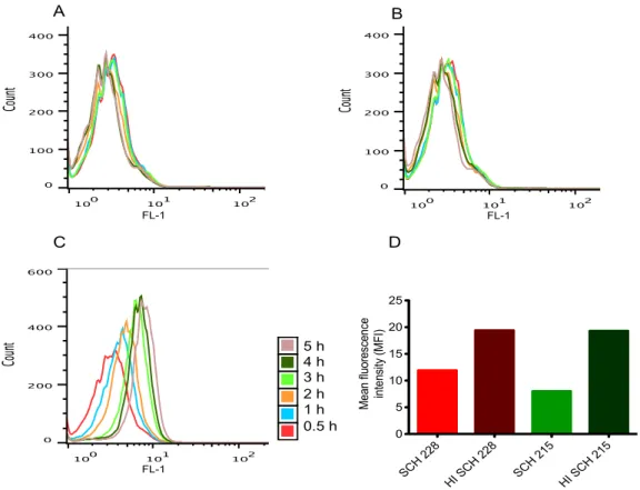
Figure 2 SCH 228 and SCH 215 M. avium inhibit phago-lysosomal processing in human neutrophils.
Human neutrophils were infected with C12-FDG-coated SCH 228 (A) or SCH 215 (B) or heat-inactivated mycobacteria (D) for 15 min at 37°C (ratio 1:20). Neutrophils fed with C12-FDG-coated magnetic beads (C) were used as positive control for this assay. After removal of extracellular mycobacteria, cells were incubated and fluorescence was analyzed by flow cytometry at the indicated time points (color scheme). Phagosomal processing is shown by the shift in the fluorescence over time. Processing of C12-FDG-coated heat-inactivated mycobacteria (HI) vs intact mycobacteria was analyzed after 2 h post-infection and represented as the mean fluorescence intensity (D). Results are representative of three independent experiments.
A B
C
5 h 4 h 3 h 2 h 1 h 0.5 h
D
SCH 228
HI SCH 228
SCH 215
HI SCH 215 0
5 10 15 20 25
Mean fluorescence intensity (MFI)
FL-1 FL-1
FL-1
Virulent mycobacteria are known to reside within the phagosome of infected macrophages by inhibiting phago-lysosomal fusion thereby preventing their degradation (Rohde et al., 2007a; Rohde et al., 2007b; Russell, 2001; Russell et al., 2010b; Sweet et al., 2010). We therefore, asked whether inhibition of phago-lysosomal fusion in SCH 228 and SCH 215- infected human neutrophils could be responsible for the observed bacterial survival (CFU/ml). For this purpose, we employed C12 FDG (5-Dodecanoylaminofluorescein Di-β- D-Galactopyranoside), a substrate for
β-galatosidase enzyme, present in the lysosome, tostudy phagosomal processing using FACS (Robinson et al., 2008). Mycobacteria coated with C12-FDG were used to infect neutrophils and chased for up to 5 h. Magnetic beads coated with C12- FDG served as a positive control in this assay. As shown in Fig. 2C, neutrophils fed with C12-FDG coated magnetic beads showed an increase in fluorescence over time suggesting that the galactopyranoside coating on magnetic beads is cleaved by lysosome resident
β-galatosidase upon phago-lysosome fusion. On the contrary, neutrophils infectedwith C12-FDG coated SCH 228 or SCH 215
M. aviumdid not show such increase in fluorescence indicating that lysosomes did not fuse with the mycobacteria containing phagosomes (Fig. 2A and B). In order to test whether these isolates actively inhibit phago- lysosomal fusion, we used heat-inactivated mycobacteria for this assay. Neutrophils infected with C12-FDG coated heat-inactivated mycobacteria showed increased fluorescence compared to live mycobacteria suggesting that heat inactivated mycobacteria are not able to inhibit phago-lysosomal fusion as compared to live mycobacteria (Fig. 2D). Together, these data show that SCH 228 and SCH 215
M. avium clinical isolates prevent killing in humanneutrophils by actively inhibiting the fusion of lysosomes with the phagosome.
2.2 M. avium SCH 228 and SCH 215 dampen the activation of p38 MAPK in human neutrophils
Clearance of pathogens by neutrophils upon phagocytosis requires efficient degranulation, production of reactive oxygen species (ROS) and phagosomal processing, which is aided by the induction of appropriate cytokines (Sabroe et al., 2005; Sabroe et al., 2003; Segal, 2005;
Winterbourn and Kettle, 2013; Yoshimura and Takahashi, 2007). Activation of p38 MAPK
in neutrophils is important for various functions including degranulation, increased ROS,
cytokine induction, etc. (Lee et al., 1994; Mocsai et al., 2000; Peroval et al., 2013; Thomas and Schroder, 2013; Zu et al., 1996; Zu et al., 1998). Therefore, we analyzed whether these clinical isolates have any influence on the activation of p38 MAPK pathway, by western blotting. The activation of p38 MAPK in infected neutrophils is reduced in the early time points from 15 min till 60 min (Fig. 3). Notably, this reduction in the activation of the pathway is stronger in SCH 228 infected neutrophils than in SCH 215-infected cells. Thus both SCH 228 and SCH 215 M. avium clinical isolates differentially dampen the activation of p38 MAPK signaling pathway.
Figure 3 SCH 228 and SCH 215 dampen the phosphorylation and activation of p38 MAPK in human neutrophils.
Mycobacteria-infected human neutrophils were lysed at the indicated time points and phosphorylation of p38 MAPK was assessed by immuno-blotting using anti-phospho p38 MAPK antibody as described in Materials and Methods.
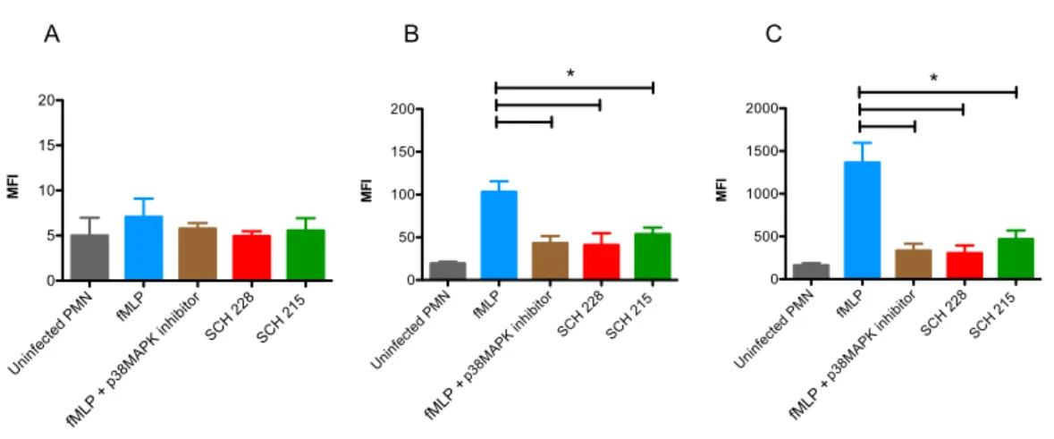
2.3 SCH 228 and SCH 215 M. avium dampen p38 MAPK mediated degranulation in human neutrophils
Killing of intracellular pathogens by neutrophils involves both oxidative and non-oxidative mechanisms. Degranulation, the non-oxidative mechanism is an important feature of neutrophils in the defense of both extra- and intracellular pathogens and is mediated by p38 MAPK activation (Borregaard, 2010; Borregaard et al., 2007; Faurschou and Borregaard, 2003; Prince et al., 2011; Segal, 2005; Thomas and Schroder, 2013; Winterbourn and Kettle, 2013). Expression and fusion of contents from various granules with phagosomes result in intra-phagosomal degradation of pathogens. Therefore, we investigated whether the observed interference of SCH 228 and SCH 215
M. aviumwith the phosphorylation and activation of p38 MAPK impacts on the degranulation process. For this purpose, human
0 15 30 60 120 15 30 60 120 min SCH 215 SCH 228
Phospho- p38 MAPK β-actin
A B
C
5h 4h 3h 2h 1h 0,5h
SCH 228 HI SCH 228
SCH 215 HI SCH 215 0
5 10 15 20 25
Mean fluorescence intensity (MFI)
D
neutrophils were infected with either SCH 228 or SCH 215 and cell surface expression of CD35, CD63, and CD66b was measured for assessing degranulation of secretory, azurophilic, and specific granules, respectively, by flow cytometry. The N-formylated peptide, fMLP, was used as a positive control for the p38 MAPK-mediated degranulation.
Fig. 4 shows the cell surface expression of granule specific markers in human neutrophils at 15 min. fMLP induced cell surface expression of both CD66b and 35 (Fig. 4B and C; blue bar) but not CD63 (Fig. 4A, blue bar). In striking contrast, both isolates dampened the expression of CD66b and 35 markers in infected human neutrophils by
≈66 and 50%,respectively.
This inhibition in degranulation in
M. avium-infected neutrophils was similar to thedegranulation pattern in cells pre-incubated with p38 MAPK inhibitor (SB203580) for 30 min (brown bar) before being stimulated with fMLP indicating the role of p38 MAPK in degranulation. These data show that both SCH 228 and SCH 215 dampen p38 MAPK mediated degranulation in human neutrophils.
Figure 4 SCH 228 and SCH 215 M. avium dampen p38 MAPK mediated degranulation in neutrophils.
Neutrophils were infected with mycobacteria SCH 228 (red) or SCH 215 (green) at a ratio of 1:10 for 15min and infected cells were analyzed for the cell surface expression of granule specific markers CD63 (A), CD35 (B) and CD66b (C) for assessing degranulation of azurophilic, secretory and specific granules, respectively, by flow cytometry. Neutrophils were incubated in the presence of HBSS buffer (control; grey), fMLP (10 µM) for 15 min (blue) or p38 MAPK inhibitor (2 µM) for 30 min prior to stimulation with fMLP for 15 min (brown).
Data represent mean
±
SEM of three independent experiments (*p <0.05).Uninfected PMN fMLP
p38MAPK inhibitor
SCH 228 SCH 215 0
5 10 15 20
MFI
Uninfected PMN fMLP
p38MAPK inhibitor
SCH 228 SCH 215 0
500 1000 1500 2000
*
MFI
Uninfected PMN fMLP
p38MAPK inhibitor
SCH 228 SCH 215 0
50 100 150 200
*
MFI
A B C
fML P +
fMLP +
fMLP +