1 2 3 4 5 6 7 8 9 10 11 12 13 14 15 16 17 18 19 20 21 22 23 24 25 26 27 28 29 30 31 32 33 34 35 36 37 38 39 40 41 42 43 44 45 46 47 48 49 50 51 52 53 54 55 56 57
Long-Lived Triplet Excited State Accessed with Spin–Orbit Charge Transfer Intersystem Crossing in Red Light-
Absorbing Phenoxazine-Styryl BODIPY Electron Donor/Acceptor Dyads
Yu Dong,
[a]Ayhan Elmali,
[b]Jianzhang Zhao,*
[a]Bernhard Dick,*
[c]and Ahmet Karatay*
[b]Orthogonal phenoxazine-styryl BODIPY compact electron do- nor/acceptor dyads were prepared as heavy atom-free triplet photosensitizers (PSs) with strong red light absorption (ɛ=
1.33 × 10
5M
1cm
1at 630 nm), whereas the previously reported triplet photosensitizers based on the spin-orbit charge transfer intersystem crossing (SOCT-ISC) mechanism show absorption in a shorter wavelength range (
<500 nm). More importantly, a long-lived triplet state (τ
T=333
μs) was observed for the newdyads. In comparison, the triplet state lifetime of the same chromophore accessed with the conventional heavy atom effect (HAE) is much shorter (τ
T=1.8
μs). Long triplet statelifetime is beneficial to enhance electron or energy transfer, the
primary photophysical processes in the application of triplet PSs. Our approach is based on SOCT-ISC, without invoking of the HAE, which may shorten the triplet state lifetime. We used bisstyrylBodipy both as the electron acceptor and the visible light-harvesting chromophore, which shows red-light absorp- tion. Femtosecond transient absorption spectra indicated the charge separation (109 ps) and SOCT-ISC (charge recombina- tion, CR; 2.3 ns) for BDP-1. ISC efficiency of BDP-1 was determined as
ΦT=25 % (in toluene). The dyad BDP-3 was used as triplet PS for triplet-triplet annihilation upconversion (upcon- version quantum yield
ΦUC=1.5 %; anti-Stokes shift is 5900 cm
1).
1. Introduction
Triplet photosensitizers (PSs) are compounds showing intersys- tem crossing (ISC) to populate triplet excited state upon photoexcitation. Much attention has been paid to the design of new triplet PSs and for their applications in photo-redox catalytic organic reactions,
[1]photodynamic therapy (PDT),
[2]H
2production by photocatalytic water splitting,
[3]triplet-triplet
annihilation upconversion (TTA-UC),
[4]and photovoltaics.
[5]The triplet state production is via ISC, a spin forbidden non-radiative electronic transition. A traditional strategy of enhancing ISC is to introduce transition metal atoms such as Ru, Ir, Pt or other heavy atoms, such as I or Br.
[6]However, drawbacks of this heavy atom effect (HAE) are the high cost of the synthesis, and the toxicity of the compounds. Moreover, the triplet state lifetime of these PSs is shortened since the HAE enhance not only the ISC of S
1!T
n, but also the T
1!S
0ISC process.
[7]Charge recombination (CR)-induced ISC was known for electron donor/acceptor dyads with a long and rigid linker between donor and acceptor.
[8]A large distance, thus a weak electronic coupling and a small electron exchange energy (J), is indispensable for the radical pair ISC (RP ISC) mechanism in these conventional electron donor/acceptor dyads. This type of ISC is based on the hyperfine interaction enhanced
1CT
!3CT process (CT: charge transfer), followed by
3CT!locally excited triplet state (
3LE) internal conversion (given the purpose is to access the
3LE, not the long-lived CT state). However, these conventional electron donor/acceptor dyads are difficult to prepare, and generally the molecular structures are not optimized for triplet PS preparation, and the visible light- harvesting ability is poor. When the linker between the electron donor and acceptor was reduced in length, for instance by a direct link between donor and acceptor in compact dyads, the electronic coupling and the electron exchange energy increase, and RP ISC is inhibited.
[9]Recently, efficient ISC was observed for the CR in some compact electron donor/acceptor dyads via the so-called spin orbit charge transfer intersystem crossing (SOCT-ISC).
[10]The ISC process requires conservation of the total angular momentum,
[a] Y. Dong, Prof. J. ZhaoState Key Laboratory of Fine Chemicals School of Chemical Engineering Dalian University of Technology
E-208 West Campus, 2 Ling Gong Road, Dalian 116024, China E-mail: zhaojzh@dlut.edu.cn
[b] Dr. A. Elmali, Dr. A. Karatay Department of Engineering Physics Faculty of Engineering
Ankara University
06100 Beşevler, Ankara, Turkey E-mail: Ahmet.Karatay@eng.ankara.edu.tr [c] Prof. B. Dick
Lehrstuhl für Physikalische Chemie
Institut für Physikalische und Theoretische Chemie Universität Regensburg
Universitätsstr. 31, 93053 Regensburg, Germany E-mail: Bernhard.Dick@chemie.uni-regensburg.de
Supporting information for this article is available on the WWW under https://doi.org/10.1002/cphc.202000300
An invited contribution to a Special Collection in Honor of O. Poizat
© 2020 The Authors. Published by Wiley-VCH Verlag GmbH & Co. KGaA.
This is an open access article under the terms of the Creative Commons Attribution Non-Commercial NoDerivs License, which permits use and distribution in any medium, provided the original work is properly cited, the use is non-commercial and no modifications or adaptations are made.
1 2 3 4 5 6 7 8 9 10 11 12 13 14 15 16 17 18 19 20 21 22 23 24 25 26 27 28 29 30 31 32 33 34 35 36 37 38 39 40 41 42 43 44 45 46 47 48 49 50 51 52 53 54 55 56 57
i. e. the sum of orbital angular momentum and spin angular momentum.
[10]Given the electron donor and acceptor moieties in a dyad adopt orthogonal orientation, the change of molecular orbital angular momentum of the CR will offset the change of electron spin angular momentum of ISC.
[10b]There- fore, conservation of angular momentum is satisfied in the ISC of an orthogonal electron donor-acceptor dyad. Hence the SOCT-ISC is efficient in orthogonal electron donor/acceptor dyad. It should be pointed out some electron donor/acceptor dyads undergo twisted intramolecular charge transfer (TICT) may also show the charge recombination induced ISC.
Advantages of these novel compact electron donor/accept- or dyads are their simple molecular structure and long triplet state lifetimes. These features are important for applications of triplet PSs in photocatalysis and PDT. Several chromophores have been used for preparation of electron donor/acceptor compact dyads showing SOCT-ISC, such as acridinium,
[10a]anthracene,
[10b]perylene,
[12]BODIPY,
[10c,13]and perylenemonoi- mide/perylenediimide,
[14]etc. However, triplet PSs based on SOCT-ISC showing red light absorption were rarely reported.
[15]In some cases, long-lived
3CT state was observed for the compact dyad.
[16]On the other hand, although BODIPY-derived triplet PSs showing red light absorption have been reported, for instance the 2,6-diiodostyrylBodipy,
[17]and the 2,6- diiodoazaBodipy,
[18]the triplet state lifetimes of these red light- absorbing triplet PSs are short (~ 1.8
μs), which is a cleardisadvantage for the applications in PDT,
[7b]TTA-UC,
[4d,19]or
photocatalysis, etc.
[20]In these applications, the intermolecular electron transfer or triplet energy transfer efficiency increases with longer triplet state lifetime of the PSs.
Inspired by the previous results, herein we selected styryl BODIPY as the electron acceptor and red-light-absorbing chromophore, and phenoxazine (PXZ) as the electron donor, in order to design compact, orthogonal electron donor/acceptor dyads as novel heavy atom-free triplet PSs showing red light- absorption and long-lived triplet states (Scheme 1). PXZ has been used in thermally activated delayed fluorescence materials (TADF),
[21]and photovoltaics.
[1a]Compared with the previously used phenothiazine (PTZ. oxidation potential
EOX= +0.21 V vs.
Fc/Fc
+), PXZ has a more planar
π-conjugated structure anddifferent redox properties (oxidation potential
EOX= +0.36 V vs.
Fc/Fc
+).
[22]It may provide different solvent polarity-dependency for the SOCT-ISC. To obtain more PSs with red light-absorption, large
π-conjugated carbazole moiety is attached at the 2,6-positions of BODIPY (BDP-3, Scheme 1), which may change the triplet state lifetime or triplet state energy, and finally the ISC efficiency.
The photophysical properties of the dyads were studied by steady-state and time-resolved transient spectroscopies. The CS and CR were studied with femtosecond transient absorption spectra, and the triplet state spectra and lifetimes were studied with nanosecond transient absorption spectra. The new heavy atom-free triplet PSs were used for TTA-UC, and larger anti- Stokes shift was achieved (0.68 eV) than the recently reported
Scheme 1.Synthesis of the Compounds. a)n-C4H9Br, DMF, KOH, N2, stirred at RT for 2 h, yield: 60 %; b) POCl3, DMF, N2, 90°C, 2 h, yield: 65 %; c) 2,4-
dimethylpyrrole, TFA, DDQ, TEA, BF3·Et2O, DCM, RT, N2, yield: 6 %; d) aryl aldehyde,p-toluenesulfonic acid, piperidine, 15 min, yield: 42 % for R=H and 13 % for R=CN; e) NIS, DCM, RT for 2 h, yield: 89 %; f) KI, KIO3, acetic acid, 85°C, 10 min, 39 %; g)n-C4H9Br, DMSO, NaH, N2, stirred at RT for 2 h, yield: 90 %; h) PdCl2(PPh3)2, PPh3, CuI, TEA, TMSA, N2, 80°C, 6 h; then stirred at RT over night, yield: 70 %; i) PdCl2(PPh3)2, PPh3, CuI, TEA and THF, N2, 65°C, 2 h, yield: 60 %.
1 2 3 4 5 6 7 8 9 10 11 12 13 14 15 16 17 18 19 20 21 22 23 24 25 26 27 28 29 30 31 32 33 34 35 36 37 38 39 40 41 42 43 44 45 46 47 48 49 50 51 52 53 54 55 56 57
PXZ-Bodipy dyads showing green light absorption (the anti- Stokes shift is ca. 0.36 eV).
[22]2. Results and Discussion
2.1. Molecular Structure Designing Rationales
Phenoxazine (PXZ) is used as electron donor. Styryl BODIPY is selected as electron acceptor and red-light-absorbing chromo- phore. The cyano groups attached to the styryl BODIPY moiety in BDP-2 will enhance the electron withdrawing ability, which may produce a more efficient charge transfer. The PXZ moiety was attached at the 8-position (meso- position) of styryl BODIPY.
Therefore, the steric hindrance imposed by 1,7-methyl groups on the styryl BODIPY will restrict the rotation of PXZ, thus the dyads will adopt a perpendicular orientation between donor and acceptor. Styryl BODIPY shows strong red light-harvesting ability. The
π-conjugated structure ofBDP-3 may lead to different triplet state properties, such as triplet state lifetime and triplet state energy. All molecular structures were fully characterized (refer to Experimental section and Supporting Information).
A single crystal of BDP-1 was obtained by slow diffusion between
n-hexane and DCM. The molecular structure deter-mined by single crystal X-ray diffraction of BDP-1 is presented in Figure 1. The dihedral angle between the electron donor (PXZ) and the acceptor (styryl BODIPY) is 71.4
°, which is slightly different from the result of DFT calculation (89.7
°), refer to later section. It also shows less orthogonality compared with the reported PTZ-styryl BODIPY dyad ( 81.9
°).
[15]The possible reason is that the better planarity of PXZ compared to PTZ weakens the conformational restriction between the PXZ and styryl BODIPY moieties. The deviation from coplanar geometry of the styryl moieties is 10.9
°and 28.4
°due to the
π-πstacking in the single crystal, because the DFT optimization of the ground state geometry indicated planar geometry. The struc- ture of the styryl BODIPY moiety of BDP-1 is more twisted than
the reported PTZ-styryl BODIPY molecule (10.9
°and 28.4
° vs°
4.7
°and 1.2
°).
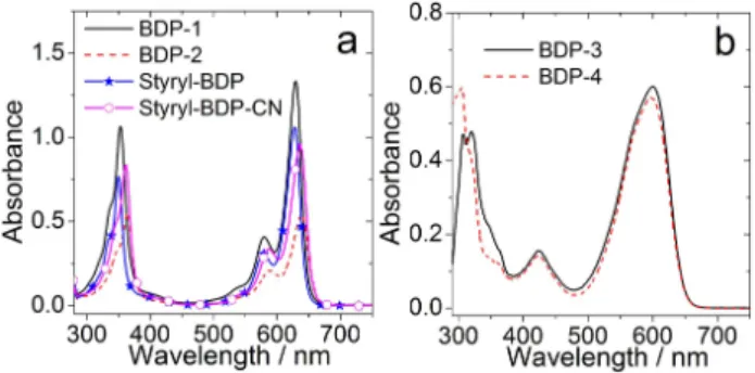
[15]2.2. UV-Vis Absorption and Fluorescence Emission Spectra
The UV-Vis absorption spectra of the compounds were studied (Figure 2). The dyad BDP-1 shows similar absorption compared with the reference Styryl-BDP in the region of 550–700 nm, which indicates negligible electronic coupling between the electron donor (PXZ) and the electron acceptor (styryl BODIPY) at the electronic ground state. A similar result was observed for BDP-2, the absorption is similar as that of Styryl-BDP-CN.
However, the absorption band of BDP-2 is slightly red-shifted compared to BDP-1. The UV-Vis absorption of BDP-3 and BDP-4 were also studied (Figure 2b). For BDP-3, a broad absorption band centered at 600 nm was observed, which shows the same absorption profile as the reference compound BDP-4. We conclude that the electronic coupling between donor and acceptor is also weak at the ground state of BDP-3. These features are similar to those observed for the BDP-PXZ dyads.
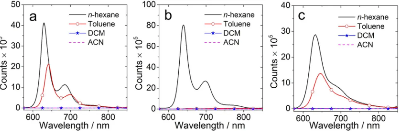
[22]Fluorescence spectra in different solvents were studied (Figure 3). For BDP-1 (Figure 3a), structured emission bands centred at 630 nm and 685 nm were observed, which is similar to the unsubstituted compound Styryl-BDP (refer to the Supporting Information, Figure S11b). The fluorescence quan- tum yield (Φ
F) was determined as 73 % in
n-hexane (Table 1),which is similar to Styryl-BDP (Φ
F=77 %, Table 1). The results indicate that CT is inefficient in a non-polar solvent. However, the fluorescence is significantly quenched in more polar solvents. For instance, the
ΦFin toluene is almost half of that in
n-hexane and it further decreased to 0.2 % in acetonitrile (ACN)(Table 1). We attributed the fluorescence quenching to the electron transfer and formation of CT state, and the CT state is a dark state. Similar results were obtained for the dyad BDP-2 (Figure 3b), but quenching of the fluorescence is more signifi- cant in polar solvents. For instance, the fluorescence quantum yield is 35.4 % in
n-hexane but it decreases to 0.5 % in toluene(Table 1). In toluene, a broad and red-shifted emission band centred at 768 nm was observed, along with the LE emission band at 651 nm (Figure 4a). However, the emission at longer
Figure 1.Single-crystal structure ofBDP-1with 50 % thermal ellipsoids.
Hydrogen atoms are omitted for clarity.
Figure 2.UV-Vis absorption spectra of compounds a)BDP-1,BDP-2,Styryl- BDPandStyryl-BDP-CN; b)BDP-3andBDP-4.c=1.0 × 10⁵M in toluene.
20°C.
1 2 3 4 5 6 7 8 9 10 11 12 13 14 15 16 17 18 19 20 21 22 23 24 25 26 27 28 29 30 31 32 33 34 35 36 37 38 39 40 41 42 43 44 45 46 47 48 49 50 51 52 53 54 55 56 57
wavelength was quenched in polar solvent (Supporting Information, Figure S10b). Considering the relative fluorescence emission intensity and the quantum yields, the CT state of BDP- 2 is in principle also a dark state.
In toluene, the fluorescence is quenched both for BDP-1 and BDP-2 compared with the reference compounds Styryl- BDP and Styryl-BDP-CN. Furthermore, it is further quenched for BDP-2 compared with BDP-1 in toluene (Figure 3a). We attribute the significant quenching of the fluorescence of BDP- 2 as compared to BDP-1 to the electron-withdrawing CN groups, and the more significant CT in BDP-2. For BDP-3, the emission was quenched obviously in polar solvents, indicating an efficient CT. Compared with the reference compound BDP-4, the emission was slightly quenched in non-polar solvents such as
n-hexane and toluene (Figure 4b and Table 1). However, theemission was quenched further in polar solvents, for instance,
Figure 3.Fluorescence emission spectra of the compounds in different solvents. a)BDP-1; b)BDP-2, and c)BDP-3. Optically matched solutions were used, i. e.all the sample solution show the same absorbance at the excitation wavelength (A=0.195),λex=570 nm, 20°C.
Table 1. The photophysical properties of compounds.
Solvent[a] λabs[b](ɛ[c]) λF[d] τF[e] ΦF[f] ΦΔ[g] τT[h] ΦT[i]
BDP-1 HEX 620 (1.40) 629 5.4 0.732 –[l] –[l] –[l]
TOL 630 (1.33) 641 4.9 0.393 0.23 333.2[k] 0.25
ACN 619 (1.23) 631 2.9 0.002 –[l] –[l] –[l]
BDP-2 HEX 629 (0.57) 640 4.3 0.354 0.05 –[l] –[l]
TOL 639 (0.54) 651/768[j] 2.6/2.3[j] 0.005/0.018[j] 0.18 382.2[k] 0.22
ACN 628 (0.53) 625 1.5 0.003 –[l] –[l] –[l]
BDP-3 HEX 597 (0.67) 633 3.0 0.484 0.04 394.4[k] 0.04
TOL 600 (0.60) 647 2.4 0.299 0.12 392.7[k] 0.13
ACN 590 (0.60) 629 1.4 0.003 –[l] –[l] –[l]
BDP-4 HEX 596 (0.66) 630 2.9 0.462 0.03 278.8[k] 0.05
TOL 597 (0.57) 641 1.9 0.360 0.04 244.0 0.04
ACN 587 (0.60) 631 0.07 0.022 –[l] –[l] –[l]
Styryl-BDP HEX 619 (1.15) 630 5.2 0.768 –[l] –[l] –[l]
TOL 628 (1.06) 641 4,7 0.768 –[l] –[l] –[l]
ACN 617 (1.04) 631 5.2 0.763 –[l] –[l] –[l]
Styryl-BDP-CN HEX 627 (0.78) –[l] –[l] –[l] –[l] –[l] –[l]
TOL 636 (0.97) 649 4.2 0.652 –[l] –[l] –[l]
ACN 627 (0.96) 639 4.4 0.612 –[l] –[l] –[l]
[a]ET(30) values are HEX (31.0), TOL (33.9) and ACN (45.6), in kcal mol 1. [b]c=1.0 × 10⁵M, in nm. [c] Molar absorption coefficient.ɛ=105M1cm1. [d]
Fluorescence wavelength, in nm. [e] Fluorescence lifetime,λex=635 nm, in ns,c=1.0 × 10⁵M. [f] Absolute fluorescence quantum yield. [g] Singlet oxygen quantum yield, methylene blue (MB)as standard (ΦΔ=0.57 in DCM). [h]Triplet state lifetime, inμs. [i] Triplet quantum yield, methylene blue (MB)as standard (ΦT=0.50 in methanol). [j] The transition of1CT!S0. [k] Intrinsic triplet state lifetimes. Obtained by fitting of the experimental curves based on the kinetic model with triplet-triplet-annihilation self-quenching effect considered.[22][l] Not observed.
Figure 4.Comparison of the fluorescence emission spectra of the com- pounds in toluene: a)BDP-1,BDP-2,Styryl-BDPandStyryl-BDP-CN; b)BDP- 3andBDP-4. Optically matched solutions were used (A=0.195),
λex=570 nm, 20°C.
1 2 3 4 5 6 7 8 9 10 11 12 13 14 15 16 17 18 19 20 21 22 23 24 25 26 27 28 29 30 31 32 33 34 35 36 37 38 39 40 41 42 43 44 45 46 47 48 49 50 51 52 53 54 55 56 57
ΦF
is 0.001 and 0.105 in DCM for BDP-3 and BDP-4, respectively (Supporting Information, Table S2).
The fluorescence decay traces were studied using the time- correlated single-photon counting (TCSPC) detection method (Figure 5 and Supporting Information, Figure S12). The fluorescence lifetimes of BDP-1 show solvent polarity depend- ency (Figure 4a). In non-polar solvents (n-hexane and toluene), the decay trace is mono-exponential. However, it shows a biexponential decay in polar solvents, such as ACN. The average lifetimes decrease from 5.4 ns (in
n-hexane), to 2.9 ns (a shortcomponent of 0.06 ns with a population ratio of 30 % and a longer component of 4.2 ns, in 70 %) in ACN, meanwhile the fluorescence quantum yield decreased by 366-fold (Figure 3a and Table 1). Similar results are observed for BDP-2 (Figure 5b and Table 1). The decay kinetics of BDP-2 shows a sharper decrease along with increasing solvent polarity (Figure 5b). The lifetimes are also shorter than BDP-1 in the same solvent (Table 1). For instance, the lifetime of BDP-2 shows a bi- exponential decay with a short component of 0.12 ns (60 %) and a longer component of 3.5 ns (40 %). The fluorescence lifetime of BDP-1 and BDP-2 are 4.9 ns and 2.6 ns in toluene, respectively, which indicates that the CS of BDP-2 is more efficient than that of BDP-1. We propose the bi-exponential decay is attributed to the existence of an equilibrium between the emissive state with a dark state (CT state), or electron transfer probability for the molecules at the
1LE state, and the probability is less than unity.
[6b]For BDP-3, a similar conclusion can be obtained (Supporting Information, Figure S12a). The
fluorescence in
n-hexane is mono-exponential with a lifetime of3.0 ns. However, in ACN it turned to a bi-exponential decay with an average lifetime of 1.4 ns (with a short component of 0.10 ns (70 %) and a longer component of 4.4 ns (30 %)), indicating the fluorescence quenched further in polar solvents.
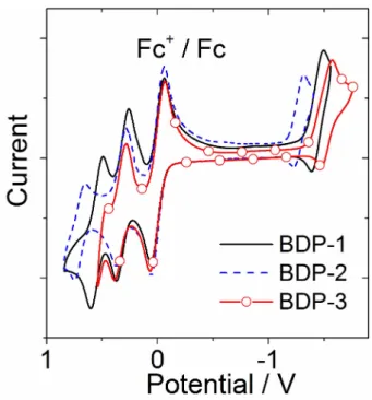
2.3. Electrochemical Studies
The electrochemical properties of the dyads were studied by cyclic voltammetry (Figure 6, Table 2, Supporting Information, Figure S22 and Table S4). For BDP-1, the reversible oxidation wave at
+0.32 V (vs Fc/Fc
+) is attributed to the PXZ moiety, another quasi-reversible oxidation wave at
+0.55 V is attributed to the styryl BODIPY moiety, as is the quasi-reversible reduction wave at 1.44 V. Therefore, PXZ is more likely the electron
Figure 5.Fluorescence decay traces of the compounds in different solvents.a)BDP-1at 650 nm and b)BDP-2at 650 nm.λex=635 nm,c=1.0 × 10⁵M.
20°C. The IRF curves of the spectrometer are also presented.
Figure 6.Cyclic voltammogram ofBDP-1,BDP-2andBDP-3. Ferrocene (Fc) was used as internal reference. In deaerated DCM containing 0.10 M Bu4N [PF6] as supporting electrolyte. Scan rates: 100 mV/s.c=1.0 × 103M, 20°C.
Table 2. Redox potentials, driving forces of charge separation (ΔGCS), charge recombination (ΔGCR), and the energy of the CSS of the compounds (ECSS) in different solvents.[a]
E(ox)[e][V] E(red)[e][V] ΔGCS[eV][f] ECSS[eV]
HEX TOL DCM ACN HEX TOL DCM ACN
BDP-1[b] +0.55 1.44 0.09 0.03 0.36 0.44 2.05 1.93 1.60 1.51
+0.32
BDP-2[c] +0.70 1.26 0.004 0.13 0.47 0.57 1.93 1.80 1.45 1.35
+0.33
BDP-3[d] +0.34 1.52 0.25 0.29 0.36 0.38 1.73 1.70 1.62 1.59
[a] Cyclic voltammetry in N2saturated DCM containing a 0.10 M Bu4NPF6supporting electrolyte; Pt electrode was used as counter electrode; working electrode is glassy carbon electrode; Ag/AgCl couple as the reference electrode.E00is the energy difference between the potential minima, approximated with the crossing point of UV-Vis absorption and fluorescence emission spectra after normalization. [b]E00=1.96 eV. [c]E00=1.92 eV. [d]E00=1.98 eV. [e] The value was obtained by setting the oxidation potential of Fc+/Fc as 0. [f] The redox potentials are approximated based on the redox potential measured in DCM.
1 2 3 4 5 6 7 8 9 10 11 12 13 14 15 16 17 18 19 20 21 22 23 24 25 26 27 28 29 30 31 32 33 34 35 36 37 38 39 40 41 42 43 44 45 46 47 48 49 50 51 52 53 54 55 56 57
donor and styryl BODIPY serves as electron acceptor. The reduction wave of BDP-2 was observed at 1.26 V, indicating the stronger electron accepting ability of the styryl BODIPY moiety with cyano groups attached, as compared with styryl BODIPY. Similar results were obtained for BDP-3. Only one reversible oxidation wave at
+0.34 V and one quasi-reversible reduction wave at 1.52 V were observed, indicating that PXZ serves as electron donor and the BODIPY moiety serves as electron acceptor. Compared with the previously reported BDP- PXZ dyads, the oxidation potential of PXZ is similar (0.32 V vs 0.36 V, vs Fc/Fc
+).
[6b]The reduction potentials of the styryl BODIPY moieties of BDP-1 ( 1.44 V) and BDP-2 ( 1.26 V) moves anodically than that of the BDP-PXZ dyad ( 1.65 V, vs Fc/Fc
+), indicating the styryl BODIPY moieties are stronger electron acceptors than the parent BODIPY.
The Weller equations (Supporting Information, Equations S1–S4) were used to determine the energy of the charge separated state (CSS) and the driving forces of intramolecular charge separation (ΔG
CS).
[8c]The results are summarized in Table 2 (For detailed information please refer to the Supporting Information).
The calculation of
ΔGCSindicates that charge separation is thermodynamically forbidden in
n-hexane due to the positive ΔGCSvalue (
+0.09 eV) for BDP-1, which agrees with the unquenched fluorescence of styryl BODIPY moiety in
n-hexane(Φ
F=73.2 % in Table 1). However, thermodynamically allowed charge separation is possible in other more polar solvents according to the negative
ΔGCSvalues of BDP-1, which also
agrees with fluorescence quenching of the styryl BODIPY moiety (Table 1). For BDP-2 and BDP-3, the
ΔGCSvalues are all negative, indicating charge separation is thermodynamically allowed in both nonpolar and polar solvents. For the previously reported BDP-PXZ dyads, the
ΔGCSvalues are all more negative.
[22]For instance, the
ΔGCSvalues are 0.40 eV and 0.74 eV in toluene and ACN, respectively. For BDP-1, the
ΔGCSis 0.44 eV in ACN, indicating the CS driving force is weaker for BDP-1. It may reduce the CS and SOCT-ISC efficiency.
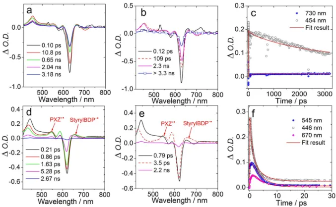
2.4. Femtosecond Transient Absorption Spectroscopy
Femtosecond transient absorption spectra (fs TA) were meas- ured in order to reveal the excited state dynamics of the dyads.
For BDP-1, the triplet state signal was observed in toluene (Figure 7), but no triplet state signal was observed in ACN.
Hence we assume that the CS and CR are both much faster in ACN. In toluene, the intense ground state blenching (GSB) band at 630 nm was immediately generated upon excitation (Fig- ure 7a). An excited state absorption (ESA) band in the range of 420–550 nm increased in intensity in less than 1 ps. This ESA band is attributed to the S
1!S
ntransition, and the increasing process may be due to vibrational relaxation. The negative band at 700 nm is assigned to stimulated emission (SE) of the localized singlet state (
1StyrylBDP*). However, no obvious absorption band of the PXZ radical cation (around 540 nm, Supporting Information, Figure S23b)
[23]and the styryl BODIPY
Figure 7.Femtosecond transient absorption spectra ofBDP-1. a) Transient absorption spectra in toluene.λex=630 nm,c=1.0 × 10⁵M, b) species-associated difference spectra (SADS), and c) decay traces at selected wavelengths. d) Transient absorption spectra ofBDP-1in ACN (λex=625 nm,c=1 × 10⁵M), e) species-associated difference spectra (SADS), and f) Decay traces at selected wavelengths. SADS were obtained by global fitting in sequential model. 20°C.
1 2 3 4 5 6 7 8 9 10 11 12 13 14 15 16 17 18 19 20 21 22 23 24 25 26 27 28 29 30 31 32 33 34 35 36 37 38 39 40 41 42 43 44 45 46 47 48 49 50 51 52 53 54 55 56 57
radical anion (around 670 nm, Supporting Information, Fig- ure S23a) were observed.
One possible reason is that the CS efficiency is not very high (Φ
F=0.39 in toluene). Furthermore, the radical anion absorp- tion of styryl-BDP in the range of 650–680 nm (Supporting Information, Figure S23a and S23b, obtained by spectroelec- trochemistry), overlaps with ESA signals of the singlet state of Styryl-BDP (
’Figure 7a and Supporting Information,
’Figure S21).
Moreover, the signal of the radical cation of PXZ also overlaps with the singlet state signal of styryl-BDP in the range of 530–
550 nm (
’Figure 7a and Supporting Information,
’Figure S21).
Thus, we can’t distinguish the characteristic
1styryl-BDP* and the
1CSS states unambiguously. The fs TA spectra of the reference compound Styryl-BDP were also recorded in toluene (Supporting Information, Figure S21). No fast decay of the GSB or the SE bands were observed, indicating that no CS exist in Styryl-BDP. Therefore, the CS is most likely responsible for the fast decay of BDP-1 in toluene.
Species-associated difference spectra (SADS) obtained by global fitting were used to analyse the photophysical processes (Figure 7b). The species with the shortest lifetime (0.12 ps) is assigned to the unrelaxed S
1state. The species with a lifetime of 109 ps displays the characteristic styryl BODIPY ESA signals (Supporting Information,
’Figure S21) and is assigned the relaxed S
1state, indicating that CS process takes 109 ps.
Subsequently, the slow CR takes 2.3 ns, and a long-lived species with infinite lifetime (on the time scale of the fs-TA experiment) is obtained, which is attributed to the triplet state. The final species is assigned as the T
1state on account of showing a weak ESA band in the range 650-750 nm (T
1!T
nabsorption) and a GSB band at 630 nm, which is in agreement with ns TA data (Figure 8a). Therefore, we conclude that the triplet state is generated by the CR and the SOCT-ISC (CR) takes 2.3 ns.
In ACN, no triplet state signal was observed (Figure 7d). The ESA band located in the range of 440–570 nm is attributed to the S
1!S
nabsorption. Due to the higher CS efficiency, absorption bands of the styryl-BDP radical anion and the PXZ radical cation were more obvious. The absorption bands centered at 670 nm and 545 nm were attributed to styryl-BDP
*and PXZ
+*, respectively, which is in agreement with the results of spectroelectrochemical studies (refer to the Supporting Information, Figure S23a and S23b). Based on the SADS, we determined the time constants of the CS and CR as 0.79 ps and 3.5 ps, respectively (Figure 7e), which are faster than the CS (109 ps) and CR (2.2 ns) in toluene.
For BDP-2, the fluorescence was quenched significantly in toluene (Φ
F=0.005 vs. 0.393 of BDP-1). In toluene, a GSB band centered at 640 nm and a strong ESA band centered at 468 nm (Supporting Information, Figure S19a) were observed, which are assigned to the S
1state. Subsequently, an absorption band at 670 nm intensified along with the decreasing ESA band at 469 nm, which is attributed to the formation of [StyrylBDP- CN]
*(Supporting Information, Figure S23c). Finally, along with the decreasing of the absorption band centered at 670 nm, the ESA signal in the range of 650-750 nm become stronger, which is characteristic for the T
1!Tnabsorption of BDP-2, corrobo- rated by the ns TA spectra (Supporting Information, Fig-
ure S13a). Therefore, we conclude that ISC occurs via CR. Based on the results from SADS (Supporting Information, Figure S19b), we determine that CS takes place in 1.2 ps, which is much faster than in BDP-1 (109 ps in toluene). Following the CS, slow CR (SOCT-ISC) continuing for 1.6 ns leads to the generation of the triplet state of BDP-2. In the polar solvent ACN, results similar to those of BDP-1 were obtained (Supporting Information, Fig- ure S19e). Faster CS (0.3 ps) and CR (1.6 ps) are observed. No triplet state formation was observed, which agrees with the lack of singlet oxygen photosensitizing of the dyad BDP-2 (Table 1).
Different results were obtained for BDP-3 (Supporting Information, Figure S19). Upon excitation, the GSB band was observed in the range of 550–750 nm. It is broader than the UV- Vis absorption, which is attributed to the overlap of the GSB and the SE band. According to spectroelectrochemical results (Supporting Information, Figure S24), we determine the absorp- tion of the radical anion and the radical cation centered at 580 nm and 540 nm, respectively. Based on the SADS analysis and the fast decay of ESA at 480 nm, we determine that CS takes 3.2 ps in toluene. The lifetime of the
1CT state in toluene is ca. 4.0 ns. No triplet state formation was observed within the time resolution of our instrument setup.
2.5. Nanosecond Transient Absorption Spectroscopy: Triplet State Properties
Nanosecond transient absorption spectra were used to study
the triplet state production of the dyads (Figure 8 and
Figure 8.Nanosecond transient absorption spectra of compounds in de- aerated toluene. a) Transient absorption spectra ofBDP-1at different delay time. b) The decay trace at 630 nm,λex=620 nm,c=5.0 × 10⁶M. c) Transient absorption spectra ofBDP-3and d) the decay trace at 600 nm, λex=590 nm,c=1.0 × 10⁵M, 20°C. The intrinsic lifetime was obtained by fitting of the decay curves with a kinetic model that takes into account of the triplet-triplet-annihilation effect.[22]1 2 3 4 5 6 7 8 9 10 11 12 13 14 15 16 17 18 19 20 21 22 23 24 25 26 27 28 29 30 31 32 33 34 35 36 37 38 39 40 41 42 43 44 45 46 47 48 49 50 51 52 53 54 55 56 57
Supporting Information, Figure S13). For BDP-1, two negative peaks centered at 340 nm and 630 nm were observed upon pulsed laser excitation. In agreement with the UV-Vis absorption spectra (Figure 2a), these bands are assigned as GSB bands.
Moreover, several ESA bands centred at 380 nm, 500 nm and 700 nm, respectively, were observed (Figure 8a). These features are typical of the styryl BODIPY triplet state transient absorption spectra.
[24]Therefore, we conclude that the triplet state is localized on the styryl BODIPY moiety, and it is not a CT state.
Similar results were obtained for BDP-2 (In toluene, Supporting Information, Figure S13a). The GSB and ESA bands of BDP-2 are both slightly red shifted compared with BDP-1 due to the extension of
π-conjugation structure. Recently with a PTZ-naphthalimide dyad, we observed a CT state.
[16]Note the ESA bands overlap with the GSB bands.
The apparent triplet state lifetimes (τ
T) were determined as 270.0
μs forBDP-1 and 242.7
μs forBDP-2, respectively, at a specific concentration. However, triplet-triplet annihilation (TTA) will quench the triplet state and shorten the triplet state lifetime, especially for the compounds showing strong absorp- tion at the excitation wavelength, high ISC efficiency and long- lived triplet state. Therefore, the intrinsic triplet state lifetime was determined by fitting the decay traces at two different concentrations with a kinetic model including TTA self- quenching.
[25]The intrinsic triplet state lifetime of BDP-1 obtained with this kinetic model is
τT=333.2
μs, which is muchlonger than the apparent triplet state lifetime
τT=270.0
μs,indicating TTA quenching. For BDP-2, the intrinsic triplet state lifetime (τ
T=382.2
μs) is also longer than the experimentalvalues (242.7
μs). Notably the triplet state lifetime ofBDP-1 is prolonged 185-fold (τ
T=333.2
μs) as compared to the tripletstate of the same parent chromophore, i. e. bisstyryl-BODIPY, but accessed by the HAE in 2,6-diiodostyryl BODIPY (1.8
μs).[17]These results demonstrated one of the advantage of using the compact electron donor/acceptor dyads as heavy atom-free triplet PSs, i. e. the triplet state lifetime becomes much longer than that accessed with the conventional HAE.
[15]Long-lived triplet state lifetime are important for photocatalysis, PDT and TTA-UC.
The ISC efficiency depends on the solvent polarity (Table 1).
For instance, the triplet state quantum yield (Φ
T) for BDP-1 is 25 % in toluene (Table 1). However, in other solvents, no triplet state formation was observed. Similar results were observed for BDP-2. Therefore, we conclude that the ISC mechanism is based on charge recombination for BDP-1 and BDP-2. Due to the short distance between electron donor/acceptor and strong electron coupling, SOCT-ISC is the most likely mechanism, instead of RP ISC. The poor SOCT-ISC in polar solvent may be due to the fast CR to the ground state (S
0state), because the CR occurs normally in the Marcus inverted region, i. e. the lower CT state energy in polar solvent will accelerate CR to the S
0state, thus inhibiting SOCT-ISC.
[6b]For BDP-3, the characteristic GSB and ESA signals are similar as the reference compound BDP-4 (Figure 8c and Supporting Information, Figure S13c), which demonstrates that the triplet state is also localized on the BODIPY moiety. The intrinsic triplet state lifetime was determined as 392.7
μs in toluene (Table 1).Theoretical computations were performed to study the ESA bands of T
1!T
ntransitions (Supporting Information, Fig- ure S18). For BDP-1, the ESA bands centered at 700 nm, 500 nm and 375 nm are attributed to the T
1!T
4, T
1!T
10and T
1!T
22transitions, respectively (Supporting Information, Figure S18a).
Similar results were obtained for BDP-2. The ESA band at 735 nm is attributed to the T
1!T
4transition and the bands at 500 nm and 395 nm were assigned to the T
1!T
10and T
1!T
22transitions, respectively (Supporting Information, Figure S18b).
The results of the calculations for BDP-3 deviated from the experimental results (Supporting Information, Figure S18c).
However, strong overlap between ESA and GSB bands can shift the apparent band maxima considerably. The ESA band at around 750 nm is assigned to the T
1!T
10transition, the other two bands at around 460 nm and 380 nm were assigned to the T
1!T
12and T
1!T
26transitions, respectively.
Triplet-triplet energy transfer (TTET) was used to determine the triplet state energy of the dyads. BDP-1 and BDP-2 were used as triplet energy donors. Rubrene (E
T1=1.14 eV)
[26]and 1- chloro-9,10-bis(phenylethynyl)anthracene (CBPEA.
ET1=1.20 eV)
[4c]were selected as triplet energy acceptor. The triplet state lifetime of BDP-1 wasn’t quenched (260.2
μs) in thepresence of 4 eq. CBPEA compared with that in the absence of CBPEA (272.3
μs) (Supporting Information, Figure S14b andS14d), which indicates the T
1state energy of BDP-1 is lower than that of CBPEA. In contrast, the triplet state lifetime of BDP- 1 was significantly quenched in the presence of 4 eq. rubrene (37.0
μs), indicating that the T1state energy of BDP-1 is higher than 1.14 eV. Therefore, we estimate that the T
1state energy of BDP-1 is in the range of 1.14 ~ 1.20 eV. A similar result was obtained for BDP-2 (Supporting Information, Figure S15), the T
1state energy of BDP-2 is in the range 1.14 ~ 1.20 eV. The results are also similar to those obtained by DFT calculation. The calculated T
1state energies are 1.06 eV and 1.03 eV for BDP-1 and BDP-2, respectively.
Due to the different
π-conjugated structure ofBDP-3 compared with styryl BODIPY (Scheme 1), we assume the T
1state energy of BDP-3 is different from BDP-1 and BDP-2. We
selected BODIPY (E
T1=1.69 eV)
[27]and 9,10-diphenylanthracene
(DPA) (E
T1=1.77 eV)
[28]as triplet energy acceptors and BDP-3 as
the energy donor. The triplet state lifetime of BDP-3 decreased
from 402.0
μs to 339.1μs in the presence of BODIPY (Support-ing Information, Figure S16). Meanwhile, a new GSB band
appeared at around 500 nm, which is assigned to the GSB band
of BODIPY. The decay at 500 nm is composed of two
components, the increasing component is attributed TTET
between BDP-3 and BODIPY. These results demonstrate that
the T
1state energy of BDP-3 is higher than 1.65 eV. On the
contrary, no reduction of the triplet state lifetime of BDP-3 was
observed when DPA was used as the energy acceptor
(Supporting Information, Figure S17). Therefore, we conclude
that the T
1state energy of BDP-3 is in the range of 1.65 ~
1.77 eV.
1 2 3 4 5 6 7 8 9 10 11 12 13 14 15 16 17 18 19 20 21 22 23 24 25 26 27 28 29 30 31 32 33 34 35 36 37 38 39 40 41 42 43 44 45 46 47 48 49 50 51 52 53 54 55 56 57
2.6. DFT Computations
The ground-state geometries of the dyads were optimized (Figure 9). The relative orientations between electron donor (PXZ) and acceptor (BODIPY chromophore) are all nearly orthogonal for all the dyads, which should be beneficial for SOCT-ISC. For instance, the dihedral angle between PXZ and styryl BODIPY in BDP-1 is 89.7
°, very close to orthogonal geometry. The
π-conjugated structure of styryl BODIPY moietyof BDP-1 shows minor distortion (by 1.8
°and 1.7
°at the two arms, respectively) (Figure 9a). These distortions are smaller than those observed in the single crystal structure (10.9
°and 28.4
°), the discrepancy may be due to the packing effect in the single crystal. The previously reported SOCT-ISC dyad BDP-PXZ has a similar dihedral angle between PXZ and BODIPY as found for BDP-1 (85.6
°vs 89.7
°).
[22]For BDP-2, the relative orientations between the PXZ and the styryl BODIPY moieties are also orthogonal, and the distortions of the styryl moieties are 1.6
°and 1.5
°(
’Figure 9).
However, for BDP-3, the BODIPY chromophore moiety has better planarity (Supporting Information, Figure S25). The potential energy surfaces (PES) of the dyads against the torsional angles between electron donor and acceptor were also constructed (Supporting Information, Figure S26). For the three dyads, the thermally accessible dihedral angles between the electron donor and acceptor are all in the range of 65
°~ 113
°. The range is similar (ca. 70
°~ 110
°) for the reported PTZ- Styryl BODIPY dyads.
[15]The frontier molecular orbitals of the dyads are presented in Figure 10. For BDP-1, the lowest unoccupied molecular orbital (LUMO) is confined to the styryl BODIPY moiety, and the highest occupied molecular orbital (HOMO) is localized on the PXZ moiety, indicating that electron transfer is possible. Slight delocalization was observed. For BDP-2, the HOMO and LUMO are exclusively localized on the PXZ and styryl BODIPY moieties, respectively. For BDP-3, a similar result was observed. The MOs demonstrate that the attachment of electron withdrawing groups may alter the HOMO and LUMO energies. Compared
with BDP-1, the HOMO energy of BDP-2 decreases from 4.90 eV to 5.02 eV and the LUMO energy shows a similar change (from 2.69 eV to 3.09 eV). The lack of overlap of the MO leads to more significant fluorescence quenching in BDP-2.
The triplet state spin density surfaces were studied at the optimized triplet state geometries (Figure 11). The spin un- paired electrons are localized on the styryl BODIPY moiety for BDP-1 and BDP-2, which agrees with nanosecond transient absorption spectra (Figure 8). The spin density surfaces of the radical anion and the radical cation of BDP-1 were also studied (Figure 12). The spin density of the radical anion is entirely restricted to the styryl BODIPY moiety. On the contrary, the spin density surface of the radical cation is completely localized on the PXZ moiety.
These results further imply that PXZ serves as electron donor and styryl BODIPY as the electron acceptor. Similar results were obtained for BDP-2 and BDP-3 (Supporting Information, Figure S29).
2.7. Application of the Dyads in TTA Upconversion
Recently, the application of heavy atom free triplet PSs on triplet-triplet annihilation upconversion (TTA-UC) has attracted particular interest.
[29]Traditional heavy atom-free triplet PSs with absorption in the red range show low triplet energies, such as methylene blue (1.44 eV)
[30]and 2,6-diiodostyryl BODIPY (ca. 1.13 eV).
[31]Due to the low triplet state energy of BDP-1 (1.14 ~ 1.20 eV) and BDP-2 (1.14 ~ 1.20 eV), the dyad BDP-3 was selected as the triplet PS of TTA-UC. The dyad BDP-3 has high triplet state energy (1.65 ~ 1.77 eV) and long intrinsic triplet state lifetime (392.7
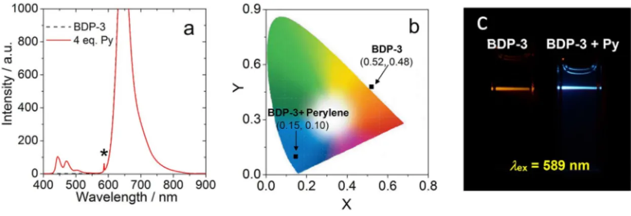
μs in toluene), which is ideal for applicationfor TTA-UC. TTA-UC was studied with BDP-3 as PS and perylene as annihilator (Figure 13). Upon 589 nm cw laser excitation, only fluorescence of BDP-3 was observed in absence of annihilator.
A new emission peak appear in the region of 420–520 nm after addition of 4.0 eq. annihilator, which is attributed to the upconverted fluorescence of perylene (Figure 13a). The anti- Stokes shift of this TTA-UC system is 5905 cm
1, and the upconversion quantum yield (Φ
UC) is 1.5 % (in toluene). The anti-Stokes shift of BDP-3 is comparable to the previously reported SOCT-ISC PSs applied in TTA-UC (ca. 3276–
5900 cm
1).
[13a,22,32]The power dependence of the upconversion emission intensity was measured (Supporting Information, Figure S31).
The integrated upconversion emission intensity increased al- most linearly rather than quadratically with the excitation laser power, indicating efficient TTET and TTA for the TTA-UC system.
Finally, the intermolecular TTET was studied by Stern- Volmer analysis of the quenching by monitoring the triplet state lifetime of BDP-3 (Supporting Information, Figure S32 and Table S5). The Stern-Volmer quenching constant (K
SV=1.74 × 10
6M
1) and quenching efficiency (f
Q=40.0 %), indicating that intermolecular TTET is efficient.
The photophysical processes of BDP-1 are summarized in Scheme 2. Upon excitation at 630 nm, the dyad is excited to the S
1state (LE state localized on styryl BODIPY moiety). The
Figure 9.Optimized ground state geometry and selected dihedral angles ofBDP-1andBDP-2calculated by DFT at the B3LYP/6-31G(d) level with Gaussian 09W.
1 2 3 4 5 6 7 8 9 10 11 12 13 14 15 16 17 18 19 20 21 22 23 24 25 26 27 28 29 30 31 32 33 34 35 36 37 38 39 40 41 42 43 44 45 46 47 48 49 50 51 52 53 54 55 56 57
following CS leads to generation of a CS state, especially in polar solvents.
ΔGCSis positive and therefore CS is inhibited in
n-hexane, resulting in unquenched fluorescence of the styrylBODIPY moiety (Table 1). In toluene, the fluorescence of the LE state is quenched by CS (rate constant 109 ps), subsequent CR
leads to population of T
1(
3LE state). According to DFT
calculations, SOCT-ISC to yield T
3is also possible because the
two states have similar energies,
[10b]Ultrafast T
3!T1internal
conversion will nevertheless populate T
1. The CR (ISC) rate
constant was determined as 2.3 ns by femtosecond transient
Figure 10.Selected frontier molecular orbitals and energies (in eV) of the compounds calculated by DFT at the B3LYP/6-31G(d) level with Gaussian 09W, based on the optimized ground state structures. Isovalue=0.02.1 2 3 4 5 6 7 8 9 10 11 12 13 14 15 16 17 18 19 20 21 22 23 24 25 26 27 28 29 30 31 32 33 34 35 36 37 38 39 40 41 42 43 44 45 46 47 48 49 50 51 52 53 54 55 56 57
absorption spectra. The triplet quantum yield
ΦTis 25 % in toluene. The energies of
1CSS in more polar solvents are much lower (1.60 eV in DCM and 1.51 eV in ACN, respectively), which may facilitate the CR back to the ground state, because the CR is normally in Marcus inverted region of electron transfer.
[6b]For instance, CR is much faster in ACN (3.5 ps) than in toluene (2.3 ns). Therefore, no triplet state signal was observed in DCM and ACN.
Similar photophysical processes are summarized for BDP-2 (Supporting Information, Figure S33). CS occurs within 3.4 ps.
The triplet state is confined to the styryl BODIPY moiety. It is produced by SOCT-ISC, with time constant of 1.4 ns (in toluene), and triplet state quantum yield
ΦT=22 % (in toluene). For the dyad BDP-3, fast CS (3.2 ps) was observed, as well as a slow CR (4.0 ns). However, no triplet state signal was observed in femtosecond transient absorption spectra, due to the slow CR (SOCT-ISC) kinetics.
3. Conclusions
In summary, we prepared a series of phenoxazine-styryl BODIPY compact electron donor/acceptor dyads as novel heavy atom- free triplet photosensitizers (PSs), based on the newly devel- oped spin-orbit charge transfer intersystem crossing (SOCT-ISC) strategy. The striking property of the new triplet PSs is the strong absorption of red light (ɛ
=1.33 × 10
5M
1cm
1at 630 nm), and the long triplet state lifetime (τ
T=333
μs),prolonged by a factor of 180 compared to the triplet state of the same styryl BODIPY chromophore but accessed by the conventional heavy atom effect (τ
T=1.8
μs). Femtosecondtransient absorption spectra show that the charge separation and SOCT-ISC (charge recombination) take 109 ps and 2.3 ns, respectively. The triplet state energies of the dyads BDP-1 and BDP-2 are 1.14 ~ 1.20 eV and BDP-3 is 1.65 ~ 1.77 eV, deter- mined by triplet energy transfer studies. The dyad BDP-3 was used as triplet PSs for triplet-triplet annihilation upconversion (upconversion quantum yield
ΦT=1.5 % in toluene; anti-Stokes shift is 5905 cm
1). Our results are useful for the design of novel heavy atom-free triplet PSs showing red light-absorption and more importantly long-lived triplet states. These triplet PSs are useful for photo-redox catalytic organic reaction, photodynamic therapy, and triplet-triplet-annihilation photon upconversion.
Figure 11.Isosurfaces of spin density of a)BDP-1and b)BDP-2at the optimized triplet state geometries. Calculation was performed at the UB3LYP/6-31G(d) level with Gaussian 09W. Isovalue=0.0004.
Figure 12.Isosurfaces of spin density ofBDP-1at the optimized a) radical anion and b) radical cation geometries. Isovalue=0.0004. The calculation was performed at the UB3LYP/6-31G(d) level with Gaussian 09W.
Figure 13.TTA upconversion withBDP-3as the triplet PS and perylene as the acceptor,λex=589 nm.ΦUC=1.5 %. a) Upconversion emission spectra. b) CIE diagram, and c) photographs ofBDP-3alone and the upconversion. All spectra were measured upon excitation of the solution with the same 589 nm continuous wave laser (power density: 125 mW/cm2). Data in diagrams (b) and (c) were measured with band-pass filter (transparent in the range 380-560 nm).
c(BDP-3)=1.0 × 10⁵M,c(perylene)=4.0 × 10⁵M, in deaerated toluene, 20°C.
1 2 3 4 5 6 7 8 9 10 11 12 13 14 15 16 17 18 19 20 21 22 23 24 25 26 27 28 29 30 31 32 33 34 35 36 37 38 39 40 41 42 43 44 45 46 47 48 49 50 51 52 53 54 55 56 57
Experimental Section
Materials and Equipment
All compounds used for synthesis are analytically pure. Solvents for synthesis were freshly dried before using.1H and13C NMR spectra were recorded on Bruker 400/500 MHz spectrometers. HRMS (high resolution mass spectra) were recorded with MALDI-TOF-MS and ESI-MS spectrometers. Fluorescence spectra were recorded on a FS5 spectrofluorometer (Edinburgh Instrument Ltd, UK). UV-Vis spectra were recorded on a 8453 A UV-Vis spectrophotometer (Agilent Ltd, USA). The time-resolved emission spectra were recorded on a OB920 luminescence lifetime spectrometer (Edin- burgh Instrument Ltd, UK).
Synthesis of BDP-1
Compound 3 (50 mg, 0.1 mmol), benzaldehyde (42.4 mg, 0.4 mmol), p-toluenesulfonic acid (PTSA) (15 mg, 0.08 mmol) and piperidine (0.5 mL) were dissolved in dry toluene (5 mL) and heated to 140°C, then water generated during the reaction was removed from reaction mixture along with removal of toluene by distillation.
The crude product was dissolved in DCM and washed with water.
The organic layer was dried over anhydrous Na2SO4and the solvent was removed with a rotary evaporator under reduced pressure. The crude product was purified by column chromatography (silica gel, PE/DCM=1/1, v/v) to obtainBDP-1as dark blue solid (28 mg, yield:
42 %). M.p.>250°C. 1H NMR (400 MHz, CDCl3): δ=7.77–7.73 (dd, 2H), 7.86 (d, 4H,J=8.0 Hz), 7.41 (t, 3H,J=6.0 Hz), 7.35–7.29 (m, 3H), 7.25 (s, 1H), 2.55 (t, 2H,J=6.0 Hz), 6.85 (t, 1H,J=8.0 Hz), 6.72–6.65 (m, 5H), 6.57–6.53 (m, 3H), 3.55 (t, 2H,J=8.0 Hz), 1.77 (s, 6H), 1.73–
1.68 (m, 2H), 1.53–1.47 (m, 2H), 1.06 (t, 3H,J=8.0 Hz). MALDI-TOF- HRMS:m/z[M]+Calcd for C43H38BF2N3O+:m/z=661.3076, found:m/
z=661.3055.
Synthesis of BDP-2
CompoundBDP-2was prepared by a similar method used forBDP- 1. Dark blue solid was obtained (4.5 mg, yield: 13 %). M.p.>250°C.
1H NMR (400 MHz, DMSO-d6):δ=8.38 (s, 2H), 8.23 (d, 2H,J=8.0 Hz), 7.92–7.61 (m, 13H), 7.07 (s, 2H), 6.87–6.68 (m, 6H), 3.63 (s, 2H), 1.75
(s, 6H), 1.63–1.55 (m, 2H), 1.47–1.42 (m, 2H), 0.97 (t, 3H,J=8.0 Hz).
MALDI-TOF-HRMS: m/z [M]+ Calcd for C45H36BF2N5O+: m/z= 711.2981, found:m/z=711.2998.
Synthesis of BDP-3
Under N2 atmosphere, compound 4 (30 mg, 0.04 mmol) and 7 (80 mg, 0.32 mmol) was dissolved in mixed solvent TEA/THF (10 mL, 1 : 1, v/v). Then PdCl2(PPh3)2 (7 mg), PPh3 (5 mg) and CuI (3.8 mg) were added and the reaction was heated to 60°C for 3 h. The product was washed with water after cooling to room temperature.
The organic layer was collected and dried over anhydrous Na2SO4. The crude product was purified by column chromatography (silica gel, PE/DCM=2/1, v/v) to obtainBDP-3as dark violet solid (24 mg, yield: 60 %). M.p.>250°C.1H NMR (400 MHz, DMSO-d6):δ=8.38 (s, 2H), 8.23 (d, 2H, J=8.0 Hz), 7.66–7.58 (m, 6H), 7.51–7.48 (m, 2H), 7.25–7.22 (m, 2H), 6.92 (s, 3H), 6.79–6.71 (m, 4H), 4.42 (t, 4H, J= 8.0 Hz), 3.65 (t, 2H,J=6.0 Hz), 2.69 (s, 6H), 1.86 (s, 6H), 1.76 (t, 4H, J=6.0 Hz), 1.62 (t, 2H,J=6.0 Hz), 1.50–1.44 (m, 2H), 1.33–1.27 (m, 4H), 0.99 (t, 3H,J=6.0 Hz), 0.88 (t, 6H,J=8.0 Hz). MALDI-TOF-HRMS:
m/z [M]+ Calcd for C65H60BF2N5O+: m/z=975.4859, found: m/z= 975.4841.
Femtosecond Transient Absorption Spectra
The femtosecond transient absorption spectra were performed on a Ti:sapphire laser amplifier-optical parametric amplifier system with 52 fs pulse duration and 1 kHz repetition rate (Spectra-Physics, Spitfire Pro XP, TOPAS) and a commercial setup of an ultrafast transient absorption spectrometer (Spectra Physics, Helios). The excitation wavelength was determined by steady UV-Vis absorption spectra. Perpendicular angle between the probe and the pump beam polarization direction was used. The Surface Xplorer and Glotaran software were used for processing the experimental data after chirp correction.
Nanosecond Transient Absorption Spectra
Nanosecond transient absorption spectra were recorded on a LP980 laser flash photolysis spectrometer (Edinburgh Instruments, Ltd UK). All the sample solutions are deaerated with N2 for ca.
15 min before measuring. The samples were excited with a nano- second pulsed laser (OpoletteTM, the wavelength is tunable in the range of 210–2400 nm. OPOTEK, USA). The typical laser power is 5 mJ per pulse. The signal was digitized on a Tektronix TDS 3012B oscilloscope. The data were analyzed with the L900 software. The intrinsic triplet state lifetime was obtained by fitting of the decay traces of the compounds at different concentrations with a kinetic model that takes into account of the triplet-triplet-annihilation quenching effect.[22]
Single Crystal X-Ray Crystallography
X-ray diffraction data were collected on a Bruker SMART APEX-II CCD diffractometer (MοKαradiation,λ=0.71073 Å) at 200 K using the SMART and SAINT programs. The structure was solved by direct method of SHELXTL-97 and refined by full-matrix least-squares using the SHELXL-2014 program on a PC. H atoms were generated geometrically. Detailed crystallographic data and structure refine- ment parameters are available in the Supporting Information, Table S1. The crystallographic data for the structure can be obtained from the Cambridge Crystallographic Data Centre via www.ccdc.cam.ac.uk with CCDC number: 1990853.
Scheme 2.Photophysical processes ofBDP-1. Energy of1CSS were obtained by the electrochemical characterization; Triplet state energies were obtained from TD-DFT calculation at the B3LYP/6-31G(d) level with Gaussian 09 W.
Triplet state lifetime is the intrinsic lifetime, obtained by fitting of the decay traces with a kinetic model with the TTA effect considered.[22,25]
![wavelength [nm]](data:image/gif;base64,R0lGODlhAQABAIAAAP///wAAACH5BAEAAAAALAAAAAABAAEAAAICRAEAOw==)