Biochemical Characterization of the Human m 6 A-Methyltransferase Complex
DISSERTATION ZUR ERLANGUNG DES DOKTORGRADES DER NATURWISSENSCHAFTEN (DR. RER. NAT.)
DER FAKULTÄT FÜR BIOLOGIE UND VORKLINISCHE MEDIZIN DER UNIVERSITÄT REGENSBURG
vorgelegt von Sam Ringle
aus
Pirmasens
im Jahr 2017
Der Promotionsgesuch wurde eingereicht am:
15.02.2017
Die Arbeit wurde angeleitet von:
Prof. Dr. Gunter Meister
Unterschrift:
Sam Ringle
"In den grundlegenden Fragen muss man naiv sein.
Und ich bin der Meinung, dass die Probleme der Welt und der Menschheit ohne Idealismus nicht zu lösen sind. Gleichwohl glaube ich, dass man zugleich realistisch und pragmatisch sein sollte."
Helmut Schmidt
Abstract/Summary
N 6 -methyladenosine (m 6 A) is one of the most prevalent RNA modifications present within messenger RNAs (mRNAs), with an average of 3-5 modified adenosines per transcript. The methyltransferase-like protein 3 (METTL3) and methyltransferase-like protein 14 (METTL14) have been reported to form a sta- ble dimeric complex that is responsible for the formation of m 6 A within mRNAs. The METTL14 subunit of this methyltransferase complex has been proposed to be the catalytic subunit. This conclusion was, however, drawn from analyzing each subunit separately and not within the context of the assembled complex. Here, point mutations within METTL3 resulted in the complete loss of catalytic activity within the binary complex, whereas no such effect was observed when mutating METTL14. In addition to this, cross-linking experiments using tritiated S-adenosyl-methionine (SAM) and recombinantly purified METTL3/14 suggest that METTL3 is also the only subunit that can efficiently bind to the methyl donor. A third aspect that was analyzed in this work was if the METTL3/14 complex can specifically recognize its RNA substrate. Electrophoretic mobility shift assays (EMSAs) that were conducted suggest that the pro- tein complex mostly binds to polyanionic molecules in an unspecific manner and that target specificity is introduced by other means.
During the experimental phase of this work, not much was known about the architecture of the methyl- transferase. Here,experiments using truncation constructs of METTL14 provide evidence that the MT- A70 domain of this protein is essential for the dimerization with METTL3. An additional protein, namely the Wilms’ tumor 1-associating protein (WTAP), has been reported to stably associate with the methyl- transferase complex through a direct interaction with METTL3. This interaction between METTL3 and WTAP was also examined in this work. The data collected from immunoprecipitation experiments could demonstrate that the first N-terminal α -helix within METTL3 binds to a small protein region within WTAP’s N-terminus. Closer examination of these identified interaction surfaces led to the hypothesis that most likely coiled-coils promote the observed METTL3-WTAP interaction.
All of the above investigated proteins are localized within the cell nucleus. Many nuclear proteins rely
on a nuclear localization signal (NLS) for them to be translocated across the nuclear envelope. Here,
mutational analysis revealed that WTAP and METTL3 each possess a classical NLS sequence that is func-
tional. For METTL14, this work provides evidence that METTL14 does not have a NLS embedded within
its primary sequence and that its localization depends on the heterodimerization with METTL3.
Contents
1 Introduction 1
1.1 The epitranscriptome . . . . 1
1.2 Eukaryotic internal mRNA modifications . . . . 2
1.2.1 Inosine . . . . 3
1.2.2 5-methylcytidine . . . . 5
1.2.3 Pseudouridine . . . . 6
1.2.4 N 1 -methyladenosine . . . . 7
1.2.5 N 6 -methyladenosine . . . . 8
1.3 Topology of m 6 A within mRNA . . . . 9
1.4 Molecular and biological impact of m 6 A in mRNAs . . . . 11
1.4.1 m 7 G-independent translation promoted by m 6 A . . . . 11
1.4.2 m 6 A switch . . . . 11
1.4.3 m 6 A recognition by the YTH domain family - translating m 6 A into function . . . 12
1.4.3.1 YTHDF2 . . . . 13
1.4.3.2 YTHDF1 . . . . 14
1.4.3.3 YTHDC1 . . . . 16
1.5 Proteins shaping the m 6 A methylome . . . . 17
1.5.1 m 6 A-demethylases - modulators of the m 6 A landscape . . . . 17
1.5.1.1 Fat Mass and Obesity-Associated Protein (FTO) . . . . 18
1.5.1.2 Alkylation repair homolog 5 (ALKBH5) . . . . 19
1.5.2 METTL3-METTL14 complex - the m 6 A-writer . . . . 20
1.6 Aim of the thesis . . . . 23
2 Results 24 2.1 Structure prediction of the human METTL3/14 complex . . . . 24
2.2 Purification and functional analysis of the human METTL3/14 complex . . . . 25
2.2.1 Expression and purification of human METTL3/14 . . . . 26
2.2.2 METTL3 can efficiently bind SAM within the METTL3/14 dimer . . . . 27
2.2.3 Large-scale purification and refinement of the METTL3/14 complex . . . . 28
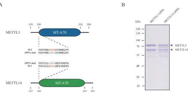
2.2.4 Construction of mutant variants of the METTL3/14 complex . . . . 30
2.2.5 Methyltransferase activity relies solely on the MT-A70 domain of METTL3 . . . 31
2.2.6 The METTL3/14 complex displays no clear substrate-binding specificity . . . . . 32
2.3 Mapping of protein-interacting regions of METTL3, METTL14 and WTAP . . . . 34
2.3.1 A short helix at the N-terminus of METTL3 is sufficient to interact with WTAP . 34 2.3.2 Mapping of the METTL3-interacting region of WTAP . . . . 37
2.3.3 Investigating METTL3-METTL14 dimerization . . . . 40
2.4 Investigating nuclear import of the METTL3/14 complex and WTAP . . . . 43
2.4.1 Both WTAP and METTL3 possess a predicted and functional NLS . . . . 43
2.4.2 The interaction with METTL3 is essential for the nuclear import of METTL14 . . 45
3 Discussion 47 3.1 A revised view on METTL3/14 catalysis . . . . 47
3.2 Dimerization of METTL3 and METTL14 . . . . 49
3.3 Substrate-binding of the METTL3/14 complex . . . . 51
3.4 WTAP-METTL3 interaction . . . . 53
3.5 Cellular localization of the core methylation machinery components . . . . 55
4 Outlook 58 5 Material and Methods 60 5.1 Material . . . . 60
5.1.1 Chemicals and Consumables . . . . 60
5.1.2 Oligonucleotides . . . . 60
5.1.3 Vectors . . . . 63
5.1.4 Antibodies . . . . 64
5.1.5 Buffers and media . . . . 64
5.1.6 Cell lines and bacteria . . . . 64
5.1.6.1 Cell lines . . . . 64
5.1.6.2 Bacteria . . . . 65
5.2 Methods . . . . 66
5.2.1 Cloning of DNA constructs . . . . 66
5.2.1.1 Polymerase chain reaction (PCR), restriction digestion and ligation . 66 5.2.1.2 Site-directed mutagenesis . . . . 67
5.2.1.3 Heat-shock trans formation of E. coli . . . . 67
5.2.1.4 Isolation of plasmid DNA from E. coli . . . . 68
5.2.1.5 Sanger sequencing . . . . 68
5.2.2 Cell culture . . . . 69
5.2.2.1 Cultivation of human cell lines . . . . 69
5.2.2.2 Cultivation of Sf21 cells . . . . 69
5.2.3 Protein based methods . . . . 69
5.2.3.1 Expression of recombinant proteins in HEK 293T using Ca 3 (PO 4 ) 2 . . 69
5.2.3.2 Expression of recombinant proteins in HeLa cells using Lipofectamine®2000 70 5.2.3.3 Expression of recombinant proteins in Sf21 cells . . . . 70
5.2.3.4 Whole-cell lysate preparation from human cell lines . . . . 70
5.2.3.5 Immunoprecipitation . . . . 71
5.2.3.6 Lysate preparation from Sf21 cells expressing GST-METTL3 and METTL14 71 5.2.3.7 HPLC purfication of GST-METTL3/14 . . . . 72
5.2.3.8 GST-pulldown . . . . 72
5.2.3.9 Methanol chloroform precipitation . . . . 73
5.2.3.10 Sodium dodecyl sulfate polyacrylamide gel electrophoresis (SDS-PAGE), Coomassie staining, western blotting and silver staining . . . . 73
5.2.3.11 In vitro m 6 A methylation assay . . . . 75
5.2.3.12 SAM-binding assay . . . . 75
5.2.3.13 Electrophoretic mobility shift assay (EMSA) . . . . 76
5.2.3.14 Cross-linking-MS analysis of METTL3/14 . . . . 77
5.2.4 RNA based methods . . . . 78
5.2.4.1 Denaturing urea PAGE and blotting of RNA . . . . 78
5.2.4.2 T7 in vitro transcription . . . . 78
5.2.5 Immunfluorescence microscopy . . . . 79
5.2.6 Bioinformatic methods . . . . 80
5.2.6.1 Nucleotide and protein sequences of METTL3, METTL14 and WTAP used in this work . . . . 80
5.2.6.2 In silico secondary structure predication of proteins using Psipred . . 80
5.2.6.3 In silico predication of coiled-coil structures within proteins using COILS 80 5.2.6.4 In silico prediction of nuclear localization signals using Eukaryotic Lin-
ear Motif resource tool . . . . 81 5.2.6.5 Quantification of gel bands using ImageJ . . . . 81
6 Appendix 82
List of Tables 86
List of Figures 87
List of Abbreviations 89
References 92
1. Introduction
1.1 The epitranscriptome
The genetic information encoded in DNA and RNA is build-up by the four different bases adenine, guanine, cytosine and thymine, or in case of RNA, uracil. Early analysis of the total pool of RNA syn- thesized by the cell, termed the transcriptome, has revealed that other bases besides the canoni- cal ones can be found in large quantities. To date, over 140 different base modifications have been identified in RNA and arise through modification reactions of the "standard" bases that occur post- transcriptionally. 1 Some examples for non-canonical nucleosides present in RNAs are pseudouridine ( Ψ ), inosine, 5-carbamoylmethyluridine and N 6 -methyladenosine (m 6 A). Additionally to the presence of a variety of bases, it has been reported that both the ribose and the phosphate moiety within RNA can undergo different modifications as well. 2–6 This collection of different RNA modifications adds a second layer of information onto the transcribed genetic information and has coined the term of the epitranscriptome. 7–9
It has been proposed that several of these modifications found in different RNA species influence the
stability or structure of the RNA. For instance, 5-methylcytosine (m 5 C) can be found in several different
transfer RNAs (tRNA). Publications have shown that the presence of this modification at the C38 posi-
tion of the tRNAs Asp GTC , Val AAC and Gly GCC , catalyzed by the protein Dnmt2 (also known as Trdmt1),
enhances the stability of the RNA under normal and stress conditions. 10–13 An example for the struc-
tural influence of an RNA modification is the pseudouridylation of tRNAs. It has been reported that this
modification can rigidify the tRNA structure by locking the ribose of Ψ in the C3’-endo conformation. 14, 15
The importance of RNA modifications is, however, not only confined to the structure and stability of
certain transcripts. It has been reported that non-canonical bases are vital for the function of different
ribonucleoprotein complexes. Ψ is again a prominent example. This base modification influences the
binding affinity of the ribosome towards tRNAs. Therefore, a depletion of pseudouridylation leads to a
reduction of translation efficiency and fidelity. 16–18 Another base modification that has an influence on
translation is 7-methylguanine (m 7 G) that forms the "methyl-cap" of mRNAs. 19–21 This modification is
established at the 5’ end of mRNAs and is directly and specifically bound by a protein termed eukaryotic
initiation factor 4E (eIF4E). 22–24 eIF4E together with eIF4A and eIF4G forms a complex (termed eIF4F)
that is essential for the recruitment of the 43S pre-initiation complex to the mRNA. 25–28 m 7 G-binding
1. Introduction
facilitated by eIF4E is considered the rate-limiting and vital step in the initiation of protein synthesis. 29 Besides translation, the m 7 G-cap is also responsible for the nuclear export of mRNAs and is considered a general stability factor for these transcripts. 30–33 Taken together, these examples highlight the necessity and importance of RNA modifications and their contribution to a variety of cellular processes.
Even though the large repertoire of different modifications have been known now for over half a century, the exact molecular modes of action for many of them remain elusive. A great majority of these non- canonical RNA building blocks have been identified and studied in tRNAs and ribosomal RNAs (rRNA).
Analysis of RNA modifications was mostly restricted to these two ncRNA species, as these transcripts are highly abundant in the cell and larger quantities can be easily extracted. It was not until recently that more sensitive methods have been developed, allowing the detection and deeper analysis of RNA modifications in all classes of transcripts present in a cell.
1.2 Eukaryotic internal mRNA modifications
It was long thought that eukaryotic mRNAs harbored only a handful of modifications and that their
presence was restricted to the 5’- and 3’-end of the transcript. The m 7 G-cap and 2’-OH methylation
of the first two transcribed nucleotides are the most commonly known representatives of mRNA mod-
ifications. 34–39 However, recent publications could show that some of the same modifications found
in ncRNAs were also present in mRNAs of different eukaryotes. 40, 41 So far the nucleosides inosine, Ψ,
m 5 C, N 1 -methyladenosine and m 6 A have been identified in internal sequences of mRNAs (Fig. 1.1).
1. Introduction
Inosine
(I) N
6-methyladenosine
(m
6A) Pseudouridine
( )
5-methylcytidine
(m
5C) N
1-methyladeosine
(m
1A)
RNARNA
RNA RNA
RNA RNA
RNA RNA
RNA RNA
Figure 1.1 Chemical structure of base modifications found in the internal sequences of eukaryotic mRNAs The nucleosides in- osine, N
6-methyladenosine, pseudouridine, 5-methylcytidine and N
1-methyladenosine have been found in internal sequences of eukaryotic mRNAs. All modifications are established by highly specialized enzymes that act upon the mRNA posttranscrip- tionally.
1.2.1 Inosine
Insosine is an RNA modification that has been extensively studied in mRNA during the last two decades.
The base adenine is converted into hypoxanthine during a process that has been termed A-to-I RNA
editing. This process has been first described to be involved in the unwinding of exogenous double-
stranded RNA (dsRNA) in crude extracts derived from Xenopus laevis oocytes, embryos or even from
mammalian cells. 42–44 This activity was distinct from that observed in the modification of tRNAs and
of the purine catabolism. Later, proteins belonging to the adenosine deaminase acting on RNA (ADAR)
family were identified as the enzymes responsible for the specific activity observed in Xenopus . 45, 46
The presence of ADAR-mediated formation of inosine in mRNA remained a matter of controversy until
several reports have shown that this RNA modification was indeed harbored in mRNAs and showed a
tissue-specific abundance. 47–49 Inosine was later readily mapped within mRNAs via high-throughput
sequencing technology and the fact that this non-canonical base is recognized as a guanosine during
reverse transcription (RT). 50 Usage of this method led to the identification of 1600 genes. 48 A to I
1. Introduction
editing seems to be a stochastic event, as no specific enrichment of this modification can be found within defined regions of mRNA, such as the 5’- or 3’-untranslated region (UTR).
ADAR1 and ADAR2 are the best studied representatives of the ADAR protein family. Investigation of both enzymes have shed light on how mRNA editing is achieved and what are the biological consequences of this event. Both ADAR1 and 2 have been shown to localize in the nucleus and modify messenger tran- scripts. 51–53 In addition to its presence in the nucleus, ADAR1 can also be found in the cytoplasm. 54, 55 Due to this distribution pattern, the event of A-to-I editing can occur in both cellular compartments. 56 A prerequisite for the deamination reaction promoted by ADARs is the presence of a double-stranded region within the mRNA. These double-stranded regions normally span a length of more than 20 base pairs (bp) and can be formed inter- or intramolecularly. 57 This is the minimal requirement towards the target for an efficient removal of the amino group at the C6 position of adenine. However, some sort of specificity is needed to explain substrates that are either edited exclusively by ADAR1 or ADAR2, whereas others are modified by both enzymes. 58–60 The necessity of double-stranded regions make certain transcripts to ideal substrates such as mRNAs that harbor repetitive elements (such as Alu ele- ments) in their UTRs. 61,62 These sequences derived from transposable elements tend to form secondary structures with double-stranded regions and can, therefore, be targeted by ADARs. Other sites have also been identified within the coding region of mRNAs. Editing events in this region can lead to amino acid recoding, as inosine is recognized as a guanine by the translation machinery. 63–65
Through the event of recoding, some ADAR-targeted transcripts give rise to proteins with a single amino acid substitution. These in return can have a great impact on the function of the protein. 66, 67 The pres- ence of inosine within 3’-UTR suggests that these deamination sites might have a regulatory potential.
Indeed reports have shown that this type of RNA editing can influence the cellular localization of an
mRNA or even its stability. 68–70 Beside the regulation of a single transcript, a single A-to-I editing event
can influence multiple transcripts. This can be explained through the modification of mRNA transcripts
that give rise to a microRNA (miRNA). A deamination of an adenosine within the seed sequence leads
to the regulation of different target transcripts compared to the unmodified miRNA. 71 As these molec-
ular consequences suggest, A-to-I RNA editing can have a significant biological impact. Studies have
shown that the inactivation of ADARs can lead to severe phenotypes such as embryonic lethality or
death shortly after birth. 58, 72, 73 Human diseases have been as well associated with the mutations of
ADAR proteins which lead to neural dysfunction or alteration of skin pigmentation. 74, 75
1. Introduction
1.2.2 5-methylcytidine
Methylation at the C5-position of cytosine is a prominent and well studied modification in the field of epigenetics. 76 The molecular consequence of this DNA modification is mostly repression of gene expression and is essential in several processes such as genomic imprinting, stem cell differentiation and transposon silencing. 77–80 This modification is, however, not restricted to DNA. m 5 C has been found and studied in different ncRNAs such as tRNAs and rRNAs. In 2012, this modification has been detected with the help of bisulfite sequencing in different human mRNA transcripts as well. 81 8495 methylated cytosines were mapped and found to be enriched within the 3’-UTR of messengers. Controversial to this finding, another transcriptome-wide sequencing method could not validate these initial findings. 82 This study involved the treatment of cells with 5-azacytidine coupled with immuno precipitation of the RNA that incorporated the cytidine analog with an anti-azacytidine antibody. The authors did not selectively enrich for mRNAs in their experimental design. Therefore, the absence of these transcripts in their sequencing data could be the result of a reduced sensitivity compared to the mRNA enrichment strategy and the bisulfite sequencing approach used by Squires et al. 81 Since these initial publications, there have been no further transcriptome-wide m 5 C-sequencing reports published. Therefore, the exact number of transcripts and abundance of this mRNA modification has to be further validated.
The methyltransferase(s) responsible for establishing the cytosine methylation is also a matter of de-
bate. Evidence has been provided that the tRNA methytransferases NSun2 and DNMT2 methylate the
5’- and 3’-UTR of different mRNAs. 81, 83 A different study concentrating on NSun2 reported that this
protein does not bind mRNAs except for one, namely NSun2. 84 Due to the fact that the methyltrans-
ferase(s) has not been clearly identified, little is known about the exact biological function of m 5 C in
mRNAs. The purposed enrichment of this modification in 3’-UTRs, based on Squires et al. , would sug-
gest a regulatory role. 81 So far, only a single study hints to a stabilizing effect on the p16 INK4 mRNA. 83
A different study using cells that carry a loss of function mutation in one or both known RNA m 5 C-
methyltransferases (NSun2 and DNMT2) showed no difference in mRNA expression or stability when
compared to their wild-type counterparts. 13 This suggests that m 5 C does not have a global stabilizing
effect on mRNA transcripts. Further investigations are needed in order to identify the exact methytrans-
ferase(s) that establishes cytosine methylation. Furthermore, efforts have to be made in resolving the
conflicting claims that are present in the field concerning the abundance, presence and function of m 5 C
within mRNA.
1. Introduction
1.2.3 Pseudouridine
Pseudouridine is one of the more recent RNA modifications found within the internal sequences of mR- NAs. Three independent studies mapped this modification within mRNAs using a sophisticated high- throughput sequencing protocol. 85–87 All three groups enriched mRNAs and treated the RNA with N- cyclohexyl-N’-(2-morpholinoethyl)carbodiimide metho-p-toluene sulfonate (CMC) to selectively label pseudouridines. The bulky carbodiimide moiety leads to a termination of the reverse transcription re- action one nucleotide after the pseudouridylated site. 88 This approach allowed to map pseudouridines in 41 to 238 protein coding transcripts in yeast and between 89 to 300 mRNAs in humans, depending on the study and the bioinformatic pipeline used to analyze the sequencing data.
These transcriptome-wide mapping studies could also elegantly identify the enzymes responsible for establishing Ψ within messengers. Interestingly, mRNAs share the same modifying proteins as tRNAs and rRNAs, namely the pseudouridine synthase (PUS) enzymes. 85–87 Isomerization of the uridine moi- ety can be catalyzed by PUS proteins using two different strategies. One arm of this protein family binds snoRNAs of the H/ACA box family that functions as a guide. 89, 90 The protein Cbf5p in Saccharomyces cerevisiae and NAP57 in humans is the catalytic subunit of this PUS RNP complex. 91–93 The other arm of the PUS family can generate Ψ in a snoRNA independent manner. 94–97 In the transcriptome-wide stud- ies, depletion of either Cbf5p or Nap57 abolish certain pseudouridine sites within mRNAs, whereas other sites were unaffected. The latter suggests that the other sites are modified via the snoRNA-independent mechanism. Deletion strains of S. cerevisiae identified the proteins Pus1-4p, Pus6p, Pus7p and Pus9 as the responsible enzymes for the pseudouridylation in mRNA without the necessity of a nucleic acid as a guide. 85, 87 In humans, so far only the proteins Pus4 and Pus7 have been linked to the isomerization of uridine in messengers. 87
Mapping of Ψ could show that the modification is not enriched within a certain region of a mRNA tran-
script but seem to be evenly distributed. 85 To date, little is known about the biological implications of
this mRNA modification. Due to the capacity of Ψ to facilitate unusual base pairing, the presence of
this modification within an open reading frame (ORF) could lead to a recoding event similar to those
observed for inosine (see 1.2.1). An in vitro study using artificial mRNA containing Ψ could show that
this non-canonical base can lead to the recoding of a stop-codon into an amino acid and, thus, leads to
the synthesis of a longer polypeptide. 98 Interestingly, mRNA pseudouridylation seems to be a regulated
process. Schwartz and colleagues could show that upon heat-shock of S. cerevisiae cultures, Pus7p is
translocated from the nucleus into the cytoplasm. After relocation, Pus7p effectively pseudouridylates
1. Introduction
mRNAs leading to a heat shock specific profile. 87 The induction of pseudouridylation has also been shown in human cell lines upon serum starvation, underlining that this modification can be induced via external stimuli and this can be mediated into a biological response. 85 Further studies will be needed to investigate other conditions that induce pseudouridylation, its biological impact and how this modi- fication is translated into a response.
1.2.4 N 1 -methyladenosine
N 1 -methyladenosine (m 1 A) is the youngest member of internal mRNA modifications. All what is known to date about this mRNA base modification has been described by two independent groups in 2016. 99, 100 Both groups applied a transcriptome-wide and antibody-based approach to investigate the topology of m 1 A within mRNA. Each protocol first enriches for poly-A RNAs that are subsequently chemically fragmented to approximately 100 to 150 nucleotides (nts). RNAs containing m 1 A are then specifically enriched using an antibody raised against the modification. After immunoprecipitation, the antibody- bound RNA is retrieved and subjected to a transcriptome-wide RNA sequencing protocol, in order to identify regions containing m 1 A. A drawback of this method used in both studies is that the position of m 1 A cannot be determined at a nucleotide resolution. Sequencing of anti-m 1 A enriched RNA fragments allowed the annotation of "m 1 A-peaks", which span a region between 55 and 150 nts. Nevertheless, this approach could identify between 887 and 4151 mRNA transcripts that harbor this modification. Addi- tionally, the authors mapped the methylation at the N1 position of adenine within the 5’-UTR of human mRNAs and/or in close vicinity (approx. 25 nt up- or downstream) of the start codon. 99 On average, only a single m 1 A peak is present on a methylated transcript. This typical pattern is also seen in mRNAs from mice suggesting conservation between humans and rodents. 99 This pattern is, however, not con- served in all eukaryotes. Sequencing of the m 1 A methylomes from Schizosaccharomyces pombe and S.
cerevisiae revealed a different profile. Within these organisms methylation is mostly found within the ORF of mRNAs, whereas the flanking UTRs display a lower abundance. 99
The m 1 A-methyltransferase(s) for mRNA has not been identified yet. So far, the potential candidates are
enzymes that have been shown to methylate ncRNAs such as tRNA or rRNA. 101, 102 The transcriptome-
wide analysis of m 1 A could not reveal a consensus sequence that could narrow down potential can-
didates. The data provided from the two studies could show that a prerequisite for methylation are
GC-rich regions. It has been experimentally demonstrated that the potential enzyme establishing m 1 A
utilizes S-adenosyl-methionine (SAM) as methyl donor for the methylation reaction. 99 Interestingly, a
potential demethylase has already been identified that can revert the reaction. This is an interesting
1. Introduction
feature as modifications such as pseudouridylation, cytosine methylation and deamination of adenine have so far been reported to be permanent marks. ALKBH3 is the enzyme responsible for the demethy- lation of m 1 A and is a member of the AlkB protein family and most likely utilizes an oxidation reaction to remove the methyl-group as already described for other RNA demethylases (see 1.5.1.1). A knock-out cell line of this demethylase has revealed numerous m 1 A peaks that are normally removed within wild type cells. This in return suggests some sort of specificity as only certain methylation sites/transcripts are targeted by the protein.
The fact that m 1 A is a reversible modification and present in the 5’-UTR of mammals suggest that this methylated adenosine could be a regulatory element involved in different biological pathways. It has been shown by the two sequencing studies that the m 1 A-methylation patterns respond to different stress stimuli such as heat shock, serum/glucose-starvation and oxidative-stress. This suggests that the methylation profile is differentially regulated during normal and stressed conditions. The direct biolog- ical effects of m 1 A have yet to be investigated. On a molecular level, this base modification interrupts the Watson-Crick base pairing interface and generates a positive charge at the N1 position under physio- logical conditions. 103 This modification can therefore facilitate the remodeling of RNA structures, which has already been demonstrated in tRNAs. 104, 105 This in return could then lead to a biological response.
It is also imaginable that both the positive charge and the methyl group could be a potential binding substrate for certain proteins (see 1.4.3) or repel others that would bind the RNA in an unmethylated state. Future investigation will be needed to explore all the proteins that establish, read and modulate this type of methylation and the biological function they promote.
1.2.5 N 6 -methyladenosine
The most prevalent internal modification found within mRNA is m 6 A. Using a quantitative liquid chro-
matography - mass spectrometry approach, it has been shown that this modification contributes to 0.5
to 1 % of the total adenosine pool incorporated into mRNA. 106, 107 The discovery that m 6 A is such an
abundant mRNA modification lead to a renaissance in the field of RNA modifications. Investigations on
this adenosine methylation also coined the term of the epitranscriptome. Due to its importance in the
field, the next chapters discuss several points such as the topology, the biological consequence and the
proteins that establish, remove and bind to this modification.
1. Introduction
1.3 Topology of m 6 A within mRNA
The presence of m 6 A in RNA has been known since the nineteen-fifties and -sixties. 108, 109 This modifi- cation has mostly been detected in different ncRNA species such as tRNAs, small nuclear RNAs (snRNAs) and rRNAs. 110 Studies conducted several years later could show that m 6 A is also present in mRNAs. 111–114 However, analysis on the distribution of this modification was limited due to the lack of appropriate methods. Until recently, the only known messenger that has been shown to harbor m 6 Aand which could be biochemically validated was the bovine mRNA encoding for prolactin. 115
In 2013, a first method was developed to map the presence of m 6 A on a transcriptome-wide scale. 40, 41 The protocol first utilizes an anti-m 6 A antibody to enrich methylated RNA fragments of about 100 nts . Next, the antibody-bound RNA is retrieved and then submitted to high throughput sequencing to iden- tify m 6 A-peaks within mRNAs (similar to the approach used to map m 1 A, see 1.2.4). The combination of immunoprecipitation and RNA sequencing has been termed m 6 A-seq and allowed the identification of m 6 A in transcripts derived from more than 7000 human and 3400 mice genes. S. cerevisiae and Ara- bidopsis thaliana are two other model organisms whose m 6 A-profile has been investigated using the same antibody-based approach. A total of approximately 1200 methylated mRNAs could be identified for yeast whereas 6300 transcripts were found to be methylated in A. thaliana . 116, 117 All transcriptome- wide studies could clearly demonstrate that the presence of m 6 A in mRNA is a common theme con- served from lower to higher eukaryotes. Due to the low resolution of these transcriptome-wide stud- ies, efforts have been made to improve this antibody-based approach. By cross-linking the m 6 A-specific antibody with the RNA via UV irradiation either induces point mutations (C to T transitions) or produces truncations during the reverse transcription reaction. 118 These signatures allow to pin-point the exact location of the methylated adenosine within an RNA.
The fact that a great portion of coding transcripts are methylated suggest that m 6 A is an abundant mod-
ification. Strikingly, an average mRNA transcript harbors 3 to 5 m 6 A-nucleotides. 41, 112 This makes m 6 A
a more prominent mRNA modification than the m 7 G-cap that is a typical mRNA hallmark. Sequenc-
ing data from human and mice revealed that m 6 A could be found around the transcription start (TSS),
5’-UTR, ORF and 3’-UTR. Subsequent normalization of the data to a"standardized" mRNA could show
that the distribution of this methylated adenosine is not stochastic. Peaks of m 6 A were highly enriched
around the stop codon and within the 3’-UTR (Fig.1.2). 40,41 A similar enrichment pattern around the stop
codon has been observed in yeast as well. However, within yeast m 6 A-methylation is only present dur-
ing meiosis and not present during its vegetative state. 116,119 In plants an additional enrichment around
1. Introduction
the start codon is observed. 117 In certain mRNA transcripts, a m 6 A derivative can be found in close proximity of the methyl-cap (1 or 2 nts away) that additionally carries a methylation at the 2’OH of the ribose. This modification is, however, established by a different machinery than the one catalyzing the formation of m 6 A within the internal sequence of the mRNA. 120 Therefore, this derivative represents an independent modification with distinct function(s).
Methylation at the N6-position of adenosine does not occur at random but within a degenerated con- sensus sequence. The sequence that is recognized by the m 6 A-methyltransferase complex is RRACH (A = methylated A; R = purine; H = A, C, or U). 40,41 Even though this sequence is not very well defined and can be found throughout the whole messenger, only methylation sites present in the 3’-portion of mRNAs are readily modified. The underlining mechanism on how only these sites are recognized and preferably methylated is currently debated. The absence of an exact methylation sequence and the fact that not all potential sites are methylated indicate that other factors such as RNA structure or the cou- pling to a certain biological process might be needed for targeting of the responsible methyltransferase to these site.
Figure 1.2 Distribution of m
6A within mRNA Normalizing m
6A-sequencing data to a model mRNA reveals that the most
methyled adenosines are enriched in close vicinity of the stop codon. This enrichment spans a region of approximately 200
nt up- and down stream of the stop codon (figure adapted from Dominissini et al.
41)
1. Introduction
1.4 Molecular and biological impact of m 6 A in mRNAs
After identifying N 6 -methyladenosine as one of the most abundant internal mRNA modifications found in humans, questions have arose concerning the impact of this methylated base within cells. Even though the field on m 6 A has only been revived in 2012, some ground breaking work has been conducted linking m 6 A methylation to different biological pathways. The next sections are a non-exhaustive list of some important m 6 A-dependent mechanisms that have an influence on several cellular processes.
1.4.1 m 7 G-independent translation promoted by m 6 A
The abundance of m 6 A within the 5’-UTR has been reported to be minor compared to the 3’ region of a mRNA transcript (see 1.3). Nevertheless, two recent studies could show that even though less fre- quently deposited in the 5’-UTR the presence of this modification can have a significant cellular impact.
These reports demonstrate that a single m 6 A within the 5’-UTR can promote cap-independent transla- tion. 121, 122 How can m 6 A facilitate translation in a cap independent manner? Meyer et al. could demon- strate that eIF3 can bind directly to methylated adenosines. As a consequence of this interaction, eIF3 can then recruit the 43S complex to the methylated 5’-UTR of the transcript and initiate translation. 122 Interestingly, the same group gathered some evidence for the physiological importance of this mecha- nism in the cell. Upon heat shock, cap-dependent translation is down regulated for most mRNAs within a cell. 123 The expression of a protein termed Heat-shock protein 70 (Hsp70) is normally induced by this stimulus and is translated in a cap-independent fashion. 124 Hsp70 functions as a chaperon and is thought to promote the proper folding of proteins during heat shock. 125 The mRNA of Hsp70 contains an m 6 A within its 5’-UTR and translation of this transcript is methylation dependent. 122 In addition to Hsp70, other mRNAs are methylated at their 5’-UTRs during heat-shock. This observation was confirmed by Zhou and colleagues, who demonstrated the de novo methylation of 5’-UTRs of different mRNAs during this type of stress. 121 Taken together, these studies provide an indication that m 6 A-methylation could be a vital mechanism to guarantee that translation of a subset of mRNAs during heat shock.
1.4.2 m 6 A switch
Initial studies could show that m 6 A does not perturb the Watson-Crick interface and, thus, does not
alter classical base pairing. 126 Nevertheless, m 6 A has been shown to influence RNA structure. A mech-
anism termed m 6 A-switch regulates the binding of a nuclear protein termed heterogeneous nuclear
1. Introduction
ribonucleoprotein C (HNRNPC). 127 Local hairpin foldings within mRNAs containing both a m 6 A- and a HNRNPC binding site are normally not accessible for the RNA binding protein. Methylation of the hair- pin reorganizes the structure and allows the binding of HNRNPC. This in return influences splicing of the transcript harboring the methylated hairpin. The m 6 A status of the RNA can be reverted through specialized enzymes (see 1.5.1.1). The removal of the methylation from the RNA allows it to fold back to its default state and, again, prevents the binding of HNRNPC. This capability of the RNA to undergo either an accessible or inaccessible state dependent on the presence or absence of m 6 A, defines the switching character of this phenomenon.
1.4.3 m 6 A recognition by the YTH domain family - translating m 6 A into function
Several ideas have been proposed on how a single methyl group at the N 6 position of an adenine can be translated into a molecular or cellular response. One concept postulates that upon methylation, the binding of different RNA binding proteins is prevented either directly or by modulating the surround- ing RNA structure, as discussed in the previous section. Another interesting concept was proposed by Dominissini and colleagues. They hypothesized that certain proteins might exist with binding proper- ties specific for m 6 A. 41 Upon binding, these proteins could then induce a down-stream process that results in a molecular/cellular effect. These "reader" proteins would, therefore, function as translators of the modifications they bind. This concept resembles a similar mechanism found within proteins that efficiently bind DNA containing 5-methylcytosine (e.g. MeCP2). 128 RNA pull-down experiments using m 6 A-methylated RNA were conducted, coupled with mass spectrometry, to find proteins with binding properties specific for this modification. 41 Within these experiments, the YTH domain family (YTHDF) members YTHDF2 and YTHDF3 were identified to bind the methylated RNA but not the unmethylated control.
As indicated by their name, both proteins harbor a domain that has been termed the YT521-B homology (YTH) domain. Early assumptions assigned the m 6 A-binding ability to this protein domain, since both YTHDF2 and YTHDF3 only have this domain in common. Structural analysis of the founding member of this protein family YT521-B (also known as YTH domain containing protein 1 (YTHDC1)) and functional analysis of YTHDF2 could provide experimental evidence that the YTH domain indeed binds specifically to m 6 A. 129–131 To date, the human YTH family consists of five members: YTHDF1-3, YTHDC1 and YTHDC2.
Homologs of these proteins have been found throughout different eukarya, ranging from complex or-
ganisms such as humans, rodents and insects down to simple representatives such as Hydra vulgaris
and yeast. 130 So far, only the human YTH proteins YTHDF1, YTHDF2 and YTHDC1 have been shown to
1. Introduction
functionally associate with m 6 A. The next sections briefly summarize the functional roles of these RNA binding proteins.
1.4.3.1 YTHDF2
As one of two YTH proteins identified in the initial RNA-pulldown experiment conducted by Dominissini et al. , this protein has provided the first insight on which biological functions m 6 A can promote. 41 YTHDF2 is mostly localized within the cytoplasm where it encounters and binds methylated mRNAs.
After binding, YTHDF2 destabilizes the transcript by localizing the RNA into a sub-cytoplasmic structure termed processing-bodies (P-bodies). 129 P-bodies are cytoplasmic foci in which mRNAs can either be stored or degraded. 132,133 The overall protein organization of YTHDF2 can be divided into two functional parts. The N-terminal region of this protein is proline-, glutamine- and asparagine-rich (P/Q/N-rich) and is most likely responsible for P-body localization. 129 The C-terminal half of YTHDF2 bears the YTH domain and was shown to efficiently enrich m 6 A-methylated mRNA. This study provided first direct evidence for a YTH domain-m 6 A interaction. Taken together, in the cytoplasm m 6 A can function as a mRNA desta- bilization signal that is promoted by YTHDF2. This interaction can directly regulate the half-life of the mRNA and the population size of the translatable transcripts. By influencing the translatable pool, this in return has an influence on the number of mRNAs that can be associated with polysomes (Fig. 1.3).
Figure 1.3 YTHDF2 destabilizes m
6A-methylated mRNAs and influences their half-life After transcription, m
6A methylation
and mRNA-processing/transport, mRNAs can either engage in translation or can be bound by YTHDF2. This YTH protein inter-
acts with methylated mRNA via its YTH-domain and localizes the bound transcript into P-bodies. This transport is facilitated
by the P/Q/N-rich domain of the protein. Within these foci, cytoplasmic RNAs can be either stored or degraded. The latter
effect results in a shorter half-life of mRNAs targeted by YTHDF2. Figure adapted from Wang2014a
1291. Introduction
Additionally to this finding, some evidence has been collected that YTHDF2 can help promote cap- independent translation upon heat shock. A recent study showed that YTHDF2 can shuttle into the nucleus under stress conditions. 121 Within this compartment, it is assumed that YTHDF2 binds to m 6 A present in the 5’-UTR of (pre-)mRNAs. This interaction protects these methylated sites from demethy- lation that would otherwise occur within unstressed cells, as YTHDF2 is normally not present in the nucleus. After export into the cytoplasm, the methylated 5’-UTR can engage in cap-independent trans- lation that is promoted through the interaction of eIF3 with the methylated adenosine that was (pre- sumably) protected by nuclear YTHDF2 (see 1.4.1).
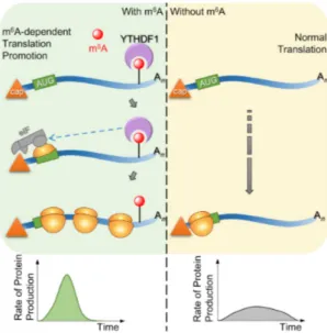
1.4.3.2 YTHDF1
YTHDF1 is the second YTH domain containing protein whose function within the cell was investigated
in more detail. Like YTHDF2, YTHDF1 is localized within the cytoplasm and interacts with mRNAs har-
boring m 6 A-methylated 3’-UTRs. This binding is carried-out with the YTH domain that resides within
the C-terminal half of the protein. 134 In contrast to YTHDF2, the N-terminal portion of YTHDF1 does
not destabilize the bound transcripts, but rather promotes their translation (Fig. 1.4). 134 This molecular
effect is facilitated by YTHDF1 through the interaction with eukaryotic initiation factors and in particular
with eIF3. However, this 3’-UTR dependent induction of translation seems to be a different mechanism
than the one promoted solely by eIF3 binding to the 5’-UTR during heat shock. The process of the for-
mer is not completely investigated, whereas the latter case relies on the direct interaction between m 6 A
and eIF3 (see 1.4.1). Another vital requirement for the YTHDF1-dependent mechanism seems to be the
formation of a closed looped mRNA structure prior to translation, which is promoted by eIF4G. 134, 135
This conclusion was made by Wang et al. through experiments using internal ribosome entry site (IRES)
reporters that either omitted or required eIF4G for efficient enhancement of translation promoted by
YTHDF1. 134 Translation is a multi-stage process involving several steps such as assembly of the 43S com-
plex on the mRNA, AUG scanning and initiation and elongation of translation. 135 The current model for
YTHDF1-promoted translation is that this protein interacts with initiation factors to increase translation
efficiency of target mRNAs. Translation efficiency is defined as the quotient of protein synthesis and
mRNA abundance. Thus, YTHDF1 enhances the protein out-put of methylated transcripts compared to
non-methylated ones without the necessity of increasing transcription.
1. Introduction
Figure 1.4 YTHDF1 promotes translation in a m
6A-dependent manner Messenger transcripts that have experienced m
6A methylation can undergo YTHDF1-promoted translation. For this process, YTHDF1 binds to the methylated 3’-UTR of mRNAs and interacts with translation initiation factors such as the eIF3 complex. This protein-protein interaction is vital to enhance translation efficiency resulting in an increase of protein synthesis while the abundance of target mRNAs remains the same.
Figure adapted from the graphical abstract of Wang et al.2015
134A central question that can be asked is: can the mechanisms promoted by YTHDF1 and YTHDF2 both
coexist within the cell? If they are both spatiotemporally present within a cell, which one is more dom-
inant or how are theses two processes regulated? Wang and colleagues demonstrated that targets
shared by both proteins can experience enhanced translation (YTHDF1) and display shorter half-lifes
(YTHDF2). 134 Shared target mRNAs of both YTH proteins are bound at different time points during their
life cycle. These transcripts are preferably first bound by YTHDF1, resulting in higher protein synthesis,
before they are bound by YTHDF2, targeting them for storage or degradation in P-bodies. This tempo-
ral separation of both m 6 A-promoted mechanisms allow a complex regulation of both translation and
mRNA stability. With such a regulation network, one could imagine that such processes enable a quick
response to different cellular or molecular stimuli that occur during various events such as stem cell
differentiation, gametogenesis and embryonic development.
1. Introduction
1.4.3.3 YTHDC1
YTHDC1 (also known as YT521-B) was the founding member of the YTH-domain protein family. Before the association of the YTH-domain with m 6 A-binding, studies indicated an involvement of this protein in alternative splicing. 136 This protein was found in subnuclear foci termed YT-bodies. 136–138 These foci are claimed to represent so-called transcription centers and are in close proximity to other nuclear structures such as coiled bodies and nuclear speckles. 137 Through structural analysis it has been demon- strated that the YTH-domain that resides in the C-terminus of YTHDC1 can efficiently bind m 6 A facilitated by the highly conserved tryptophan residues at the amino acid positions 380 and 431. 130, 131
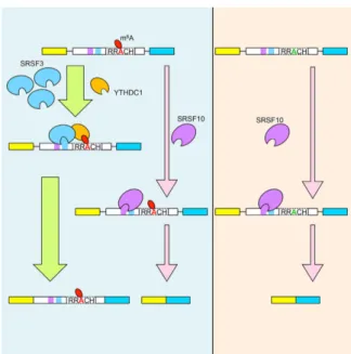
Recently, a mechanism was reported in which YTHDC1 can regulate alternative splicing in a m 6 A- depen- dent fashion (Fig. 1.5). 139 With its N-terminal half, YTHDC1 can bind to the serine/arginine-rich splicing factors (SRSF) SRSF3 and SRSF10. The binding sites of all three proteins have been demonstrated to re- side within the boundaries of 5’- and 3’-splice sites. In addition to this, m 6 A methylation peaks can also be found within these regions, indicating a possible interconnection between mRNA methylation and splicing. The formation of a SRSF3/YTHDC1 complex promotes exon inclusion. This is facilitated through the binding of YTHDC1 to a methylated splice site which in return allows SRSF3 to enforce the inclusion of the targeted exon.
Interestingly, binding of YTHDC1 to SRSF10 results in an antagonistic effect. The formation of this hetero-
dimeric complex results in the suppression of the splicing reaction promoted by SRSF10 alone (exon skip-
ping). Not only does YTHDC1 influence the function of both SRSFs, but also their localization. While the
import of SRSF3 into nuclear speckles is stimulated by YTHDC1, the opposite is observed for SRSF10. This
import into nuclear speckles is another indication that YTHDC1 is involved in splicing, as many splicing
factors (e.g. various SRSFs) are enriched in these subnuclear structures. 140–143 The underlining mecha-
nism on how this selective import is facilitated remains enigmatic. This YTH-protein is the third repre-
sentative that clearly underlines that "reader"-proteins are essential to translate the m 6 A-methylation
into a specific function but are not the only mechanism on how this modification can influence cellu-
lar physiology. Further challenges within the field will be to identify more proteins with maybe novel
m 6 A-binding domains and to functionally validate these.
1. Introduction
Figure 1.5 YTHDC1’s involvement in alternative splicing m
6A methylation sites in close proximity of a potential splice site can be bound by the nuclear YTH protein YTHDC1 that forms a complex with SRSF3. The splicing factor binds its own sequence located near a methylated m
6A-site. Binding of this complex to both sites (m
6A methylation and SRSF3-binding) facilitates the inclusion of the methylated exon in the spliced transcript. This process is antagonized by a different splicing factor termed SRSF10. This protein leads to exon-skipping upon binding to its sequence of an unmethylated transcript and/or blocking the binding of the YTHDC1/SRSF3 complex. Figure adapted from the graphical abstract of Xiao et al. 2016
1391.5 Proteins shaping the m 6 A methylome
1.5.1 m 6 A-demethylases - modulators of the m 6 A landscape
Even though m 6 A has been known to be present within mRNA since the sixties, the field recently has
attracted great attention through the discovery of specialized enzymes that can revert the methylation
at the N6-position of adenine. Investigations conducted on these "erasers" were, together with ad-
vances in mapping of m 6 A in different RNAs, the driving force for the renaissance of the m 6 A field. So
far, two proteins have been identified that can demethylate RNAs containing m 6 A. This finding suggests
that m 6 A in addition to other RNA modifications is reversible. This resembles several processes found
within the field of epigenetics, for instance DNA-methylation or histone modifications. 144
1. Introduction
1.5.1.1 Fat Mass and Obesity-Associated Protein (FTO)
The fat mass and obesity-associated protein (FTO) was the first enzyme identified with m 6 A-demethylase activity. 106 It was, however, first presumed that this protein is a DNA/RNA repair enzyme, since FTO has been shown to be related to the AlkB protein family and is able to oxidize 3-methylthymine in single- stranded DNA and 3-methyluracil in ssRNA. 145–148 As suggested by its name, certain human variants of FTO have been associated with an increased body-mass-index (BMI). 149, 150 Strikingly, genetic studies carried out in Fto knock-out (Fto -/- ) mice have demonstrated that the inactivation of the Fto-gene leads to a reverse phenotype, the reduction of total body mass. 151 A proposed hypothesis is that the lack of Fto in the mice leads to a higher energy expenditure. This in return is the cause for the observed reduction in body weight. Some evidence for this hypothesis has been provided by a study indicating that FTO might function as a nutrient sensor that influences amino acid levels within the cell. 152 The hypothesis of the involvement of the RNA demethylase in energy homeostasis is further supported by the observation that FTO is essential for adipogenesis via the regulation of mRNA splicing 153
FTO is predominantly found within the nucleus as a putative nuclear localization signal (NLS) within its N-terminus has been predicted in silico but has not been functionally validated so far. 146, 154 Different reports have, however, indicated that FTO can shuttle between the nucleus and the cytoplasm. 152, 155 It has yet to be determined what the underlining mechanism is and what the biological relevance of this trafficking between compartments is. Within the nucleus, FTO localizes into nuclear speckles and thus is present in the same nuclear sub-compartment as YTHDC1 (see 1.4.3.3) and the methyltransferase that establishes m 6 A within mRNAs (see 1.5.2).
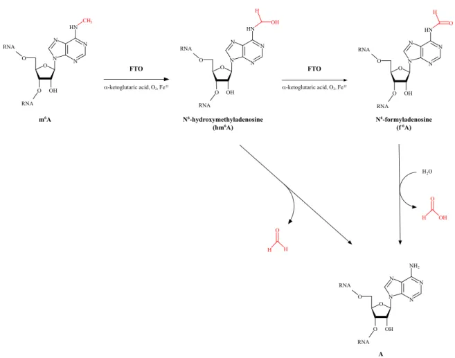
FTO utilizes oxygen, α -ketoglutarate and non-haem bound Fe II to catalyze the oxidation of m 6 A. This
reaction resembles that of Ten-eleven translocation (TET) proteins that convert m 5 C in DNA into cyto-
sine. 156–159 Another similarity between this RNA- and the DNA-demethylases is that the oxidation of the
methylated base is a step-wise process. 160 FTO has been shown to convert m 6 A first into N 6 -hydroxy
methyladenosine (hm 6 A) and then N 6 -formyladenosine (f 6 A). The latter can then be hydrolyzed by the
surrounding water in the cell to give rise to an unmethylated adenosine (Fig.1.6). 161
1. Introduction
m6A N6-hydroxymethyladenosine
(hm6A) N6-formyladenosine
(f6A)
A
FTO FTO
α-ketoglutaric acid, O2, FeII α-ketoglutaric acid, O2, FeII