Role of proteolipid protein (PLP/Dm20) and polyunsaturated fatty acids in normal and pathological central nervous system
Inaugural-Dissertation
zur
Erlangung des Doktorgrades
der Mathematisch-Naturwissenschaftlichen Fakultät
der Universität zu Köln
vorgelegt von Raju Chintha
aus Warangal, India
Köln, 2012
The present work has been done in the Molecular Neuroscience Laboratory of Medical Faculty of the University of Cologne/Köln under the direct supervision of
Prof. Dr. Dr. Dr. h.c. mult. Wilhelm Stoffel.
REFEREES/ BERICHTERSTATTER
1. Berichterstatter: Prof. Dr. Dr. Dr. h.c. mult. Wilhelm Stoffel 2. Berichterstatter: Prof. Dr. Günter Schwarz
Vorsitzender: Prof. Dr. Siegfried Roth
Tag der mündlichen Prüfung: 04 December 2012
I thank Professor Dr. Dr. Dr. h.c. mult. Wilhelm Stoffel for giving this opportunity to do my PhD thesis under him and for the very interesting PhD thesis topic. I am also very thankful for his generous support, patience and for his immense guidance and comments throughout my PhD work.
My sincere thanks to Prof. Dr. Günter Schwarz for evaluating my PhD thesis work and being in my PhD thesis committee, and I also thank Prof. Dr. Siegfried Roth for being in my PhD thesis committee and for evaluating my PhD thesis work credentials.
My sincere thanks and gratitude towards all my lab members: Frau Erika Binczek, Frau Brigitte Handwerk, Frau Barbara Holz, Frau Britta Jenke, Dr. Ina Hammels und Frau Inga Schmidt- Soltau for creating wonderful atmosphere in the lab.
Once again, I am grateful to Dr. Ina Hammels for friendly in depth subject discussions, unending chats, and for German language help during my PhD. I would like to thank Frau Erika Binczek for her incredible teachings and discussions on molecular biology and biochemical protocols and techniques. I thank Frau Britta Jenke for Pronuclear Injections, Frau Barbara and Frau Inga Schmidt-Soltau for helping me in the cell culture laboratory, and last but not least Frau Brigitte Handwerk for caring my laboratory mice in the animal house.
I also thank to our collaborators at Institute for Anatomy in helping us by giving access to their electron microscope and in supplying behavioral physiology protocols, I am thankful to Milteny Biotech (Bergisch Gladbach, Cologne) for Macs Kits and wonderful technical comments and assistance when ever needed. I thank Ingo Voigt, Max-Planck Institute for Ageing, Cologne, for his assistance in pronuclear and blastocyst injections, and I also thank Analytical department, CMMC Institute, Cologne, for DNA sequencing
My heartily, personal thanks to Dr. Praveen Mamidala, Ohio University, USA, for being as my well wisher in all respects of my professional life and helping me in accomplishing my PhD goal. I also thank him for giving me his wonderful in time suggestions whenever I needed.
I thank Dr. Rehan Villani, CMMC, University of Cologne, Germany, for critically reading some parts of my thesis and for her valuable comments.
I also thank Jun. Prof. Dr. Thalapalli Shirisha Sai for her invaluable time to time discussions on organic chemistry and on lipid chemistry related topics, and for the immense support.
Last but not least, I thank my sweet family especially to the little girl Madella Jeevana Priya Mani Goud, for their incredible constant moral support and never ending unconditional love.
Dedicated to my beloved
father Chintha Venkataswamy Goud and mother Kalavathi
Index
1 Introduction…... 1
1.1Central nervous system... 1
1.2 Oligodendrocytes…... 2
1.2.1 Targeting of myelin components in oligodendrocytes...3
1.2.2 Nestin and MBP expression... 4
1.3 CNS Myelin... 5
1.4 CNS Myelin; Proteolipid protein (PLP)... 9
1.4.1 PLP gene transcripts and isoproteins... 10
1.4.2 Cys residues and hydrophobic properties... 11
1.4.3 PLP related myelin disorders; PMD and SPG2... 13
1.5 CNS Myelin; Phospho- and sphingolipids... 14
1.5.1 Fatty acid composition of phospholipids... 16
1.5.2 Classification of fatty acids and the polyunsaturated fatty acids (PUFAs) synthesis cascade... 18
1.5.3 PUFAs in the CNS... 20
1.5.4 Retinal morphology... 21
1.5.5 Fads2 knock-out mouse (PUFAs deficient mouse model)... 22
1.6 Transgenic mouse models... 22
1.6.1 Gene targeting... 22
1.6.2 Pronuclear microinjection... 23
1.7 Site-directed mutagenesis... 24
2 Aim of the thesis... 25
3 Results... 26
3.1 Construction of mutant cDNA pool of PLP and Dm20... 26
3.1.1 Site-directed mutagenesis (SDM) and Overlap extension PCR... 26
3.1.2 DNA sequencing... 28
3.2 In silico Kyte-Doolittle hydropathy plots... 30
3.3 EGFP fused WT-PLP, Mut-PLP, and Mut-Dm20; Intracellular trafficking... 33
3.3.1 Cloning and validation of WT-PLP-EGFP, Mut-PLP-EGFP and Mut- Dm20- EGFP... 33
3.3.2 Mut-PLP-EGFP and Mut-Dm20-EGFP compared to WT-PLP-EGFP trafficking in COS-7 ... 34
3.3.3 WT-PLP-EGFP and Mut-PLP-EGFP trafficking in PLP null oligodendrocytes... 37
3.3.3.1 Isolation and transfection of PLP-null murine oligodendrocytes... 37
3.3.3.2 WT-PLP-EGFP and Mut-PLP-EGFP membrane targeting in oligodendrocytes……… 37
3.4 Mut-PLP expression under the control of Nestin-enhancer; generation of a transgenic mutant... 39
3.4.1 Validation of Nestin enhancer function in OLN93 cells... 39
3.4.2 Trans-gene cloning and pronuclear injection... 41
3.5 Generation of knock-in mouse mutant of PLP/Dm20 devoid of di-sulfide bridges ... 43
3.5.1 Knock-in targeting construct and targeting strategy... 43
3.5.2 ES cells electroporation and ES clones screening... 45
3.5.3 Verification of a positive ES clone... 46
3.5.4 Mutated Cys residues validation by DNA sequencing... 47
3.5.5 Karyotyping of positive ES clone... 48
3.5.6 Embryo manipulation... ... 48
3.5.7 Chimera validation and breeding... ... 49
3.6 PLP and PUFAs pivotal role in the CNS. ... 50
3.6.1 Generation of plp-/-fads2-/- double mutant mouse (DM)... 50
3.6.2 Genotyping and validation of DM... 51
3.6.3 Phenotyping of DM... 53
3.6.4 Gene expression analysis by semi-quantitative RT-PCR... 55
3.6.5 Brain and myelin protein analysis... 56
3.6.6 Myelin quantification... 57
3.6.7 Immunohistochemical stainings...58
3.6.8 Lipid analysis... 60
3.6.8.1 Brain lipid analysis... 60
3.6.8.2 Brain fatty acid profile... 61
3.6.8.3 Brain phospholipids and glycosphingolipids fatty acid profile ... 63
3.6.8.4 Myelin lipid and fatty acid profile analysis... 64
3.6.9 Morphology of the myelin sheath... 66
3.6.10 Retinal morphology and function... 67
3.6.11 Behavioral physiology... 69
3.6.11.1 Rotarod Task (Wallace et al., 1980)... ... 69
3.6.11.2 Horizontal-Rod Task (Wallace et al., 1980)... ... 70
3.6.11.3 Grid Task... 71
3.6.11.4 Open field Task (Barclay et al., 1982)... 72
3.6.11.5 T-Maze Task ... 72
3.6.11.6 Morris Water-Maze Task (Morris et al., 1984)... 73
4 Summary of Results... 76
5 Discussion... 80
5.1 Cys residues are critical for PLP and Dm20 targeting to the Cell surface... 80
5.1.1 Similar trafficking of Mut-PLP and Mut-Dm20 in COS-7 cells and PMD... ... 81
5.1.2 Differential trafficking in vitro and in vivo... 81
5.1.3 Cys-mut PLP (Mut-PLP) expression in early CNS developmental stages... 82
5.1.3.1 Validation of Nestin enhancer regulation of Mut-PLP... 82
5.1.3.2 Generation of a transgenic mouse expressing Mut-PLP regulated by the Nestin enhancer region... 82
5.2 Knock-in mouse mutant of PLP/Dm20-lacking disulfide bridges... 83
5.3 The role of PLP and PUFA in the CNS... 84
6 Materials and methods... 88
6.1 Cell culture methods... 88
6.1.3 Electroporation and Selection of ES cells... 89
6.1.4 Karyotyping of ES cells... 90
6.1.5 Cryopreservation of ES cells and COS-7 cells... 91
6.1.6 Thawing and expansion of ES-cells and COS-cells... 91
6.1.7 COS-7 cell culture and transfection... ... 91
6.1.8 Embrynoic fibroblasts culture... 91
6.1.9 Lipofection (DNA-Lipofectamine 2000), Electroporation of COS-7 and OLN93 cells... ... 92
6.1.10 Stable selection of COS-7 or OLN93 cells... 92
6.2 Molecular biology methods... 92
6.2.1 Plasmid-DNA preparation... 92
6.2.2 Extraction of DNA from agarose gels... 93
6.2.3 DNA isolation from ES cells or COS-7 cells... 93
6.2.4 Genomic DNA isolation from mouse tail... 93
6.2.5 Restriction analysis/digestion... 94
6.2.6 Ligation... 94
6.2.7 Preparation of Competent Cells and transformation of plasmid DNA. 94 6.2.8 Polymerase chain reaction (PCR)... 95
6.2.9 DNA sequencing and Synthesis of oligonucleotide...96
6.2.10 RNA preparation... 96
6.2.11 RT-PCR (Reverse transcription-PCR) ... 96
6.2.12 Radioactive DNA labeling... 97
6.3 Biochemical methods... 97
6.3.1 Protein quantification... 97
6.3.2 SDS-polyacrylamide gel electrophoresis (SDS-PAGE)... 98
6.3.3 Myelin preparation (Based on sucrose gradient separation)... 98
6.3.4 Lipid analysis... 98
6.3.5 Gas Chromatography–Mass Spectrometry (GC-MS)... 99
6.4 Immunological methods...100
6.4.1 Western blotting ... 100
6.4.2 Immunostaining of cells ... 101
6.4.3 Mouse brain fixation for cryo-sectioning... 101
6.4.3.1 Cryo-sections immunohistochemical staining... 102
6.4.4 Mouse brain fixation for paraffin sectioning... 102
6.4.4.1 Paraffin sections immunohistochemical and hematoxylin-eosin staining... 103
6.5 Electron microscopy... 103
6.6 Methods for generating mice mutants (Transgenic and Knock-in mutants)...103
6.6.1 Mice maintenance... 103
6.6.2 Microinjection... 104
6.6.3 Vasectomized males...104
6.6.4 Blastocyst injection... 104
6.7 Behavioral physiology... 105
6.7.1 Rotarod... 105
6.7.2 Horizontal rod and Grid task... 105
6.7.3 T-maze... 106
6.7.4 Open field...106
6.7.5 Morris water maze (Morris et al., 1984)... 106
6.8 Bioinformatic programs and servers... 107
7 Appendix... 108
7.1 Plasmids used in the present study...108
7.2 Oligonucleotides... 108
7.3 Abbreviations... 110
8 References... 113
Zusammenfassung (Abstract in German language)………... 126
Abstract………... 128
1
1 Introduction
1.1 Central nervous system
The CNS of vertebrates consists of the brain, spinal cord and retina. The brain is made up of extensive and complex networks of neurons and their supporting cells termed as glial cells.
Neurons are interconnected by synaptic junctions and are involved mainly in conduction of rapid action potential signals along their axons. Compared to neurons, glial cells are more abundant in the CNS i.e., accounting for approximately 65% of cells in rodent brain and 90% in humans (Pfrieger und Barres, 1995). Virchow in 1846 for the first time described glial cells as neuroglia and later they were more extensively described by Ramon y Cajal and Rio-Hortega in 1913.
Glial cells are predominantly involved in supporting neurons and maintaining metabolic homeostasis in the CNS. They are broadly classified into astrocytes, oligodendrocytes, and microglia, based on their functional significance. Glial cells including astrocytes and oligodendrocytes are derived from ectodermal tissue of the developing embryo, whereas microglial cells are derived from hematopoietic precursors.
Astrocytes are star shaped cells and are majorly populated among the glial cell population within the brain. They are distributed in the grey and white matter of the brain. Immature astrocytes are called radial glia, which play a major role in neuronal migration during fetal development.
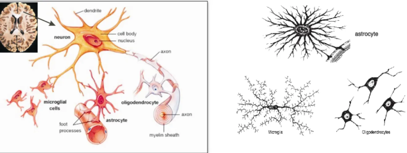
Astrocytes are mainly involved in regulating the external chemical environment in the brain such as recycling neurotransmitters released during synaptic transmission, and also provide growth factors to neurons. Astrocytes contact blood capillaries and the nodes of Ranvier (Figure 1) and are also involved in angiogenesis (Montgomery et al., 1994). Microglia functions as brain macrophages of the CNS and account for 15% of total glial cells. They are mainly involved in phagocytosis (Compston et al., 1997) and are typically in the resting state and are activated under conditions like CNS inflammation, trauma, and infections. Microglial cells are the resident macrophages in the brain. Oligodendrocytes are involved in the myelination of axons in the CNS, whereas Schwann cells are involved in PNS.
Figure 1: Neuron and glial cells in the brain. Left; Oligodendrocytes wrapping myelin sheath around neuronal axons, astrocytes are attached to blood capillaries, neurons and microglial cells, Right;
Individual glial cells i.e., astrocytes, microglia and oligodendrocytes (modified from MHHE science resource)
1.2 Oligodendrocytes
Rio-Hortega first described oligodendrocytes and microglia in 1928. Like neuronal progenitors, early oligodendrocytes progenitors are derived from neuroepithelial precursors of the ventricular zone of the brain and lumen of the spinal cord (Reynolds and Wilkin, 1988; Levine and Goldman, 1988). Mature oligodendrocytes develop from their early progenitors broadly in four stages i.e., early progenitor cells (O2A cells), late progenitor cells (A2B5, O4 cells), premature oligodendrocytes and mature oligodendrocytes (Figure 2). All these differentiation stages undergo several morphological changes starting from single or bipolar to multiple projections, and express cell specific antigen markers such as NG2, O4, GalC, MBP, and PLP (Figure 2) along with several other intracellular changes (Wia Baron, 2003). The early glial progenitors exhibit lineage plasticity such as neural stem cell like features of differentiation into neuronal or any of the glial cell types (Kondo and Raff, 2000c), whereas O4 progenitors (OL-progenitors) are the start of the committed oligodendrocyte lineage. Immature-oligodendrocytes express galactocerebrosides and other myelinating proteins such as MBP and Dm20 which are transported and enriched at the filopodial-like projections of the plasma membrane, whereas they are not capable of myelinating axons. Pre or mature-oligodendrocytes are involved in myelinating axons with their plasma membrane derived filopodia-like structures.
Oligodendrocytes are also capable of expressing myelin genes and develop filopodia-like projections in the devoid of external neuronal signals under in vitro conditions (Pfeiffer et al.,
3
Figure 2: Developmental stages of oligodendrocytes from early progenitors. Different cell specific gene markers expressed during oligodendrocyte maturation; OL=Oligodendrocyte. (Modified from Back SA et al., 2007)
1.2.1 Targeting of myelin components in oligodendrocytes
Oligodendrocytes have highly specialized mechanisms for sorting and targeting myelin protein and lipid components to the growing polarized plasma membrane/myelin membrane.
Oligodendrocytes share similarities with polarized epithelial cells, such as MDCK cells, in the underlying mechanisms of protein trafficking to the plasma membrane (Kramer et al., 2001).
Polarized cells like MDCKs are therefore suitable in vitro cell culture models for studying the underlying mechanisms of myelin proteins trafficking. For instance, transfection studies in MDCK cells showed differential trafficking among myelin proteins; PLP is sorted to the apical membrane and MOG sorted to the basolateral membrane (Kroepfl and Gardinier, 2001). Among the two isoforms of MAG i.e., S-MAG and L-MAG, L-MAG is targeted to both apical and basolateral compartments, whereas S-MAG is targeted only to the apical domain (Minuk and Braun, 1996).
In oligodendorcytes, PLP is transported from the ER to the cell surface via mechanisms including vesicular transport, endosome, and transcytosis. Polarized trafficking of myelin proteins and lipids, such as PLP and GalC, may include initial sorting at trans-Golgi or in endosomes accordingly and then transported to specific plasma membrane domains directly or via cytoskeleton anchored transport in a spatial and temporal manner. MBP and MOBP’s mRNA isoforms are transported near to the plasma membrane where actually their protein translation is initiated (Barbarese et al., 1995). CNP is transported anchored to the cytoskeleton, such as actin filaments and microtubules, to the non-compact myelin membrane regions (Laezza et al., 1997).
The exact trafficking routes of myelin components, especially mutated myelin proteins, and their mechanisms of plasma membrane targeting are still under investigation.
1.2.2 Nestin and MBP expression
Nestin protein is an intermediate filament protein that is specifically expressed in neural tube, neuroepithelium stem cells and in glial precursors (Lendahl et al., 1990). Nestin is also expressed in neural stem cells and in its subsequent generations of neuronal progenitors and glial progenitors (Zimmerman et al., 1994, Johansson et al., 2002). In the oligodendroglial lineage, nestin expression levels are gradually down-regulated from immature-oligodendrocyte to mature oligodendrocytes (Vittorio Gallo and Regina C Armstrong, 1995). The nestin gene contains four exons and three introns and is highly conserved among human, rat and mouse (Lendhal U et al., 1990; Lothian et al., 1999). The second-intron enhancer of the nestin gene is particularly critical as it contains regulatory elements proposed to regulate gene expression specifically in the neural stem cells and in subsequent oligodendrocyte progenitors (Zimmermann et al., 1994, Lothian et al., 1999).
Of the two major myelin proteins synthesized in oligodendrocytes, PLP and MBP, MBP has a stronger promoter for transcription initiation than PLP. 1.5kb of intron-I and partial-exon-I of MBP gene sequence containing the MBP promoter has been well characterized in a transgenic mouse mutant (Kimura et al., 1989). Although MBP is a marker for mature-oligodendrocytes, an MBP isoform is also detected in early embryonic developmental stages, on both the protein and mRNA level, and is called embryonic-MBP or E-MBP (Mathisen et al., 1993; Nakajima et al., 1993). As characterized in wild-type mice, E-MBP expression is observed in early embryonic stages, postnatal day-7, day-20 (active-myelination phase) and day-60 (matured- myelination phase) (kimura et al., 1989).
5 1.3 CNS Myelin
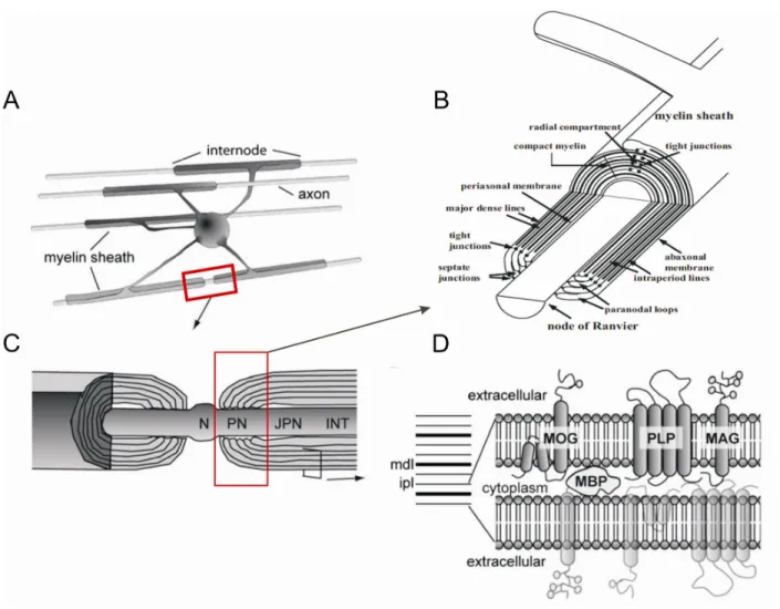
Myelin has been first described and termed by Rudolf Virchow in 1854 and its ultra structure first studied by Sjoestrand in 1947 by electronic microscopy. Myelin is a highly regulated plasma membrane extension of oligodendrocytes in the CNS, and Schwann cells in the PNS. In the CNS, the plasma membrane of oligodendrocytes or myelin is spirally wrapped around the axons of neurons in multiple locations termed internodes (Figure 3.A). Periodic interruptions between two internodes that leave the axon unmyelinated are termed nodes of Ranvier (N), and the edges of the internode are termed paranodes (PN) (Figure 3 C, B).
Internodes and myelin compaction
Spirally wrapped internodal myelin layers are compacted due to van der Waals, and electrostatic forces between polar head groups of phospho- and sphingolipids, as well as interactions of phospholipids with cytosolic MBP and integral membrane proteins such as PLP. In electron micrographs myelin layers appear as periodic structures of electron dense lines termed major- dense lines (MDL) and less electron-dense lines termed intraperiod lines (IPL) (Figure 3.D).
MDLs represent the tight opposition of cytoplasmic sides while IPLs represent the extracellular side of the myelin membrane. Myelin proteins such as PLP, MAG and MOG are integrated into the cholestrol-phospho- and sphingolipid bilayer of the myelin membrane (Figure 3D). The periodicity distance between two IPL is 170 ± 7 Å. Occasionally the compact cytoplasm, i.e.
MDL, is interrupted and compaction is lost, forming funnel like longitudinal structures along the MDL, called Schmidt-Lanterman clefts however these are very rare in CNS and more abundant in PNS.
Paranodes and junctions
Paranodes are the end points of internode (Figure3 C, B). The periaxonal space of the myelin is sealed from the outside milieu and connected to the axon via septate-like junctions (Einheber et al., 1997; Saher et al., 2011) (Figure 3B). The paranodal loops or junctions are connected with the axons by tight junction proteins such as OSP or Claudin-11 (Figure 3 B). Paranodal junctions are critical for efficient action potential propagation and in ion channel clustering (Schafer et al., 2004). The outer most part of paranode is termed juxtaparanodal (JPN) region.
Nodes of Ranvier
Louis-Antoine Ranvier discovered regularly spaced unmyelinated gaps (approximately 1 µm in length) termed nodes of Ranvier (Figure 3.B). They harbor voltage-gated sodium (Na+) ion channels that are engaged in conduction of action potentials. Myelinated axons have salutatory conduction in the range of 1 m/s to over 100 m/s, 0.5m/s to 15 m/s in unmyelinated axons (Mutschler, 1991), thus enhancing the conductance 10-fold compared to unmyelinated axons.
Figure 3: Schematic representation of CNS myelin. A. An oligodendrocyte myleinating multiple axons. B. Zoomed view depicting paranaodal junction and its molecular arrangement C. Zoomed view representing node of Ranvier (N), paranodal junction (PN) juxtaparanodal junction (JPN) and internode (INT), and D. Zoomed view of myelin membrane extracellular side and cytoplasmic arrangement, intraperiod lines (IPL), myelin dense line (MDL), and embedded myelin proteins in the myelin
7 Myelin proteins
Myelin accounts for 30% of the dry weight of the brain. Myelin is a lipid rich membrane, with 70-80% lipids and 20-30% proteins. The myelin membrane is composed of unusual sphingolipid classes and high cholesterol and myelin specific proteins.
The most abundant proteins of myelin are proteolipid protein (PLP) and myelin basic protein (MBP) constituting 50% and 30%, respectively, together making 80% of whole myelin protein composition. PLP and MBP localization in the myelin membrane was first identified based on immunolocalization studies (Hartman et al., 1982). Other myelin proteins are: 2’, 3’-cyclic nucleotide-3‘-phosphodiesterase (CNP) 4-5%, myelin associated glycoprotein (MAG) 1%, and myelin oligodendrocyte glycoprotein (MOG) and myelin-oligodendrocyte basic protein (MOBP)
>1% each (S. A. Kalwy and Ross Smith, 1994). Recently several hundreds of distinct proteins of low range concentrations were detected in the myelin through gel based proteome analysis (Werner et al., 2007).
Proteolipid protein (PLP) is further discussed in section 1.4.
Myelin basic protein (MBP)
MBP has several splice variants which differ in different species, four in humans, and six in mice, with protein sizes ranging from 14 to 24kDa (Newman et al.,1987). MBP contains a high number of basic amino acid residues and is a hydrophilic protein localized in the cytoplasm of the myelin membrane (MDL) (figure 3 D). As MBP is highly positively charged, it may interact with the cytoplasmic side myelin membrane, thereby contributing to compaction and stabilization of the myelin membrane (Figure 3 D) (Morell et al., 1994). Although MBP null mice (shiverer mutant or myelin deficient mutants) show compact myelin around the axons, its hypomyelination causes severe neurological defects including tremors and seizures (Bird et al., 1978). Several types of post-translational modifications occur on MBP such as acetylation, phosphorylation, methylation and citrullination (Wood and Moscarello, 1989). Futhermore methylation of MBP might be important for compaction of myelin sheath (Campagnoni et al., 1993). MBP is also present in PNS which accounts for 5-15 % of total proteins.
Myelin-associated glycoprotein (MAG)
MAG is an integral membrane glycoprotein with a molecular mass of 100 kDa. It has two isoforms; L-MAG and S-MAG. MAG is involved in axon-glial contacts and signaling, and axonal maintenance (Poltorak et al., 1987). The majority of MAG is located in periaxonal regions of myelin (Webster et al., 1983). Its structural role in the myelin sheath is not clear.
Myelin-oligodendrocyte glycoprotein (MOG) and other proteins
MOG is an integral membrane glycoprotein which was first identified as one of the antigens in experimental autoimmune encephalomyelitis (EAE). MOG is highly glycosylated and is believed to act as an adhesion molecule. MOG is also involved in signal transduction pathways, and lipid- protein sorting (Kim and Pfeiffer, 1999).
Other myelin proteins such as: CNP is located in the cytoplasm and one of the earliest expressed proteins during myelination. Although CNP is named after its function and believed to have a structural role, its functional role in CNS remains to be elucidated. Adult CNP null mice showed axonal swellings and axonal degeneration. The double mutant plp-/-cnp-/- mice showed progressive axonal loss (J.M. Edgar et al., 2009). Other myelin proteins such as myelin- associated oligodendrocytic basic protein (MOBP) and tight junction proteins like oligodendrocyte specific protein (OSP)/Claudin-11 are minor components among myelin proteins with undefined functions. Due to various transgenic mouse mutants, it is now clear that the tight junction proteins including OSP protein are important in proper CNS myelination and function.
Myelin proteins and respective phenotypes
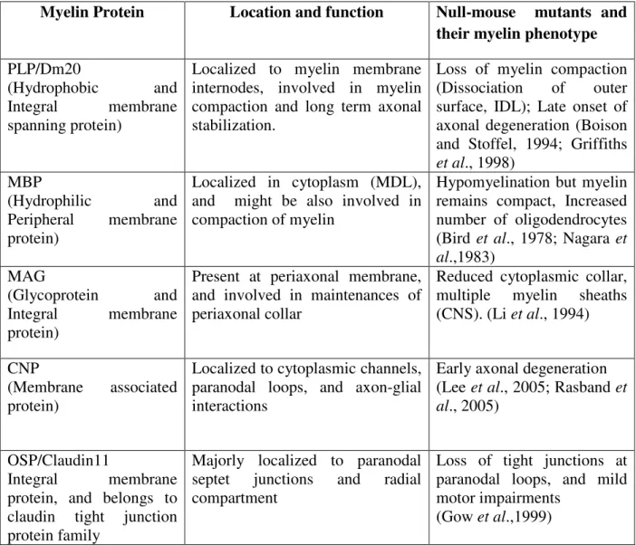
Myelin is a complex unique membrane, and many proteins are specific to myelin. Many studies have been made during the past two decades also by employing transgenic mice. Various myelin proteins, respective knockout mouse mutants, and their phenotypes are summarized in the table 1.
9
Myelin Protein Location and function Null-mouse mutants and their myelin phenotype PLP/Dm20
(Hydrophobic and
Integral membrane spanning protein)
Localized to myelin membrane internodes, involved in myelin compaction and long term axonal stabilization.
Loss of myelin compaction (Dissociation of outer surface, IDL); Late onset of axonal degeneration (Boison and Stoffel, 1994; Griffiths et al., 1998)
MBP
(Hydrophilic and
Peripheral membrane protein)
Localized in cytoplasm (MDL), and might be also involved in compaction of myelin
Hypomyelination but myelin remains compact, Increased number of oligodendrocytes (Bird et al., 1978; Nagara et al.,1983)
MAG
(Glycoprotein and Integral membrane protein)
Present at periaxonal membrane, and involved in maintenances of periaxonal collar
Reduced cytoplasmic collar, multiple myelin sheaths (CNS). (Li et al., 1994)
CNP
(Membrane associated protein)
Localized to cytoplasmic channels, paranodal loops, and axon-glial interactions
Early axonal degeneration (Lee et al., 2005; Rasband et al., 2005)
OSP/Claudin11
Integral membrane protein, and belongs to claudin tight junction protein family
Majorly localized to paranodal septet junctions and radial compartment
Loss of tight junctions at paranodal loops, and mild motor impairments
(Gow et al.,1999)
Table 1: Summary of the major myelin proteins and their functional significance in myelin
1.4 CNS Myelin; Proteolipid Protein (PLP)
In 1951 Folch and Lees isolated PLP, the major protein accounting approximately 50% of total brain myelin protein. PLP cDNA has been cloned in 1986 in our laboratory (Schaich et al., 1986).
1.4.1 PLP gene transcripts and isoproteins
The PLP (plp1) gene is 16 kb (Diehl et al., 1986) and is located on the X-chromosome, Xq22 in human and H2C in mice. The PLP gene is comprised of seven exons (Diehl et al., 1986; Stoffel et al., 1985) and has a protein coding region of 828 bps that encodes 276 amino acids (Stoffel et al., 1982). PLP has a splice variant called Dm20. Dm20 mRNA lacks the last 105 bps of exon 3 of PLP mRNA and thereby 35 amino acids in the cytosolic loop of PLP (Nave et al., 1987). PLP is expressed primarily in oligodendrocytes where it is integrated into the plasma membrane. PLP has been extensively studied for more than 20 years, on the molecular and structural level, and regarding its role in myelin morphology specifically in relation towards CNS myelin disorders.
PLP is highly conserved. Specific point mutations in PLP result in the severe form of Pelizaeus–
Merzbacher disease (PMD) (Weimbs et al., 1990) or spastic paraplegia type 2 (SPG2) (Koeppen and Robitaille, 2002), whereas the absence of PLP does not result in PMD nor in myelin defects but only in loss of myelin compaction (Boison and Stoffel., 1994) and axonal degeneration in adults (Griffith et al., 1998). Strikingly, the wild-type PLP gene over-expression in mouse models (i.e., PLP transgenic mouse model) revealed that PLP over-expression alone imparts toxicity in the oligodendrocytes and thus causes myelin defects that could result in PMD condition, demonstrating the importance of PLP gene dosage (Karim et al., 2007).
In addition to the classical PLP and its isoform Dm20, Bongarzone et al., 1999 showed an additional exon and translational site between exon-I and exon-II of the classical PLP gene. The derived mRNA can also be alternatively spliced into two new isoforms called sr.PLP and sr.Dm20. Their corresponding protein products contain an additional 11 amino acids. These novel isoforms (i.e., sr.PLP and sr.Dm20) accounts to 2% of total myelin protein composition, restricted to the somata of oligodendrocytes and neurons, and not detected nor trafficked to the myelin membrane.
Structure and Protein conservation
PLP is the highly hydrophobic and integral transmembrane protein of the myelin membrane. The complete topology of PLP was unraveled by biochemical studies by Weimbs and Stoffel, 1992 and Wahle and Stoffel, 1998. PLP contains four transmembrane spanning hydrophobic domains connected by three hydrophilic loops in which two loops (EC-I and EC-II) are located in the extra-cytosolic space and one loop in the cytosolic space. The NH2 and COOH terminal domains are located on the cytosolic side (Weimbs and Stoffel, 1992; Gow et al., 1994; Greer et al., 1996). PLP is phylogenetically highly conserved (Schliess and Stoffel, 1991), and is identical at
11
Dm20 and other proteins such as M6 glycoproteins share, to a certain extent, homology in their transmembrane domains and Cys positions (Greer et al., 2001). PLP amino acid residues are conserved especially with regard to their point mutations among mammals and other species (Schliess and Stoffel, 1991).
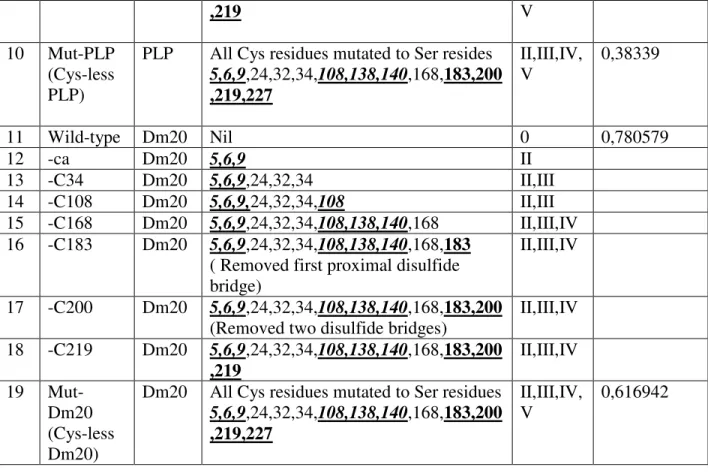
1.4.2 Cys residues and hydrophobic properties
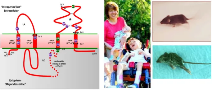
Of fourteen Cys residues in PLP, six at amino acid positions 5, 6, 9, 108, 138 and 140 are involved in post-transacylation modification, four Cys residues are involved in formation of disulfide bridges i.e., Cys183-Cys227 and Cy200-Cys219, and the remaining four Cys residues at amino acid positions 24, 32, 34 and 168 are located in the hydrophobic domains in the myelin membrane (Weimbs and Stoffel, 1992; Wahle and Stoffel, 1998).
PLP is also soluble in organic solvents such as chloroform/methanol due to its strong hydrophobic nature. It is composed of more than 50% of apolar (hydrophobic) residues that are spanned in the phospholipid bilayer as four hydrophobic membrane domains (Wahle and Stoffel, 1998; Stoffel et al., 1984; Weimbs and Stoffel, 1992). PLP also undergoes post-translational modification i.e., acylation where fatty acid (usually palmitic acid) is covalently bound to thiol groups of Cys residues of PLP. Acylated fatty acids of PLP might interact with the adjacent myelin phospholipid bilayer and also contribute to the hydrophobic nature of PLP (Weimbs and Stoffel, 1992). Alterations in PLP such as point mutations, deletions, duplication, missense mutation, over-expressions lead to disturbance in the PLP topology or conformation and abnormal targeting in oligodendrocytes. PLP misfolding may result in oligodendrocytes apoptosis and thus myelin disorders such as PMD and SPG2.
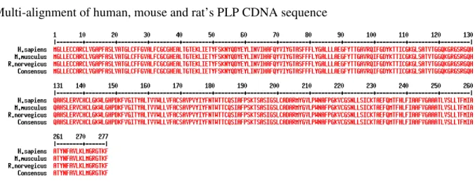
Multi-alignment of human, mouse and rat’s PLP CDNA sequence
Figure 4: Alignment of human, mouse and rat protein sequences. 100% homology (consensus) is
observed among human (H.sapiens), mouse (M.musculus), and Rat (R. norvegicus) on protein sequence.
‘Align’ software was used for alignment and respective protein sequences were obtained from ensemble database.
Figure 5: Topology of proteolipid protein (PLP) and its isoform Dm20. Dm20 lacks 35 amino acids from 116 to 150 in the hydrophilic loop of PLP (absence of violet color dashed line). PLP/Dm20 contains four hydrophobic membrane spanning domains, one intracellular and two extracellular loops, two disulphide bridges in the extra-cytosolic-II loop and post-translational modifications (transacylation on Cys residues).
Point mutations, disulfide bonds, and cell surface targeting
PLP/Dm20 is synthesized in oligodendrocytes on membrane bound polysomes and via Golgi vesicular transport it is targeted to the plasma membrane. Calnexin, an ER chaperone, binds to the newly synthesized PLP in the ER and contributes in its proper folding and ER quality control (Swanton et al., 2003). Properly folded PLP exits the ER and interacts with lipid rafts for vesicular transportation to the cell surface, whereas misfolded PLP (due to point mutations) is stably coupled with calnexin and does not exit the ER. Moreover stable binding of PLP with calnexin chaperone further prohibits ER-protein degradation, thus accumulating PLP in the ER, causing ER stress and further triggering apoptosis via unfolded-protein response (UPR), which in turn may lead to PMD or SPG2. The impairment of the PLP association with lipid rafts, due to its misfolding, might perturb its normal transport leading to a condition called as SPG2
13
during its intracellular transport excess PLP constituting lipid rafts are trapped in the lysosome which triggers oligodendrocyte apoptosis, a situation observed in dysmyelination (Simons et al.
2002). In less severe forms of PMD or SPG2, PLP is trapped in the ER, but its isoform Dm20 is targeted normally to the cell surface, indicating that PLP and Dm20 are differentially targeted, which correlates with the extent of PMD pathology.
The proximal (Cys183-Cys227) and outer disulphide bridges (Cys200-Cys219), located in EC-II loop of PLP/Dm20 are critical in trafficking. Their functional role under in vivo conditions specifically in cell surface trafficking of PLP/Dm20 and in myelin phenotype is still unknown. In the heterologous COS-7 cells of in vitro system, PLP, lacking the proximal disulphide bridge, is retained in the ER and is not detected on the cell membrane. Therefore the proximal disulphide bridge is involved in ER quality control. In contrast, PLP lacking the outer disulphide bridge passed ER quality control and appeared on the cell membrane similar to the wild-type PLP.
Therefore the outer disulphide bridge is not involved in ER quality control. PLP devoid of both disulphide bridges is ER retained (Dhaunchak and Nave, 2007). Interestingly, there is also a human case report demonstrating naturally occurring point mutation at Cys219Tyr deleting the outer disulfide bridge of PLP. This point mutation traps PLP in the ER and expresses the connatal PMD phenotype (Fukumura et al., 2010).
1.4.3 PLP related myelin disorders; PMD and SPG2
Naturally occurring mouse mutants such as rumpshaker mouse containing a point mutation (I186T) in PLP (Schneider et al., 1992), Jimpy mouse mutant with an A242V point mutation in PLP, and a PLP transgenic mouse with extra PLP copies (Redhead et al., 1994), exhibited severe myelin defects such as dysmyelination and hypomyelination. Some of these myelin defects resulted in PMD or SPG2 diseases. The main molecular mechanism that underlies PMD and SPG2 is impaired targeting of PLP to the oligodendrocyte plasma membrane (Seitelberger, 1995;
Werner et al., 1998) and oligodendrocyte apoptosis. The different trafficking of PLP and Dm20 in oligodendrocytes is also a determining factor influencing PMD severity. Both isoforms, when retained in the ER, result in severe forms of connatal PMD. When PLP alone is retained in the ER and Dm20 is targeted normally to the oligodendrocyte cell surface a less severe or classical form of PMD is observed (Gow and Lazzarini, 1996).
PMD or SPG2 mouse models exhibit phenotypes such as neuromotor defects including leg and arm muscle atrophy, seizures, tremors, cognitive and behavioral defects. They die prematurely within few weeks after birth (Yool et al., 2000). The PMD mouse models closely resemble human PMD on the molecular level, such as mutations in PLP. PMD patients exhibit mild to
severe abnormalities starting from their first postnatal year with gradual progression; delayed cognitive development, spasticity or muscle weakness, partially controlled neck and head movements and involuntary eye moments. Patients with the severe form of SPG2 exhibit defects in motor development including partial extent of cognitive defects of PMD.
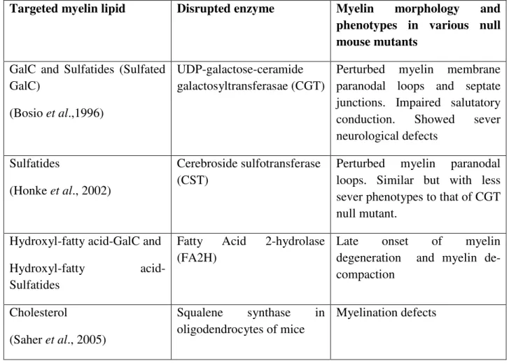
Figure 6: PLP with some of the point mutations which lead to PMD/SPG2. Children effected with PMD, and the severely affected PMD (PLP jimpy, A242V) mouse model. (Pictures adapted and modified from Grossi et al., 2011, CURE-PMD organization).
1.5 CNS Myelin; Phospho- and sphingolipids
Lipids constitute 70% of the total CNS myelin dry mass, with galactocerebrosides (GalC) being the most abundant lipid class. Major lipid types in the whole brain and in myelin (white matter) are cholesterol, phospholipids, and glycosphingolipids which are present in a ratio of 4:3:2 (Norton and Cammer, 1984). Phospholipids of myelin contain 70% phosphatidylethanolamine as plasmalogens compared to 5% in liver. Phospholipids are asymmetrically distributed in the myelin membrane; wherein mostly phosphoglycerides are arranged in the cytoplasmic leaflet of myelin MDL and galactosphingolipids (GalC and sGalc) at the exoplasmic face of the myelin IDL (Figure 3.D). The important role of lipids and proteins in the myelin membrane was verified
15
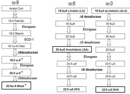
Targeted myelin lipid Disrupted enzyme Myelin morphology and phenotypes in various null mouse mutants
GalC and Sulfatides (Sulfated GalC)
(Bosio et al.,1996)
UDP-galactose-ceramide galactosyltransferasae (CGT)
Perturbed myelin membrane paranodal loops and septate junctions. Impaired salutatory conduction. Showed sever neurological defects
Sulfatides
(Honke et al., 2002)
Cerebroside sulfotransferase (CST)
Perturbed myelin paranodal loops. Similar but with less sever phenotypes to that of CGT null mutant.
Hydroxyl-fatty acid-GalC and Hydroxyl-fatty acid- Sulfatides
Fatty Acid 2-hydrolase (FA2H)
Late onset of myelin degeneration and myelin de- compaction
Cholesterol
(Saher et al., 2005)
Squalene synthase in oligodendrocytes of mice
Myelination defects
Table 2: Important lipids of myelin, their enzymes, and their role in myelin morphology
The retina, a highly organized CNS structure, is rich in phospholipids and is essential for visual functions. Phospholipids of retina consists 54% of phosphatidylethanolamine (PE) and 30-40%
phosphatidylcholine (PC), which constitutes mainly PUFA at both sn1 and sn2 positions.
Impairment in PUFAs composition may alter visual function. Retina morphology and function, is further discussed in section 1.5.4.
1.5.1 Fatty acid composition of phospholipids
Phosphoglycerides have a glycerol backbone, and sphingolipids have a sphingosine back bone.
In phosphoglycerides; two fatty acids, generally a saturated and an unsaturated or polyunsaturated fatty acid (PUFA), are bound as esters at the sn-1 and sn-2 of the glycerol backbone. In sphingolipids; only single fatty acid chain, generally a saturated or un- or polyunsaturated, is linked with an amide bond to the sphingosine back bone. In phosphoglycerides, a polar head group such as choline, ethanolamine, serine and inositol, is linked by a phosphate ester bond to a third carbon (C3) of the phosphoglyceride back bone.
Thus, based on the type of polar head group attached, the phosphoglycerides are termed accordingly as phosphatidylcholine, phosphatidylethanolamine, phosphatidylserine, and phosphatidylinositol. Sphingolipids contain polar head groups such as phosphocholine in sphingomyelin, and glucose or galactose polar head groups in cerobroside (GluC/GalC).
The basic structure of membrane phospholipids is depicted in the figure 7.
The appropriate nature and composition of saturated and unsaturated or polyunsaturated fatty acids in phospholipids of cell membranes are critical in maintaining respective membrane conformation, stability, fluidity and dynamics. Besides structural roles, fatty acids also serve in a variety of functions such as energy reserves, precursors of eicosanoids, and regulators of myelin gene expression in the brain. Deficiency or lack of essential fatty acids (EFAs) in mammals causes physiological disorders and is incompatible with life.
17
Figure 7: Myelin membrane composed of asymmetrically distributed phospholipids, and major myelin proteins. Zoomed box: depicting a phospholipid; phosphatidylcholine with its saturated and unsaturated hydrophobic fatty acid tails and the hydrophilic choline polar head group (Adopted and modified from Uschkureit, 2000).
1.5.2 Classification of fatty acids and the polyunsaturated fatty acids (PUFAs) synthesis cascade
Fatty acids are broadly classified into saturated and unsaturated fatty acids based on the number of carbon-carbon double bonds. Saturated fatty acids have no carbon-carbon double bonds, e.g.
palmitic acid (16:0) and stearic acid (18:0). They are mostly involved in energy storage in the form of triglycerides in adipose tissue and are also major structural components in membrane phospholipids. Unsaturated fatty acids are classified into monounsaturated fatty acids (MUFAs) and polyunsaturated fatty acids (PUFAs). MUFAs contain only a single carbon-carbon double bond, e.g., oleic acid (18:1, n-9). PUFAs are further classified into omega-9, omega-6, and omega-3-fatty acids (Figure 8) and contain more than one carbon-carbon double bond. PUFAs are the important structural constituents of membrane phospholipids; their carbon-carbon double bonds introduce kinks in the membrane phospholipids. The degree of double bonds or unsaturation in the phospholipid fatty acid chains may increase membrane fluidity and decrease membrane melting point and transition temperature. PUFAs such as arachidonic acid (AA), docosahexanoic acid (DHA), are also involved in cell signaling and are precursors for variety of metabolic processes. According to the omega (ω/n) classification system, the carbon-carbon double bond numbering starts from the methyl carbon terminal.
19 Polyunsaturated fatty acid biosynthesis
In mammals, the majority of PUFAs are acquired from diet and by transforming from their precursors essential fatty acids (EFAs) such as linoleic acid (18:2, n-6) and alpha-linolenic acid (18:3, n-3) endogenously via specific fatty acid synthases, elongases, and desaturases.
In mammals, delta-desaturases such as delta-9, delta-5, and delta-6 introduce double bonds at defined positions in respective fatty acids. Delta-9 desaturases, also called stearoyl-CoA desaturase-1 (SCD), introduce the double bond at the 9, 10 position from the carboxylic terminal in 18:0 fatty acid (Figure 9) thus generating 18:1 (n-9) monounsaturated fatty acid. Four SCD isoforms have been identified in mouse and they are tissue-specific. The SCD1 isoform is expressed in liver whereas SCD2 is brain specific, and SCD3 and SCD4 isoforms are skin and heart specific, respectively. In the PUFA synthesizing cascade, elongase is involved in extending the fatty acid chain.
PUFAs (n-3 and n-6) are synthesized by desaturation from their respective precursor fatty acids in the ER membrane by ER membrane-bound desaturases i.e., delta-5 desaturase also called as Fads1, and delta-6 desaturase also called as Fads2. Fads1 introduces at position 5 the cis double bond in their respective precursor fatty acyl chains, Fads2 at position 6 of EFAs in the initial and further steps of the PUFA biosynthetic pathway (Figure 9). Lack of fads2 enzyme disrupts PUFA biosynthesis, thereby abolishing synthesis of endogenous the major PUFAs such as AA (20:4 n- 6), EPA (20:5, n-3), and DHA (22:6, n-3), along with very long chain-PUFAs (Stoffel et al., 2008; Stroud et al., 2009).
Figure 9: The cascade of PUFA biosynthesis by desaturases and elongases in mammals.
1.5.3 PUFAs in the CNS
PUFAs and very long chain-PUFAs (VL-PUFAs) are the major components in phospholipids of the myelin membrane in the CNS. Among the PUFAs, AA and DHA are the major constituents of the CNS brain and in retinal phospholipids in contrast to other cell membranes. It is known that AA and DHA are highly conserved and are critical for normal physiological functions in the brain and retina (Carrie et al., 2000a). AA and DHA are also precursors of eicosanoids and docosanoids, respectively, and also carry out a myriad of pharmacological functions.
Deficiency of PUFAs may disrupt membrane bilayer conformation, cell polarity and tight junctions, which might in turn disrupt respective physiological functions. This has been shown in Sertoli cell polarity and in blood testis-barrier, and in granulosa cells of ovarian follicles (Stoffel et al., 2008).
The supplementation of omega-3 and omega-6 PUFAs for brain development, especially in early myelin developmental stages, is an active research area in nutritional and brain physiology (Yehuda et al., 2002). Miller et al., 1984 demonstrated that a lack of essential fatty acids (EFAs) results in a decrease of total myelin protein content to 25%, lipid phosphorous content to 22%, brain weight to 7% and a reduction in body weight up to 40%. PUFAs play an important role in myelinogensis by influencing myelin gene expression levels (Clarke, 2001). Eicosapentaenoic acid (EPA) supplementation to glial cells in vitro up-regulated PLP gene expression levels via cAMP-mediated pathways (Salvati et al., 2004), whereas under DHA supplementation PLP gene expression is up-regulated via retinoid X receptor signaling pathway (Jang et al., 2004). In vivo, injecting DHA and EPA into rat brain stimulated several myelin proteins such as PLP, MBP, MOG and CNP, via down-regulating CREB (cAMP-response element-binding) activity due to decreased levels of CREB phosphorylation (Salvati et al., 2008). In clinical studies PUFAs supplementation has been promising in remyelination, especially in multiple sclerosis (MS) with demyelinating disorder. PUFAs are also used in minimizing neurotoxin levels in schizophrenia.
In mammals, perturbed behavioral physiology such as impaired cognitive ability and motor coordination has been observed in EFA deficient conditions (Vinot et al., 2011; Chengwei He et al., 2009).
Retinal photoreceptor rods and cones outer segments (OS) consist of thousands of vertically staked membrane disks that are made up of lipoprotein bilayers enriched with DHA and AA (Bazan et al., 1993). Electroretinography (ERG) is used to assess retinal photoreceptor function and retinal photo-transduction cascade which is triggered in response to light (Weisinger et al., 1996a). ERG output is the direct measurement of the visual function of the retina in the presence of light. LC-PUFA deficiency may affect the rod outer membrane and RPE membrane and thus may impair normal visual functions (Jeffrey et al., 2001).
21 1.5.4 Retinal morphology
In vertebrate embryonic development, the retina and optic nerve originate as an outgrowth of the developing brain. The vital role of the retina is in the conversion of light signals into nerve impulses that are passed along the optic nerve to different visual centers of brain. Outer segments of retinal photoreceptors and retinal synapses are highly enriched in PUFAs, specifically DHA.
The retinal pigment epithelium (RPE) is involved in nutrient transport and removal of waste products from photoreceptors by the apical microvilli. The basal surface of the RPE interacts with underlying Bruch membrane (Figure 10). RPE is separated from the choriocapillaris (CC) by Bruch membrane (Figure 10). The outer segments (OS) are made up of sensory cilia and a series of membrane stacks composed of 60% of DHA, which are important in light conduction.
DHA depletion may result in retinal degeneration and functional impairments.
Figure 10: Morphology of the retinal layers (Human). Right upper diagram depicts the layers of the retina: CC, choriocapillaris, BM, Bruch’s membrane; RPE, retinal pigment epithelium; ap, apical processes or RPE apical microvilli; os, outer segments; C, cones, R, rods; M, Müller cells. Right lower image: electron micrograph of human retinal RPE region (adapted and modified from Web vision-retina)
1.5.5 Fads2 knock-out mouse (PUFAs deficient mouse model)
The fads2 (Delta6) null mouse mutant has been generated in our lab (Stoffel et al., 2008) to investigate the functional significance of PUFAs deficiency without depleting their precursor EFAs (LA and ALA). The fads2 null mouse lack the synthesis of all PUFAs including AA and DHA along with their derivatives such as eicosanoids and LC-PUFA (Stoffel et al., 2008, Stroud et al., 2009). Since DHA and AA are the major representatives among PUFAs in the brain and retina, lack of endogenous PUFAs biosynthesis especially AA and DHA in the fads2 null mouse mutant enables us to investigate the significance of PUFAs in membrane structure and function focusing on the CNS i.e., brain and retina.
The generation of gene targeted mouse mutants (transgenic mice), in which key enzymes of lipid biosynthesis have been deleted are central to the key strategy to unravel the role of specific lipid functions. These models are similar to the mouse models in which key protein structures of CNS have been deleted.
1.6 Transgenic mouse models
A very important method for analyzing gene function, from its role in normal development to pathology, is possible by generating gene manipulated mouse models, in which a specific gene of interest is deleted, modified or over-expressed. Genetically engineered mouse mutants can be obtained by several ways, such as by gene targeting (Capecchi et al., 1994) in which the endogenous copy of a gene is partly or completely deleted by homologous recombination, or by pronuclear injection technique (Gordon and Ruddle, 1983) where a gene can be over-expressed at random loci in the genome.
1.6.1 Gene targeting
The technique of gene targeting involves the mutation of a specific gene locus by the homologous recombination in embryonic stem cells. It is most often employed for generating gene knock-out or knock-in mutants. In the gene knock-out approach, specific gene function is usually eliminated by deleting partly or completely the endogenous gene by homologous recombination, whereas in a gene knock-in approach, instead of eliminating gene function the gene is manipulated as desired, e.g., by introducing point mutations in specific loci.
23
In the gene targeting technique, initially a gene-targeting construct with the desired modifications such as point mutations, partial gene deletions, insertion of fluorescent gene markers etc., is engineered. This is then cloned into a gene targeting vector and further transfected into pluripotent ES cells. The mutated homologous gene fragment then recombines with the endogenous gene counterpart in the ES cells (Thomas and Capecchi, 1986). Increased length of the exogenous targeting homologous arms will increase the frequency of homologous recombination with the endogenous locus (Hasty et al., 1991b). The targeting vector also contains one or more selection markers such as a neomycin resistance gene and a thymidine kinase gene from herpes simplex virus (HSV-TK). Positive or negative selection markers allow for screening of homologously recombined clones from a pool of clones (Mansour et al., 1988).
Successfully homologously recombined positive ES clones will be verified by PCR and Southern blot hybridization. Positive ES clones are then injected into mouse embryos at the blastocyst stage (Bradley et al., 1984) and re-implanted in to pseudo-pregnant foster mice. Thus obtained chimeric progeny are verified by PCR and DNA sequencing. The verified chimeras can be further bred to generate germ line chimeric heterozygotes and further to homozygous mutants.
1.6.2 Pronuclear Microinjection
The pronuclear micro-injection technique is fast and robust for generating transgenic mouse mutants to investigate gene function in vivo. An exogenous gene or cDNA from the appropriate gene, termed transgene, is introduced into the pronucleus of fertilized murine eggs by microinjection. The transgene is a DNA fragment usually containing a tissue-specific enhancer, promoter, and the gene-of- interest (gene or protein coding CDNA). In this technique, multiple transgene copies are stably integrated into the mouse genome at random loci. Pronuclei injection technique can be used to investigate gene gain-of-function (Babinet, 2000), and gene localization by tagging or fusing gene of interest with the appropriate reporter gene such as EGFP or E. coli beta-galactosidase. The transgene can also micro-injected into the fertilized eggs from a specific genetic background such as a null-mutant. The main disadvantage of the transgenic technique is the position effect, i.e., the pronuclear injected transgene integrates randomly and therefore may disturb the function of other genes. Hence large numbers of founder mice have to be analyzed to assess the true function of the injected transgene.
1.7 Site-directed mutagenesis
The site-directed mutagenesis (SDM) technique is used to introduce mutations (point or multiple), insertions and deletions at desired positions in a DNA fragment, by using synthetic oligonucleotides (Itakura et al., 1984; Carter, 1986). In SDM, the DNA sequence that has to be manipulated is employed as a template in the PCR reaction with mismatched nucleotide containing primers, and these primers are then extended on the template synthesizing new DNA fragments incorporating the mutations or mismatches. Among various methods of PCR-based mutagenesis, basic strategies employed within this thesis work are conventional PCR mutagenesis, which introduces mutations towards the 5’end and 3’ ends of the desired template DNA fragment, and overlap-extension PCR, which introduces mutations at more than one desired location within the target DNA template fragment.
In overlap-extension PCR, multiple mutations are introduced in a single PCR reaction by employing more than one set of primers containing nucleotide mismatches (mutations). It can also be employed by combining two individual DNA fragments at their terminal overlapping sequence.