Bacterial contamination rates in extracorporeal photopheresis
Irene Pamler,
1,†Eva Richter,
1,†James A. Hutchinson ,
2Viola Hähnel ,
1Ernst Holler,
3André Gessner,
4Ralph Burkhardt ,
1and Norbert Ahrens
1,5BACKGROUND:Extracorporeal photopheresis (ECP) is an immunosuppressive treatment that involves leukocyte apheresis, psoralen and UV light treatment, and subsequent reinfusion. Patients treated with ECP are usually immunosuppressed. Bacterial contamination therefore poses a much unwanted risk, but incidence data are lacking.
PATIENTS AND METHODS:We screened all 1922 consecutive ECP procedures scheduled within a roughly 3-year period for eligibility. Those with missing data on ECP method (inline or offline) or type of venous access (peripheral or central) were excluded. ECPs with complete aerobic and anaerobic microbial testing of baseline patient blood samples (n = 1637) and of ECP cell concentrates (n = 1814) were included in the analysis.
RESULTS:A test for microbial contamination was positive for 1.82% of the cell concentrates, with central venous access was the most significant risk factor for the contamination (odds ratio = 19). Patient blood samples were positive in 3.85% of cases, but no patients became septic. Staphylococcus spp. were most abundant, and products with bacterial contamination did not cause side effects after reinfusion. There were no significant differences in contamination rates between inline and offline ECP.
CONCLUSION:Thesefindings stress the importance of sterile procedures and the benefits of using peripheral over central venous access for reducing the risk of bacterial contamination in ECP.
E xtracorporeal photopheresis (ECP) is a therapeutic procedure consisting of leukocyte apheresis, treat- ment of the collected cells with 8-methoxypsoralen (8-MOP), ultraviolet A (UVA) light irradiation, that causes covalent binding of 8-MOP with DNA, and subse- quent reinfusion of treated product back to the patient with- out storage.1 This therapy adds to transplantation tolerance or in autoimmune diseases and is typically applied for patients that are refractory to
first line therapy and that are often subject to a complex immunosuppressive regimen.
2
The required medical devices are available as com- bined equipment with apheresis and UVA irradiation in a single machine (inline ECP), or as separate devices (of
fline ECP). The latter requires that the user connects the leukapheresis bag with the UV irradiation bag, as the former is not UVA permeable. The components may be connected prior to the procedure (closed ECP), or during the proce- dure while the patient is connected (open ECP, sometimes classi
fied differently by regulatory authorities). Possible combinations of these are closed inline, closed of
fline, and
From the1Institute for Clinical Chemistry and Laboratory Medicine, the2Department of Surgery, the3Department of Hematology and Oncology, and the4Institute of Clinical Microbiology and Hygiene, University Hospital Regensburg, Regensburg, and5Institute for Laboratory Diagnostics, Microbiology, and the Transfusion Medicine, Sozialstiftung Bamberg, Germany.
Address reprint requests to:Norbert Ahrens, University Hospital Regensburg, Institute for Clinical Chemistry and Laboratory Medicine Franz-Josef-Strauss-Allee, 11 93053 Regensburg, Germany; e-mail: norbert.ahrens@ukr.de.
This is an open access article under the terms of the Creative Commons Attribution-NonCommercial-NoDerivs License, which permits use and distribution in any medium, provided the original work is properly cited, the use is non-commercial and no modifica- tions or adaptations are made.
†These authors contributed equally to this project and should
be considered co-first authors.
Received for publication December 6, 2019; revision received February 27, 2020, and accepted March 1, 2020.
doi:10.1111/trf.15801
© 2020 The Authors. Transfusion published by Wiley Periodicals, Inc. on behalf of AABB.
TRANSFUSION2020;60;1260–1266
open of
fline ECP. Apart from this, both ECP types require connections to apply 8-MOP.
To start with ECP, leukapheresis requires access to peripheral veins or a central venous catheter (CVC). Periph- eral venous access used to cause bacterial contamination in 1
–2% of healthy blood donors until the advent of pre- donation sampling, a preventive measure that introduced a bag to the tubing system for the initial 15
–30 mL of the donation process.
3,4This in combination with improved skin disinfection lowered the rate of microbial contamina- tion to approximately 0.03%.
5,6In autologous stem cell transplant (ASCT), on the other hand, bacterial contamina- tion rates of up to 4.5% are still common.
7,8The affected patients are treated under antibiotic protection, as there is suf
ficient time between donation and reinfusion to obtain diagnostic test results.
9In ECP, however, bacterial contami- nation typically remains undetected, because the ECP prod- uct is quickly reinfused without bacterial testing.
Undetected bacteria could, in principal, have severe conse- quences. Up to now, data on bacterial contamination rates in ECP are lacking. Therefore, we retrospectively collected and analyzed data from sterility testing of ECP procedures performed at our institution to get to know the incidence of bacterial contamination in ECP patients and products.
PATIENTS, MATERIALS, AND METHODS Patients
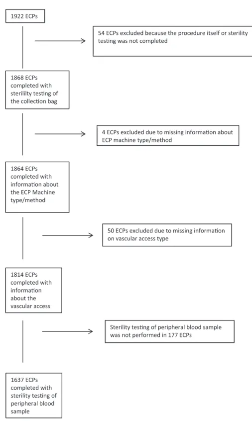
A total of 1922 ECP procedures scheduled at our hospital from September 26, 2012 through August 31, 2015 were screened for eligibility (Table 1). Most of the patients (79%) had suffered graft-versus-host disease (GvHD) after autolo- gous stem cell transplantation and received treatment on the basis of their clinical symptoms for several weeks to sev- eral months. Patients with other conditions like cutaneous autoimmune diseases were usually treated for longer periods. Treatment frequencies were adjusted to clinical needs. Treatments that met eligibility criteria were included in the analysis as outlined in Fig. 1.
ECP treatment protocol
Treatment was carried out as either closed inline or open off- line ECP as described above.
10Closed inline photopheresis can be administered with a single needle and was therefore preferentially but not exclusively used for patients with lim- ited venous access. Other patients received of
fline ECP.
Photopheresis was carried out as described previ- ously.
10Brie
fly, standardized open of
fline ECP and closed inline ECP procedures were used.
11Open of
fline ECP was performed with the Cobe Spectra (Cobe, Terumo BCT), Spectra Optia (Optia, Terumo BCT) or the Amicus (Fresenius Kabi) device using acid citrate dextrose (ACD-A) for anticoagulation. Heparin was additionally used if clotting was observed. Patients received calcium as required. ECP
was delivered via a central or peripheral venous access depending on the condition of the patient
’s veins.
Required connections in open of
fline ECP were between patient and apheresis tubing set, for sampling and 8-MOP application, to the UV irradiation bag, and back to the patient.
Transfusion sets with 200
μm
filters were used for the latter.
Closed inline ECP was performed with the Uvar XTS (Therakos). There were no connection steps apart from venous punctuation and sampling with 8-MOP addition.
Sterility testing
Blood samples for sterility testing were taken from the peripheral or central venous line before the start of aphere- sis. Samples from the cell concentrates were taken before 8-MOP addition and UV illumination at the bedside, as closed inline ECP does not allow removal of the bag. Treat- ments and sampling were performed in controlled environ- ment equipped with H13
filtered ventilation. Aerobic and anaerobic culture bottles (BD Bactec Standard Anaerobic/F and Aerob/F, respectively) were incubated for 7 days at 30-32°C with a sample volume of 3-5 mL and 7-10 mL, respectively. Positive cultures were isolated and differenti- ated by matrix-assisted laser desorption ionization-time of
flight (MALDI TOF, Bruker Daltonik GmbH Life Sciences) mass spectrometry, and antibiotic resistance testing was performed using the BD Phoenix system.
Statistical analysis
Data was collected in Microsoft Excel 2010, and R was used to calculate statistical signi
ficance using the Wilson score interval with continuity correction using its built-in prop.test function.
TABLE 1. Patient characteristics
Patients* 68
Sex (male [%] / female [%]) 57,4%/42,6%
Age [years] 53 (2–73)
Diagnoses*
Acute GvHD 29
Chronic GvHD 25
Sézary’s disease 4
Atopy 1
Scleroderma 4
Psoriasis 1
Crohn’s disease 1
Bronchiolitis obliterans following lung transplantation
3 Number of treatments per patient 19 (1–136) Total number of completed treatments 1868
Closed inline ECP (Uvar XTS) 25.3%
Open offline ECP (Cobe) 71.7%
Open offline ECP (Optia) 1.9%
Open offline ECP (Amicus) 0.8%
Missing data on method 0.2%
Numbers represent median value with range in parentheses unless otherwise indicated.
* Total number.
RESULTS Bacterial contamination rate
Complete information on the ECP method (online or off- line), type of vascular access (central or peripheral), and
bacterial contamination rate (sterility testing) was available for 1814 ECP cell concentrates. We identi
fied a positive microbial test results in a total of 33 ECP cell concentrates (1.82%). Central venous access, which was required in 20%
Fig. 1. Patient recruitment and causes of drop out.
of the ECP procedures, was more frequently associated with bacterial contamination of ECP cell concentrates (7.38%
positive) than peripheral access (0.41% positive, Table 2, p < 2.2
×10
−16, odds ratio 19). Patient blood samples col- lected before the start of the procedure were available in 1637 cases. From these, 63 tested positive, with signi
ficant differences between central and peripheral venous access (Table 2, p = 0.0026, odds ratio 2.3).
Overall, we found 73 cases of 1637 treatments, in which sterility testing was positive either in cell concentrates and/or patient blood samples. The rate of overlap between procedures with positive ECP cell concentrates and positive patient blood sample was low (16 of 73, 22%). In an addi- tional 10 procedures, only the cell concentrate was positive (14%), and in another 47 instances, only the patient blood sample was positive (64%).
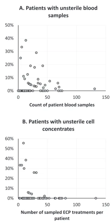
Patient blood samples had a signi
ficantly higher contam- ination rate than samples from cell concentrates (3.85% of 1637 vs. 1.82% of 1814 respectively, p = 0.00044). Contamina- tion proportions in patients
’blood and in cell concentrates were not equally distributed. The frequency of positive sam- ples for patients
’blood and for cell concentrate samples was unequally distributed (Fig. 2). Patients with fewer ECP treat- ments tended to have more frequent unsterile
findings.
Of note, we did not detect signi
ficant differences between inline and of
fline ECP with regard to bacterial detection rates in ECP cell concentrates or patient blood samples (p = 0.068 and p = 1, respectively, Table 2). In addi- tion, none of the bacterially contaminated products caused adverse events upon reinfusion.
Bacterial species distribution
Samples testing positive for microbiological contamination exhibited a large and heterogenous variety of germs. The majority of the identi
fied bacteria or fungus belong to the human skin
flora, mouth
flora, and/or intestinal
flora:
Staphylococcus epidermidis, Propionibacterium acnes, Acinetobacter Iwof
fi, Actinomyces odontolyticus, Staphylo- coccus capitis, Micrococcus luteus, Staphylococcus aureus, Staphylococcus hominis, Citrobacter freundii, Candida guilliermondii, Enterobacter cloacae, Prevotella bivia, Haemophilus parain
fluenzae, Staphylococcus haemolyticus,
TABLE 2. Microbial detection rates*for (A) offline versus inline ECP and (B) peripheral versus central venous access
ECP products Patient blood
Percent positive 95% CI n Percent positive 95% CI n
(A)
Offline ECP 2.18% (1.50–3.13%) [30/1378] 3.93% (2.94–5.22%) [48/1220]
Inline ECP 0.69% (0.18–2.17%) [3/436] 3.60% (2.10–6.00%) [15/417]
(B)
Peripheral access 0.41% (0.17–0.95%) [6/1448] 3.11% (2.27–4.23%) [41/1319]
Central venous access 7.38% (5.00–10.68%) [27/366] 6.92% (4.49–10.44%) [22/318]
* Detection rates refer to different sample populations.
Fig. 2. Proportion of unsterile samples from peripheral or central venous blood (A) or from ECP cell concentrates (B) per patient.
Staphyloccous simulans, Staphylococcus lugdunesis, Enterococ- cus faecium, Klebsiella pneumoniae, and Capnocytophaga ochracea.
In addition, some species are environmental bacteria that can be found in soil, air, and/or water: Spores, Micro- coccus luteus, Bacillus pumilus (resistant to UV light), Can- dida guilliermondii, Enterobacter cloacae, and Pseudomonas aeruginos. Interestingly, Bacillus altitudinis is a germ which was
first isolated from cryogenic tubes used for collectingair samples from high altitudes.
12Five patient blood samples contained two different bacte- ria (Acinetobacter Iwoffii and Staphylococcus aureus, Staphylo- coccus aureus and Staphylococcus epidermidis, Staphylococcus simulans and Staphylococcus epidermidis, Staphylococcus lugdunensis and Staphylococcus capitis, Prevotella bivia and Haemophilus parainfluenza). In one of these cases, the cell concentrate also tested positive, but only for one bacterial spe- cies (Prevotella bivia).
The distribution of detected bacterial species separated by ECP system type is shown in Table 3.
DISCUSSION
In this large retrospective study, we found an overall micro- bial contamination rate of 1.82% in the cell concentrates of extracorporeal photopheresis. This
figure is comparable to that of apheresis procedures for the collection of other blood components, such as platelets and hematopoietic stem cells.
Bacterial contamination rates in platelet concentrates from healthy donors reportedly range from 0.01 to 0.2%.
13The frequency of positive culture tests that failed in con
fir- mation testing is usually higher, i.e., 0.11 to 0.72%.
5,14,15Negative con
firmation testing can occur due to inappropri- ate diagnostic techniques and may re
flect contamination with low numbers of bacteria in the inoculum or with bacte- ria that require special growth conditions. We did not dis- tinguish between these factors and included all positive
findings. Thus, the initial positive frequencies are techni- cally closer to those observed in our patients.
Bacterial contamination rates in hematopoietic stem cell apheresis range from 0.2 to 24%, averaging about 3%.
16Higher frequencies in patients may be caused by additional handling steps during the procedure. These include disin- fection as well as the diversion of the
first milliliters after venipuncture for predonation sampling which, however, has been shown to substantially decrease the contamination rate.
17The type of venous access used is another difference between healthy blood donors and patients undergoing apheresis. In our study, a central venous catheter (CVC) was required in 20% of ECP procedures, and CVC use was associated with a 19-fold higher risk for cell concentrate contamination. Though none of the patients became obvi- ously septic at the time of treatment, these
findings illustrate the risk of bacteremia in patients with a central venous line.
Microbial contamination risk was not evenly distributed among the patients. We found a subgroup of patients with a high frequency of contaminations up to a maximum of 56%
of the cell concentrates. Patients with high contamination rates had a comparable low treatment number, e.g., because of uncontrollable grade IV graft-versus-host disease. In addi- tion, central venous catheter contamination contributed to positive
findings. Bacterial contamination in these patients indicates therefore at least in part the severity of the underly- ing disease.
Bacterial contamination may be impacted by the type of centrifugation. Recently intermittent
flow with the Amicus (Fresenius Kabi) was shown to be disadvantageous in platelet apheresis compared to continuous
flow apheresis using the Trima (Terumo BCT).
18,19This disfavors inline ECP that uses intermittent
flow technique. On the other hand, the buffy coat layer of bacteria is unknown. Bacteria could distribute freely in plasma, or they could sediment together with red blood cells and platelets. Intermittent
flow apheresis using the Latham bowl technique as in Therakos devices, in contrast to the technique used within the Amicus, collects less selected
TABLE 3. Bacterial species in ECP cell concentratesand in patients’blood Closed inline ECP*
Open offline
ECP* Patient’s blood† Peripheral venous access
Staphylococcus epidermidis – 4 13
Staphylococcus aureus – 1 13
Prevotella bivia – 1 1
Propionibacterium acnes Spore
Acinetobacter Iwoffii Actinomyces odontolyticus Staphylococcus capitis Micrococcus luteus Staphylococcus hominis Bacillus altitudinis Bacillus pumilus Bacillus subtilis Citrobacter freundii Haemophilus parainfluenza Staphylococcus
haemolyticus Staphylococcus simulans Staphylococcus lugdunensis
–– –– –– –– –– –– – ––
–– –– –– –– –– –– – ––
1 1 1 1 2 2 1 1 1 1 1 1 3 1 1 Central venous access
Staphylococcus epidermidis 3 12 11
Staphylococcus capitis – 7 5
Pseudomonas aeruginosa – 1 1
Enterococcus faecium – 2 –
Klebsiella pneumoniae Micrococcus luteus Staphylococcus hominis Candida guillermondii Enterobacter cloacae Capnoctyophaga ochracea
–– –– ––
1 –– –– 1
1 1 1 1 1 –
* Data from 1814 procedures.
†Data from 1637 procedures.
cell suspensions in general,
10,20though there are exceptions to this.
21Inferior collection ability of inline ECP could trans- late to a reduced bacterial enrichment. In addition, this method processes lower blood volumes, thus further decreas- ing the possibility of contamination in bacteremic patients.
Higher collection ability of of
fline ECP methods trans- lates to successful single day treatments.
22Inline ECP, in contrast, requires treatments on two adjacent days.
In our study, no signi
ficant differences were observed between microbial contamination in inline and of
fline ECP.
Patients were not randomized and preferentially treated off- line, if a central venous access was available. Central venous lines, in contrast to venous canula, are handled in a sterile way. However, this is more than outweighed by the risk from inapparent catheter infections. The contamination risk was thus increased in of
fline ECP in our study.
The difference in microbial contamination rates between ECP cell concentrates (1.82%) and patient blood samples (3.85%) might be explained by the fact that the lat- ter samples were collected before apheresis and, thus, had the same effect as predonation sampling. In addition, the apheresis technology itself could have contributed to bacte- rial depletion to some degree as described above.
We found 24 different bacterial species with Staphylococcus spp. being most abundant. Most of the bacteria we detected were part of human skin
flora. These may indicate a contamina- tion by the handling steps. However, these may also indicate that patients for ECP with skin diseases cannot be disinfected successfully with standard procedures. Contamination with the same bacterial type in bag and patient supports this explanation.
The type of bacterial contamination is of relevance from a clinical point of view, as antibiotic susceptibility depends in part on the species. From a technical point of view, there is no contamination that can be regarded as acceptable. There are no commensal bacteria in cell suspensions.
Regardless of the cause of microbial contamination, it is highly unwanted. Patients referred for ECP are usually severely immunosuppressed like in solid organ transplanta- tion or graft-versus-host disease. Their risk for septic com- plications is therefore increased. On the other side, bacterial contamination of ECP is questionable for two reasons. First, ECP cell concentrates are not stored but reinfused immedi- ately after UV treatment, thus eliminating the chances for bacterial replication. Second, 8-MOP injection with subse- quent UV light exposure acts like a pathogen inactivation that reduces bacterial growth potential by several log units.
23Thus, it can be assumed that any relevant bacterial replication potential is effectively reduced in ECP treatment.
Pathogen inactivation by ECP, however, is speculative.
It is therefore mandatory to avoid contamination as much as possible. Disinfection before venipuncture or central line connection was done following a standardized protocol in this study. Patients in need of ECP frequently have skin con- ditions that are prone to bacterial infections as illustrated by the
findings of this study. Thus, effective skin disinfection
techniques such as those de
fined for whole blood donation should also be used in ECP.
24In addition, the presented data do clearly favor peripheral access for ECP, as contami- nation by central venous access is by far the most signi
ficant risk for bacterial contamination.
This study is the
first to evaluate bacterial contamina- tion in ECP cell suspension bags and in ECP patients. Steril- ity sampling was scheduled for every treatment within the study period, and all patients were considered for inclusion.
Most patients and treatments could be included, but drop- outs could have impacted the results to some degree. Data have also to be interpreted with caution, as patient and treatment factors with potential impact on contamination, such as venous access and therefore type of ECP, were cho- sen on a clinical basis and not according to this study.
Though it is not possible to conclude on ECP types, it seems reasonable to recognize the contamination risk for ECP in general, especially for patients with central venous access.
The relevance of these
findings is unclear, as ECP com- prises always pathogen inactivation and cell suspensions would not be stored. This disfavors bacterial testing. The clinical relevance, however, is unclear, and clinical follow up was helpful. In addition, the clinical relevance of inap- parently contaminated catheters in these frequently immu- nosuppressed patients calls for further studies.
ACKNOWLEDGMENTS
Support for this work was funded by the Deutsche Forschungsgemeinschaft (DFG, German Research Foundation), project ID B13–TRR221 given to AG and EH.
We would like to thank the team of operators and physicians who conducted the photopheresis procedures for their help, sup- port, and excellent handling of the whole ECP process.
CONFLICT OF INTEREST
The authors have disclosed no conflicts of interest.
REFERENCES
1. Schneiderman J. Extracorporeal photopheresis: cellular therapy for the treatment of acute and chronic graft-versus-host dis- ease. Hematology Am Soc Hematol Educ Program 2017;2017:
639-44.
2. Schwartz J, Padmanabhan A, Aqui N, et al. Guidelines on the use of therapeutic apheresis in clinical practice-evidence-based approach from the Writing Committee of the American Society for Apheresis: The Seventh Special Issue. J Clin Apher 2016;31:
149-338.
3. Chassaigne M, Vassort-Bruneau C, Allouch P, et al. Reduction of bacterial load by predonation sampling. Transfus Apher Sci 2001;24:253.
4. Liumbruno GM, Catalano L, Piccinini V, et al. Reduction of the risk of bacterial contamination of blood components through diversion of thefirst part of the donation of blood and blood components. Blood Transfus 2009;7:86-93.
5. Walther-Wenke G, Wirsing von König CH, Däubener W, et al.
Monitoring bacterial contamination of blood components in Germany: effect of contamination reduction measures. Vox Sang 2011;100:359-66.
6. Hillyer CD, Josephson CD, Blajchman MA, et al. Bacterial con- tamination of blood components: risks, strategies, and regulation:
joint ASH and AABB educational session in transfusion medicine.
Hematology Am Soc Hematol Educ Program 2003;1:575-89.
7. Schwella N, Zimmermann R, Heuft HG, et al. Microbiologic contamination of peripheral blood stem cell autografts. Vox Sang 1994;67:32-5.
8. Kozlowska-Skrzypczak M, Bembnista E, Kubiak A, et al. Micro- bial contamination of peripheral blood and bone marrow hematopoietic cell products and environmental contamination in a stem cell bank: a single-center report. Transplant Proc 2014;46:2873-6.
9. Klein MA, Kadidlo D, McCullough J, et al. Microbial contami- nation of hematopoietic stem cell products: incidence and clin- ical sequelae. Biol Blood Marrow Transplant 2006;12:1142-9.
10. Brosig A, Hähnel V, Orsó E, et al. Technical comparison of four different extracorporeal photopheresis systems. Transfusion 2016;56:2510-9.
11. Ahrens N, Geissler EK, Witt V, et al. European reflections on new indications for extracorporeal photopheresis in solid organ transplantation. Transplantation 2018;102:1279-83.
12. Shivaji S, Chaturvedi P, Suresh K, et al. Bacillus aerius sp. nov., Bacillus aerophilus sp. nov., Bacillus stratosphericus sp. nov.
and Bacillus altitudinis sp. nov., isolated from cryogenic tubes used for collecting air samples from high altitudes. Int J Syst Evol Microbiol 2006;56:1465-73.
13. Prax M, Bekeredjian-Ding I, Krut O. Microbiological screening of platelet concentrates in Europe. Transfus Med Hemother 2019;46:76-86.
14. Schrezenmeier H, Walther-Wenke G, Müller TH, et al. Bacterial contamination of platelet concentrates: results of a prospective
multicenter study comparing pooled whole blood-derived platelets and apheresis platelets. Transfusion 2007;47:644-52.
15. Vuk T, Barišic M, Hecimovic A, et al. Bacterial contamination of blood products at the Croatian Institute of Transfusion Med- icine: results of eleven-year monitoring. Transfus Med 2012;22:
432-9.
16. Störmer M, Wood EM, Schurig U, et al. Bacterial safety of cell- based therapeutic preparations, focusing on haematopoietic progenitor cells. Vox Sang 2014;106:285-96.
17. Satake M, Mitani T, Oikawa S, et al. Frequency of bacterial contamination of platelet concentrates before and after intro- duction of diversion method in Japan. Transfusion 2009;49:
2152-7.
18. Bravo M, Shaz BH, Kamel H, et al. Detection of bacterial con- tamination in apheresis platelets: is apheresis technology a fac- tor? Transfusion 2015;55:2113-22.
19. Eder AF, Dy BA, DeMerse B, et al. Apheresis technology cor- relates with bacterial contamination of platelets and reported septic transfusion reactions. Transfusion 2017;57:
2969-76.
20. Bueno JL, Alonso R, Gonzalez-Santillana C, et al. A paired trial comparing mononuclear cell collection in two machines for further inactivation through an inline or off- line extracorporeal photopheresis procedure. Transfusion 2019;59:340-6.
21. Piccirillo N, Putzulu R, Massini G, et al. Inline extracorporeal photopheresis: evaluation of cell collection efficiency. Transfu- sion 2019;59:3714-20.
22. Cid J, Carbasse G, Suarez-Lledo M, et al. Efficacy and safety of one-day offline extracorporeal photopheresis schedule processing one total blood volume for treating patients with graft-versus-host disease. Transfusion 2019;
59:2636-42.
23. Schlenke P. Pathogen inactivation technologies for cellular blood components: an update. Transfus Med Hemother 2014;
41:309-25.
24. McDonald C, McGuane S, Thomas J, et al. A novel rapid and effective donor arm disinfection method. Transfusion 2010;50:
53-8.