Analysis of the Immune Cell Infiltrates and Biomarkers during acute Gastrointestinal
Graft vs Host Disease
Dissertation
zur Erlangung des Doktorgrades der Biomedizinischen Wissenschaften
(Dr. rer. physiol.) der
Fakultät für Medizin der Universität Regensburg
vorgelegt von Sakhila Ghimire aus Kathmandu, Nepal
im Jahr 2016
SELBSTÄNDIGKEITSERKLÄRUNG
Ich, Ghimire Sakhila, geboren am 16 Februar 1988 in Kathmandu, Nepal erkläre hiermit, dass ich die vorliegende Arbeit ohne unzulässige Hilfe Dritter und ohne Benutzung anderer als der angegebenen Hilfsmittel angefertigt habe. Die aus anderen Quellen direkt oder indirekt übernommenen Daten und Konzepte sind unter Angabe der Quelle gekennzeichnet. Insbesondere habe ich nicht die entgeltliche Hilfe von Vermittlungs- bzw. Beratungsdiensten (Promotionsberater oder andere Personen) in Anspruch genommen.
Die Arbeit wurde bisher weder im In- noch im Ausland in gleicher oder ähnlicher Form einer anderen Prüfungsbehörde vorgelegt.
Ort, Datum eigenhändige Unterschrift des Promovenden
Declaration
I, Sakhila Ghimire, born on 16th February 1988 in Kathmandu, Nepal, hereby confirm that my doctoral thesis entitled ‘Analysis of immune cell infiltrates and biomarkers during acute gastro-intestinal graft vs. host disease’ is the result of my own work. I did not receive any help or support from commercial consultants. All sources and/or materials applied are listed and specified in the thesis.
Furthermore, I confirm that this thesis has not yet been submitted as a part of another examination process neither in identical nor in similar form.
Date Signature
The present work was carried out from April 2013 to September 2016 at the Department of Internal Medicine III at the University Hospital Regensburg.
Die vorliegende Arbeit entstand im Zeitraum von April 2013 bis September 2016 an der Klinik und Poliklinik für Innere Medizin III des Universitätsklinikums Regensburg.
I dedicate this doctoral thesis to my mother and my motherland.
जननी जन्मभूममश्चस्वर्गादपिर्रीयसी
Mother and motherland are superior to Heaven
TABLE OF CONTENTS
LIST OF FIGURES ... V LIST OF TABLES ... VII LIST OF ABBREVIATIONS... VIII
1 INTRODUCTION ... 1
1.1 Hematopoietic Stem Cell Transplantation (HSCT) ... 1
1.1.1 Selection of stem cell donors ... 2
1.1.2 Sources of Stem Cells ... 3
1.1.3 Conditioning therapy for HSCT ... 4
1.2 Graft versus Host Disease (GvHD) ... 7
1.2.1 Pathophysiology of acute GvHD: a three-step model... 7
1.3 Regulatory T cells (Tregs) in GvHD ... 11
1.3.1 Tregs in immune balance ... 11
1.3.2 Tregs in pre-clinical model of Stem Cell Transplantation. ... 12
1.3.3 Tregs in clinical Hematopoietic Stem Cell Transplantation ... 13
1.3.4 Approaches to induce regulatory T cells after HSCT ... 13
1.3.5 Tregs in GvHD: First-In-Man Clinical Trial... 14
1.4 Indoleamine-2,3 dioxygenase (IDO) in GvHD ... 15
1.5 Innate lymphoid cells (ILC) in GvHD ... 16
1.6 IL-17 in GvHD ... 18
1.7 Microbial metabolites in GvHD ... 19
1.8 Conclusion ... 21
2 RESEARCH OBJECTIVES ... 22
3 MATERIALS ... 23
3.1 Equipments ... 23
3.2 Consumables ... 24
3.3 Media, buffers and solutions ... 25
3.4 Chemicals ... 26
3.5 Enzymes, kits, and reagents for molecular biology... 26
3.6 Antibiotics ... 27
3.7 Molecular weight standards ... 27
3.8 Oligonucleotides for qRT-PCR ... 27
3.9 PCR Primers ... 28
3.9.1 Primers for RT-qPCR ... 28
3.9.2 Primers for Fluidigm microarray (digital qPCR) ... 28
3.10 Antibodies ... 30
3.10.1 Antibodies for Immunohistochemistry and Immunofluorescence ... 30
3.10.2 Antibodies for Flow cytometry ... 31
3.10.3 Antibodies for Elisa ... 32
3.10.4 Antibodies for Western Blot ... 32
3.11 Databases and Softwares ... 33
4 METHODS ... 34
4.1 Patient samples collection ... 34
4.1.1 Ethics and consent... 34
4.1.2 Clinical and Histological Information ... 34
4.1.3 Patient characteristics ... 34
4.2 Immunohistochemistry and Immunofluorescence ... 36
4.2.1 Sample collection and storage ... 36
4.2.2 Immunohistochemistry ... 37
4.2.2.1 Preparation of citrate buffer ... 38
4.2.2.2 Preparation of 1x wash buffer ... 38
4.2.2.3 Preparation of DAB substrate ... 39
4.2.2.4 Preparation of 0,3% Sodium hypochlorite-Solution ... 39
4.2.3 Double Immunofluorescence ... 39
4.3 Quantitative Real-Time PCR ... 39
4.3.1 Biopsies collection and storage ... 40
4.3.2 RNA extraction ... 40
4.3.3 Reverse Transcription PCR (RT-qPCR) ... 40
4.3.4 Quantitative Real-Time PCR (qPCR) ... 41
4.4 Fluidigm Array ... 42
4.4.1 RNA isolation and cDNA synthesis ... 43
4.4.2 Pre-amplification ... 43
4.4.3 Digital qPCR with Fluidigm ... 45
4.5 Cell Culture ... 46
4.5.1 Isolation of Monocytes ... 46
4.5.2 Freezing and thawing of cells ... 47
4.5.3 Dendritic cell culture ... 47
4.5.4 Mixed Leukocyte Reaction... 48
4.6 Fluorescence Activated Cell Sorting ... 49
4.7 Enzyme Linked Immunosorbent Assay (ELISA) ... 50
4.8 Protein Analysis ... 50
4.8.1 Protein Isolation ... 50
4.8.2 SDS-Polyacrylamide-Gel Electrophoresis ... 52
4.8.3 Western Blot analysis and Immunostaining ... 54
4.9 Statistical Analysis ... 55
5 RESULTS ... 56
5.1 Analysis of immune cell infiltrates during acute gastro-intestinal (GI) graft-versus- host disease (GvHD). ... 56
5.1.1 Analysis of CD4+ cell infiltrates and CD8+ cell infiltrates during acute GI-GvHD ... 56
5.1.2 Foxp3+ cells increase during acute GI-GvHD and are produced by CD4+ cells and/or CD8+ cells ... 59
5.1.3 IDO+ cells increase during acute-GI-GvHD and correlates with Foxp3 expression .. 66
5.1.4 IL-17+ cells decrease during acute GI-GvHD and are produced by non-T cells ... 69
5.2 Analysis of gene profiles during acute GI-GvHD (in collaboration with Medical University of Göttingen) ... 76
5.2.1 Gene expression by fluidigm array correlates with gene expression by qPCR ... 76
5.2.2 Differential gene expression during acute-GI-GvHD ... 77
5.3 Immunomodulatory effects of bacterial metabolite Indoxyl 3-sulfate (I3S): implications for GvHD ... 85
5.3.1 I3S does not induce apoptosis in monocyte derived DCs ... 85
5.3.2 I3S does not alter survival of mature DCs ... 87
5.3.3 I3S alters LPS-induced changes in surface marker expression of mature DCs ... 87
5.3.4 I3S alters LPS-induced pro-inflammatory and anti-inflammatory cytokines in monocyte derived mature DCs ... 88
5.3.5 I3S alters IL-12 pathway to mediate anti-inflammatory and immunoregulatory effect by mDCs ... 89
5.3.6 I3S treated monocyte derived mDCs suppress the proliferation and cytokine production of antigen-specific T cells. ... 91
6 DISCUSSION AND CONCLUSION ... 93
6.1 Immune cell infiltrates during acute GI-GvHD ... 93
6.1.1 Infiltration of CD4+ T cells and CD8+ T cells during acute GI GvHD ... 93
6.1.2 Infiltration of Foxp3+ and IDO+ cells during acute GI GvHD ... 95
6.1.3 Infiltration of IL-17+ cells during acute GI GvHD ... 98
6.2 Differential gene regulation during acute GI GvHD ... 100
6.3 Role of bacterial metabolites in GI GvHD ... 102
6.4 Conclusion and Perspective ... 105
8 ZUSAMMENFASSUNG ... 112 9 REFERENCES ... 114 10 ACKNOWLEDGEMENTS ... 123
List of Figures
Figure 1.1: Overview of myeloablative and non-myeloablative conditioning
regimens ... 5
Figure 1.2: Conditioning mediated tissue damage (phase I) ... 8
Figure 1.3: Donor T cell priming and differentiation (phase II) ... 9
Figure 1.4: Target cell apoptosis: the effector phase (phase III) ... 10
Figure 1.5: Tryptophan degradation by IDO ... 16
Figure 1.6: Development of Innate Lymphoid Cells. ... 18
Figure 1.7: Clinical intervention of gut microbiota ... 21
Figure 4.1: 96*96 Dynamic Array IFC sample and assay inlets ... 43
Figure 4.2: Plate Layout for Mixed Lymphocyte Reaction ... 49
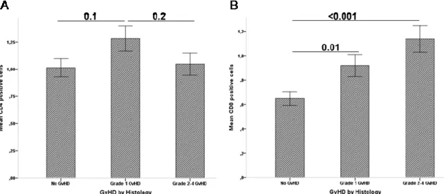
Figure 5.1: Infiltration of CD4+ T cells and CD8+ T cells in gut during acute GI- GvHD ... 57
Figure 5.2: Survival curve in relation to epithelial apoptosis. ... 58
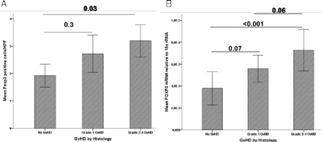
Figure 5.3: Infiltration of Foxp3+ cells and expression of FOXP3 mRNA in GI tract during acute GI-GvHD. ... 59
Figure 5.4: Foxp3 staining in thymus tissue. ... 61
Figure 5.5: CD4 staining in the thymus tissue. ... 61
Figure 5.6: Foxp3+CD4+ double staining of thymus tissue. ... 62
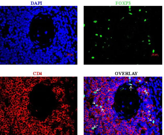
Figure 5.7: Foxp3+CD4+ double staining of colon tissue of GvHD patient. ... 63
Figure 5.8: Foxp3+CD4+ double staining of colon tissue of GvHD patient. ... 64
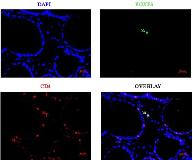
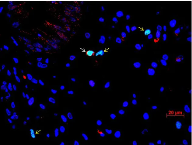
Figure 5.9: Foxp3+CD8+ double staining of colon tissue of GvHD patient. ... 65
Figure 5.10: Quantification of CD4+Foxp3+ cells and CD8+Foxp3+ cells by double immunofluorescence. ... 66
Figure 5.11: Infiltration of IDO+ cells and expression of IDO1 mRNA in GI tract during acute GI-GvHD. ... 68
Figure 5.12: Infiltration of IL-17+ cells and expression of RORC mRNA in GI tract during acute GI-GvHD. ... 70
Figure 5.13: IL-17 protein correlates with RORC mRNA and Treatment Related Mortality (TRM) occurs more frequently in patients with low IL- 17/RORC expression. ... 71
Figure 5.14: IL-17 and CD4 staining in colon tissue. ... 72
Figure 5.15: IL-17+ cells analysis by two independent analyzers. ... 72
Figure 5.16: Establishment of IL-17+CD4+ double staining in colon biopsy of Crohn‘s disease patient. ... 73
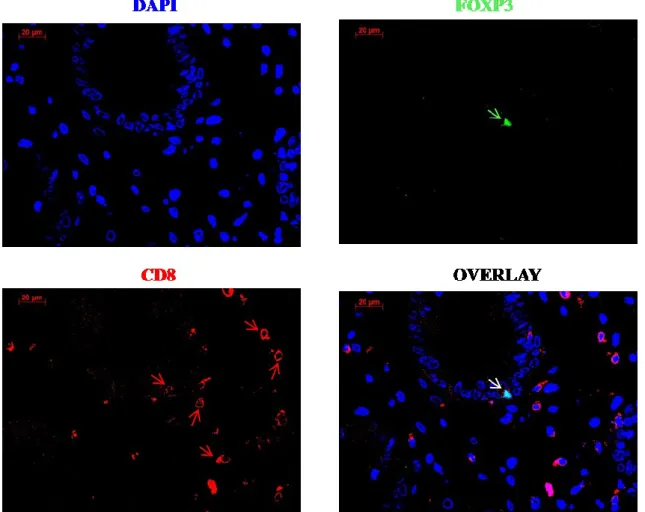
Figure 5.17: IL-17+CD4+ (A) and IL-17+CD3+ (B) double staining of colon tissue of ASCT patient. ... 74
Figure 5.18: : IL-17+CD117+ double staining of colon tissue of ASCT patient. .. 75
Figure 5.19: Alteration of CYP27A1 and VDR gene expression during acute GI- GvHD. ... 79 Figure 5.20: CYP27A1 mRNA correlates with VDR mRNA and TRM occurs
Figure 5.21: Survival curve in relation to median CYP27A1 and VDR mRNA
expression. ... 81
Figure 5.22: Alteration of CYP27B1 gene expression during acute GI-GvHD. . 82
Figure 5.23: Alteration of PXR gene expression during acute GI-GvHD. ... 83
Figure 5.24: Alteration of PXR gene expression after transplantation in response to types of gut decontimation. ... 84
Figure 5.25: Apoptosis measurement of LPS stimulated mature DCs by Annexin V/7AAD staining. ... 86
Figure 5.26: Effect of I3S on survival of human monocyte-derived mature DCs. ... 87
Figure 5.27: Impact of I3S on surface marker expression of human monocyte- derived DCs. ... 88
Figure 5.28: Downregulation of LPS-induced pro-inflammatory cytokines and upregulation of anti-inflammatory cytokine by I3S treated mature DCs. ... 89
Figure 5.29: I3S mediates immunosuppression by altering IL-12 pathway but not AhR pathway. ... 90
Figure 5.30: Effect of I3S on iKB expression. ... 91
Figure 5.31: Co-culture of I3S treated mDCs with allogeneic T cells. ... 92
Figure 7.1: Pathophysiology of acute GvHD. ... 108
Figure 7.2: Pathophysiology of acute GvHD. ... 111
List of Tables
Table 1.1: Diseases Commonly Treated with Allogenic Hematopoietic Stem Cell
Transplantation ... 2
Table 1.2: Frequently used Conditioning Regimens in various Transplant Centre Worldwide ... 6
Table 1.3: Innate IL-17 producing cells ... 19
Table 4.1: RT-qPCR reaction composition ... 41
Table 4.2: Cycling protocol for RT-qPCR ... 41
Table 4.3: Preparation of 500 nM (10X) pooled STA Primer Mix ... 43
Table 4.4: STA Reaction solution ... 44
Table 4.5: Exonuclease I Reaction solution ... 44
Table 4.6: Preparing Sample Pre-Mix and Samples for Gene Expression using Fluidigm Dynamic Arrays ... 45
Table 4.7: Preparing the Assay Mix ... 45
Table 4.8: Priming and Loading the Dynamic Array IFC ... 46
Table 4.9: Elutriation parameter and cell types ... 46
Table 4.10: Protein isolation Buffer A ... 51
Table 4.11: Protein isolation Buffer B ... 51
Table 4.12: Protein isolation Buffer C ... 52
Table 4.13: Preparation of SDS sample buffer (2X) ... 52
Table 4.14: SDS-PAGE stock solutions ... 52
Table 4.15: SDS-PAGE gel mix solutions ... 52
Table 4.16: Required buffers and solutions for SDS gel preparation ... 53
Table 4.17: Required buffers and materials for transferring protein for immunodetection ... 54
Table 5.1: Correlation table of CD4+ and CD8+ infiltrates with crypt loss and epithelial apoptosis in the gut of ASCT patients ... 58
Table 5.2: Correlation table of Foxp3+ cell infiltrates and FOXP3 mRNA in the gut of ASCT patients. ... 60
Table 5.3: Correlation table of IDO with Foxp3 (protein and mRNA) infiltrates and neutrophil infiltrates. ... 68
Table 5.4: Correlation of gene expression by digital PCR and conventional qPCR. ... 77
Table 5.5: Differential gene experssion during acute GI-GvHD. ... 77
List of Abbreviations
aGI-GvHD acute Gastro Intestinal Graft vs. Host Disease aGvHD acute Graft versus Host Disease
AML Acute myeloid leukemia
ANOVA Analysis of variance APCs Antigen Presenting Cells
APS Ammonium persulfate
ATG Anti Thymocyte Globulin
BM Bone Marrow
BW Body Weight
CARD Caspase Recruitment Domain
CD Cluster of Differentiation
cDNA complementary DNA
cGvHD chronic Graft versus Host Disease CLL Chronic lymphocytic leukemia
CY Cyclophosphamide
DANN Deoxyribonucleic acid
DAPI 4',6-diamidino-2-phenylindole
DCs Dendritic cells
DMSO Dimethyl sulfoxide
dNTPs 2'-deoxyribonucleosid-5'-triphosphate
dsDNA Double stranded DNA
ECL Enhanced chemiluminescence
ECP Extracorporeal Photophoresis EDTA Ethylene diamine tetra acetic acid ELISA Enzyme-linked immunosorbent assay
FCS Fetal calf serum
FFPE Formalin fixed-paraffin-embedded FITC Fluorescein isothiocyanate
Flu Fludarabine
Foxp3 Forkhead Box Protein 3
GAPDH Glyceraldehyde 3-phosphate dehydrogenase G-CSF Granulocyte colony stimulating factor
GM-CSF Granulocyte -monocyte colony-stimulating factor GvHD Graft versus Host Disease
GvL Graft versus Leukemia
GvT Graft versus Tumor
Gy Gray
HLA Human Leukocyte Antigen
HRP Horse radish peroxidase
HSCs Hematopoietic Stem Cells
HSCT Hematopoietic Stem Cell Transplantation
I3S Indoxyl 3-sulfate
IDO Indoleamine 2,3-dioxygenase
IFNγ interferon gamma
Ig Immunoglobulin
IL Interleukin
IL-17 Interleukin-17
ILCs Innate Lymphoid Cells
iTregs induced Tregs
IκB Inhibitor of kappa B
LPS Lipopolysaccharide
MA Myeloablative
mHags Minor Histocompatibility Antigens MHC Major Histocompatibility Complex miRs/miRNAs micro RNAs
MMF Mycophenolate Mofetil
MP Methyl prednisolone
mRNA messenger RNA
MUD Mutual Unrelated Donor
NF-κB Nuclear factor κB
NK Natural Killer
NKT Natural Killer T cell
NMA Non-Myeloablative
NOD2 Nucleotide oligomerization domain 2
nTregs natural Tregs
PAMP Pathogen associated molecular pattern
PBS Phosphate buffered saline
PBSC Peripheral Blood Stem Cells
PCR Polymerase chain reaction
PRR Pathogen recognition receptor qRT-PCR Quantitative real-time PCR
Reg3a Regenerating islet-derived protein 3 alpha RIC Reduced intensity conditioning
RISC RNA-Induced Silencing Complex
RNA Ribonucleic acid
RORγT Retinoic acid receptor related orphan receptor 3
RT Room temperature
rRNA ribosomal RNA
s.e.m standard error of mean
SCID Severe combined immunodeficient
SDS-PAGE Sodium dodecyl sulfate polyacrylamide gel electrophoresis SNP Single nucleotide polymorphism
T-bet T-box transcription factor TBI Total Body Irradiation
TBS Tris buffer saline
TBST TBS + Tween 20
TCR T Cell Receptor
TLI Total Lymphocyte Irradiation
TLR Toll Like Receptor
TNF Tumor Necrosis Factor
Tregs Regulatory T cells
1 Introduction
Since the early beginnings, in the 1950’s, hematopoietic stem cell transplantation (HSCT) is being performed as the major curative therapy for several life- threatening hematological and genetic disorders. Huge advances have been made in the past 60 years for successful transplantation and to increase the quality of life of patients, yet, complete success is hard to achieve due to the frequent occurrence of a secondary disease called graft versus host disease (GvHD). The number of allogenic hematopoietic cell transplantations continues to increase with more than 25,000 allogenic transplantations performed annually1.
1.1 Hematopoietic Stem Cell Transplantation (HSCT)
Hematopoietic stem cells (HSCs) are pluripotent stem cells in the bone marrow (and fetal liver) that can give rise to all lineages of blood cells, including lymphocytes2. Mature blood cells are produced continuously by differentiation and expansion of hematopoietic stem cells. These stem cells have a unique capacity of self-renewal: they produce some daughter cells that do not differentiate, but replace and maintain the stem cell pool throughout individual’s life. Leukemic cells arise from malignant stem cells that usually originate via mutation of normal stem cells. Most of the leukemic cells have limited capacity for proliferation and are continuously replenished by leukemic stem cells. Only 1 in 1 million leukemic blasts appears to be a true stem cells, according to the capacity to propagate and sustain human leukemia in immunologically susceptible mice3. Chemotherapy may eradicate the majority of leukemic blasts; leukemic stem cells, however, are quiescent and insensitive to therapy4, and will result in recurrence. Some malignant stem cells survive even a lethal dose of total-body irradiation (TBI) and chemotherapy which is given as a preparative regimen for HSCT. The recurring malignant cells may be eliminated by immunologically active donor T cells and this phenomenon is called Graft vs Leukemia (GvL) effect5. Therefore HSCT
serves as promising therapy to get rid of leukemia and keep the immune system in balance but at the cost of GvHD.
HSCT is an intensive curative therapy for several malignant and non-malignant diseases1. Following conditioning with irradiation or chemotherapy to eradicate leukemic cells and render the recipient receptive of new stem cells, patients are given infusion of hematopoietic stem cells from a suitable donor6.
Table 1.1 summarizes some diseases commonly treated with allogenic hematopoietic stem cell transplantation7.
Table 1.1: Diseases Commonly Treated with Allogenic Hematopoietic Stem Cell Transplantation
Cancers Other Diseases
Acute myeloid leukemia Acute lymphoblastic leukemia Chronic myeloid leukemia Myelodysplastic syndrome Myeloproliferative disorders Non-Hodgkin’s lymphoma Hodgkin’s disease
Chronic lymphocytic leukemia Multiple myeloma
Juvenile chronic myeloid leukemia
Aplastic anemia
Paroxysmal nocturnal hemoglobinuria Fanconi’s anemia
Blackfan-Diamond anemia Thalassemia major
Sickle cell anemia
Severe combined immunodeficiency Wiskott-Aldrich syndrome
Inborn errors of metabolism
1.1.1 Selection of stem cell donors
In order to achieve successful stem cell transplantation, it is crucial that both donor and recipient have a high degree of match in Human Leukocyte Antigen (HLA), a highly polymorphic protein that differs from individual to individual, encoded by Major Histocompatibility Complex (MHC). Class I HLA (A,B & C) are expressed on almost all nucleated cells and platelets whereas class II HLA proteins (DR, DQ & DP) are primarily expressed on cells that arise from hematopoietic stem cells such as B cells, monocytes and dendritic cells, but can be induced on other cell types during immune response1. The HLA system
encodes structurally homologous cell surface glycoproteins which present peptides to the immune cells that trigger an immune response and are encoded by chromosome 6, characterized by a high degree of allelic polymorphism. The strongest transplant reactions occur when the HLA of the donor and the recipient are incompatible7. Therefore the best donor for HSCT would be an HLA matched sibling. When a recipient does not have any siblings, a matched unrelated donor (MUD) is chosen based on at least HLA-A, -B, -C and –DRB1 alleles. Allele compatibility for the HLA-A/B/C/DRB1//DQB1 loci is defined as a 10/10 match and considered as the standard donor requested by many transplant centers8. Despite HLA identity between donor and recipient, almost 40% of patients receiving HLA-identical grafts develop GvHD due to genetic differences that lie outside of the HLA-loci, in terms of minor histocompatibility antigens (mHags)1. mHags are immunogenic peptides derived from polymorphic cellular proteins.
These peptides bind to HLA antigens and are recognized by allogenic T cells.
1.1.2 Sources of Stem Cells
HSCs can be obtained either directly from bone marrow, from peripheral blood or from umbilical cord blood. Since marrow stem cells detach continuously and circulate in the peripheral blood, peripheral blood stem cells (PBSCs) are the most convenient source of hematopoietic stem cells. In allogenic stem cell transplantation, PBSCs are thought to have better and rapid hematopoietic reconstitution and have the potential to reduce disease recurrence9 . However, PBSCs contain more T lymphocytes than the bone marrow, thus increase the incidence and prolong the treatment of chronic GvHD (cGvHD)10,11.
The use of cord blood as an alternative to donor stem cells has provided a promising alternative when transplantation is urgent or when the suitable donor is not found7. Blood from the umbilical cord and placenta of newborn child is rich in hematopoietic stem cells but the numbers of cells are limited. Infection is a common problem after cord blood transplantation since hematological and immune reconstitution is slower in cord blood transplantation. However this type of transplantation requires less-stringent HLA matching12 compared to marrow or peripheral blood.
1.1.3 Conditioning therapy for HSCT
For successful HSCT, a first criterion is that the graft gets enough space and support for cell proliferation and differentiation. Therefore, the existing host stem cells must be eradicated in order for donor stem cells to engraft properly for which conditioning therapy is necessary prior to HSCT. Secondly, it is crucial that recipients are immunocompromised to prevent rejection of the incoming donor cells by the host immune system. The administered conditioning regimen suppresses the host immune system and thus, allows donor stem cells to home in the bone marrow microenvironment without the risk of graft rejection. Finally and most importantly, the conditioning therapy eradicates the underlying disease and provides long-term disease control or at least reduces leukemic cells to a level which allows final elimination by GvL effects. This is particularly important for patients with hematological malignancies.
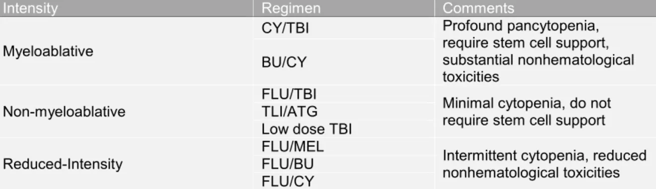
Although there is no full agreement on meticulous classification of conditioning treatments, generally accepted definitions are of three types: myeloablative conditioning, non-myeloablative and reduced-intensity conditioning13. The diagrammatic overview of conditioning is shown below in Figure 1.1.
Figure 1.1: Overview of myeloablative and non-myeloablative conditioning regimens
Lightning bolts in yellow represent total body irradiation and the ovals in red represent chemotherapy5.
Myeloablative (MA) conditioning is of high-dose intensity consisting of a single agent or combination of agents that eradicate the patient’s hematopoietic cells in the bone marrow and induce long-lasting trilineage aplasia. This strategy includes TBI and/or alkylating agents, at doses that will not allow autologous hematologic recovery resulting in profound pancytopaenia within days from the time of administration13. Pancytopaenia is life-threatening and fatal unless patients’
hematopoiesis is restored by infusion of hematopoietic stem cells.
Non-myeloablative (NMA) conditioning can be defined as a regimen that will cause minimal cytopenia, little early toxicity and does not require hematopoietic
stem cell support13,14. Nevertheless, NMA conditioning regimens are immunosuppressive to the extent that, when followed by granulocyte-colony stimulating factor (G-CSF) mobilized PBSC or BM infusion, donor lymphohematopoietic cells can engraft with at least mixed donor/recipient chimerism15. The final elimination of host hematopoiesis is then achieved by graft- versus-hematopoietic and GvL effects of the donor immune cells resulting eventually in full donor chimerism14.
Reduced intensity conditioning (RIC) regimens try to fill the gap between MA and NMA conditioning therapies and are used for the majority of patients nowadays.
The concept of RIC is based on the idea of preventing the high toxicity and mortality associated with MA conditioning regimens in patients with advanced age or relevant comorbidities16. The goal of RIC is not always complete tumor eradication and thus, complete destruction of host hematopoiesis but sufficient control of the underlying disease by cytotoxic therapy followed by the immune- mediated effects of donor graft cells16.
Table 1.2 summarizes the current, most frequently used conditioning regimens for allogeneic HSCT.
Table 1.2: Frequently used Conditioning Regimens in various Transplant Centre Worldwide
Intensity Regimen Comments
Myeloablative
CY/TBI Profound pancytopenia,
require stem cell support, substantial nonhematological toxicities
BU/CY
Non-myeloablative
FLU/TBI Minimal cytopenia, do not require stem cell support TLI/ATG
Low dose TBI Reduced-Intensity
FLU/MEL Intermittent cytopenia, reduced nonhematological toxicities FLU/BU
FLU/CY
Abbreviations: CY = cyclophosphamide; TBI = total body irradiation; BU = busulfan; FLU = fludarabine; TLI = total lymphoid irradiation; ATG = anti- thymocyte globulin; MEL = melphalan.
1.2 Graft versus Host Disease (GvHD)
GvHD is the most recognized complication after HSCT. The first recognition of GvHD was in 1956 in a murine model. Barnes et al.. demonstrated that irradiated mice infused with the allogenic marrow and spleen cells recovered from radiation injury and aplasia but they developed diarrhea, weight loss, skin changes and liver abnormalities. Mice subsequently died due to “secondary disease”17. This phenomenon was subsequently recognized as graft versus host disease (GvHD).
A decade later, in 1966, Billingham postulated three crucial requirements for the development of GvHD:
i) the transplanted graft must contain immunologically competent cells,
ii) the recipient must be incapable of rejecting or eliminating transplanted cells and,
iii) the recipient must express tissue antigen that are not present in the transplant donor thus the recipient antigen be recognized as foreign by donor cells18.
Today it is realized that the immunocompetent cells are T lymphocytes that are present in the stem cell inoculum and are required to mount an effective immune response19. A normal immune system is able to reject T cells from a foreign donor, but when the patient immune system is compromised by the use of various immune-ablative agents (chemotherapy and/or radiotherapy), the recipient is incapable of rejecting transplanted cells20. Previously it was believed that acute GvHD occurs within day 100 after transplantation and chronic GvHD occurs beyond day 100 and that the most affected organs at the onset of GvHD are skin (81%), gastrointestinal tract (54%) and liver (50%)21. Now it is clear that acute GvHD can occur after day 100 as late acute GvHD (e.g. after cessation of immunosuppression or after donor lymphocyte infusion) or cause an overlap syndrome of both acute and chronic GvHD22.
1.2.1 Pathophysiology of acute GvHD: a three-step model
Acute GvHD has been attributed to three stages. Initially there is tissue damage due to conditioning which in turn activates the host antigen presenting cells
Finally, in the efferent phase, cellular and inflammatory factors work together to destroy the target organs.
a. conditioning-mediated tissue damage: damaged host tissue release danger signals that includes pro-inflammatory cytokines like tumor necrosis factor (TNF) and interleukin-1 (IL-1)23, which can activate host APCs, ultimately activating donor T cells infused in the stem cell inoculum24,25. Conditioning-mediated damage to the GI tract remains the main concern as the GI tract allows systemic translocation of microbial products like lipopolysaccharide (LPS) and pathogen associated molecular patterns (PAMPs) that greatly amplify host APC activation26, leading to T cell activation.
Figure 1.2: Conditioning mediated tissue damage (phase I)
Radiotherapy and/or chemotherapy damage the gastrointestinal mucosa which causes the translocation of DAMPS and PAMPS leading to the release of pro- inflammatory cytokines27.
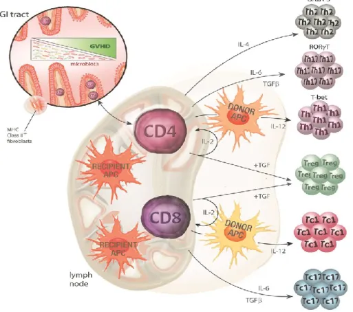
b. donor T cell activation (the afferent phase): graft versus host disease occurs when donor T cells become activated and respond to HLA differences on recipient’s cells1. Experimental models have proved that the host APCs are necessary and sufficient to activate donor T cells and initiate GvHD25,28. Donor T cell can recognize alloantigen either on host APC, known as direct antigen presentation29 or on donor APCs, known as indirect presentation30. T cell response depends on the disparity between the donor and the recipient with
regard to major and minor HLA1. CD4+ T cells respond to the variations in MHC class II molecules (HLA-DR, DQ, DP) and CD8+ T cells respond to the variations in MHC class I molecules (HLA-A, B, C)31. Even though the transplantation is carried out with a HLA matched donor, GvHD occurrence has been reported to occur due to differences in minor HLA32.
Figure 1.3: Donor T cell priming and differentiation (phase II)
Both donor and recipient APCs are actively involve in antigen presentation.
Activation of T cells in the presence of various cytokines determines the fate of T cell differentiation. Th2, Th17 and Th1 marked by the transcription factors GATA- 3, RORyt and T-bet respectively27.
c. target cell apoptosis (the efferent phase): in this phase, both innate and adaptive immune cells work synergistically to aggravate the T cells mediated inflammation. Cellular mediators such as cytotoxic T lymphocytes (CTLs) and natural killer (NK) cells use Fas/Fas ligand (FasL) pathway and perforin/granzyme pathway to lyse the target cells33,34. Furthermore, inflammatory cytokines synergize with CTLs, resulting in further tissue injury and possible target organ dysfunction1. In addition, microbial products like LPS, which are released during conditioning, leak through a damaged intestinal mucosa and skin, and stimulate mononuclear cells (monocytes/macrophages) to secret inflammatory cytokines.
This leads to amplification and propagation of the so-called “cytokine storm”1 and to the destruction of epithelial cells mostly in the GI tract.
Figure 1.4: Target cell apoptosis: the effector phase (phase III)
Liver, lung, skin and most pronouncedly gut are the target tissue of T cells27,35. In the last 10 years, this concept has been largely extended and a more differentiated view has been adapted. It became clear that the microbiota of epithelial tissues are major players influencing epithelial integrity and local immune tolerance by commensal bacteria and millions of metabolites are produced to maintain epithelial homeostasis 36,37.
In context with the concept of microbiota as important players, researchers have recognized the increasing importance of regulatory immune cells which normally balance immune reactions. Regulatory T cells expressing the transcription factor
Foxp3 occur as natural, thymus derived regulatory cells and are able to prevent alloreactions38. On epithelial surfaces, induced peripheral Tregs try to dampen acute inflammation39. Foxp3+ T cells act in cooperation with numerous newly identified regulatory populations such as invariant NKT cells40, myeloid derived suppressor cells and a whole new set of innate immune cells such as innate lymphoid cells41.
.
1.3 Regulatory T cells (Tregs) in GvHD
Tregs are a subset of CD4+ T cells whose function is to suppress immune responses and maintain self-tolerance42. A transcription factor called FOXP3, a member of the fork head family of transcription factors, is critical for the development and function of Tregs and is used as a definite marker to identify Tregs42,43. Tregs are produced in the thymus as a functionally mature subpopulation of T cells and can also be induced from naive T cells in the periphery44.
Natural Tregs (nTregs), derived from the thymus are characterized by the co- expression of CD4, CD25 and Foxp3, collectively represented as CD4+CD25+Foxp3+ Tregs45. Induced or adaptive Tregs (iTregs) are generated in the peripheral lymphoid organs in presence of transforming growth factor beta (TGF-B)46.
1.3.1 Tregs in immune balance
Tregs are known to downregulate immune responses by a) production of inhibitory cytokines and b) a contact-mediated effect on APCs. Tregs produce the anti-inflammatory cytokine- interleukin 10 (IL-10), that inhibits the production of IL- 12 by activated DCs and macrophages47,48. IL-10 also inhibits the expression of co-stimulators and class II MHC molecules on DCs and macrophages thus inducing tolerance of immune system47-49. Another anti-inflammatory cytokine produced by Tregs, TGF-β, inhibits the proliferation and effector functions of T
of functionally distinct subsets of T cells, stimulates production of immunoglobulin A (IgA) antibodies, promotes tissue repair after local immune and inflammatory reactions subside, conferring Tregs mediated immune reconstitution47-50. Tregs play a major role in regulation of epithelial inflammation and are strongly influenced by the interaction with the epithelial microbial environment36,51.
1.3.2 Tregs in pre-clinical model of Stem Cell Transplantation.
Tregs play an indispensable role in both solid organ transplant tolerance and in allograft tolerance after HSCT. In rodents and humans, a subpopulation of thymus derived naïve CD4+ T cells that co-express the IL-2Ralpha chain, CD25, has potent suppressor activity. Tregs mediate transplantation tolerance in experimental models of skin, solid organs52 as well as tolerance to bone marrow allografts53. HSCT and conditioning can cure malignant and non-malignant hematological disorders ,but at the same time the treatment efficacy is highly limited due to GvHD1. Regulatory T cells have received considerable attention in recent science due to their ability to suppress the proliferation of conventional T cells and prevent GvHD in animal models when added to donor grafts containing conventional T cells, hence suppressing GvHD54. Using a mouse model, Edinger and co-workers have shown that CD4+CD25+ Tregs suppress GvHD after bone marrow transplantation without abrogating the graft versus-leukemia (tumor) (GvL or GvT) effect38 supporting the importance of Tregs in allogenic HSCT. It has been demonstrated that the adoptive transfer of Tregs preserved thymic and lymphoid architecture of the host and hence accelerate post-transplant T cell immune reconstitution in a murine GvHD model55.
Tregs are long believed to be a subset of CD4+ T cell compartment. Recently, CD8+ Treg population was reported and ws shown to be capable of suppressing T cell responses in an experimental model of autoimmunity56. In terms of GvHD, Robb et al.. demonstrated that CD8+Foxp3+ Tregs suppressed GvHD and attenuated GvHD mortality after bone marrow transplantation (BMT) in mice model57.
1.3.3 Tregs in clinical Hematopoietic Stem Cell Transplantation Many researchers have focused on evaluating the status of Tregs after HSCT, since they play an important role in the amelioration of GvHD. Using peripheral blood of patients after transplantation, Li et al. demonstrated that the frequency of CD4+CD25+ Tregs was significantly downregulated in patients with severe acute or chronic GvHD 58. They also showed that a decreased level of CD4+CD25+ Tregs was correlated to increased severity of GvHD 58. While majority of studies focused on blood-derived Tregs, there is little information on Tregs isolated from intestinal tissues due to the lack of availability of repeated gut biopsies without diagnostic purpose. Using immunoenzymatic labeling, Rieger et al. were the first to demonstrate that the infiltrating Tregs decreased the signs of acute and chronic GvHD in intestinal mucosa 59. They showed that the acute and chronic GvHD patients had a complete lack of counter regulation indicated by a Foxp3+/CD8+ T cell ratio in patients with GvHD identical to that of healthy individuals, while this ratio was increased in patients without GvHD59.
However, for the first time, Lord et al. demonstrated a contradictory result showing that Foxp3+ cells were nither decreased in blood nor in gastrointestinal tissues and that the frequency of Tregs did not correspond to the disease incidence or severity 60. They rather found that the Foxp3+ cells were significantly upregulated in GvHD intestinal mucosa when compared to non GvHD mucosa60. This finding is further supported by Ratajczak et al. who observed an increased proportion of CD4+Foxp3+ cells in Grade 2-4 GvHD patients compared to grade 0- 1 GvHD patients61. Part of these conflicting results is again due to the lack of possibility to discriminate between natural and induced Tregs. It may well turn out that nTregs are decreased in GvHD while iTregs try to compensate for exaggerated inflammation.
1.3.4 Approaches to induce regulatory T cells after HSCT
Tregs are crucial to induce tolerance and maintain immune homeostasis. A major challenge to use Tregs as a therapy is their relative scarcity in blood (0.5-1% of CD4+CD25 bright T cells)62. In 2011, Hippen et al. showed two individual reports
regarding generation of induced Tregs in large scale63 and ex vivo expansion of natural Tregs64, both method focused on the development of large scale expansion protocols for Tregs with higher cellular yield so that they can ultimately be used as GvHD therapy or for GvHD-prevention63,64. Using chronic GvHD subjects, Matsuika and co-workers reported that daily administration of low-dose IL-2 induced selective expansion of functional Tregs, improved cGvHD, restored CD4+ T cells homeostasis, and promoted the re-establishment of immune tolerance65. This suggests that low-dose IL-2 could be a potential therapy to restore immune balance after HSCT.
Furusuwa and colleagues reported that the clostridia products like short chain fatty acid (SCFA) especially butyrate can induce the differentiation of colonic Tregs in-vitro and in vivo in mice66 providing strong evidence of the necessity of host-microbiome interaction to establish immunological tolerance and homeostasis in gut. Moreover, Mathewson and colleagues reported that restoring clostridia metabolites or the strain itself modulated intestinal epithelial cell integrity and mitigated GvHD in mice36. Of note, Baban and colleagues reported indoleamine 2,3-dioxygenase (IDO) as a potent inducer of Tregs and an inhibitor of Th1 cell subset67. Later it was reported that Tregs generation by IDO takes place via aryl hydrocarbon receptor (AhR)68. Interestingly, it was recently observed that AhR engages in long-term regulation of systemic inflammation only in presence of IDO69 suggesting a triangular positive feedback loop between Tregs, IDO and AhR. AhR is a ligand activated transcription factor that is activated in presence of xenobiotic compounds and bacterial metabolites such as indole and its derivate70. Taken together, these findings strongly suggest that right balance of gut microbiome is crucial to induce Tregs for intestinal tolerance.
1.3.5 Tregs in GvHD: First-In-Man Clinical Trial
The first clinical trial reported was an adoptive transfer of ex-vivo expanded CD4+CD25+CD127- Tregs after HLA-identical sibling HSCT. Transfer of Tregs resulted in a reduction of the steroids administered, increase in levels of circulating Tregs and a decrease in inflammatory cytokine levels in the peripheral blood (PB)71. Another “first-in-human” clinical trial was reported in double
umbilical cord blood transplantation (UCBT) in 23 patients, who received in-vitro expanded Tregs derived from partially HLA-matched third-party UCB units. There was an almost significant reduction of the incidence of grade II-IV acute GvHD controls without transfusion of Tregs. No toxicities were observed, no infections, relapse or early mortality which suggested that UCB Tregs could be a potential tool for preventing acute GvHD72. Taken together these early trials suggest the potential of Tregs as candidates to ameliorate GvHD and establish immune reconstitution after transplantation in larger clinical trials.
1.4 Indoleamine-2,3 dioxygenase (IDO) in GvHD
IDO is an intracellular enzyme that catalyzes the first and rate-limiting step of essential amino acid tryptophan catabolism73. This immunosuppressive enzyme is expressed by APCs and parenchymal cells and is further inducible by inflammation74. Depletion of tryptophan and the increase of tryptophan metabolites (namely kynurenines) via IDO inhibit T cell proliferation and ultimately favor T cell apoptosis. Moreover, tryptophan starvation and the presence of kynurenines induces the conversion of naïve T cells and regulatory T cells75. Of interest, Mezrich and co-workers demonstrated that kynurenine mediated Tregs generation takes place via AhR activation68. Immunosuppressive properties of IDO have been well defined in maternal-fetal acceptance, tumor immunity, autoimmunity and chronic infections76. Figure 1.5 shows IDO mediated tryptophan degradation and effect on T cells77.
Figure 1.5: Tryptophan degradation by IDO
IDO induce tryptophan degradation which results in inhibition of T-cell proliferation, increase in T-cell apoptosis, and de novo formation of Tregs77.
In the context of GvHD, experimental models have well established IDO as a critical regulator of GvHD. IDO knock-out mice showed increased colon GvHD and reduced survival after allogenic stem cell transplantation when compared to wild-type mice74. In a clinical context, GvHD patients tend to have increased IDO production which reflects a reactive release of immunosuppressive mediators in GvHD related inflammation, that is perhaps initiated to balance immune reactions78. IDO is well-known to induce peripheral tolerance by increasing regulatory T cells79,80. In terms of protective effect in GvHD, apart from Tregs and IDO positive cells, a new type of immune cell-innate lymphoid cells has gained substantial importance in the past few years.
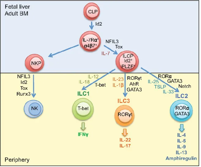
1.5 Innate lymphoid cells (ILC) in GvHD
ILCs are a recently identified family of mononuclear hematopoietic cells that preserve epithelial integrity and tissue immunity throughout the body 81. These cells are derived from Common Lymphoid Progenitor (CLP) and combines innate and adaptive modes of function 82. With recent advances in understanding the development and proliferation of ILCs, they are classified into 3 groups. ILCs group 1, ILCs group 2 and ILCs group 3.
The group 1 ILCs comprises cells that produce type 1 cytokines like IFN-y 83 and aredependent on transcription factor T-bet for development . Initially, conventional (c) NK cells were classified in group 1 ILC since they express T-bet and produce significant amount of IFN-y. However, cNK cells turn out to differ from other group 1 ILC subset as they are developmentally dependent on Eomesodermin (Eomes, a T box transcription factor related to T-bet) but not on T-bet 84.
Innate lymphoid cells that predominantly produce type 2 cytokines are termed as group 2 ILCs or ILC2s 85. ILC2s produce very high amount of IL-5 and IL-13, and also IL-9, IL-4 and GM-CSF86,87. These cytokines are vital for mucus production from goblet cells, eosinophils induction, muscle contraction, mastocytosis 88,89 and ILC2 produced amphiregulin is crucial for tissue repair90. ILC2s are developmentally dependent on the transcription factors retinoic acid receptor- related orphan receptor-a (RORa) 91 and GATA- binding protein 3 (GATA3) 92.
Group 3 ILCs are defined by the expression of transcription factor RORT (RORC) for their development and expression of NK cell activating receptor NKp46 but are distinct from NK cells93-95. ILC3s are able to produce cytokines IL- 17A and /or IL-22 85. ILC3s are divided into Lymphoid Tissue inducer (LTi) cells that expresses IL-17 and IL-22, natural cytotoxicity receptor (NCR) positive IL-22 producing ILC3s and NCR negative IL-17 producing ILC3s 83.
ILC3s has gained substantial focus ever since the discovery that IL-22 producing ILC3s are indispensable for mucosal immunity95,96. In particular, ILC3s are the major source of IL-22 cytokine that are protective to epithelial cells in the intestine
97. These cell types are activated directly by bacterial metabolites via aryl hydrocarbon receptor (AhR) or indirectly through myeloid cells mediated cytokine IL-23 98. Studies of ILC3s on human gut are highly missing due to the lack of specific marker for ILCs, however, mice experiments have revealed that ILC3s play a crucial role in gut immunity by directly inducing epithelial cell proliferation, promoting epithelial cell mediated production of anti-inflammatory cytokines and antimicrobial peptides like Reg3a, preventing dissemination of commensal bacteria, and suppressing microbiota-specific pro-inflammatory CD4+ T cell responses 99. Figure 1.6 shows the development of innate lymphoid cells100
Figure 1.6: Development of Innate Lymphoid Cells.
In fetal liver or in adult bone marrow, ILCs differentiate from common lymphoid progenitor (CLP) in presence of transcription factor Id2. NK cell precursor gives rise to innate NK cells and ILC precursor gives rise to T-bet dependent ILC1s, GATA-3 dependent ILC2s and RORyt dependent ILC3s100.
1.6 IL-17 in GvHD
Ubiquitously known as Th-17 cell cytokine, IL-17 is a pro-inflammatory cytokine known to mediate protection again extracellular pathogens, and promotes inflammatory pathology in autoimmune disease 101. The inflammatory properties of IL-17 have gained a substantial importance in development of GvHD in recent years102-104. IL-17 production is regulated by transcription factor RORT 105, an orphan nuclear receptor that drives the differentiation of Th17 cells.
Although IL-17 is defined as Th-17 cytokine, Th-17 pathway is inadequate to explain the early IL-17 mediated immunity that have crucial roles during stress
responses and host defense especially when the IL-17 mediated immune pathway is induced within hours in response to epithelial cell injury or activation of pattern recognition receptors (PRRs)106-108, and this short time is not enough for the development of Th-17 cells. Therefore it is possible that the innate source of IL-17 exists and perhaps these cells in parts mediate regulation instead of inflammation. Furthermore, the inflammatory role of IL-17 in development of GvHD has been now challenged since the discovery of innate lymphoid cells 83. In brief, table 1.2 presents an overview of some IL-17 producing cells 109.
Table 1.3: Innate IL-17 producing cells
1.7 Microbial metabolites in GvHD
Human gastro-intestinal tract harbors trillions of microbes that are fundamental for well-being of their host. These microbes are necessary to educate and discipline the human immune system. The diversity and complexity of the gut microbiota can be evaluated by using 16S ribosomal RNA (rRNA) sequencing 110 technology. By now it is not surprising to see that the alteration of microbiome is
associated with cutaneous problems, gastric associated diseases, colorectal cancer, inflammatory bowel disease, liver complications, obesity, rheumatoid arthritis, and many more in the queue 111. In the context of allogenic stem cell transplantation, 16S rRNA sequencing revealed the major microbiome shift in the course of ASCT 112. Interestingly, there was huge predominance of Enterococcus species in patient with GvHD compared to non-GvHD patients 112. Surprisingly, authors also shed light on the decrease of microbial metabolite indoxyl sulfate which occurred as a consequence of loss of bacterial diversity 112. A more recent study proposed the loss of bacterial diversity and the decrease in urinary indoxyl sulfate in ASCT patients was associated with higher transplant related mortalityand poor survival 113. , detection of higher indoxyl sulfate in urine of patients was associated with presence of clostridia species 113. Collectively these finding suggests that the balanced microbial population and thereof, the bacterial metabolites may be important for immune mediated complications like GvHD.
Other bacterial metabolites like butyrate, a short chain fatty acid that is produced by bacteria as a result of dietary fiber degradation, has been implicated in maintaining integrity of intestinal epithelial cell and reduces the severity of GvHD
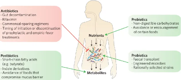
36. Among several short chain fatty acid receptors, butyrate receptor GPR109a is known to be involved in butyrate mediate suppression of inflammation114. GPR109a signaling promotes anti-inflammatory properties in myeloid cells thereby generating regulatory T cells, promotes IL-18 production by epithelial cell which collectively suppress inflammation and promotes immune regulation114. Moreover, bacterial metabolite indole has also been shown to strengthen epithelial barrier and reduce mucosal inflammation115,116. Due to immense importance of balanced microbial diversity, it has been suggested that prebiotics, probiotics and post biotics approach could benefit the patients while sparring antibiotic approach 37. Figure 1.8 shows the diagrammatic representation of possible clinical intervention of gut microbiota 37
Figure 1.7: Clinical intervention of gut microbiota
Green box shows approaches to maintain healthy and balanced gut microbiome.
Red box shows factors to destroy and alter microbiome37.
1.8 Conclusion
Despite of broad understanding, a major issue in treatment of especially aGvHD is that most approaches are started too late, when major changes have already severely damaged the target tissue. Therefore, biomarkers allowing early identification of patients at high risk are needed. A handful of biomarkers have been discovered which might be used to guide treatment in the future 117.
The pathophysiology of aGvHD seems to be much more complex than our understanding due to the rising fact that bacterial diversity and bacterial metabolites plays crucial role in immune balance. Almost 1.5 million of bacterial metabolites are believed to be present in human immune system, effectively regulating both immune reaction and immune tolerance, ultimately deciding the fate of immune homeostasis. It is now of crucial importance to better understand the role of commensals in human health and therefor perhaps modulating bacteria and bacterial metabolites could be established as an alternative safe approach in future to improve the quality of life of GvHD patients.
Finally, practice of stem cell transplantation differs from countries, and within a same country, it differs from transplantation institutes. Approaches aiming at standardization of diagnosis and treatment are urgently needed and addressed by several consensus projects118,119.
2 Research Objectives
Graft versus host disease (GvHD) accounts for significant morbidity and mortality after allogenic stem cell transplantation (ASCT). Early diagnosis of the disease provides clinicians a better strategy for treatment of patients to improve the quality of life after ASCT. Histological evaluation of apoptotic changes in the biopsies of affected organs, especially skin and gastrointestinal (GI) tract is so far the gold standard for the diagnosis of GvHD. Apoptosis of target organs are largely mediated by donor immune cells that infiltrates to skin and GI tract. The overall aim of this study was to analyze immune cell infiltrates and biomarkers during acute GI GvHD.
The specific aims of this dissertation were
1) to assess immunoregulation in intestinal biopsies of patients after allogeneic SCT in relation to presence of GvHD and outcome by two different approaches.
First, CD4+ and CD8+ T cell infiltrates in GI tract of patients after ASCT were quantified using single antibody immunohistochemistry (IHC). In order to analyze functional changes at tissue level, Foxp3, IDO and IL-17 was accessed with IHC.
Further characterizations of cellular source of Foxp3+ and IL-17+ cell were performed by double immunofluorescence. In a second approach, PCR was performed to analyze FOXP3, IDO and RORC at transcriptional level in order to confirm results obtained by protein expression.
2) to assess whether tissue markers of the GI tract can be used as biomarkers in patients. In addition to the described analyses, a panel of carefully selected messenger RNAs was analyzed in GI tract of transplanted patients in collaboration with Medical University Göttingen to identify possible new biomarkers of GvHD and outcome.
3) to characterize the functional role of the bacterial metabolite indoxyl sulfate with respect to immunoregulation in cultured dendritic cells in order to highlight the functional importance of bacterial metabolites.
3 Materials
3.1 Equipments
Autoclave Technomara, Fernwald, Germany
Bioanalyzer 2100 Agilent Technologies, Böblingen, Germany CASY Cell Counter Innovatis/Roche, Basel, Switzerland
Centrifuge Sigma 2 Sartorius, Göttingen, Germany Electrophoresis equipment Biometra, Göttingen, Germany ELISA plate reader MWG Biotech, Ebersberg, Germany EVOS Cell Imaging System Life Technologies, Carlsbad, CA, USA FACS Calibur flow cytometer BD Biosciences, Franklin Lakes, NJ, USA
Forceps Aesculap, Tuttlingen, Germany
Fluidigm Fluidigm, South San Francisco, CA, USA
Heat sealer Eppendorf, Hamburg, Germany
Hemocytometer Marienfeld, Lauda-Königshofen, Germany
Incubators Heraeus, Hanau, Germany
Laminar Flow Cabinet Clean Air Telstar, Woerden, The Netherlands
Microscopes Zeiss, Jena, Germany
Multipipettor Multipette plus Eppendorf, Hamburg, Germany
NanoDrop 1000 Thermo Fisher Scientific, Schwerte, Germany PCR Thermocycler PTC-200 MJ-Research/Biometra, Oldendorf, Germany
pH Meter Knick, Berlin, Germany
Pipetboy Integra Biosciences, Fernwald, Germany
Pipettes Gilson, Milddleton, WI, USA
Pipettes Eppendorf, Hamburg, Germany
Power supplies Biometra, Göttingen, Germany
Realplex Mastercycler epGradient Eppendorf, Hamburg, Germany Rocking plattform HS250 IKA Labortechnik, Staufen, Germany
Sonifier 250 Branson, Danbury, CT, USA
TissueFAXS TissueGnostics GmBH, Vienna, Austria
TissueLyser Qiagen, Hilden, Germany
Vortexer Scientific Industries Ink., Bohemia, NY, USA Water purification system Millipore, Eschborn, Germany
Waterbath Julabo, Seelstadt, Germany
Western blot chamber Biometra, Göttingen, Germany
3.2 Consumables
Cell culture flasks Costar, Cambridge, MA, USA Cell culture plates BD, Franklin Lakes, NJ, USA
Cell scrapers Sarstedt, Nümbrecht, Germany
Combitips for Eppendorf multipette Eppendorf, Hamburg, Germany
Cryo tubes Corning, Corning, NY, USA
Heat sealing film Eppendorf, Hamburg, Germany
HyperfilmTM ECL GE Healthcare, Chalfont St Giles, UK Immobilon-P PVDF membrane Millipore, Schwalbach, Germany Micro test tubes (0.5 ml, 1.5 ml, 2 ml) Eppendorf, Hamburg, Germany Micropore filters Sartorius, Göttingen, Germany Microtiter plates (6, 12, 24, 96 wells) Costar, Cambridge, MA, USA Microtiter plates for ELISA Costar, Cambridge, MA, USA PCR plate Twin.tec 96 well Eppendorf, Hamburg, Germany
Petri dish Falcon, Heidelberg, Germany
Pipette tips Eppendorf, Hamburg, Germany
Plastic pipettes Costar, Cambridge, MA, USA
Polystyrene test tubes Falcon, Heidelberg, Germany Syringe Filters, sterile Sartorius, Göttingen, Germany
Syringes and needles Becton Dickinson, Heidelberg, Germany Tubes (5 ml, 15 ml, 50 ml, 225 ml) Falcon, Heidelberg, Germany
Whatman® Chromatography Paper Sigma-Aldrich, St. Louis, MO, USA
3.3 Media, buffers and solutions
2-Mercaptoethanol Gibco/Life Technologies, Carlsbad, CA, USA
Acrylamide Carl Roth, Karlsruhe, Germany
APS Merck Millipore, Billerica, MA, USA
Aqua Braun
Bovine serum albumin Sigma-Aldrich, St. Louis, MO, USA
CasyTON Roche, Basel, Switzerland
DMSO Sigma-Aldrich, St. Louis, MO, USA
FACS clean BD Biosciences, Franklin Lakes, NJ,
USA
FACS flow BD Biosciences, Franklin Lakes, NJ,
USA
FACS rinse BD Biosciences, Franklin Lakes, NJ,
USA
Fetal calf serum Gibco/Life Technologies, Carlsbad, CA, USA
Hydrogen peroxide Merck, Darmstadt, Germany
HCl Carl Roth, Karlsruhe, Germany
Isopropanol Braun
L-Alanyl-L-Glutamine Merck Millipore, Billerica, MA, USA
Methanol Thermo Fisher Scientific, Waltham, MA,
USA Mounting media for
immunohistochemistry
Thermo Fisher Scientific, Waltham, MA, USA
Nuclease-free water Gibco/Life Technologies, Carlsbad, CA, USA
RPMI 1640 Gibco/Life Technologies, Carlsbad, CA, USA
TEMED Sigma-Aldrich, St. Louis, MO, USA
Triton X100 Sigma-Aldrich, St. Louis, MO, USA
Tween 20 Sigma-Aldrich, St. Louis, MO, USA
Trypsin-EDTA PAN Biotech, Aidenbach, Germany
3.4 Chemicals
All chemicals were purchased from Sigma-Aldrich (Taufkirchen, Germany) or Merck Millipore (Darmstadt, Germany) unless otherwise mentioned.
3.5 Enzymes, kits, and reagents for molecular biology
Agilent RNA 6000 Nano Kit Agilent Technologies, Santa Clara, CA, USA
AmershamTM ECLTM Prime Western Blotting
Detection Reagent
GE Healthcare, Chalfont St. Giles, UK
Aportinin Roche, Mannheim, Germany
Bio-Rad protein assay Bio-Rad, Munich, Germany
dNTPs Roche diagnostics, Mannheim,
Germany
Ethidium bromide Sigma-Aldrich, St. Louis, MO, USA Foxp3 transcription factor staining
buffer set
eBioscience, San Diego, CA, USA
GM-CSF Berlex, Seattle, USA
LPS Enzo Life Sciences, Farmingdale, NY,