Mechanism in Gram-negative Bacteria
Dissertation
Zur Erlangung des akademischen Grades doctor rerum naturalium
(Dr. rer. nat.) im Fach Biologie eingereicht an der
Mathematisch-Naturwissenschaftlichen Fakultät I der Humboldt Universität zu Berlin
von
Kim Stephanie Dohlich
Präsident der Humboldt Universität zu Berlin Prof. Dr. Jan-Hendrik Olbertz
Dekan der Mathematisch-Naturwissenschaftlichen Fakultät I Prof. Stefan Hecht, Ph.D.
Gutachter: Prof. Arturo Zychlinsky, Ph.D.
Prof. Dr. Kai Matuschewski Prof. Dr. Gerhard Wolber Tag der mündlichen Prüfung: 17.12.2013
Two roads diverged in a yellow wood, And sorry I could not travel both And be one traveler, long I stood And looked down one as far as I could To where it bent in the undergrowth;
Then took the other, as just as fair, And having perhaps the better claim, Because it was grassy and wanted wear;
Though as for that the passing there Had worn them really about the same, And both that morning equally lay In leaves no step had trodden black.
Oh, I kept the first for another day!
Yet knowing how way leads on to way, I doubted if I should ever come back.
I shall be telling this with a sigh Somewhere ages and ages hence:
Two roads diverged in a wood, and I - I took the one less traveled by, And that has made all the difference.
Robert Frost (1920)
This work has partially been published as a research manuscript:
Dohlich K, Zumsteg AB, Goosmann C, Kolbe M (2014) A Substrate-Fusion Protein Is Trapped in- side the Type III Secretion System Channel in Shigella flexneri. PLoS Pathog 10(1): e1003881.
doi:10.1371/journal.ppat.1003881
Das Typ III Sekretionssystem (T3SS) ist ein makromolekularer Proteinkomplex den Gram- negative Bakterien nutzen um in einem Schritt Effektorproteine (Effektoren) aus dem Zytosol über die Doppelmembran zu sekretieren. Für viele Bakterien ist das T3SS ein essenzieller Virulenzfaktor, der es ihnen erlaubt mit ihrem Wirt zu interagieren und diesen zu manip- ulieren. Charakteristisch für das T3SS ist die membrangebundene strukturelle Komponente, der sogenannte Nadelkomplex. Der Nadelkomplex ähnelt in seiner Struktur einer Spritze, deren Basalkörper die bakteriellen Membranen und das Periplasma durchspannt und einer Nadel, die vom Basalkörper aus dem Bakterium hervorragt. Basierend auf dem Modell einer Spritze wird angenommen, dass Effektoren entfaltet und anschließend durch Basalkörper und Nadelkanal sekretiert werden. Trotz der kontinuierlichen Forschung an T3SS entbehrt dieses Modell einer experimentellen Grundlage und der Sekretionsmechanismus ist nicht vollständig erklärt.
Ziel der Arbeit war es, eine experimentelle Basis für den Sekretionsmechanismus des T3SS zu schaffen um die Funktionsweise des T3SS besser zu erfassen. Um zu verstehen, wie das T3SS Effektoren sekretiert, wurden zunächst Fusionsproteine konstruiert, welche aus einem Effektor und einem stabil gefalteten Knotenprotein bestehen. Die Effektorfunktion wird durch die Fusion nicht beeinflusst und der Knoten faltet nativ innerhalb des Fusion- sproteins. Aufgrund der Position des Knotens in der Fusion ist davon auszugehen, dass der Knoten während der Sekretion nicht entfalten kann. Die Effektordomäne wird sekretiert, der Knoten verbleibt jedoch im Kanal und verstopft das T3SS. Zusätzlich wurden die blockierten Nadelkomplexe mittels Immunodetektion im Elektronenmikroskop untersucht und das Fu- sionsprotein am isolierten Nadelkomplex lokalisiert. Nach dem derzeitigen Stand der Forschung und unserem Wissen ist diese Arbeit die erste Visualisierung von Effektorfusionen an isolierten Nadelkomplexen. Die Effektorfusion wird N-terminal voran durch den Kanal sekretiert, wobei der Kanal das Substrat umschließt und gegen Proteasen und chemische Modifikationen ab- schirmt. Die Ergebnisse dieser Arbeit untermauern ein elementares Verständnis über die Funktionsweise des T3SS und liefern eine vielversprechende Strategie für zukünftigein situ- Strukturanalysen. Dieser Ansatz lässt sich auch auf andere Proteinsekretionssysteme über- tragen, bei welchen Substrate vor dem Transport entfaltet werden müssen.
bacteria to interact with and manipulate their respective host. A characteristic structural fea- ture of the T3SS is the needle complex (NC). The NC resembles a syringe with a basal body spanning both bacterial membranes and a long needle-like structure that protrudes from the bacterium. Based on the paradigm of a syringe-like mechanism, it is generally assumed that effectors are unfolded and secreted from the bacterial cytoplasm through the basal body and needle channel. Despite extensive research on T3SS, this hypothesis lacks experimental ev- idence and the mechanism of secretion is not fully understood.
This work aimed to provide an experimental basis for the model of the T3SS mechanism for a better comprehension of how secretion by T3SS works. In order to elucidate details of the effector secretion mechanism, fusion proteins consisting of an effector and a bulky protein containing a knotted motif were generated. The functionality of the effector do- main within the fusion was tested, as well as the folding status of the knotted domain. It is assumed that the knot cannot be unfolded during secretion of the chimera. Consequently, these fusions are accepted as T3SS substrates but remain inside the NC channel and obstruct the T3SS. Furthermore, the obstructed T3SS channel was analyzed by immuno-labeling com- bined with electron microscopy. This is, to our best knowledge, the first time effector fusions have been visualized together with isolated NCs and it demonstrates that effector proteins are secreted directly through the channel with their N-terminus first. The channel physi- cally encloses the fusion protein and shields it from a protease and chemical modifications.
These results corroborate an elementary understanding of how the T3SS works and provide a powerful tool forin situ-structural investigations in the future. This approach might also be applicable to other protein secretion systems that require unfolding of their substrates prior to secretion.
1 Introduction 8
1.1 Shigella- a model for type III secretion . . . 8
1.2 Type III Secretion Systems: Syringes mediating bacterial virulence . . . 11
1.2.1 Architecture of T3SS . . . 13
1.2.2 The repertoire of T3SS substrates . . . 14
1.2.3 Through the eye of a needle: Effector secretion by T3SS . . . 16
1.3 Fusion proteins as tools for secretion analysis . . . 17
1.4 Knotted proteins - Non-linear folds as common motifs . . . 19
1.5 Injection revisited: Does the syringe function as a syringe? . . . 20
1.6 Aim of the study . . . 22
2 Results 24 2.1 Knotted proteins - candidates for blocking T3 secretion . . . 24
2.2 Purified IpaB-Knot induces pyroptosis in macrophages . . . 26
2.3 The Knot is folded within IpaB-Knot . . . 28
2.3.1 Fluorescence spectrometry . . . 28
2.3.2 Protease treatment reveals a folded core including the trefoil knot . . . . 29
2.4 Plasmid-encoded IpaB-Knot: effects on hypersecretion . . . 34
2.4.1 Induced expression in∆ipaB . . . 34
2.4.2 Induced expression in∆ipaD . . . 34
2.5 Insertion ofknotinto pWR100 . . . 37
2.6 IpaB-Knot does not complement IpaB in invasion . . . 37
2.7 IpaB-Knot influences regulation of effector secretion . . . 38
2.8 IpaB-Knot impedes T3SS secretion . . . 39
2.8.1 The effect of IpaB-Knot on effector secretion . . . 39
2.8.2 Expression ofknothas no effect . . . 41
2.9 Needle complexes co-purify with IpaB-Knot . . . 42
2.10 Localization of IpaB-Knot at isolated needle complexes (overall NC population) . 43 2.11 Detection of IpaB-Knot C-terminus in occupied NCs . . . 44
2.11.1 Production of humanized IpaB mAB . . . 45
2.11.2 Simultaneous detection of IpaB-Knot N- & C-terminus . . . 47
2.12 The T3SS channel shields parts of IpaB-Knot . . . 48
2.12.1 The fusion protein is inaccessible to TEV protease . . . 48
2.12.2 Chemical modification is limited by the presence of the NC . . . 49
2.13 Initial single particle analysis of NCs with IpaB-Knot . . . 50
3 Discussion & Perspectives 52 3.1 IpaB-Knot is a functional fusion protein . . . 52
3.2 T3 secretion is attenuated by IpaB-Knot . . . 54
3.3 IpaB-Knot stably interacts with the NC . . . 57
6
4 Materials & Methods 65
4.1 Kits, Chemicals & Machinery . . . 65
4.2 Buffer solutions . . . 67
4.3 Bacteria & DNA . . . 68
4.4 Enzymes . . . 72
4.5 Cellular Methods . . . 72
4.5.1 General cell culture . . . 72
4.5.2 Cytotoxicity assay . . . 73
4.5.3 Gentamicin protection assay . . . 73
4.6 Molecular Methods . . . 74
4.6.1 Polymerase Chain Reaction (PCR) . . . 74
4.6.2 Gene Targeting with linear DNA products . . . 74
4.6.3 Conventional agarose gel electrophoresis . . . 75
4.6.4 Standard cloning of plasmid constructs . . . 75
4.6.5 Cloning and expression of murine Ig genes . . . 75
4.7 Microbiological & Biochemical Methods . . . 76
4.7.1 Bacterial Lifestyle . . . 76
4.7.2 Electrocompetent cells . . . 77
4.7.3 Transformation of bacteria . . . 77
4.7.4 Secretion assay . . . 77
4.7.5 Induction of T3SS with Congo Red . . . 78
4.7.6 SDS PAGE and Western blot . . . 78
4.7.7 In-gel visualization of SDS PAGE . . . 79
4.7.8 Protein expression inE. coliBL21 . . . 79
4.7.9 Purifying proteins from heterologous expression . . . 80
4.7.10 Fluorescence spectroscopy . . . 81
4.7.11 Limited proteolysis and mass spectrometry (MS) . . . 81
4.7.12 Isolation of needle complexes . . . 81
4.7.13 TEV protease protection assay . . . 82
4.7.14 Addition of PEG moieties by crosslinking (PEGylation) . . . 83
4.8 Imaging & Bioinformatics . . . 83
4.8.1 Immuno-electron Microscopy (iEM) . . . 83
4.8.2 Nearest-neighbor analysis . . . 84
4.8.3 cryo-electron microscopy (cryo-EM) . . . 84
4.8.4 Single particle-analysis . . . 85
5 References and Tables 86 6 Appendix 101 6.1 Abbreviations . . . 102
6.2 Sourcecode for R: Nearest-neighbor analysis . . . 104
1.1 Shigella - a model for type III secretion
Shigella are the causative agent of bacillary dysentery (Shigellosis). They are Gram- negative γ-proteobacteria which are rod-shaped, non-flagellated and thus non-motile (Peng et al., 2009). The genusShigellacontains four species, namelyS. dysenteriae,S. flexneri,S.
sonneiandS. boydii, all of which are able to cause disease in humans which areShigellas only natural hosts. First descriptions date back to the late 19thcentury where the Japanese scien- tist Kiyoshi Shiga reported a bacterium being responsible for a dysenteric epidemic with over 89.000 cases of which more than 22.000 were fatal. Shigellaare very similar toEscherichia coli, but were classified into a different genus because of their medical significance in the 1940s. In fact, they are so similar that sequence analysis revealedShigella being clones of E. colispecies and that enteroinvasiveE. coliandShigellashare a common pathovar (Lan &
Reeves, 2002). Although distinct phylogeny is kept merely for traditional and medical rea- sons, bacteria will be regarded as independent species throughout this work.
The lack of clean water, hygiene and malnutrition are contributing factors to higher in- cidents in developing countries. In addition, the bacteria are highly contagious: oral up- take of 10-100 bacteria is sufficient to cause disease (DuPont et al., 1989), which is at least partially because of their resistance to acidic conditions (Gorden & Small, 1993; Jennison &
Verma, 2007). With regards to acquisition of antibiotic resistances (von Seidleinet al., 2006;
Shiferawet al., 2012), infections withShigellaeare in need for alternative medical strategies, especially since there is no vaccine available yet (Sansonetti, 2006; Phalipon et al., 2008).
In the developed world, S. sonnei is the predominant isolate (77% of all cases) whereas S.
flexneriis endemic in developing countries and the most frequent isolate of bacillary dysen- tery worldwide (WHO on diarrhoeal diseases/Shigellosis, 2009). Shigella-caused dysentery
8
the lumen through the apical side but have to cross the epithelium and infiltrate epithelial cells from the basolateral side.
1
2
3
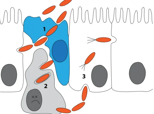
Figure 1.1: Early steps in Shigella flexneri infection. Bacteria (orange) traverse the epithelial cell line (white) through M-cells (blue, 1) where they encounter macrophages (grey). The bacteria then escape the phagolysosome and induce pyroptosis (2). Subsequently, bacteria evade the macrophage and enter the ep- ithelial cells from the basolateral side, where they hijack the actin network for actin-based motility (3).
Three major steps take place before invasion of epithelial cells byS. flexneri: First, bac- teria cross the epithelium through microfold cells (M-cells) that constantly sample macro- molecules and bacteria from the gut lumen (Wassef et al., 1989; Perdomoet al., 1994)(Fig.
1.1, 1). Next, resident macrophages engulf the bacteria which subsequently escape the phagosome and activate Caspase-1. By induced cell death, the bacteria evade phagocytic killing (2) (Zychlinsky et al., 1992, 1994; Schroeder & Hilbi, 2008; Senerovic et al., 2012).
Membrane disruption of macrophages leads to release of mature Interleukin-1β, thereby stimulating the inflammatory response. As a consequence, tissue destruction further desta- bilizes the epithelial lining which in turn favors bacterial infiltration of the lamina propria (Perdomoet al., 1994). The bacterium invades the epithelium by stimulating phagocytosis (Highet al., 1992), escapes the phagosome and hides inside the cytoplasm of epithelial cells.
After entering from the basolateral side, bacteria spread intracellularly to neighboring cells
by hijacking the actin-network of the host. This exploitation of host factors is displayed by the formation of actin tails which confer motility to the bacteria (Fig. 1.1,3) (Mounieret al., 1992; Vasselon et al., 1992; Prevost et al., 1992). Once inside a host cell, S. flexneri repli- cates remarkably fast with a doubling time of 40 min, pointing out the efficient adaptation of the bacterium to the host environment (Sansonettiet al., 1986; Rayet al., 2009).
Most of these steps depend on a functional Type III Secretion System (T3SS) which is encoded on an extrachromosomal 200 to 220 kb plasmid (Venkatesanet al., 2001; Buchrieser et al., 2000). This plasmid is present in all virulentShigella flexneriisolates (Sansonettiet al., 1982). Specifically a 31 kb core region, called the entry region, is indispensable and sufficient for invasion of epithelial cells (Maurelliet al., 1985). This region encodes genes for structural components of the T3SS, most of the substrates, chaperones and regulators. Apart from its role as a highly prevalent human pathogen, Shigella flexneri is a model organism for understanding the T3SS.
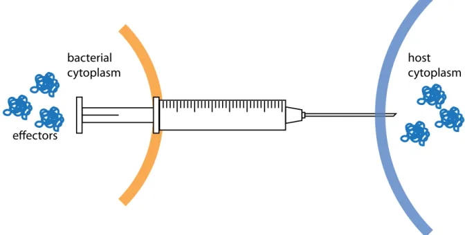
The T3SS is metaphorically described as a molecular syringe thatS. flexneri and other Gram-negative bacteria use to inject effector proteins into their hosts (Fig. 1.2). The T3SS enables delivery of effector proteins into eukaryotic cells and is an important instrument in host-pathogen interaction (Cornelis, 2006; Galan & Wolf-Watz, 2006). Some of the worst pandemics in human history, for example plague and cholera, were caused by bacteria that are known to utilize a T3SS. Recently, the T3SS has been also focused on as it might serve as a target for novel anti-infective therapies (Clatworthyet al., 2007).
bacterial cytoplasm
host cytoplasm
effectors
Figure 1.2: Paradigm of type III secretion.Bacterial effector proteins (blue) are secreted across the bacterial membrane (orange) and injected into the host cell (light blue). The T3SS (syringe) forms a continuous channel connecting both cytoplasms.
More than 20 years ago, the T3 secretion pathway was discovered by analysis ofYersinia outer proteins (Yops) as it was distinguished from the type I transporters and the sec-dependent type II pathway (Salmond & Reeves, 1993). The independence from a Sec-signal peptide as seen for type I but also the dependency on a different gene cluster for secretion like type II secretion led to the definition of a third bacterial secretion pathway.
By the end of the 20thcentury, the structural component had been identified inSalmonella and Shigella and isolated from the bacterial envelope (Kubori et al., 1998; Blocker et al., 2001). This syringe-like needle complex (NC) is the structural hallmark of the T3SS pathway.
The T3SS bridges the gap between the bacterium and the host cell as it is thought to provide
1 µm
Figure 1.3: Type III needle complexes in the bacterial cell wall.Artificially coloured transmission electron- micrograph of a bacterial cross-section. The needle complexes embedded into the bacterial membranes (orange) are highlighted. Image courtesy of Ulrike Abu Abed, Diane Schad and Michael Kolbe.
a continuous channel between the bacterial and host cytosol (Büttner & He, 2009). Consist- ing of more than 25 structural proteins, the syringe is a complex export system spanning the bacterial membranes and the periplasm (Fig. 1.3).
Both pathogens and symbionts use T3SS to establish a trans-kingdom relationship with one or more eukaryotic organisms by secreting proteins via the T3SS pathway, which are stored in the bacterial cytoplasm prior to secretion (Hueck, 1998; Sotoet al., 2006; Preston, 2007; Büttner & He, 2009). These proteins are collectively termed effectors, which includes general effectors that target pathways in the host cell, and translocators that play an ad- ditional role in effector translocation into the host cytoplasm. Effectors are thought to be delivered unidirectionally in a single step into the host cytoplasm, where they then manipu- late host cell biology, metabolism and defense pathways (Rosqvistet al., 1994; Ghosh, 2004;
Dean, 2011).
T3SS are thought to have appeared in bacteria just after the evolution of eukaryotes as these systems are dedicated to trans-kingdom interaction (Troisfontaines & Cornelis, 2005).
Since then, the T3SS have been shaped by evolution depending on the pathogens or sym- bionts niche. The T3SS was most likely spread by horizontal gene transfer, resulting in a phylogeny tree completely different from the 16S rRNA-based tree which assigns bacterial phyla. The T3SS is very similar to the flagellar apparatus in Gram-negative bacteria and it is assumed that both systems share a common ancestor (Galan & Collmer, 1999; Erhardt et al., 2010). Based on sequence analysis of non-flagellar and flagellar T3SS proteins, seven T3SS subfamilies have been identified (Troisfontaines & Cornelis, 2005). Despite the distribu-
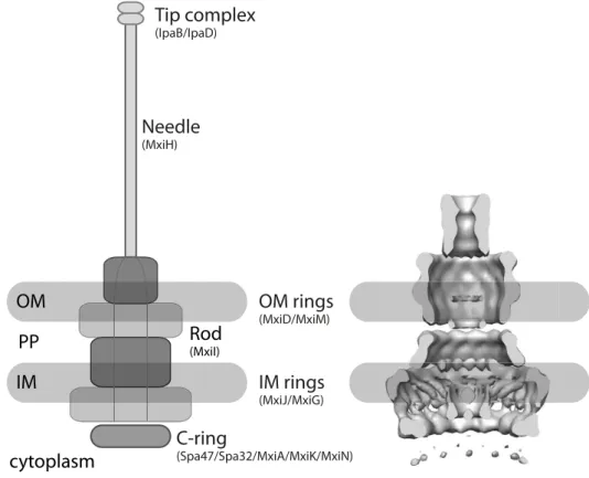
1.2.1 Architecture of T3SS
The NC structure is remarkably conserved among the T3SS-positive bacteria. Of the seven structural families, the Inv-Mxi-Spa subfamily including T3SS from S. flexneri and Salmonella entericaand the Ysc subfamily fromYersiniahave been extensively studied (Tro- isfontaines & Cornelis, 2005). Cryo-EM analysis of the NC structure of Salmonella enterica serovar Typhimurium SPI-1 and S. flexneri revealed structural details up to subnanometer resolution (Schraidt & Marlovits, 2011; Hodgkinsonet al., 2009). Assembly of the NC occurs in a stepwise manner starting with embedding protein rings into the bacterial membrane (Galan & Wolf-Watz, 2006; Chatterjeeet al., 2013). The first proteins to build the NC are pi- lotins and secretins, which are secreted via the sec-pathway across the inner membrane and are essential building blocks for type III and also type II or type IV secretion systems (Bayan et al., 2006). The pilotin helps with insertion of the secretin into the outer bacterial mem- brane which in turn provides a structural basis for attachment of additional protein multimers (Bayanet al., 2006; Chatterjeeet al., 2013). Rings of variable stoichiometry are stacked on top of each other until the membrane-spanning part is completed. This unit is the basal body and has an inner diameter of about 30 to 40 nm. In S. flexneri, the basal body consists of rings of MxiG and MxiJ in the inner and MxiD in the outer membrane, where MxiD is a secretin that targets the outer membrane with the assistance of the pilotin MxiM (Schuch & Maurelli, 2001). Together, these rings enclose a central tube-like structure, called the inner rod, which is formed by MxiI (Fig. 1.4).
Once the basal body is complete, the needle is formed by numerous copies of a small protein with a hairpin structure (MxiH inS. flexneri). The yet unfinished system secretes the structural needle-protein during assembly of the extracytosolic appendage (Erhardt et al., 2010). The hollow needle grows at the distal end and usually ranges between 45 and 50 nm in length, which is regulated by Spa32 in S. flexneri(Tamanoet al., 2000, 2002). The inner diameter of the needle is about 2-3 nm and is considerably smaller than the basal body.
Recently, a channel of 2.5 nm has been confirmed inSalmonella SPI-1 which is conserved in S. flexneri and thereby seems to be a universal feature of the Inv-MxiG-Spa T3SS (Loquet et al., 2012; Demers et al., 2013). The assembly of the central channel is assisted by the
Needle
(MxiH)
Tip complex
(IpaB/IpaD)
OM rings
(MxiD/MxiM)
IM rings
(MxiJ/MxiG)
C-ring
(Spa47/Spa32/MxiA/MxiK/MxiN)
OM
IM PP
cytoplasm
Rod
(MxiI)
Figure 1.4: Shigella flexnerineedle complex scheme and model.Left: Schematic of the T3SS embedded in the bacterial envelope, denotations for constituent proteins are given in brackets. Right: Model of the S.
flexneriNC in a lateral section with schematic membranes (modified from Hodgkinsonet al.(2009)). OM = outer membrane, PP = periplasm, IM = inner membrane.
cytosolic proteins MxiN and MxiK, as mutants lacking these proteins did not form secretion- competent needles (Jouihri et al., 2003). These proteins interact with the ATPase (Spa47 in S. flexneri) which is an essential component of the basal body and is part of a cytoplasmic ring (C-ring). Through the interaction with cytoplasmic and structural T3SS components, the C-ring is thought to orchestrate assembly and substrate secretion (Morita-Ishiharaet al., 2006).
1.2.2 The repertoire of T3SS substrates
Compared to the conserved structural part, the repertoire of effectors is highly diverse as they shape the interaction between bacterium and host (Preston, 2007). Effector proteins thereby constitute the armory of pathogenic and symbiotic bacteria and they often mimic or modify host factors (Galan, 2009). Phosphorylation and ubiquitinylation pathways are com- mon targets for bacterial effectors that covalently modify host factors, including pathways regulating the actin network and immune response (Dean, 2011). The variety of interactions underlines the flexibility in bacterial adaptation to widespread ecological niches where effec- tors serve as an extensive toolbox to support the bacterium in the host environment.
formatic studies developed prediction tools for T3SS signal sequences based on amino acid composition and structure prediction (Arnoldet al., 2009; Lower & Schneider, 2009; Samu- dralaet al., 2009; Yanget al., 2010), although the signal itself is not completely characterized yet. Many effectors require chaperones for their cytoplasmic stability which bind to their N- terminal region downstream of the signal peptide. For theS. flexnerieffector and translocator IpaB, two chaperone binding domains have been identified (Lunelli et al., 2009; Lokareddy et al., 2010), the first of which overlaps with the secretion signal (Fig. 1.5). To prevent ag- gregation or effector activity in the bacterium, IpaB stays bound to the chaperone IpgC in a partially unfolded conformation. Without IpgC, IpaB is degraded and recombinant expression of IpaB requires co-expression of IpgC (Menardet al., 1994b; Pageet al., 1999; Lunelliet al., 2009).
IpaB
IpgC IpgC
1 15 45 51 72 effector/transloctor domains 580
S
Figure 1.5: Schematic of the effector and translocator IpaB. IpaB has an N-terminal signal in the first 15-20 amino acids that overlaps with the first chaperone binding domain (IpgC binding site). An additional IpgC binding site is further downstream and followed by effector domains. Numbers indicate amino acids, S = signal sequence.
For reaching the host cytoplasm, effectors require the help of translocator proteins. Translo- cators are thought to mediate contact with the host-cell membrane where they insert to form a pore and mediate translocation of other effectors (Mülleret al., 2008). Therefore, translo- cators like IpaB and IpaC inS. flexneriare also situated at or recruited to the tip of the needle forming a complex that is thought to sense and establish contact with the host (Blockeret al., 1999; Westet al., 2005; Espinaet al., 2006; Veenendaalet al., 2007) (Fig. 1.4). IpaD assists translocon formation and interacts with the needle subunit MxiH and IpaB, thereby connect- ing the two (Johnsonet al., 2007; Epleret al., 2012). IpaB is a multifunctional protein and has, in addition its translocator activity, at least four different eukaryotic targets. First, it activates Caspase-1 in phagocytes, thereby stimulating an inflammatory response (Hilbi et al., 1998;
Zychlinsky et al., 1994). Second, it has been shown that IpaB can oligomerize to form ion channels in the endolysosomal membrane. Subsequent leakage of potassium activates the
IPAF inflammasome and leads to pyroptosis (Senerovicet al., 2012). Third, there is evidence for IpaB mediating disruption of the Golgi network, possibly by its CD44/cholesterol-binding activity (Skoudyet al., 2000; Lafontet al., 2002; Mounieret al., 2012). Last but not least, IpaB can arrest the host cell cycle by interacting with Mad2L2, an activtiy that possibly decelerates the otherwise rapid epithelial cell turnover (Iwaiet al., 2007).
1.2.3 Through the eye of a needle: Effector secretion by T3SS
Since it is energetically costly to express the T3SS and effectors, the T3SS is strictly reg- ulated at the level of transcription and secretion. T3SS components are only synthesized at 37 °C and effectors are stored within the cytosol in an inactive state until a host cell is encountered (Menard et al., 1994a). The tip complex formed by IpaB and IpaD regulates secretion and in the absence of host cells Shigella leaks minimal amounts of Ipa proteins into culture supernatants (Parsot et al., 1995). Mutants of eitheripaBoripaDfail to estab- lish a regulatory tip complex and hypersecrete effectors without induction. The interaction between IpaB and IpaD is crucial, as deletion of the 3 C-terminal amino acids in IpaB causes a hypersecretory phenotype (Roehrich et al., 2010). Secretion can be artificially inducedin vitro by addition of Congo Red (Bahraniet al., 1997), although it remains unclear how the dye acts on the T3SS. WhenS. flexneriestablishes contact with a host cell, effector pools of IpaB and IpaC are released entirely into the host cell with a half-time of five minutes (Enninga et al., 2005). Other effectors are not transcribed until IpaB and IpaC are secreted, as they are regulated by a feedback loop mediated by IpgC. When IpaB and IpaC are secreted, IpgC is released and can associate with MxiE. IpgC and MxiE together induce transcription of a second set of effectors, termed late effectors (Parsot, 2009). Timely regulation of effector expression provides an efficient way to maintain a secretion hierarchy that is adapted to the pathogens needs.
Besides temporal regulation, hierarchy of effector secretion occurs through substrate specificity (Ghosh, 2004; Barisonet al., 2013). InS. flexneri, Spa32 is involved in the switch from basal body components to needle subunits, while MxiC regulates the switch to translo- cator secretion after the needle is completed (Magdalenaet al., 2002; Botteauxet al., 2009;
Martinez-Argudo & Blocker, 2010). Furthermore, a recently discovered protein complex in Salmonella termed sorting platform was proposed to order substrates prior to secretion by
conserved in other T3SS and shown to interact in Shigella (MxiN, MxiK and Spa47 (Jouihri et al., 2003)), but thus far, function of this complex has only been investigated inSalmonella.
Once an effector reaches the entry port of the NC, effectors are required to unfold in order to fit the narrow dimensions of the needle. The inner diameter of the needle is 2.5 nm which only allows secretion of secondary structures or simple structural motifs. As effectors are shown to be folded prior to secretion (Wilharm et al., 2004b), activity of an unfoldase is re- quired. This function is mediated by the ATPase which associates with the NC and the sorting platform (Akeda & Galan, 2005; Lara-Tejeroet al., 2011).
The driving mechanism of effector secretion is not fully elucidated. The ATPase, which localizes close to the entry port of the NC in the bacterial cytoplasm, has been shown to be essential for secretion and it is possible that it might be fueling the secretion process.
Secretion also depends on a proton motive force (PMF) as the PMF inhibitor CCCP abolished secretion in Yersinia andShigella (Wilharmet al., 2004a; Schroeder et al., 2007). A recent alternate model proposed rotation of the needle involved in effector secretion (Ohgitaet al., 2013). Based on the similarity to the rotating flagellar export apparatus, structural compo- nents of the NC needle tip in Pseudomonas aeruginosahave been labeled and rotation has been observed by microscopy. Whether this is a general feature of secretion mechanism is not clear. However, rotation fits into the context of the needle being a helical structure with effectors moving in a screw-like manner through the channel (Wilharmet al., 2007).
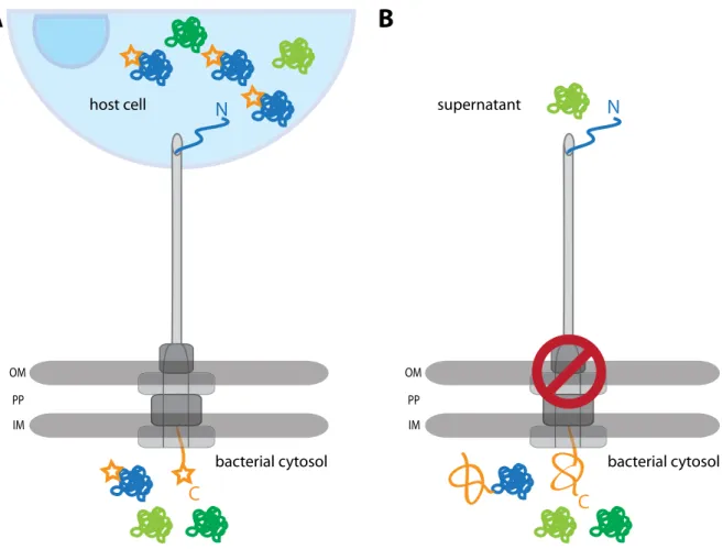
1.3 Fusion proteins as tools for secretion analysis
Substrate proteins fused to reporter domains are powerful tools for investigations of se- cretion mechanisms. The substrate, an effector in case of T3SS, is a carrier for the reporter which serves as a readout for translocation or localization studies (Fig. 1.6A). InShigella, this approach has been used to study kinetics of secretion with effectors that were labeled with small fluorescent dyes or linked to small enzymatic domains (Schesseret al., 1996; Enninga et al., 2005). Another possibility is to specifically inhibit transport or system assembly by
fusing domains that clearly exceed the channel dimensions and cannot be unfolded prior to secretion (Fig. 1.6B). This leads to obstruction of the channel, preventing secretion of other effectors. Consequently, only effectors that were secreted before the channel was occluded are found in culture supernatants or the host.
bacterial cytosol bacterial cytosol
C
PP IM OM
host cell
N
A
supernatant
C N
B
PP IM OM
Figure 1.6: Monitoring secretion with engineered effectors.(A) Schematic of secretion with labeled effec- tors (blue molecule with star) that can be transported into the host. (B) Schematic of effector impassable for the T3SS that blocks secretion of other effectors, effector in the supernatant (light green) was transported before the channel was obstructed. OM = outer membrane, PP = periplasm, IM = inner membrane.
The T3SS with its narrow channel has been the subject of many studies with either re- porter fusions or blocking fusions for gaining mechanistic insights. Many of these experi- ments have been carried out inYersiniawhere the T3SS was first discovered. The N-terminal secretion signal was defined by fusion of truncated effectors to β-galactosidase or alkaline phosphatase (Michiels & Cornelis, 1991) and could be further specified for YopE fusions where the N-terminal 11 amino acids were sufficient for secretion (Sory & Cornelis, 1994; Schesser et al., 1996).
Other experiments have attempted to obstruct the channel using murine dihydrofolate reductase (DHFR) as a fusion partner for effectors. This approach derived from studies in
tochondrial import channel (Eilers & Schatz, 1986). InYersinia, DHFR was fused to YopE and the fusion protein was secreted very little already in the absence of methotrexate (Lee &
Schneewind, 2002). Additional studies revealed that effector fusions including the signal se- quence and the chaperone binding domain were secreted depending on the presence of the cognate chaperone SycE (Feldmanet al., 2002). In line with these findings was the secretion of a truncated and thereby destabilized DHFR mutant. Therefore, the chaperone mediates unfolding of domains downstream of its binding site in the effector. It is noteworthy that DHFR has been shown to be folded in these fusions prior to secretion (Wilharmet al., 2004b).
This suggests that further active unfolding by the T3SS required, a task probably seen to by the ATPase and that fusion proteins are indeed unfolded by the T3SS (Wilharmet al., 2007).
In agreement with the ATPase being the potential unfolding component was another study withYersiniaeffectors fused to gluthatione-(S)-transferase. Upon blocking the T3SS, the AT- Pase was detected in effector fusion-pulldown assays (Riordanet al., 2008). In summary, the T3SS has complex unfolding capacities that at least partially depend on the ATPase.
1.4 Knotted proteins - Non-linear folds as common motifs
Our general understanding of protein folding is that a linear peptide chain folds into in- tricate structures, but, like knit-wear, can be unraveled if we pull on both termini. This un- derstanding has been challenged by the observation of proteins that do not comply with this model and have a knot as a structural motif. A knot is practically defined as a crossing of threads which remain folded when a pull is applied to both ends (Virnau et al., 2011). In particular, knots are technically defined only in closed circles where there is no option for the ends to slip and untie the knot. Therefore, algorithms for knot identification include arti- ficial joining of the protein termini, a process that resembles the circle being closed as if we pick up a string (Taylor, 2000; Yeateset al., 2007). Despite the tendency of long collapsed chains to form knots, the knotted fold in a protein seemed unlikely at first, as a computa- tional analysis of the entire protein data bank (PDB, 400 proteins at the time of the analysis) did not reveal any knots (Mansfield, 1994). This result was challenged by structural data of
anS-adenosyl methionine-synthetase that obviously contained a knotted motif (Takusagawa
& Kamitori, 1996). An improved analysis of the PDB revealed more than 10 different knot- ted proteins and already distinguished between simple trefoil knots and more sophisticated figure-of-eight knots (Taylor, 2000). By now, knotted topologies in proteins are accepted as common structural motifs and have opened new possibilities for studying protein folding dy- namics. Although the purpose of knotted folds is not clear, the fact that methyltransferases with trefoil knots have been found predominantly in extremophiles indicates that knots might confer additional structural stability (Taylor, 2007).
One example of a knotted methyltransferase is RrmA fromThermus thermophilus, a ther- mophilic eubacterium. RrmA has a trefoil-knot in the active site and the position of the knot implies that it is of a deeply-folded nature, meaning that it is unlikely to slide off the chain (Vir- nauet al., 2011; Nurekiet al., 2002). RrmA was the first protein where a deeply-folded trefoil knot was discovered. Homologs of RrmA are YibK fromHaemophilus influenzaand YbeA from Escherichia coli, which, together with RrmA and other methyltransferases containing knots, have been assigned to theα/β-superfamily of knotted proteins in the Pfam protein database (Bateman et al., 2004; Mallam & Jackson, 2007b). These proteins usually form homodimers and bind S-adenosyl methionine as a ligand in their active center. Folding studies revealed that the knotted motif can refoldin vitrowithout need for chaperones and it was suggested that the folding process takes place when the chain is largely unfolded (Mallam & Jackson, 2007a). Fusion experiments with these proteins showed that knots still form when either one or both termini are fused to ThiS from Archeoglobus fulgidus (Mallam et al., 2008). These findings underlined the intricate folding properties of knotted proteins and suggested that the knot is formed when the protein is in a largely denatured state.
1.5 Injection revisited:
Does the syringe function as a syringe?
With its defined role as a device that mediates bacterial virulence, T3SS have been the subject of extensive research in the past 25 years. Early studies with fusion proteins clearly elucidated a correlation between the fold of a domain and its ability to be secreted. Further- more, effector proteins fromYersiniawere only active when injected into the respective host
addition, a central channel across the NC was discovered by electron microscopy averaging.
The channel discovery lead to the common belief that effector proteins are delivered by the means of the NC into the bacterium in a single step, however, this hypothesis has not been confirmed experimentally.
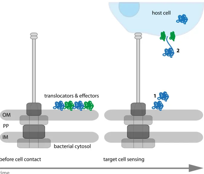
Regarding studies using effector fusions, it is still possible that these fusions constantly occupy the ATPase which hampers secretion, leading to an absence of secreted effectors interpreted as channel obstruction. Thus, these experiments do not prove secretion through the channel. Opposing the injection model, a recent study in Yersinia demonstrated the presence of effector proteins which are stored on the outer membrane of the bacterium and are found in the host cell in later stages of the infection (Akopyan et al., 2011)(Fig.
1.7). These findings contradict the model of single-step translocation from one cytoplasm to the other, although effector assembly on the bacterial surface was shown to be a T3SS- dependent process. Even if this study did not exclude the idea of secretion through the NC channel, it raised questions concerning the purpose of the syringe. An alternative model considers the syringe as an anchoring device that mediates intimate contact between the bacterium and the host. Importantly, the authors pointed out that there is no experimental evidence of effectors being secreted through the NC channel (Edgrenet al., 2012).
OM
IM PP
2
1
before cell contact target cell sensing
time
translocators & effectors
host cell
bacterial cytosol
Figure 1.7: Model of protein translocation in distinct steps. Bacterial translocators and effectors are stored on the bacterial cell wall and released only upon host cell contact (1). Translocators (green) form a pore in the host cell membrane where effectors (blue) are secreted into the target cell (2). OM = outer membrane, PP
= periplasm, IM = inner membrane. Adapted from Edgrenet al.(2012).
1.6 Aim of the study
The aim of the study was to answer the question whether or not effector proteins are secreted through the T3SS channel. Secretion through the NC has two major consequences:
First, effectors need to be unfolded in order to be secreted, as bulky domains are impassable for the narrow channel. Second, the NC channel shields the substrate from the environment as it physically surrounds the effector. We proposed that if an effector protein were fused to a stably folded knotted motif, it could not be unfolded by the T3SS during secretion. The trefoil knot-motif is a good candidate for blocking the T3SS because first, its size does not fit the re- stricted dimensions of the channel and second, it stably folds in the absence of chaperones.
The knotted protein was fused to the effector IpaB, which is one of the first effectors being secreted by the bacterium. IpaB is well characterized and protocols for protein purification
If the fusion protein were trapped in the NC, it would prevent passage of other native effector proteins. This approach also provides the opportunity to analyze the occupied NC in more detail and localize the effector protein in an isolated NC. Furthermore, combination of biochemical assays, like protease treatment and chemical modification, might elucidate whether the effector fusion protein was accessible or trapped within the NC. While the pro- tease should not be able to reach the cleavage site if buried inside the channel, the chemical modification indicates which sites of the protein are still accessible on isolated NCs. Taken together, this work aims to experimentally address the secretion mechanism in T3SS. This approach might serve as a convenient tool to study secretion mechanisms of other systems which require unfolding of their substrates.
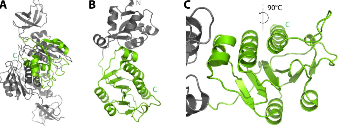
2.1 Knotted proteins - candidates for blocking T3 secretion
Selection of proteins containing knots as candidates for effector fusions was based on pro- tein size and structural data available. Both proteins were smaller than 30 kDa and contained a simple trefoil-knot in the C-terminal region (Fig. 2.1). One protein (Knot 1, Fig. 2.1A) is a hypothetical protein from the archaeal bacterium Methanobacterium thermoautotrophicum (PDB 1K3R) (Zarembinskiet al., 2003), the other (Knot 2, 2.1BandC, Fig. 2.2) a methyl trans- ferase RrmA from the eubacterium Thermus thermophilus(PDB 1IPA) (Nurekiet al., 2002).
The topology plot shows the trefoil-knot motif highlighted in green, which results from three peptide crossings in the C-terminal region of RrmA (Fig. 2.2).
C N
C
C
A B
NC
90°CFigure 2.1: 3D structures of the knotted proteins. Full length structural data of Knot 1 (A) and Knot 2 (B) with the trefoil-knot domains highlighted in green. A close-up of the knotted region inBis given inC.
As the knot locates in the C-terminal part of each protein, it was assumed that knot fold- ing should be autonomous from an N-terminal fusion to the effector IpaB. Thus, both proteins appeared to be suitable candidates for stably-folded domains that might obstruct the T3SS channel.
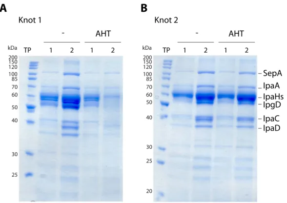
The influence on effector secretion by these proteins was tested by expressing both knot 24
Figure 2.2: Topology of Knot 2/RrmA.Topology plot of Knot 2/RrmA, adapted from Nurekiet al.(2002). The trefoil knot is highlighted in green analog to figure 2.1BandC.
protein-encoding genes without a fusion in the∆ipaBstrain and comparing secretion of in- duced and non-induced cultures before and after induction.
1 2 1 2 1 2 1 2
AHT AHT
- -
Knot 1 Knot 2
A B
kDa
50 60 70 85
40
30
25 100 120 150200
kDa
50 60 70 85
40
30 25
20 100 120 150200
TP TP
SepA IpaA IpaHs IpaC IpaD IpgD
Figure 2.3: Secretion assay of ∆ipaB with Knot 1 and Knot 2. Bacterial supernatants were analyzed by Coomasssie stain before (1) and after (2) induction time points (TP). Cells were left untreated (-) or protein expression was induced with AHT (AHT). Knot 1 is shown in (A), Knot 2 in (B).
Aliquots of culture supernatants that contain secreted proteins were precipitated with TCA and analyzed by SDS PAGE and Coomassie stain. A typical effector pattern for hypersecretory strains was seen for the ipaBmutant as effector proteins accumulated in the supernatants during the logarithmic growth phase of the culture (Fig. 2.3, lanes TP 1). The protein level was increased as bacteria were allowed to secrete an additional hour (Fig. 2.3, lanes TP 2) without induction of expression of the knots. Expression ofknot1influenced the T3SS path- way as the amount of secreted effectors was decreased after induction (Fig. 2.3A, AHT, TP
2). This decrease was not observed for the other Knot fromT. thermophilus(Fig. 2.3B, AHT).
The protein fromM. thermoautotrophicumwas therefore excluded from further experiments.
Knot 2 was chosen as a fusion partner for IpaB and is henceforth termed "Knot" (or knot, for genes). The Knot was fused to the C-terminus of IpaB, resulting in the fusion protein IpaB-Knot.
2.2 Purified IpaB-Knot induces pyroptosis in macrophages
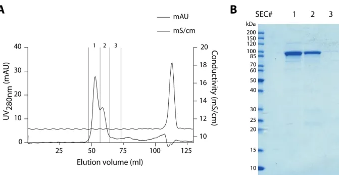
As an initial step for characterizing the fusion protein, the functionality of IpaB fused to Knot was tested using purified protein. Expression of ipaBis toxic to E. coliBL21 and toxic- ity can be circumvented by co-expression of the cognate chaperoneipgC (Lokareddyet al., 2010). IpgC stabilizes IpaB in the cytoplasm and thereby prevents IpaB degradation (Menard et al., 1994b). Therefore, IpaB-Knot was co-purified after expressing it together with ipgC in E. coli BL21. The purification strategy for IpaB is based on purifying the IpgC/IpaB com- plex viaa His-tag in IpgC and subsequent destabilization of the interaction with LDAO. This destabilization lead to elution of monomeric IpaB (Birketet al., 2007; Senerovicet al., 2012).
Additional affinity purification using Strep-Tactin resin prior to gelfiltration of the protein re- sulted in the purification of only full-length IpaB-Knot (Fig. 2.4A), as the Strep-tag of the fusion is C-terminal. Gelfiltration separated aggregated protein from monomeric IpaB-Knot (Fig. 2.4B).
To ensure that IpaB-Knot remains a functional T3SS effector, IpaBs capability to induce pyroptosis in murine bone marrow macrophages (mBMM) was analyzed using purified IpaB- Knot. IpaB is a multifunctional effector and translocator and the induction of pyroptosis is well documented (Zychlinsky et al., 1994; Senerovic et al., 2012; Lunelli et al., 2009) and can be assessed with a colorimetric assay. Upon macrophage disintegrity, lactate dehydro- genase (LDH) is released into the supernatant. Activity of LDH is quantified in a colorimetric reaction, where LDH reduces a yellow tetrazolium salt (2-(4-Iodophenyl)-3-(4-nitrophenyl)-5- phenyl-2H-tetrazolium chloride) into a red formazan product. The accumulation of red dye is proportional to the amount of cells lysed and was quantified in a 96-well plate spectrometer.
IpaB exerts cytotoxicity at concentrations as low as 0.4 µM (Senerovicet al., 2012) and a
50 75
25 100 125
0 10 20 30 40
10 12 14 16 18 20
Elution volume (ml)
kDa
50 60 70 85
40
30 25 20
15
10 100 120150 200
1 2 3
mAU SEC#
mS/cm
Conductivity (mS/cm)
UV 280nm (mAU)
A B
1 2 3
Figure 2.4: Size-exclusion chromatography and analytical SDS PAGE/Coomassie of recombinant IpaB-Knot. (A) Elution profile of IpaB-Knot. 1, 2 and 3 indicate fractions collected which were analyzed in (B).
50 25 10 50 25 bufferdetergent
0.0 0.5 1.0 1.5 2.0
Absorbance (492 nm)
IpaB-Knot BSA µg/ml
Figure 2.5: Induction of cell death in macrophages by IpaB-Knot.Cell death was measured by release of LDH from mBMM in a colorimetric assay. Error bars indicate SD, n=3 independent experiments.
similar result was obtained for IpaB-Knot, where cytotoxicity was already seen at 0.25 µg/ml (0.26 µM) (Fig. 2.5). At 0.5 µg/ml (0.53 µM), cell lysis was as efficient as disruption with 1 % detergent (Triton X-100). Since IpaB-Knot is concentrated in the presence of LDAO, the de- tergent might accumulate during protein concentration and enhance cytotoxicity. To rule out a cytotoxic effect of LDAO, the non-toxic protein bovine serum albumin (BSA, 66 kDa) was concentrated under same conditions as IpaB-Knot and added to mBMM. No cellular disinte- gration was detected in samples with 50 µg/ml (0.75 µM) BSA. Thus, disruption was caused by a functional IpaB domain in IpaB-Knot. This confirmed that IpaB maintains effector function inside the fusion protein.
2.3 The Knot is folded within IpaB-Knot
For efficient obstruction of the NC channel by IpaB-Knot, the Knot has to be tightly folded when fused to IpaB. Since IpaB is stored in complex with IpgC in the bacterial cytoplasm, the complex of IpaB-Knot and IpgC represents the conformation of IpaB-Knot prior to secretion.
As the Knot needs to fold prior to secretion, folding analysis was carried out on IpaB-Knot bound to IpgC by means of fluorescence spectrometry and limited proteolysis.
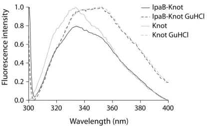
2.3.1 Fluorescence spectrometry
Fluorescence spectrometry measures the intrinsic fluorescence of the protein which largely depends on tryptophan residues and their molecular environment. Thereby, different folding states of the protein can be compared as the emission spectrum changes when tryptophans are exposed to solvent upon unfolding. The folding status of the Knot-domain in IpaB-Knot was addressed by measuring the change in tryptophan fluorescence under native and dena- turing conditions (Fig. 2.6). IpaB contains a single tryptophan whereas the Knot domain has four tryptophans, three of which are part of the trefoil knot in the C-terminal region, indicat- ing that the fluorescence is mainly derived from folding status of the Knot. Fluorescence of IpaB-Knot in complex with the chaperone IpgC was recorded. IpgC does not contain trypto- phans and was included in the blank buffer as a control.
Spectra of purified IpaB-Knot complex were compared to spectra of the Knot alone, either in buffer or in the presence of the chaotropic salt guanidine hydrochloride (GuHCl). The native
300 320 340 360 380 400 0.0
0.2 0.4 0.6
Fluorescence in
Wavelength (nm)
Figure 2.6: Fluorescence spectra of IpaB-Knot and Knot. Solid lines represent native conditions, dashed lines show spectra under the influence of 6 M guanidine hydrochloride.
Knot protein had a fluorescence emission peak at 335 nm which represents tryptophans in the folded protein (Fig. 2.6, solid grey line). The fluorescence spectrum of IpaB-Knot was similar to the native Knot spectrum with an emission peak at 335 nm (Fig. 2.6, solid black line).
This similarity of fluorescence spectra suggests folding of Knot in IpaB-Knot analog to native Knot. Treatment with 6 M GuHCl lead to unfolding represented by a red shift of emission peaks from 335 nm to 355 nm. This shift indicated a change of the molecular environment due to exposure of the respective tryptophans to solvents in both IpaB-Knot and Knot alone.
Thus, the change in fluorescence derived from GuHCl-induced structural alterations of the Knot domain confirms that this domain was folded under the tested conditions.
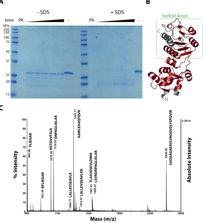
2.3.2 Protease treatment reveals a folded core including the trefoil knot
Folding of a protein can be analyzed in more detail with treatment by unspecific pro- teases. Under native conditions, the protein fold prevents cleavage by proteases. Only free structures can be accessed by the enzyme and the folded parts remain intact over time.
For the Knot protein alone, protein fragments were obtained by treatment with the non- specific protease Proteinase K and separated by SDS PAGE (Fig. 2.7A).
Already after a few minutes of incubation, treatment led to a product of approximately 25 kDa which is no further digested. The cleavage product was stable because of its native fold, as total cleavage of Knot occurred in the presence of the denaturing detergent SDS, a condition where Proteinase K is active (Hilz et al., 1975). The obtained fragment was ana- lyzed by mass spectrometry (MS) (Fig. 2.7B) and mapped on the known structure for RrmA (Fig. 2.7C). Only the terminal helices were cleaved whereas the trefoil knot remained intact.
250 150 100 75 50 37
25 20
15 kDa
- -
time
- SDS + SDS
PK PK
trefoil-knot
C
N
A
C
B
800 1160 1520 1880 2240 2600
3.3E+4
0 10 20 30 40 50 60 70 80 90
100 1343.71
1110.63
805.46 1079.59 2429.26
1561.81
961.56 1565.91
1412.87
1284.77
Mass (m/z)
Absolute Intensity
% I n tensit y
FLIEGAR RFLIEGAR NSTGVVFSLR DNPAGLIALAR LALLEVSEAVLK LALLEVSEAVLKKAAWLEAAQTQVR TLAASESEVLDWIK LSVRDNPAGLIALAR SADAAGAEAVLVAGGVDLYSPQVIRFigure 2.7: Protease K treatment of Knot under native and denaturing conditions. (A) SDS PAGE and Coomassie stain of Knot treated with Proteinase K (PK) or untreated (-) under native (-SDS) and denaturing (+SDS) conditions, where Proteinase K is still active. (B) 3D structure of Knot mapped with fragment identified by MS from the stable band in (A). The trefoil knot is indicated by a green box. (C) MS spectrum of the stable fragment. Peptides are indicated on peaks, see Fig. 2.10 for Knot sequence. MS analysis was performed by Monika Schmid, MPIIB.
alyzed by MS and MS/MS. Some peptides resulted from digestion of the IpaB domain, which, in complex with IpgC, was partially unfolded when treated with Proteinase K (Fig. 2.8, Box 1).
As seen for the Knot, polypeptides that cover almost the entire sequence of the Knot domain and the complete sequence of the trefoil-knot motif were detected (Fig. 2.8, Box 2).
1
2
- Proteinase K + Proteinase K
MW
IEF
Figure 2.8: 2D-electrophoresis of IpaB-Knot.IpaB-Knot was separated by isoelectric focus (IEF) and molec- ular weight (MW) as a second dimension without Proteinase K (left) or after Proteinase K treatment. Protein fragments after Proteinase K digest were analyzed by mass spectrometry. Electrophoresis was performed by Ursula Zimny-Arndt, MPIIB.
Subfragments from these spots were confirmed by MS/MS analysis (Fig. 2.9). y and b ions have been mapped to fragments with the respective charge retained either at the N-terminus (b) or C-terminus (y). Although the spectrum for the IKELAR-peptide was of low quality (Fig.
2.9A), the FLIEGAR peptide matched the N-terminal peptide of the Knot in IpaB-Knot (Fig.
2.9B) and the LLRDL peptide confirmed the C-terminal fragment (Fig. 2.9C). Again, only the C-terminal helix and N-terminal parts of the Knot domain, but not the trefoil knot, were cleaved, suggesting that this motif is folded and consequently inaccessible to Proteinase K (Fig. 2.10).
Fluorescence analysis and mass spectrometry after limited proteolysis substantiate a folded core domain of Knot in IpaB-Knot. This includes the trefoil-knot motif which was un- folded by GuHCl and resistant to proteolysis by Proteinase K under native conditions.
70 226 382 538 694 850 70.4
0 10 20 30 40 50 60 70 80 90
100 303.12
175.06 616.97
415.12
261.11 528.21
Mass (m/z)
% Intensity Absolute Intensity
y3
y1
b3 y5
y4
432.17 545.20
b2
374.16
70 196 322 448 574 700
1175.6
0 10 20 30 40 50 60 70 80 90
100 498.27 516.28 630.33
175.09 612.34
415.15
290.10 400.27 654.38383.25 446.21
227.13
Mass (m/z)
% Intensity Absolute Intensity
y2b2 y3
y1
b5 b4
b3
69.0 209.4 349.8 490.2 630.6 771.0
13.9
0 10 20 30 40 50 60 70 80 90 100
359.18
175.07 556.09
Mass (m/z)
% Intensity Absolute Intensity
y3
y1
b5
A
B
C
L L R D R
y1 y3 y2
b2 b3 b4
F L I E G A R
y1
b2
y5 y4 y3
b3
I K E L A R
y3 y1
b5
Figure 2.9: MS/MS analysis of IpaB-Knot. Spectra for N-terminal and C-terminal fragments of Knot after Proteinase K digest of IpaB-Knot and separation of peptides by 2D gel electrophoresis (analysis of Box 2 from Fig.
2.8). b and y anions correspond to amino acids with charge retained on the N-terminus (y) or C-terminus (b).
Analysis was performed by Monika Schmid, MPIIB.
---MRITSTANPRIKELARLL
ERKHRDSQRRFLIEGAREIERALQAGIELEQALVWEGGLNPEEQQVYAALGRVGRLALLE
VSEAVLKKLSVRDNPAGLIALARMPERTLEEYRPSPDALILVAVGLEKPGNLGAVLRSAD
AAGAEAVLVAGGVDLYSPQVIRNSTGVVFSLRTLAASESEVLDWIKQHNLPLVATTPHAE
ALYWEANLRPPVAIAVGPEHEGLRAAWLEAAQTQVRIPMQGQADSLNVSVSAALLLYEAL
RQRLLRDRLTKTHSTLSAWSHPQFEK
Strep-tag
linker
IpaB Knot
trefoil knot-motif ...
Figure 2.10: MS and MS/MS analysis of IpaB-Knot. Identified peptides by MS are highlighted in red in the Knot sequence and fragments confirmed by MS/MS are highlighted bold. The Knot is indicated by a green line above the amino acid sequence.
2.4 Plasmid-encoded IpaB-Knot: effects on hypersecretion
The fusion protein was introduced into hypersecretory strains, the∆ipaBor∆ipaDstrain in order to analyze effects on T3SS. In the initial approach, fusion genes were expressed from inducible plasmids with a tetracycline-promoter after bacteria had grown into the logarithmic phase.
2.4.1 Induced expression in
∆
ipaBThe∆ipaBstrain was complemented with plasmids containing either pipaBor pipaBknot and secretion was analyzed by SDS PAGE and silver stain (Fig. 2.11). Induced cultures were washed and splitted and leaky secretion (-CR) was differentiated from Congo Red-induced secretion (+CR) (Fig. 2.11A). Effector secretion in the ∆ipaBstrain is not affected by Congo Red-addition except for one band missing at 40 kDa. In comparison, expression of pipaB leads to absence of a doublet at 60 kDa and a band at 38 kDa. Secretion could be enhanced by Congo Red, as additional bands appeared (e.g. 50 kDa, 52 kDa, 60 kDa and 70 kDa). For complementing∆ipaBwith pipaBknot, no secretion was observed after adding Congo Red.
Secretion was analyzed in more detail by Western blotting. For the ∆ipaB + pipaB strain, IpaB was detected in supernatants after gene expression of pipaBwas induced. In this strain, secretion was enhanced in the presence of Congo Red (Fig. 2.11B). This was not observed for ∆ipaB complemented with pipaBknot, although intracellular protein levels were equal for both IpaB and IpaB-Knot (Fig. 2.11C). The intracellular chaperone DnaK in supernatants served as a marker for cellular disintegrity. DnaK was not detected in the supernatants which confirms bacterial viability in this experiment. Furthermore, IpaB-Knot was not secreted in this strain and also influenced secretion of other effectors when expressed from a plasmid.
2.4.2 Induced expression in
∆
ipaDSince the aim was to block the T3SS channel with the fusion, expressing the fusion in the background of a strain that secretes the effector IpaB in the absence of Congo Red induction was considered. The∆ipaDstrain has a hypersecreting phenotype similar to∆ipaB, secretes IpaB as well as other effectors without Congo Red (Menardet al., 1994a) and IpaB does not contribute to regulation of secretion in∆ipaD. Unlike in the∆ipaBstrain, expression of pipaB or pipaBknotshould not restore wildtype secretion, thus no Congo Red-induction is needed.
+CR -CR
+CR -CR +CR -CR
pipaB
- pipaBknot
250 150 100 75
50
37
25
kDa +CR -CR +CR -CR +CR -CR
100 75
50
100 75
50 kDa
IpaB-Knot IpaB IpaB mAB
IpaB mAB DnaK DnaK mAB
IpaB-Knot IpaB
C
100 75
50
lysates
kDa
pipaBknot pipaB
-
+CR -CR +CR -CR +CR -CR
ΔipaB ΔipaB ΔipaB pipaBknot pipaB
-
ctrl
Figure 2.11: Secretion assay of∆ipaBwith IpaB-Knot.(A) Bacterial supernatants were analyzed by silver stain after induction with AHT or without induction (∆ipaB / -). Induced samples were further grown in the presence of Congo Red (CR+) or without Congo Red (CR-). (B) Supernatants analyzed by Western blotting with IpaB and DnaK mouse monoclonal antibodies. One lysate was included as an DnaK antibody control (ctrl). (C) Bacterial lysates analyzed by Western blotting with IpaB mouse monoclonal antibody.
Expression of either pipaBor pipaBknot in ∆ipaDwas investigated in comparison to an mxiHmutant (secretion deficient) and the untransformed∆ipaDstrain. These strains served as controls to monitor effects of AHT (+) or DMF (-, vehicle), as secretion was analyzed from overnight-grown cultures in order to accumulate more effector proteins in culture su- pernatants (Fig. 2.12A).
For induced expression of pipaB, analysis of SDS PAGE and Coomassie stain showed a slight reduction in secretion compared to the ∆ipaD control but the typical pattern of se- creted effector proteins was retained (Fig. 2.12A, middle panel). With regards to the effector proteins secreted, the∆ipaDstrain with induced pipaBknot was completely deficient in se- cretion and only residual proteins were detected in the supernatant (Fig. 2.12A, right panel).
100 75 50
50 37 150
250 100 75
50 37
25 20 kDa
kDa
supernatants lysates ΔmxiH ΔipaD ΔipaD
pipaB
ΔipaD pipaBknot
ΔmxiH
ΔipaD ΔipaD piB
ΔipaD piBk
- + - + - + - +
AHT
50 37
IpaB mAB
IpaC pAB
MxiG mAB - + - + - + - + - + - + - + - +
ΔmxiH
ΔipaD ΔipaD piB
ΔipaD piBk
IB IBK
B A
Figure 2.12: Secretion assay of ∆ipaD with IpaB or IpaB-Knot. (A) Coomassie stain of precipitated culture supernatants of∆mxiH,∆ipaDand∆ipaDplus plasmid-encoded pipaBor pipaBknot. All strains tested were treated with DMF (-) or AHT in DMF (+). Supernatants were normalized. (B) Western blot analysis of strains fromA, probed with anti-IpaB mouse mAB, anti-IpaC rabbit pAB, anti-MxiG mouse mAB, piB: plasmid-ipaB, piBk:
plasmid-ipaBknot, IB: IpaB, IBK: IpaBKnot.
The effect on secretion by pipaBknot and lack of effector synthesis due to regulatory ef- fects were further differentiated. Western blot analysis of IpaB in supernatants and lysates showed that the ∆ipaD strain secreted IpaB whereas the ∆mxiH did not and both strains had equal levels of IpaB in the cytoplasm (Fig. 2.12B, supernatants and lysates, upper left panels). The level of IpaB in∆ipaDrepresents the basal intracellular amount of IpaB for this bacterial background. This level, as well as the amount of secreted IpaB, was increased by additional expression of pipaB(supernatants and lysates, middle panels). In∆ipaDexpress- ing pipaBknot, the amount of secreted IpaB as well as the intracellular level of endogenous IpaB were reduced and cytoplasmic levels of IpaB-Knot (IBK) were comparable to IpaB (IB) in ∆ipaD. Again, IpaB-Knot was not secreted. IpaC was analyzed as an additional effector protein. Levels of IpaC in supernatants and cytoplasm were reduced compared to ∆ipaD+ pipaB. This only applied to effector proteins, as levels of MxiG, a structural component of the NC basal body, were not altered in bacterial lysates.