J. Perinat. Med.
13 (1985) 31
Catecholamines in arterial and venous umbilical blood: placental
extraction, correlation with fetal hypoxia, and transcutaneous partial oxygen tension
R. Paulick, E. Kastendieck, H. Wernze
Department of Obstetrics and Gynecology, Department of Internal Medicine, University Würzburg, F. R. Germany
1 Introduction
Investigations performed in animal experiments [6, 7, 16, 17, 19] as well as in man [2, 3, 9, 10, 18, 20, 25] suggest that fetal hypoxia is associated with an increase in circulating catecholamines due to sympathoadrenal stimulation. The correlation between catecholamines and cardiovascular, respir- atory, and metabolic changes as determined during intrapartum fetal monitoring by means of cardio- tocography (CTG), fetal blood gas analysis, and measurement of the transcutaneous partial oxygen tension (tcpC^) has not yet been exhaustively investigated. The effect of fetal hypoxia on the dopamine concentration in cord blood likewise has not been explored.
These gaps in our knowledge are attributable to some extent to methodological factors. The older (fluorimetric) tests, which require blood volumes of 15—20ml, are restricted to the detection of epinephrine (E) and norepinephrine (NE) in mixed cord blood [3, 22] or venous umbilical blood [18, 23], and some studies only measured total cate- cholamines [2, 20] without further differentiation.
In contrast, the radioenzymatic single-isotope technique [8, 29] allows the specific measurement of E, NE and the third catecholamine, dopamine (D), in 0.1-0.2 ml plasma. With the advent of this method it has become possible to reassess the following questions of clinical interest:
Curriculum vitae
RENE PAULICK, M. D.f was born in January, 1956 in Hannover, Germany.
Medical studies in Würz- burg from 1974 to 1980.
He is at present assistent at the Department of Obstet- rics and Gynecology, Uni- versity of Würzburg (Head:
Prof. Dr. K.-H. WULF/
Chief area of interest: Fetal and neonatal physiology, gynecological oncology.
high values, extracted across the human pla- centa?
What interrelationship exists between the extraction rates of NE, E, and D?
2. How are fetal hypoxia, acidosis, changes in fetal heart rate (FHR), and postpartum fetal distress associated with the catecholamine levels in fetal plasma?
3. Is an increased secretion of catecholamines in the human fetus accompanied by a fall in tcpO2, as has previously been suggested though only on the basis of animal experiments [16, 19,28]?
2 Methods
1. To what extent are free catecholamines, varying The patients in our study comprised 34 parturient
over a wide range from normal to exceptionally women aged 19—34 years who after essentially
uncomplicated pregnancies (duration of preg- nancy 36—41 weeks) were delivered vaginally (30 spontaneous births, 4 vacuum extractions).
The birth weights ranged from 2450-4460 g. Free catecholamine concentrations were determined separately in arterial and venous umbilical blood following immediate clamping of the cord, and in samples of maternal blood obtained immediately after delivery from the brachial vein.
After collection the blood was at once transferred into chilled polypropylene tubes to which had been added, as anticoagulant and antioxidant, 50 μΐ of a solution containing 76 mg/ml EGTA (ethyleneglycol-bis-(j3-aminoethyl ether) Ν,Ν,Ν'- tetraacetic acid) and 48 mg/ml reduced glutath- ione. In some of the later experiments, lithium- heparin tubes (SARSTEDT No. 36 377) were used since comparative studies had shown that cate- cholamine concentrations can be measured with equal accuracy in heparinized plasma and anti- oxidant-containing heparinized plasma, but not in EDTA-containing plasma. It is important however that tubes are placed on ice without delay [36], Hemolyzed samples were discarded. After centri- fugation for 10 minutes (4000 rpm) at + 2 to + 4°C, the plasma was kept frozen at — 25 °C until assayed.
The concentrations of free NE, E, and D were assayed radioenzymatically using a modified and shortened version of the method of PEULER and JOHNSON [8, 29, 36]. The sensitivity of the method is below 1 pg/ml for NE, E, and D, with intra-assay and inter-assay coefficients of variation of approx. 3 % and 10 %, respectively.
The acid-base balance and blood gases were assessed with the TECHNICON Gas Analyzer. The base deficit in the extracellular fluid was calculated nomographically by the method of SlGGAARD- ANDERSEN from the pH and pCO
2in the umbili- cal artery, hemoglobin concentration 5 g% [33].
For interpretation of the cardiotocograph (CTG) tracings, the deceleration areas obtained during the last hour antepartum were measured plani- metrically in cm
2/h (chart speed: 1 cm/min;FHR amplitude: l c m = 20bpm). The baseline heart rate was determined every 2 minutes, followed by calculation of mean values.
In 22 parturient women the fetal tcpO
2was con- tinuously monitored With an oxygen electrode (TRANSOXODE/Hellige) during the late first stage and the second stage of labor. At the same time, the catecholamine concentrations in the umbilical vessels were measured in 14 patients. The elec- trode was applied to the fetal scalp by means of a self-adhesive tissue glue (HlSTO-ACRYL, Braun Melsungen). The temperature of the electrode was adjusted to 44 °C. After attainment of a stable level, the tcpO
2was recorded for 92 ±71 (SD) minutes on average. The mean tcpO
2values deter- mined at ΙΟ-minute intervals were used to cal- culate during the last hour before delivery the overall means. By simultaneously recording the relative local skin perfusion, distinct artefacts could be identified and excluded [14].
The statistical analysis and significance calcula- tions were performed at the Computer Center of W rzburg University (Dr. L HAUBITZ). The corre- lation coefficients were calculated using either the Spearman rank correlation test (when there was no normal distribution) or the Kendall rank correla- tion test (when 'ties' were present).
3 Results
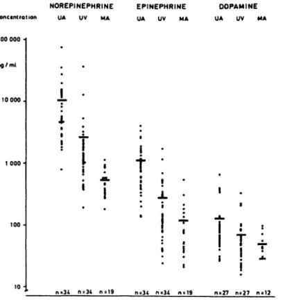
3.1 Catecholamine concentrations in arterial and venous cord blood ;placental catecholamine extrac- tion: The free catecholamine levels measured in arterial and venous cord blood may vary sub- stantially (Fig. 1). The mean NE concentration was 10,200 pg/ml (range 1,500-74,100 pg/ml) in arterial cord blood and 2,650 pg/ml (range 200- 30,700 pg/ml) in venous cord blood. The con- centrations of E in arterial and venous cord blood were 1,120 pg/ml (range 140-4,030 pg/ml) and 280 pg/ml (range 25-1,790 pg/ml), respectively.
The concentrations of circulating free D approxi- mated 130 pg/ml (range 30-660 pg/ml) in arterial cord blood, and 70 pg/ml (range 15-320 pg/ml) in venous cord blood.
The placental catecholamine extraction rate (ER),
calculated from the concentrations in the umbilical
NOREPINEPHRINE EPINEPHRINE Concentration UA UV MA UA UV MA
too ooo
pfl/ml
10000 .
OOPAMINE UA UV HA
1 000
100 -
10
I i
i ;
•: ·ι Ι• · t
» n =34 nO4 nr19 ns34 nr34 na19
! T :
i ! i
•ns27 n=27
:
•! u.
n = 12
Fig. 1. Concentrations of free NE, E and D in the umbili- cal artery (UA), umbilical vein (UV) and in maternal venous (brachial vein) blood (MA) at the time of delivery.
Epinephrine
r a t e of e x t r a c t i o n 100-1
80-
60-
20-
··.
• ·• ··
EE= 0 . 9 4 EN
rs p = 0.7.
2 oc < 0.001
20 40 60 80 100
N o r e p i nephrinc r a t e of e x t r a c t i o n
Fig. 2. Correlation between the extraction rates of E (ERE) and NE (ERNE) during placental passage. Extrac- tion rates vary between 35 % and 95 %. A highly significant correlation is demonstrated. There is no difference between the regression line and the line of identity.
artery (C^) and umbilical vein (C
uv) according to the formula
ER =
'ua100
'ua
was established as 77 ±14% (range 41-96) for NE, 76 ± 16 % (range 33-96) for E, and 33 ± 25 % (range 24-99) for D.
There was a significant correlation between the extraction rates of E and NE (Fig. 2).
The umbilical arteriovenous difference in NE and E concentration rose in proportion to the arterial catecholamine concentration (Fig. 3).
3.2 Catecholamine concentrations in maternal blood: At the time of delivery, the mean free NE concentrations in the maternal venous blood were 540pg/ml (range 180-1,120 pg/ml) (Fig. 1). In the majority of the patients, the NE levels thus exceeded the normal range of 100—450 pg/ml that had been determined using the same method in normotensive male and female subjects under con- ditions of rest [36].
The concentrations of free Ein the maternal blood, 120 pg/ml (range 20—550 pg/ml), were likewise
Catecholamines umb. art - umb. vein 100000*1
pg/ml 10000-
1000·
100·
N o r e p i n e p h r i n e log NE o r t_v cjn s l°9
= 0,94 ; 2-<0.001
- 0.11
s
o Epincphrine
*< 0.001
10 100 1000 10000 100000 pg/ml Catecholamines umbilical artery
Fig. 3. Correlation between the arterio-venous concentra- tion difference of Ε and NE and arterial concentrations.
A linear relationship is demonstrated in the range below 4.000 pg/ml for Ε and 74.000 pg/ml for NE, suggesting that the placental catecholamine extraction does not reach full saturation.
increased over the normal E levels (20—95 pg/ml).
In contrast, the mean concentrations recorded for free D, 50 pg/ml (range 30-100 pg/ml), were fairly unchanged compared to the normal range of
10-70 pg/ml.
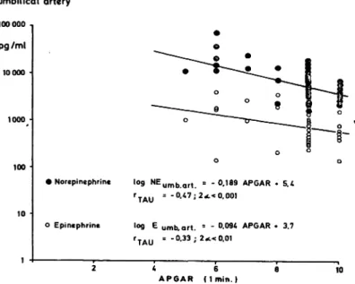
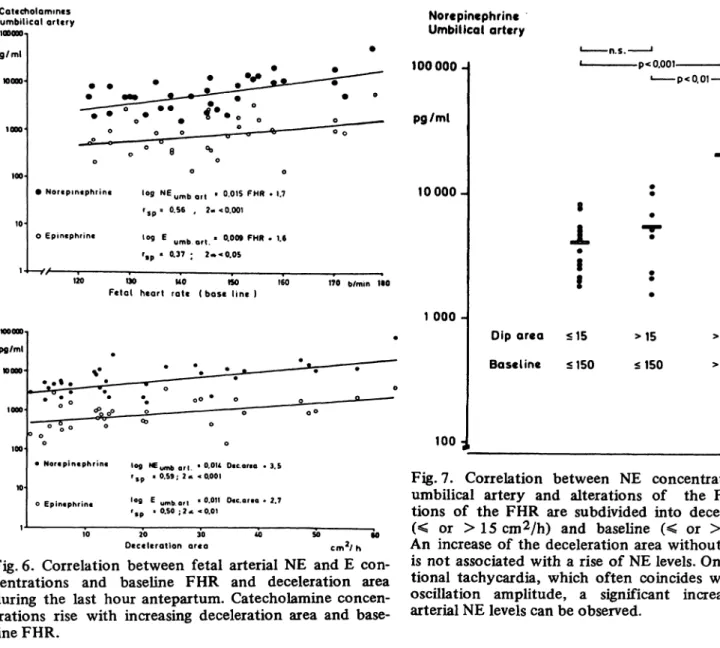
2.3 Catecholamines and fetal hypoxia: As shown in Figs. 4 and 5, there was a highly significant relationship between the umbilical arterial NE levels, neonatal status (as assessed by the 1-minute APGAR score), and metabolic acidosis (determined by pH and base deficit). In some cases, the NE concentrations in the fetal blood were exceedingly high when associated with low Apgar scores and increasing metabolic acidosis. Analysis of the FHR in the last hour preceding delivery also revealed a significant relation between the area of decelera- tion, the baseline FHR, and the umbilical arterial NE concentrations. The fetal NE concentrations were found to rise as the deceleration area and baseline FHR increased (Fig. 6).
Fig. 7 gives a more detailed description of the cor- relation that exists between deceleration area, fetal tachycardia, and arterial NE concentration. It is seen that an increase of the deceleration area (> 15 cm
2/h) is not associated with elevated NE levels. A significant rise in the fetal arterial NE
Catecholamines umbilical artery
100000
pg/ml
10000
1000
100
10
• Norepinephrinc log NEu m b a r t
o Epincphrine
TAU
log
= - 0.47; 2
-0.189 APGAR * 5.4
< 0.001
umb. art. = -
= -0.33 ; 2«c<0.01
A P G A R * 3.7
2 4 6 β 10 A P G A R ( I m i n . )
Fig. 4. Correlation between the 1-^iinute APGAR score and NE and Ε concentrations in the umbilical artery. Low APGAR scores are associated with increased catechola- mine concentrations.
Catecholamines umbilical artery 100000
pg/ml 10000
1000-
100-
10-
Norepinephrme log NE umb drt = - 3 , 3 ρ Η * 2 β
O Epinephrine
rsp = - 0.70 : 2<c < 0.001
»°9 Ε umb. art. = -2.5pH * 20.7 rs p = -0.51 ; 2-t «0.01
7,00 7.10 7.20 7.30
pH in the umbilical artery
7.40
pg/ml wooo-
1000-
100
fc /
O Norepmephrme ,og N Eu m b > Q r l =0.062 BD * 3.4
rsp s °·70 j 2* < 0.001
Ο Epmephrine ,og E umb Q f t = 0.046 B D « 2.6 rs p r O.S6 : 2 χ. < 0.001
* β 12 16 m m o l / l 20 Base deficit in the umbilical a r t e r y
Fig. 5. Correlation between fetal arterial NE and Ε con- centrations and metabolic acidosis measured by the pH and base deficit in the umbilical artery. Catecholamine concentrations start to rise with increasing metabolic acidosis.
.concentrations only occurs when tachycardia (> 150 b/min) supervenes.
Fetal arterial E concentrations as an indicator of enhanced release of the hormone from the adrenal medulla in conditions of stress, also demonstrate good correlations with the fetal parameters, although the correlation coefficients were lower than for NE (Figs. 4-6).
Unlike NE and E concentrations, the D levels in
the umbilical vessels were affected by fetal
hypoxia to a minor degree, only.
Cotccholommcs umbilical artery pg/ml
• Nortpmtphrint log NEu m b Q f t t 0.015 FHR » 1.7 rs p 8 0.56 , 2« < 0.001 Ο epintphrin« iog Ε yfnb ef| . 0.009 FHR · 1.6
r»p ' °·37 I 2·»* 0.05
-V/ , , — , , ,-_
120 190 UO «SO 160
Fetal heart rate ( b a s e l i n e ) 170 b/min 1·0
pg/ml η ooo
• Nortpintphrin« Iofl NEy^ ef| . O.OU Dtc or.o * 3.5 r* * °·59·· 2* <0·001
oEpintphrin« '°9 E umb.art ' <>·0" 0«.ar.o . 2.7 'sp ' 0.50 ; ? < < 0.01
20 30
Deceleration ar«a cmV h
Fig. 6. Correlation between fetal arterial NE and E con- centrations and baseline FHR and deceleration area during the last hour antepartum. Catecholamine concen- trations rise with increasing deceleration area and base- line FHR.
3.4 Catecholamines, transcutaneous pO
2, and transcutaneous-arterial pO
2difference: Fig. 8 illus- trates the tcpO
2values recorded during the 2 hours preceding parturition. They differ widely, ranging between 0—25 mm Hg and showing a tendency to decrease before delivery.
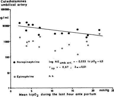
In Fig. 9, the mean tcpO
2measured during the last hour before delivery is plotted against the NE con- centration in the umbilical artery. There was a significant inverse relation with NE concentrations rising as tcpO
2values decreased. No correlation could be detected between arterial E levels and the tcpO
2.
In most of the cases (70%), the tcpO
2measured during the last hour before delivery was lower than the umbilical artery pO
2. This transcutaneous- arterial pO
2difference (tc-art pO
2-d) was depen-
Norepinephrine Umbilical artery 100000-
pg/ml
10000-
1 000 -
100
-p<0.00t- -p<0,01-
J. Τ
Dip area <15
Baseline <150 <1SO
>15 c m2/ h
>150 b/min
Fig. 7. Correlation between NE concentrations in the umbilical artery and alterations of the FHR. Altera- tions of the FHR are subdivided into deceleration area (< or >15cm2/h) and baseline (< or > 150 b/min).
An increase of the deceleration area without tachycardia is not associated with a rise of NE levels. Only with addi- tional tachycardia, which often coincides with a loss of oscillation amplitude, a significant increase of fetal arterial NE levels can be observed.
dent on the NE concentration in the fetal arterial blood (Fig. 10) and was found to increase with rising NE concentration.
Timt ontt per turn
Fig. 8. Course of tcpO2 during the last two hours before delivery. tcpO2 values vary between 0 and 25 mmHg. In some cases a high tcpO2 is recorded until delivery, while a fall in tcpO2 can be demonstrated in other cases.
Catccholamincs umbilical artery
100000η
pg/ml wooo·
1000
100
O Norepinephrine log N Eu m b a r t = - 0 , 0 3 3 tcp02**.0 rs p = - 0.67 ; 2*<0.01
o Epinephrine n. s.
0 5 10 15 20 mmHg 25 Mean tcp02 during the last hour ante partum
Fig. 9. Correlation between tcpO2 in the last hour before delivery and NE and Ε concentrations in the umbilical ' · artery. There is a significant inverse relationship: tcpC>2
decreases with increasing NE.
Catecholamines umbilical artery
100000-1
pg/ml
10000-
1000-
100
10-
O Norepinephrine log NEu m b Qrt = 0.036 a r t - tc p02 » 3.4 rs p = 0.59 ; 2*<0.05
ο Epinephrine n.s.
5 10 is 20 mmHg 25 a r t - t c p02 - difference
Fig. 10. Correlation between tc-art pO2-d in the last hour before delivery and NE and Ε concentrations in the umbilical artery. With rising NE-secretion the tc-art pO2-d increases. The correlation coefficient improved con- siderably after eliminating the value marked with an asterisk (rsp = 0.74, ρ < 0.01). This was the only patient exhibiting low umbilical artery pO2 (10.1 mmHg) at low oxygen saturation (< 20 %).
4 Discussion
4.1 Catecholamine extraction by the placenta:
The fetal artery NE and Ε concentrations were in part excessively high, mean values being four times higher than in the umbilical vein and exceeding those in the maternal venous blood 20-fold for NE, and 10-fold for E (Fig. 1). These findings suggest that the Catecholamines measured in cord blood are of fetal origin and that the placenta has a high capacity for inactivation of free Catecholamines.
At concentrations below 4000 pg/ml for Ε and 74,000 pg/ml for NE, a saturation of the placental catecholamine extraction was not detectable (Fig. 3).
It appears that the high placental catecholamine clearance is primarily attributable to metaboliza- tion of biogenic amines. Studies with radioactively labelled tracers have shown that the placental transfer accounts for only 5—10% of the umbilical arteriovenous concentration difference [24, 31].
The enzymes catechol o-methyltransferase and monoamine oxidase, which are required for degra- dation of circulating Catecholamines, have been demonstrated in placental tissue in high concen- trations [5]. It also is probable that the biologic catecholamine inactivation is additionally effected by sulfate conjugation. Phenolsulfotransferase (EC 2.8.2.1), which is necessary for the conversion of free Catecholamines to sulfated derivates has been isolated from human placental tissue [32].
Whereas umbilical artery E and NE concentrations were widely different, the extraction rates from the placenta with mean values of 75 % were found to be identical (Fig. 2). This was to be anticipated since the metabolic pathways of NE and E are identical. However, a comparison of the placental NE and E extraction with that of other organs revealed that the placental tissue occupies a unique position: on passage of Catecholamines through the liver, cardiac and skeletal muscle, the NE extrac- tion rate is invariably lower than that of E [4, 13].
The NE concentrations in the renal vein but also in
the coronary sinus may even substantially exceed
those measured in the arterial (afferent) blood
[21, 26]. These observations suggest that in tissues
with abundant sympathetic innerv tion NE is not
only extracted but may also be released from post-
ganglionic sympathetic nerve endings [21, 26]. As
a circulating hormone, E in contrast is released enlarged deceleration area coincides with fetal only from the adrenal medulla (and some brain tachycardia that the NE concentration will areas) and extracted on its passage through the increase substantially.
other organs. Hence, the different extraction rates The findings in this study provide a differentiated of NE and E may be taken to reflect the density confirmation of the relationship which exists, of sympathetic innervation and the sympathetic according to LAGERCRANTZ et al. [20], between activity of an organ other than the adrenal medulla a tachycardic baseline FHR and total catechola- [4, 13, 21, 26]. In agreement with morphologic mines in the umbilical artery: Our observation studies [30], the nearly identical placental extrac- that an increased baseline FHR is accompanied by tion rates of NE and E indicate that sympathetic a rise of both NE and E concentrations (Fig. 6) innervation is absent on the fetal side of the supports the concept that the development of placenta. tachycardia may reflect a circulatory compensa- tion for acute changes in the blood oxygen con- 4.2 Catecholamines and fetal hypoxia: Animal
tent>
as manifested bV
the hyP°
xic shock**
studies [6, 7, 16, 17, 19] and clinical investigations
drome-
[2, 3, 10, 18, 20, 25] suggest that fetal hypoxic
The above results su^
est thatdecelerations signal- shock is associated with vigorous sympathoadrenal
Iin8
acute chanS
es in fetal arterialP°2 Π
9]
cannotstimulation, as reflected by an increase in circu-
be taken to indicate theP
resence of a^Ρ
οχίοlating catecholamines, especially in NE. This con-
shocksyndrome with increased catecholamines cept receives further support from the results merely on the strength of a quantitative analysis of reported in the present paper. The finding that the frequency, depth and duration of decelerations, stimulation of NE secretion is frequently accom-
Based on the available results> tachycardia should panied by fetal hypoxia is in accord with the
beevaluated as a supplementary parameter in the observations of KANEOKA et al. [18] and LAGER-
dete'
tion of fetal hVP°
xic shock-
CRANTZ et al. [20]. The rise of the arterial E con-
The;
e is no w^
of assessin/
to™
h ich exten'
othe' centration is distinctly less pronounced and less CTG features (e.g. loss of oscillation amplitude constant. The average arterial concentration of <
an be e™P
loy
ed as a» indication of severe fetal
r
T ^ · · ,ι ι -> c * ΙΛ 4. ι hypoxia due to the heterogeneity of the CTG free D is increased only 2.5-fold over maternal ;* ,
Λ„ , η . ι
* , , , ,
TA^ ,
u. ,, , ~ changes and the small number of tracings showing blood levels. It thus becomes obvious that D
t 5„ .„ . . , ,
5 5 u,, , . ,
u, , .. loss of oscillation without tachycardia,
hardly responds to sympathoadrenal system stimu-
Jlation.
Both pathologic alterations in FHR (decelerations, 4.3 Catecholamines, transcutaneous pO
2, and tachycardia) and hypoxic acidosis/postpartum transcutaneous-arterial pO
2difference: The results depression are associated with increased arterial obtained on measurement of the tcpO
2as described NE concentration. Various investigators have by HUGH [14] for intrapartum fetal monitoring reported that analogous correlations exist between are contradictory. Some authors reported good catecholamines, acid-base balance [2, 10, 18, 20], agreement between fetal arterial and trans- and the APGAR score [18, 25]. The results on cutaneous pO
2levels [11]. However, when patho- FHR alterations and catecholamines are in part logical CTG alterations occur, tcpO
2values are conflicting [2, 18, 20]. Unlike BISTOLETTI et al. frequently lower than the pO
2determined in [2], but in agreement with KANEOKA et al. [18], arterialized fetal scalp blood [1, 15, 35]. In our the present study evidences a quantitative relation- studies, the tcpO
2recorded during the two hours ship between the deceleration area and the arterial prior to delivery, varied between 0 and 25 mm Hg catecholamine levels (Fig. 6). More differentiated (Fig. 8). In the majority of cases, the tcpO
2was analysis of the deceleration areas reveals that an lower than the arterial pO
2. Decreased tcpO
2increase of the deceleration area exceeding levels with development of a transcutaneous-
15 cm
2/h taken by itself is not associated with a arterial pO
2difference (tc-art pO
2-d) were pri- rise in arterial NE (Fig. 7). It is only when an marily seen with pathologic CTG changes.
J. Perinat. Med. 13 (1985)
What then is the explanation for this tc-art pO
2-d?
Besides the epidermal thickness and possible measuring artifacts (e.g. pressure exerted on the electrode), the cutaneous blood flow is a major factor determining the results of tcpO
2measure- ments. The tcpO
2only corresponds to the arterial pO
2when maximal vasodilatation, by warming the skin under the electrode to 42-43 °C with the heating element of the electrode, is achieved [12, 14, 27, 34]. Animal experiments have shown that the tcpO
2varies from the arterial pO
2after recur- rent hypoxic episodes. It appears that vasocon- striction of cutaneous vessels due to increased release of NE during hypoxia may be responsible for the observed tc-art pO
2-d [6, 7, 16, 17]. The heating element of the electrode obviously is not always capable of maintaining optimal cutaneous blood flow. This assumption receives further sup- port from the fall in tcpO
2observed after injec- tion of NE into experimental animals, the pO
2in the arterial blood however remaining uneffected [19,28].
In the present study, proof was furnished that in the human fetus hypoxic release of NE is closely correlated with the tcpO
2in that NE levels rise with decreasing tcpO
2(Figs. 9, 10). Stimulation of the sympathoadrenal system in hypoxic episodes causes peripheral vasoconstriction with pallor as it occurs in severely depressed neonates (so-called
"pale asphyxia")-
From a theoretical point of view, the relationship between NE and the tc-art pO
2-d should actually be nonlinear. The tc-art pO
2-d, which is low when NE levels are low, shows an initial increase with rising NE; with the appearance of severe hypoxia and a further increase in NE, the arterial pO
2like- wise starts to decrease. In the presence of severe hypoxia and a low arterial pO
2, the tc-art pO
2-d should consequently decrease again. This situation is depicted in Fig. 10 which shows considerable improvement of the correlation coefficient after elimination of one measuring point with an oxygen saturation below 20 %.
The established correlation between fetal arterial NE levels and the fall in tcpO
2lends additional support to the concept of KuNZEL and JENSEN who had pointed to the potential of tcpO
2moni- toring in the diagnosis of a fetal hypoxic shock
syndrome. These authors had shown that an increase in tc-art pO
2-d, together with the time interval during which ä
rlow tcpO
2(0—3 mm Hg, the so-called "zero time") is recorded, are useful diagnostic tools in the detection of fetal shock [15]. On the other hand, it should be born in mind that even in the presence of a greatly reduced tcpO
2the central artery pO
2, and hence the oxygen supply to vital organs unaffected by peri- pheral constriction (brain, heart, adrenal medulla), may not show a corresponding significant decrease.
Also, there is a lack of exhaustive information as to which extent artifacts may affect the accuracy of transcutaneous monitoring (e.g. reduction in skin perfusion due to caput succedaneum or pres- sure exerted by the birth canal on the electrode).
5 Clinical consequences to be considered in the diagnosis of fetal hypoxic shock syndrome at time of delivery
For the obstetrician, the early detection of pro- tracted fetal hypoxia as manifested by increased NE secretion, circulatory centralization, severe tissue hypoxia, and acidosis is of critical impor- tance. While the diagnosis of acute fetal hypoxia presenting as continuous deceleration does not cause difficulty, the detection of hypoxic shock presenting as contraction-dependent decelerations poses a greater,problem.
Based on the established interrelationship between increased catecholamine (NE) secretion and the various diagnostic parameters employed in intra- partum monitoring of the fetus, the diagnosis of hypoxic fetal shock syndrome is warranted if the following patterns are observed:
— wide and deep decelerations with an in- creased dip area and concurrent tachydardia
— severe metabolic acidosis of the fetus
— a greatly depressed tcpO
2that has fallen to a few mm Hg (excepting artifacts)
The described investigations do not provide an
answer as to how long fetal intrapartum hypoxia
can be allowed to persist without creating a risk of
late sequelae. Infants showing signs of extreme
acidosis with concurrent release of catecholamines
should therefore have a thorough follow-up.
Summary
In 34 parturient women the levels of free epinephrine (E), norepinephrine (NE), and dopamine (D) were deter- mined by a radioenzymatic method using maternal venous and umbilical arterial and venous blood. The study was conducted to investigate the relationship between fetal catecholamines and hypoxia, fetal heart rate (FHR), and transcutaneous pO2 (tcpO2). The placental catecholamine extraction rates were also calculated.
Results
1. The NE concentrations (10,200 pg/ml) and the E con- centrations (l,120pg/ml) in the fetal arterial blood were highly elevated with mean values increased 4-fold over umbilical vein values. Compared with the mater- nal venous blood, NE values were increased 20-fold, and E values 10-fold (Fig. 1). Free D concentrations in fetal arterial blood (130 pg/ml) had risen 2.5-fold over maternal levels.
These results suggest that the catecholamines meas- ured in cord blood are of fetal origin and that the placenta has a high capacity for inactivation of free catecholamines. The placental extraction rate is 77 ± 14% for NE, 76 ± 16% for E, and 33 ± 25%for D (Fig. 2). The placental extraction rates for E and NE were virtually identical; in agreement with morpho- logical studies they demonstrated absence of sympa- thetic innervation on the fetal side of the placenta.
2. Highly significant correlations were found between fetal arterial NE concentrations and the 1-minute APGAR score, pH and base deficit in the umbilical artery and alterations of the FHR (deceleration area, baseline FHR) (Figs. 4-6). Further analysis of FHR alterations (Fig. 7) reveals that an increase in decelera- tion area without tachycardia is not correlated with an increase of fetal arterial NE concentration. A signifi-
cant rise in NE was only found with additional tachy- cardia which is often associated with a loss of oscilla- tion aptitude.
Fetal arterial E concentrations were found to corre- late with the fetal parameters indicating increased adrenal secretion of the hormone during fetal stress.
However, correlation coefficients were lower than those obtained for NE (Figs. 4-6). A significant effect of fetal hypoxia on arterial and venous D levels could not be demonstrated.
3. Fetal tcpC>2 varies between 0-25 mm Hg during the last two hours before delivery (Fig. 8). In most cases tcpO2 was lower than the arterial pO2· Besides epi- dermal thickness and artifacts, skin perfusion is a major factor influencing the tcpO2 (transcutaneous arterial pO2 difference). Vasoconstriction of the cuta- neous vessels induced by increased NE secretion during hypoxia may obviously produce a fall in tcpO2. This hypothesis receives support from the demonstra- tion that the tcpO2 is correlated with the fetal arterial NE concentration: tcpO2 falls with rising NE and the tc-art pO2-d increases (Figs. 9-10). The stimulation of the sympathoadrenal system during hypoxia results in peripheral vasoconstriction as manifested by the pallor of depressed neonates ("white asphyxia").
Clinical consequences
In view of the demonstrated correlation between increased catecholamine (NE) secretion and the various parameters for monitoring fetal intrapartum conditions, fetal hypoxic shock can be taken to be present if
— wide and deep decelerations with an increased dip area occur in combination with tachycardia,
— severe fetal metabolic acidosis is present,
— tcpO2 is lowered to a few mm Hg (excluding artifacts).
Keywords: Cardiovascular system, catecholamines, dopamine, epinephrine, extraction rate, fetal heart rate, fetal shock, norepinephrine, transcutaneous pO2, transcutaneous-arterial pO2 difference.
Zusammenfassung
Katecholamine im arteriellen und venösen Nabelschnur- blut: plazentare Extraktion, Beziehungen zur fetalen Hypoxie und zum transkutanen Sauerstoffpartialdruck Bei 34 Gebärenden wurden im matern-venösen sowie arteriellen und venösen Nabelschnurblut die freie Adre- nalin-(E), Noradrenalin-(NE) und Dopaminkonzentration (D) radioenzymatisch im Plasma bestimmt, um den Zusammenhang zwischen fetaler Katecholaminkonzentra- tion, Hypoxie, fetalen Herzfrequenzveränderungen und transkutanen pO2 zu untersuchen und die plazentare Katecholaminextraktion zu bestimmen.
Ergebnisse
1. Im fetal arteriellen Blut finden sich zum Teil exzessiv erhöhte NE-( 10200 pg/ml)- und -Konzentrationen (1120 pg/ml), die im Mittel 4fach höher als in der V.
umbilicalis und beim NE 20fach und beim E l Of ach höher liegen als im mütterlichen venösen Blut (Abb. 1).
Das zirkulierende freie D (130pg/inl) ist im fetal- arteriellen Blut um das 2,5fache gegenüber dem mütterlich venösen erhöht. Diese Befunde sprechen
dafür, daß die im Nabelschnurblut gemessenen Kate- cholamine fetalen Ursprungs sind und die Plazenta eine hohe Kapazität zur Inaktivierung von freien Katecholaminen aufweist. Die plazentare Extraktions- rate beträgt für NE 77 ± 14 %, für E 76 ± 16 % und für D 33 ± 2 5 % (Abb. 2). Die bei der Plazentapassage gefundenen nahezu gleichen Extraktionsraten für E und NE weisen in Übereinstimmung mit morphologi- schen Studien darauf hin, daß in der Plazenta keine sympathische Nervenversorgung vorhanden ist.
2. Zwischen der NE-Konzentration in der A. umbilicalis und dem APGAR (nach l Minute), dem pH und Basendefizit in der A. umilicalis sowie den Verände- rungen der FHF (Dezelerationsfläche, basale Herz- frequenz) bestehen hochsignifikante Korrelationen (Abb. 4-6). Die nähere Analyse der FHF-Verände- rungen zeigt (Abb. 7), daß eine Zunahme der Dezele- rationsfläche allein ohne Tachykardie nicht mit einem Anstieg der NE-Konzentration korreliert. Erst bei zusätzlichem Auftreten einer Tachykardie, die zumeist J. Perinat. Med. 13 (1985)
mit einer Einschränkung der Oszillationsamplitude ein- hergeht, ist ein signifikanter NE-Anstieg nachweisbar.
Für die -Konzentrationen in der A. umbilicalis als Ausdruck der gesteigerten adrenalen Freisetzung des Hormons bei Streßzuständen lassen sich ebenfalls Korrelationen zu den fetalen Parametern nachweisen;
die Korrelationskoeffizienten liegen jedoch niedriger als dies für NE zutrifft (Abb. 4-6). Die fetale Hypoxie hat auf die arteriellen und venösen D-Spiegel indessen kaum einen Einfluß.
3. Der tcpO2 schwankt in den letzten beiden Stunden vor der Geburt zwischen 0 und 25 mmHg (Abb. 8). In den meisten Fällen liegt der tcpÜ2 niedriger als der arterielle pO2 (arterielle-transkutane pO2-Differenz).
Der tcpC>2 ist neben der Epidermisdicke und mögli- chen Meßartefakten (z. B. Druck auf die Elektrode) im wesentlichen abhängig von der Hautdurchblutung.
Offensichtlich kann eine Vasokonstriktion der Haut- gefäße, bedingt durch NE-Freisetzung während der Hypoxie, zu einem tcpO2-Abfall führen. Dafür spricht, daß zwischen hypoxisch bedingter NE-Ausschüttung und dem tcpÜ2 eine enge Korrelation vorhanden ist:
bei steigender NE-Konzentration fällt der tcpO2 ab und die arterielle-transkutane pO2~Differenz nimmt zu (Abb. 9, 10). Die Stimulation des sympathoadrenalen Systems be(i< Hypoxie führt zu einer peripheren Vaso- konstriktion, die zu der bei schwer deprimierten Neu- geborenen bekannten Hautblässe führt (sogenannte
„blasse Asphyxie").
Klinische Schlußfolgerungen
Aufgrund des gesicherten Zusammenhangs zwischen erhöhter Katecholamin- (besonders NE)-Sekretion und den verschiedenen diagnostischen Hypoxieparametern der fetalen Intensivüberwachung ist ein hypoxischer Schock- zustand des Feten anzunehmen, wenn
— breite und tiefe Dezeleratipnen mit großer Dezelera- tionsfläche in Kombination mit Tachykardie,
- eine schwere metabolische Azidose des Feten,
- ein auf wenige mmHg erniedrigter tcpO^ (Artefakte ausgeschlossen)
vorliegen.
Schlüsselwörter: Adrenalin, Dopamin, Extraktionsrate, fetale Herzfrequenz, fetaler Schock, kardiovaskuläres System, Katecholamine, Noradrenalin, transkutan-arterielle pO2-Differenz, transkutaner pO2.
Resume
Catecholamines chez la parturiente, dans le sang veineux maternel et dans le sang ombilical arteriel et veineux On a determine les taux d'Adrenaline libre (A), de Nor- adrenaline (NA) et de Dopamine (D) par une methode radioenzymatique chez 34 parturientes, dans le sang veineux maternel, et dans les sangs veineux et arteriel ombilicaux. Cette etude a ete realisee afin de rechercher la relation entre les catecholamines foetales et l'hypoxie, le rythme cardiaque foetal (RCF) et la pO2 transcutanee (pO2tc). On a aussi calcule le taux d'extraction des catecholamines placentaires.
Resultats
1. Les concentrations de NA (10,200 pg/ml) et de A (1,120 pg/ml) du sang arteriel foetal sont tres elevees avec une augmentation de plus de 4 fois des valeurs moyennes par rapport aux valeurs de la veine ombili- cale. Comparees aux valeurs du sang veineux maternel, les valeurs de NA sont augmentees de 20 fois, et les valeurs de A de 10 fois (Fig. 1). Les concentrations de D libre dans le sang arteriel foetal (l 30 pg/ml) sont elevees 2,5 fois au dessus des taux maternels.
Ces resultats suggerent que les catecholamines mesu- rees dans le sang du cordon sont d'origine foetale et que le placenta a une capacite elevee d'inactivation des catecholamines libres. Le taux d'extraction placentaire est de 77 ± 14 % pour la nor-adrenaline, de 76 ±16%
pour l'adrenaline, et de 33 ± 25% pour la D (Fig. 2).
Les taux d'extraction placentaire sont virtuellement identiques pour l'adrenaline et la nor-adrenaline; en accord avec les etudes morphologiques, ces resultats demontrent l'absence d'innervation sympathique au niveau de la face foetale du placenta.
2. On a trouve des correlations hautement significatives entre les concentrations arterielles foetales de NA et le
score d'APGAR ä une minute, le pH et le base deficit arteriel ombilical, et les alterations du RCF (surface de deceleration, rhythme de base) (Fig. 4— 6). En outre, l'analyse des alterations du RCF (Fig. 7) met en evi- dence qu'une augmentation des surfaces de decelera- tions sans tachycardie n'est pas correlee avec une aug- mentation arterielle foetale de NA. Une elevation significative de NA n'a ete trouvee qu'avec une tachy- cardie surajoutee, tachycardie souvent accompagnee d'une perte de Famplitude des oscillations.
On a trouve que les concentrations arterielles foetales d'A sont correlees avec les parametres foetaux indi- quant une secretion surrenalienne hormonale augmen- tee au cours du stress foetale. Toutefois, les coeffi- cients de correlation sont plus faibles que ceuxobtenus pour la NA (Fig. 4-6). On n'a pas pu demontrer d'effet significatif de l'hypoxie foetal sur les niveaux arteriels et veineux de D.
3. La pU2 tc varie de O ä 25 mm de Hg pendant les deux dernieres heures qui precedent I'accouchement (Fig. 8).
Dans de nombreux cas la pO2 tc est plus basse que la pU2 arterielle. A cöte de l'epaisseur de l'epiderme et des artefacts, la perfusion cutanee est un facteur majeur influenqant la pO2tc (difference de la pO2
arterielle transcutanee). La vasoconstriction des vaisseaux cutanes induite par une elevation de la secretion de NA au cours de Fhypoxie peut entrainer objectivement une chute de la pU2 tc.
Cette hypothese est renforcee par la demonstration que la pU2tc est correlee avec la concentration arterielle foetale de : la pO2tc diminue lors de l'augmentation de NA et la pO2 tc art6rielle augments (Fig. 9-10). La stimulation du Systeme sympathique pendant l'hypoxie entraine une vasoconstriction
peripherique, vasoconstriction dont temoigne la päleur des nouveaux-n^s ddprimrfs («asphyxie planche»).
Consequences cliniques
Dans Poptique de la correlation demontree entre 1'aug- mentation de la secretion de Catecholamines (NA) et les divers parametres de la surveillance du foetus en cours de
travail, on peut considerer qu'il existe un choc foetal hypoxique si:
— surviennent des decelerations larges et profondes avec une augmentation des surfaces de decelerations, accompagnees d'une tachycardie;
- il existe une acidose metabolique foetale severe;
- la pC>2 tc s'abaisse ä quelques mm de Hg (en dehors de tout artefact).
Mots-cles: Adrenaline, Catecholamines, choc foetal, difference de la pC>2 arterielle transcutanee, dopamine, nor-adre- naline, pU2 transcutanee, rythme cardiaque foetal, Systeme cardio-vasculaire, taux d'extraction.
Bibliography
[1] BAXI, L., L. S. JAMES: Validity of transcutaneous pO2 in human fetus. Second International Sympo- sium on Continuous Transcutaneous Blood Gas Monitoring, Zürich 1981. Dekker, New York and Basel, in press.
[2] BISTOLETTI, P., H. LAGERCRANTZ, N. O.
LUNELL: Correlation of fetal heart rate pattern with umbilical artery pH and Catecholamines during last hour of labor. Acta Obstet. Gynec. Scand. 59 (1980)213
[3] BLOUQUIT, M. F., G. STURBOIS, G. BREAT et al.:
Catecholamine levels in newborn human plasma in normal and abnormal conditions and in maternal plasma at delivery. Experimentia 35 (1979) 618 [4] BROWN, M. J., D. A. JENNER, D. J. ALLISON et al.:
Variations in individual organ release of noradrena- line measured by an improved radio enzymatic tech- nique; limitations of peripheral venous measure- ments in the assessment of sympathetic nervous activity. Clin. Sei. 61 (1981) 585
[5] CASTREN, O., S. SAARIKOSKI: The simultaneous function of catechol-0-methyltransferase and mono- amine oxidase in human placenta. Acta Obstet.
Gynec. Scand. 53 (1974) 41
[6] COHN, H. E., E. SACKS, M. A. HEYMANN et al.:
Cardiovascular response to hypoxemia and acidemia in fetal lambs. Am. J. Obstet. Gynec. 120 (1974) [7] COMLINE, R. S., J. A. SILVER, M. SILVER: Fac-817 tors responsible for the stimulation of the adrenal medulla during asphyxia in the foetal lamb. J.
Physiol. 178(1965)211
[8] DA PRADA, M-, G. S. ZÜRCHER: Simultaneous radioenzymatic determination of plasma and tissue adrenaline, noradrenaline and dopamine within the femtomole range. Life Sei. 19 (1976) 1161
[9] ELIOT, J. R., R. LAM, R. D. LEAKE et al.: Plasma catecholamine concentrations in infants at birth and during the first 48 hours of life. Pediatrics 96 (1980) [10] FALCONER, A. D., D. M. LAKE: .Circumstances311 influencing umbilical-cord plasma Catecholamines at delivery. Br. J. Obstet. Gynec. 9 (1982) 44
[11] FALL, O., M. JOHNSSON, B. A. NILSSON et al.:
A study of the correlation between the oxygen ten- sion of the fetal scalp blood and the continuous, transcutaneous oxygen tension in human fetuses
during labour. In: HUGH, A., R. HUGH, J. F.
LUCE : Continuous Transcutaneous Blood Gas Monitoring. Original Article Series - Birth Defects - The National Foundation March of Dimes, Vol. 15, 4 A. R. Liss. Inc., New York 1979
[12] FALL, ., . , B. A. NILSSON et al.: The effects of mechanical pressure and local stasis on trans- cutaneous monitoring of fetal oxygen tension. Br. J.
Obstet. Gynaec. 87 (1980) 230
[13] HENRIKSEN, J., N. J. CHRISTENSEN, H. RING- LARSEN: Noradrenaline and adrenaline concentra- tions in various vascular beds in patients with cirrhosis. Relation to hemodynamics. Clinical Physi- ology 1(1981) 293
[14] HUGH, R., A. HUGH, D. W. LUBBERS: Transcuta- neous measurement of blood pO2 (tcpO2)-method and application in perinatal medicine. J. Perinat.
Med. 1 (1973) 183
[15] JENSEN, ., W. KÜNZEL: The difference between fetal transcutaneous pO2 and arterial pO2 during labour. Gynec. Obstet. Invest. 11 (1980) 249
[16] JENSEN, A., M. HOHMANN, W. KÜNZEL: Ände- rung der Organdurchblutung und des transkutanen pO2 des Feten nach rezidivierenden Hypoxien. Arch.
Gynecol. 235 (1983)646
[17] JONES, C. T., R. O. ROBINSON: Plasma catechola- mines in fetal and adult sheep. J. Physiol. 248 (1975) [18] KANEOKA, T., H. OZONO, U. GOTO et al.: Plasma15 noradrenalin and adrenalin concentrations in feto- maternal blood: Their relations to fetomaternal endocrine levels, cardiotocographic and mechano- cardiographic values, and umbilical arterial blood biochemical profilings. J. Perinat. Med. 7 (1979) 302 [19] KÜNZEL, W., E. KASTENDIECK, C. S. KURZ et al.:
Transcutaneous pO2 and cardiovascular observations in the sheep fetus following the reduction of uterine blood flow. J. Perinat. Med. 8 (1980) 85
[20] LAGERCRANTZ, H., P. BISTOLETTI: Catechola- mine release in the newborn infant at birth. Pediatr.
Res. 11 (1977)889
[21] LEEUW, P., W. DE, H. E. FALKE et al.: Noradrena- line secretion by the human kidney. Clinical Science and Molecular Medicine 55 (1978) 85
[22] LEONETTI, G., C. BIANCHINI, G. B. PlCOTTI et al.:
Plasma Catecholamines and plasma renin activity at J. Perinat. Med. 13 (1985)
birth and during the first days of life. Clin. Sei. 59 (1980)319
[23] MESSOW-ZAHN, K., M. SARAFOFF, K. P. RIEGEL:
Stress at birth: Plasma noradrenaline concentrations of women in labour and in cord blood. Klin. Wochen- schr. 56(1978)311
[24] MORGAN, D., M. SANDLER, M. PANIGEL: Piacen-
tal transfer of catecholamines in vitro and in vivo.
Am. J. Obstet. Gynec. 112 (1972) 1068
[25] NAKAI, T., R. YAMADA: The secretion of catecho- lamines in newborn babies with special reference to fetal distress. J. Perinat. Med. 6 (1978) 39
[26] OLIVER, J., J. PINTO, R. SCIACCA et aL: Basal norepinephrine overflow into the renal veiij: effect of renal nerve stimulation. Am. J. Phyiol. 23$ (1980) [27] PAULICK, R.: Der Einnuß der Epidermisdicke aufF371 die transkutane pO2-Messung. Z. Geburtsh. Perinat.
186(1982)82
[28] PAULICK, R., W. RUNZEL: Der transkutane Sauer- stoffpartialdruck beim Meerschweinchen - Unter- suchungen zur Wirkung von Dextran 40, Hypoxie, Adrenalin und Noradrenalin. Z. Geburtsh. Perinat.
186(1982)269
[29] PEULER, J., G. JOHNSON: Simultaneous single iso- tope radioenzymatic assay of plasma norepine- phrine, epinephrine and dopamine. Life Sei. 21 (1977) 625
[30] REILLY, F. D., P. T. RUSELL: Neurohistochemical evidence supporting an abscence of adrenergic and cholinergic innervation in the human placenta and umbilical cord. Anat. Rec. 188 (1977) 277
[31] SAARIKOSKI, S.: Fate of noradrenaline in the human foetoplacental unit. Acta Physiol. Scand. 93 (1975) Suppl. 421
[32] SCHNEIDER, H.: Zum Übergang von Medikamenten von der Mutter auf den Fet. Gynäkologe 15 (1982)
1 2 2 < f
[33] SIGGAARD-ANDERSEN, O.: Therapeutic aspects of acid-base disorder. In: EVANS, F. T., T. C.
GRAY: Modern Trends in Anaesthesia. Butterworths, London 1967
[34] WALLENBURG, H.C.S., A. VERHÖFF, T. C. JAN- SEN et aL: Effects of external pressure on the fetal head on fetal transcutaneous pO2 ( 2) and arterial pO2 - an experimental study in sheep.
Second International Symposium on Continuous Transcutaneous Blood Gas Monitoring, Zürich 1981.
Dekker, New York and Basel, in press
[35] WEBER, T., N.J. SECHER: Transcutaneous fetal oxygen tension and fetal heart rate pattern preceding fetal death. Br. J. Obstet. Gynec. 87 (1980) 165 [36] WERNZE, H., P. DÜNNINGER, R. LAJTKEP: Radio-
enzymatic measurement of plasma catecholamines:
pitfalls and improvements in methodology. In press
Received August 1, 1983. Revised September 14, 1983 Accepted October 18, 1983.
Dr. R. Paulick
Department of Gynecology University of Wuerzburg Josef-Schneider-Str. 4
D-8700 Wuerzburg, F. R. Germany


