Colonic Inflammation in Mice
Julia M. Martı´nez Go´mez1, Lieping Chen3, Herbert Schwarz2*, Thomas Karrasch4*
1Department of Microbiology, Yong Loo Lin School of Medicine, National University of Singapore, Singapore, Singapore,2Department of Physiology, Yong Loo Lin School of Medicine, National University of Singapore, Singapore, Singapore, 3Department of Immunobiology, Yale University School of Medicine, New Haven, Connecticut, United States of America,4Department of Internal Medicine I, University of Regensburg, Regensburg, Germany
Abstract
Background:CD137 and its ligand (CD137L) are potent immunoregulatory molecules that influence activation, proliferation, differentiation and cell death of leukocytes. Expression of CD137 is upregulated in the lamina propria cells of Crohn’s disease patients. Here, the role of CD137 in acute Dextran-Sodium-Sulfate (DSS)-induced colitis in mice was examined.
Methods:We induced acute large bowel inflammation (colitis) via DSS administration in CD1372/2and wild-type (WT) mice.
Colitis severity was evaluated by clinical parameters (weight loss), cytokine secretion in colon segment cultures, and scoring of histological inflammatory parameters. Additionally, populations of lamina propria mononuclear cells (LPMNC) and intraepithelial lymphocytes (IEL) were characterized by flow cytometry. In a subset of mice, resolution of intestinal inflammation was evaluated 3 and 7 days after withdrawal of DSS.
Results:We found that both CD1372/2and WT mice demonstrated a similar degree of inflammation after 5 days of DSS exposure. However, the resolution of colonic inflammation was impaired in the absence of CD137. This was accompanied by a higher histological score of inflammation, and increased release of the pro-inflammatory mediators granulocyte macrophage colony-stimulating factor (GM-CSF), CXCL1, IL-17 and IFN-c. Further, there were significantly more neutrophils among the LPMNC of CD1372/2mice, and reduced numbers of macrophages among the IEL.
Conclusion:We conclude that CD137 plays an essential role in the resolution of acute DSS-induced intestinal inflammation in mice.
Citation:Martı´nez Go´mez JM, Chen L, Schwarz H, Karrasch T (2013) CD137 Facilitates the Resolution of Acute DSS-Induced Colonic Inflammation in Mice. PLoS ONE 8(9): e73277. doi:10.1371/journal.pone.0073277
Editor:David L. Boone, University of Chicago, United States of America ReceivedApril 3, 2013;AcceptedJuly 18, 2013;PublishedSeptember 4, 2013
Copyright:ß2013 Martı´nez Go´mez et al. This is an open-access article distributed under the terms of the Creative Commons Attribution License, which permits unrestricted use, distribution, and reproduction in any medium, provided the original author and source are credited.
Funding:This study was supported by grant NMRC/CNIG/1086/2012 from the National Medical Research Council, Singapore to HS. TK was supported by the German Research Foundation (DFG KA 1846/2-1) as well as the Robert-Bosch-Stiftung, Germany. The funders had no role in study design, data collection and analysis, decision to publish or preparation of the manuscript.
Competing Interests:The authors have declared that no competing interests exist.
* E-mail: phssh@nus.edu.sg (HS); Thomas.Karrasch@klinik.uni-regensburg.de (TK)
Introduction
CD137 (TNFRS9, 4-1BB) is a member of the tumor necrosis factor (TNF) receptor family, and CD137 ligand (CD137L) is a member of the TNF superfamily. CD137 and its ligand are expressed mostly on immune cells, and the CD137 receptor/
ligand system regulates activation, proliferation, differentiation and cell death of leukocytes [1,2,3,4]. CD137 is expressed activation-dependently on T cells, where it acts as a potent costimulatory molecule [5]. Crosslinking of CD137 enhances T cell activity sufficiently to induce rejection of tumors or transplants [6,7]. Surprisingly, the very same agonistic anti-CD137 antibodies have been demonstrated to ameliorate disease severity in various murine models of autoimmune diseases [8,9,10].
Human inflammatory bowel diseases (IBD) are autoimmune diseases that are characterized by chronic autoinflammatory processes within the intestinal mucosal lamina propria. TNF has been demonstrated to play a key role in human IBD [11,12,13]
and in experimental models of autoimmune colitis in mice [14,15], where TNF induces intestinal epithelial cell apoptosis during
intestinal inflammation, thereby aggravating the disease [16]. Of note, CD137L signaling has been demonstrated to induce TNF secretion [17,18], and TNF receptor 1 (TNFR1) acts as a coreceptor for CD137L and mediates CD137L signaling [19].
CD137 is induced in human IBD and in murine models of intestinal inflammation [20,21]. However, the impact of CD137 and CD137L on experimental colitis in mice as well as in human IBD is not known.
In the current study, we induced acute large bowel inflamma- tion (colitis) via Dextran Sodium Sulfate (DSS) exposure in CD1372/2and WT mice. We found that while both CD1372/2 and WT mice demonstrated a similar degree of inflammation after 5 days of DSS exposure, the resolution of colonic inflammation was significantly impaired in CD1372/2mice. This was accom- panied by increased histological signs of inflammation, increased levels of the pro-inflammatory mediators GM-CSF, CXCL1, IL- 17 and IFN-c as well as increased neutrophil recruitment and reduced macrophage numbers in the colonic lamina propria of CD1372/2mice.
Materials and Methods Mice
C57BL/6 mice were obtained from the Centre for Animal Resources of the National University of Singapore. Generation of CD1372/2 mice on a C57Bl/6 background has been described previously [22]. CD1372/2 mice were bred in-house under specific pathogen-free (SPF) conditions. All experiments were conducted at the National University of Singapore. The experi- mental protocol was approved by the National University of Singapore Institutional Animal Care and Use Committee (NUS IACUC). Institutional guidelines for animal care and use were followed throughout the experiments. At the designated time points for cell or tissue harvesting, mice were sacrificed by CO2- asphyxiation. During the course of DSS treatment, mice were checked daily for signs of distress or advanced colitis: Animals losing above 20% of their original body weight or showing signs of distress (e.g. apathy, aggressive behavior upon contact, unphysi- ological bodily posture, shaggy fur) were sacrificed immediately by CO2-asphyxiation. No additional analgetics were used during the course of the experiments.
DSS-induced Acute Colitis in Mice
DSS-induced acute colitis in mice is a well-established model of human IBD [23,24]. In pilot experiments, six to eight week-old CD1372/2 and wild-type (WT) mice were administered 3.5%
Dextran Sodium Sulfate (DSS, MW 36.000–50.000, MP Biome- dicals) in drinking water ad libitum versus tap water control over a period of 7 days. Water consumption was monitored and was comparable between the different groups. Weight loss, stool consistency as well as stool occult blood tests (Hemocare-Test, Care Diagnostica) were monitored daily to provide an assessment of colitis severity during the experiment. At day 7, mice were sacrificed, the intestines were removed, washed in ice-cold phosphate-buffered saline (PBS), and samples were harvested, fixed in 10% formalin for 24 h and embedded in paraffin to provide sections for histological scoring of inflammation severity.
To study the resolution of inflammation, CD1372/2and WT mice were administered 2.0% DSS in drinking water ad libitum versus tap water control over a period of 5 days. After 5 days of DSS exposure, all mice were switched back to tap water for an additional period of 0 to 7 days. Water consumption was monitored and was comparable between the different groups.
Weight loss, stool consistency as well as stool occult blood tests were monitored daily to provide an assessment of colitis severity during the experiment. At the designated time points (day 5, day 8 and day 11 of the experiment), mice were sacrificed, the intestines were removed, washed in ice-cold PBS, and samples were harvested, fixed in 10% formalin for 24 h and embedded in paraffin to provide sections for histological scoring of inflammation severity. Additional samples were harvested for evaluation of cytokine secretion in colonic segment cultures, as described below.
Additional sets of mice were sacrificed at the designated time points (day 5 and day 8 of the experiment) for evaluation of lamina propria mononuclear cell compartments, as described below.
Histological Evaluation of DSS-induced Colitis Severity Histological evaluation of DSS-induced colitis severity was performed as previously described [25]. Briefly, paraffin-embed- ded colonic cross-sections were stained with Hematoxylin-Eosin and evaluated under a light microscope by an experienced investigator blinded to the experimental conditions. The distal colon was evaluated since it has been demonstrated to be the most severely affected colon segment in DSS-induced colitis [23,26],
and to provide a score representing the disease severity in the entire colon [27]. Coded sections were scored using a validated scoring system developed by Cooper [23] and Dieleman [28], and modified by Williams [27] using a scale of 0 to 40. For each animal, 2 sections approximately 400mm apart were scored and averaged.
Evaluation of Cytokine Secretion in Colon Segment Tissue Cultures
Cytokine secretion was evaluated in cultures of colon segments as previously described by other groups [29,30]. Briefly, mice were sacrificed, the intestines were removed, opened lengthwise and washed in PBS. After being cut into pieces of approximately 1 cm, colon segments were shaken vigorously for 30 min in PBS. Tissue segments of approximately 50 to 100 mg were then divided into wells of a 24-well tissue culture plate and cultured in triplicates at 37uC for 24 h in 1 ml of complete medium containing penicillin/
streptomycin, gentamycin and amphotericin B. Culture superna- tants were then collected and stored at 220uC until further evaluation.
Cytokine concentrations in culture supernatants were evaluated using ELISA kits (R&D Systems) according to the manufacturer’s specifications. Cytokine concentrations are expressed per g of colonic tissue.
Isolation of IEL and LPMNC and Flow Cytometry Isolation of intestinal lymphocytes from both epithelial (IEL) and lamina propria (LPMNC) compartments of the large intestine was performed as previously described [31,32]. Briefly, mice were sacrificed, the large intestines were removed, opened lengthwise and washed in PBS. After cutting the colon into small pieces, the intestinal epithelial layer was selectively detached by treatment with 1 mM DTT in a shaker water-bath at 37uC for 30 min, followed by vortexing for 20 sec. The supernatant containing IEL and epithelial cells was then collected.
The residual tissues were incubated in a shaker water-bath at 37uC for 1 h with collagenase D (1 mg/ml) (Roche) and then disrupted mechanically through a 70mm cell strainer. IEL and LPMNC fractions were further purified by Percoll (Sigma-Aldrich) density gradient centrifugation. Mononuclear cells enriched in the interface between 70 and 30% Percoll were recovered.
Freshly isolated intestinal IEL and LPMNC single cell suspensions were washed in staining buffer (5% FBS/0.02%
NaN3 in PBS), blocked with anti-CD16/32 antibody for 5 min at RT and then stained for 30 min at 4uC with the following surface antibodies: anti-CD45-V500 (BD Pharmingen), anti-CD3 A700, anti-CD4 PB (BD Pharmingen), anti-CD8 FITC, anti-CD11b PE, anti-Ly6C APC, anti-Ly6G PE-Cy7, and with a live/dead fixable dead stain kit in the far red fluorescent APC-Cy7 (Invitrogen). Cell suspensions were then washed and analyzed using an LSRFortessa flow cytometer (BD Biosciences) and FlowJo software (Version 9.3.1, Treestar). Unless otherwise stated antibodies were from eBioscience.
Statistical Analysis
Data are expressed as mean6SEM. Prism software (Version 5, GraphPad software) was employed to analyze the data using the Student T-test. p,0.05 was considered statistically significant.
Results
CD1372/2and WT Mice Develop a Similar Disease Severity in Acute 3.5% DSS-induced Colitis
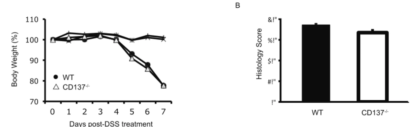
We first examined the impact of an acute 3.5% DSS challenge in CD1372/2versus WT mice, which induces acute large bowel inflammation [23,24]. Mice were given 3.5% DSS in drinking water ad libitum for a period of 7 days, after which time mice were sacrificed. The intestines were removed and sections were embedded for histological scoring of inflammation severity.
Weight loss, stool consistency as well as stool occult blood tests were used to monitor colitis severity in mice throughout the experiment.
No difference was observed in CD1372/2versus WT mice in colitis severity during the 3.5% DSS challenge as measured by weight loss (Fig. 1A), stool consistency as well as stool occult blood tests (data not shown). Histological assessment of colitis severity demonstrated a similar degree of inflammation both after 4 days of 3.5% DSS challenge (data not shown) as well as after 7 days of 3.5% DSS challenge (Fig. 1B). No histological signs of inflamma- tion or weight loss were observed in water control treated animals (Fig. 1A and data not shown). Thus, CD1372/2 and WT mice develop a similar disease severity in acute 3.5% DSS-induced colitis.
CD1372/2Mice Show Increased Weight Loss during the Resolution Phase After a 2.0% DSS Challenge
We next investigated whether CD137 influences the resolution of intestinal inflammation. CD1372/2 and WT mice were given 2.0% DSS in drinking water ad libitum for a period of 5 days, after which time mice were switched back to water to allow subsiding of the colonic inflammation. At different time points after DSS withdrawal mice were sacrificed, the intestines were removed and sections were embedded for histological scoring of inflammation severity.
CD1372/2mice demonstrated an increased weight loss during the resolution phase of intestinal inflammation after DSS withdrawal compared to WT mice (Fig. 2A). Kaplan-Meier- Analysis using a combined end-point of weight-loss above 20% of original weight/death demonstrated a significantly increased drop- out rate in CD1372/2mice as compared to WT mice after DSS withdrawal (Fig. 2B). Also the surviving CD1372/2 mice demonstrated significantly increased histological inflammation 7 days after DSS withdrawal compared to WT mice (data not
shown). Besides histological examination, shortening of the colon can be used as a measure of inflammation. We found significant colonic shortening 3 days after DSS withdrawal in both CD1372/
2and WT mice as compared to untreated mice, and significantly shorter colons in CD1372/2DSS-treated mice than in WT DSS- treated mice (Fig. 2C). These results indicate that resolution of intestinal inflammation is impaired in CD1372/2mice.
Resolution of Inflammation After DSS Withdrawal is Impaired in CD1372/2 Mice
To assess the development of inflammation after 2.0% DSS exposure, CD1372/2and WT mice were divided into two groups each. One group of mice was sacrificed after 5 days of 2.0% DSS exposure, while the other group of mice was allowed to resolve intestinal inflammation for an additional 3 days on drinking water.
CD1372/2and WT mice developed a similar degree of colitis severity after 5 days of 2.0% DSS exposure as assessed by histological criteria (Fig. 3A, B). However, while in WT mice inflammation did not progress after withdrawal of DSS, CD1372/
2mice showed a histological progression towards severe colonic inflammation (Fig. 3A, B). These histological results correlate with the increased weight loss and increased drop-out rate observed in CD1372/2 mice after 2.0% DSS exposure (Fig. 2A, B). In summary, CD1372/2and WT mice develop a similar histologic disease severity after 5 days of acute 2.0% DSS exposure, however, resolution of inflammation after withdrawal of DSS is impaired in CD1372/2mice.
2.0% DSS-induced Colitis Leads to Increased Local Colonic IFN-c, GM-CSF, CXCL1 and IL-17 Secretion in CD1372/2 Mice
Having observed a difference in the resolution of inflammation we investigated whether this is reflected and possibly attributable to a difference in the cytokine profile between CD1372/2and WT mice. Thus, mice were exposed to 2.0% DSS for a period of 5 days, or to 5 days DSS plus an additional 3 days with normal water, after which time animals were sacrificed. Local cytokine secretion in colon segment cultures was then evaluated using previously published techniques [29,30]. Interestingly, colon segments of CD1372/2 mice that were exposed for 5 days to 2.0% DSS exhibited increased IFN-csecretion compared to colon segments of WT mice with the same treatment, indicating a stronger Th1-type colonic inflammatory response in the absence of
Figure 1. CD1372/2and WT mice develop a similar disease severity in acute 3.5% DSS-induced colitis.A, Percentage of body weight loss over the course of 7 days of treatment with 3.5% DSS. B, Histology score of WT (black bars) and CD1372/2(white bars) mice treated with 3.5%
DSS for 7 days. Data are representative of two independent experiments, shown here are means6SEM, n = 15 for DSS groups, n = 5 for water control groups.
doi:10.1371/journal.pone.0073277.g001
CD137 (Fig. 4A). Additionally, the colon segments from the CD1372/2mice had an increased GM-CSF, IL-17 and CXCL1 secretion which points to an increased pro-inflammatory neutro- phil response (Fig. 4B, G, H). Of note, increased levels of IL-17 secretion after colitis induction indicate increased numbers of Th17 cells (Fig. 4G). Secretion of IL-2, which is important for growth, proliferation and differentiation of T cells, was reduced in colon segment cultures of 2.0% DSS-exposed CD1372/2 compared to WT mice, however, the difference did not reach statistical significance (Fig. 4C). The levels of TGF-ß, TNF-a, G- CSF and CXCL2 did not significantly vary between the two mouse strains (Fig. 4D–F, I). It should be emphasized that the cytokine secretion profiles were observed after 5 days of 2.0%
DSS-exposure, at a time when histologically there were no differences detectable in the severity of the colonic inflammatory response (Fig. 3A, B).
Of note, no differences were observed in systemic cytokine profiles (IL-6, IL-10, IL-13, IL-17A, IFN-c, TNF-a, GM-CSF) as measured in whole blood drawn after 5 days of 2.0% DSS- exposure as well as 8 days and 11 days post DSS treatment (data not shown). In summary, 2.0% DSS-induced colitis is accompa- nied by increased local colonic IFN-c, GM-CSF, CXCL1 and IL- 17 secretion in CD1372/2compared to WT mice.
CD1372/2Mice have Increased Numbers of Neutrophils during the Resolution of DSS-induced Inflammation
In addition to the altered cytokine profile we asked whether the different histopathology observed in WT and in CD1372/2mice was accompanied by a change in the infiltrating leukocyte subsets in the colonic mucosal lamina propria. The specific subsets of lamina propria mononuclear (inflammatory) cells contributing to the increased DSS-induced colitis were characterized in mice that received 5 days of 2.0% DSS treatment, followed by a resolution phase of 3 days. On days 5 and 8, mice were sacrificed, the large intestines were removed and the intestinal lymphocytes from both epithelial (IEL) and lamina propria (LPMNC) compartments of the large intestine were isolated as described previously [31,32], and were analyzed by flow cytometry.
There was minimal cellular infiltrate with no difference in the subsets of intestinal lamina propria mononuclear cells (LPMNC) in CD1372/2versus WT mice 5 days after 2.0% DSS exposure (data not shown). However, 3 days after withdrawal of DSS, CD1372/2 mice had significantly increased numbers of neutrophilic granu-
locytes in the LPMNC subset, reflecting the increased inflamma- tion (Fig. 5A, B). At the same time, CD1372/2 mice had significantly decreased numbers of macrophages in the IEL subset compared to WT mice (Fig. 5A, D). Since macrophages have been demonstrated to be necessary for intestinal regenerative responses to injury [33], these observations suggest an impaired colonic restitution in CD1372/2 mice. No differences were observed in the numbers of monocytes, B cells, NK cells, dendritic cells, CD4+ or CD8+T cells, neither among the LPMNC (B, C) or the IEL (D, E), nor in untreated CD1372/2versus WT mice (data not shown).
In summary, the decreased number of macrophages that are necessary for mucosal healing, together with the increased recruitment of neutrophils to the lamina propria of CD1372/2 mice may explain the increased severity of DSS-colitis in these mice.
Discussion
The current study investigated DSS-induced colitis in CD1372/
2and WT mice. We found that in the absence of CD137, mice develop a more severe colitis, suggesting that CD137 exerts an anti-inflammatory effect in this model.
Of note, both CD1372/2and WT mice demonstrated a similar degree of inflammation after 5 days of DSS exposure. However, the resolution of colonic inflammation was significantly impaired in CD1372/2 mice. This led to prolonged tissue infiltration of leukocytes, an increased neutrophil/macrophage ratio among the infiltrating cells and an increased release of the pro-inflammatory cytokines IFN-c, GM-CSF, CXCL1 and IL-17.
The reason for the more pronounced disease manifestation in the CD1372/2 mice may be due to several factors. Overexpres- sion of the granulocyte survival factor GM-CSF has been associated with suppression of neutrophil apoptosis in the absence of CD137 activation [34]. It has been shown in vitro that CD137 is constitutively expressed on neutrophilic granulocytes, and that the CD137 signaling pathway blocks the anti-apoptotic effect mediated by the1-subunit of the GM-CSF receptor in neutrophils [35]. Therefore, the more severe disease manifestation in the CD1372/2mice may be due to the absence of CD137-mediated granulocyte apoptosis leading to increased granulocyte accumu- lation and subsequent tissue damage, since mucosal inflammation in DSS-induced colitis is mediated by neutrophilic granulocytes [36,37]. In line with this hypothesis and published data, we found Figure 2. Increased susceptibility of CD1372/2mice during the resolution phase after 5 days of 2.0% DSS treatment as compared to WT mice.A, Percentage of body weight loss over the course of 5 days of treatment with 2.0% DSS water followed by 7 days of normal water. B, Kaplan-Meier-Analysis using a combined end-point of weight-loss above 20% of original weight/death in 5 days 2.0% DSS treated mice followed by 7 days of recovery phase with normal water. C, Colon length of mice untreated or treated for 5 days with 2.0% DSS followed by 3 days of normal water.
Data are representative of at least three independent experiments, shown here are means6SEM, n = 10–15 for DSS groups, n = 5 for water control groups (* p,0.05).
doi:10.1371/journal.pone.0073277.g002
increased GM-CSF in the colons of CD1372/2mice, which likely supported neutrophil accumulation in the colons. Additionally, we found increased levels of CXCL1 in colitic CD1372/2 mice, which increase neutrophil influx into the inflamed mucosal tissue.
Remarkably, increased levels of IL-17 in CD1372/2mice indicate increased numbers of Th17 cells, which is further supporting neutrophil recruitment [38]. Although the initial infiltration by neutrophils is beneficial for killing bacteria, it is presumed that it is
the persistent presence of neutrophils in the tissues that causes damage [36,37]. Of note, it has recently been demonstrated in a T-cell transfer model of colitis that GM-CSF skews hematopoietic stem and progenitor cells towards granulocyte-monocyte progen- itor cells, leading to the exacerbation of intestinal inflammation.
Importantly, colitis could be ameliorated by GM-CSF blockade [39]. Thus, the GM-CSF driven increased accumulation and Figure 3. CD1372/2and WT mice develop a similar histologic disease severity after 5 days of acute 2.0% DSS exposure, however, resolution of inflammation after withdrawal of DSS is impaired in CD1372/2mice.A, Representative histologic colonic cross-sections of mice exposed to normal water (control), 5 days of 2.0% DSS water ((+)DSS Day 5) and 5 days of 2.0% DSS water followed by 3 days normal water ((+)DSS Day 8) stained with Hematoxylin-Eosin (20-fold magnification). B, Histology score of WT (black bars) and CD1372/2 (white bars) mice untreated, treated with 2.0% DSS for 5 days or mice treated with 2.0% DSS for 5 days followed by 3 days of normal water. Data are representative of two independent experiments, shown here are means6SEM, n = 4 per group (*** p,0.001).
doi:10.1371/journal.pone.0073277.g003
prolonged presence of neutrophils in the colons of CD1372/2 mice can explain the increased tissue damage in these mice.
There is an additional function of CD137, whose absence in the CD1372/2mice is likely to contribute to the more severe disease manifestation: CD137 not only regulates the activities of mature leukocytes, it also regulates hematopoiesis, and has been shown to induce proliferation and expansion of hematopoietic progenitor cells. Concomitantly, CD137 induces myeloid differentiation, and within the myeloid lineage monocytic differentiation [40,41], especially during inflammation [42]. In fact, CD137 and G-CSF compete for driving the differentiation of myeloid progenitor cells into the monocytic and granulocytic differentiation, respectively, with an increasing G-CSF/CD137 ratio leading to a preponder- ance of granulocytes [43]. In the absence of CD137 more myeloid precursor cells would be expected to differentiate to granulocytes at the expense of monocytes and macrophages. Indeed, we not only see an increase of granulocytes in the colons CD1372/2 compared to WT mice as a response to DSS administration but also a decrease in macrophages. This increased granulocyte/
macrophage ratio could not only contribute to the more severe colitis in the CD1372/2mice through recruiting more granulo- cytes that in turn cause tissue damage but also because macrophages are the cells that coordinate wound healing [33].
With fewer macrophages being present in the colons of the CD1372/2 mice, resolution of inflammation and wound healing are expected to be delayed, and that is what we observe.
The CD137 receptor/ligand system has so far been mainly associated with proinflammatory effects. Agonistic anti-CD137 antibodies costimulate T cells, and enhance immunity leading to rejection of tumors and transplants in mice, and enhance anti- virus immune responses [5,44,45,46]. Further, bidirectional signaling exists for the CD137 receptor/ligand system, and reverse signaling of CD137L activates antigen presenting cells, which leads to a further enhancement of immune responses [47]. The proinflammatory activities of CD137 and CD137L are also evident in autoimmune diseases. The administration of antago- nistic anti-CD137 ligand antibodies to mice with collagen-induced arthritis resulted in a milder disease [48]. Further, experimental Figure 4. 2.0% DSS-induced colitis leads to increased local colonic IFN-c, GM-CSF, CXCL1 and IL-17 secretion in CD1372/2versus WT mice.Colon segment culture supernatants of CD1372/2(white bars) and WT mice (black bars) untreated (day 0), or treated either for 5 days with 2% DSS (day 5) or 5 days with 2% DSS followed by 3 days with normal water (day 8), were examined for IFN-c(A), GM-CSF (B), IL-2 (C), TGF-1(D), TNF- a(E), G-CSF (F), IL-17 (G), CXCL1 (H) and CXCL2 (I). Data are representative of two independent experiments, shown here are means6SEM, n = 4 per group (* p,0.05, ** p,0.01).
doi:10.1371/journal.pone.0073277.g004
autoimmune encephalomyelitis is more severe in mice that are deficient in CD137L [49]. However, the very same agonistic anti- CD137 antibodies that enhance anti-tumor immunity have been shown to quench autoimmunity and to ameliorate autoimmune diseases, including experimental autoimmune encephalomyelitis
and type 1 diabetes [8,9,10,50,51]. An explanation for these puzzling findings and seemingly contradictory activities has not yet been found. These examples demonstrate that the consequences of modulating the CD137 receptor/ligand system are difficult to predict in autoimmune diseases. Nevertheless, our findings in the Figure 5. CD1372/2mice have increased numbers of neutrophils in the lamina propria mononuclear cell (LPMNC) subpopulations while presenting lower macrophage numbers in the intraepithelial (IEL) subpopulation.A, Gating strategy for myeloid (top panel) and lymphocyte (lower panel) populations. B, Quantification of different immune cell populations in the LPMNC cells (B, C) and IEL cells (D, E) during the resolution phase of inflammation 3 days after withdrawal of 2.0% DSS. Data represent means6SEM of 4 independent experiments pooled together, n = 3–10 per group (* p,0.05). Mo (Monocytes), Neutro (Neutrophils), Macro (Macrophages), DCs (Dendritic cells), NK (Natural Killer cells).
doi:10.1371/journal.pone.0073277.g005
DSS colitis model suggest that neutralizing CD137 with antago- nistic anti-CD137 antibodies would not be beneficial for the treatment DSS-induced colitis and possibly also not for IBD patients. Our data suggest that agonistic anti-CD137 antibodies may be more suited to ameliorate colitis.
Author Contributions
Conceived and designed the experiments: TK HS JMMG. Performed the experiments: JMMG TK. Analyzed the data: TK JMMG HS. Contributed reagents/materials/analysis tools: LC. Wrote the paper: JMMG HS TK.
References
1. Wilcox RA, Chapoval AI, Gorski KS, Otsuji M, Shin T, et al. (2002) Cutting edge: Expression of functional CD137 receptor by dendritic cells. Journal of immunology 168: 4262–4267.
2. Wilcox RA, Tamada K, Strome SE, Chen L (2002) Signaling through NK cell- associated CD137 promotes both helper function for CD8+cytolytic T cells and responsiveness to IL-2 but not cytolytic activity. Journal of immunology 169:
4230–4236.
3. Lee SC, Ju SA, Sung BH, Heo SK, Cho HR, et al. (2009) Stimulation of the molecule 411BB enhances host defense against Listeria monocytogenes infection in mice by inducing rapid infiltration and activation of neutrophils and monocytes. Infection and immunity 77: 2168–2176.
4. Olofsson PS, Soderstrom LA, Wagsater D, Sheikine Y, Ocaya P, et al. (2008) CD137 is expressed in human atherosclerosis and promotes development of plaque inflammation in hypercholesterolemic mice. Circulation 117: 1292–
1301.
5. Thum E, Shao Z, Schwarz H (2009) CD137, implications in immunity and potential for therapy. Front Biosci 14: 4173–4188.
6. Melero I, Shuford WW, Newby SA, Aruffo A, Ledbetter JA, et al. (1997) Monoclonal antibodies against the 4-1BB T-cell activation molecule eradicate established tumors. Nature medicine 3: 682–685.
7. Shuford WW, Klussman K, Tritchler DD, Loo DT, Chalupny J, et al. (1997) 4- 1BB costimulatory signals preferentially induce CD8+T cell proliferation and lead to the amplification in vivo of cytotoxic T cell responses. The Journal of experimental medicine 186: 47–55.
8. Irie J, Wu Y, Kachapati K, Mittler RS, Ridgway WM (2007) Modulating protective and pathogenic CD4+subsets via CD137 in type 1 diabetes. Diabetes 56: 186–196.
9. Vinay DS, Kim JD, Kwon BS (2006) Amelioration of mercury-induced autoimmunity by 4-1BB. J Immunol 177: 5708–5717.
10. Mittler RS, Bailey TS, Klussman K, Trailsmith MD, Hoffmann MK (1999) Anti-4-1BB monoclonal antibodies abrogate T cell-dependent humoral immune responses in vivo through the induction of helper T cell anergy. J Exp Med 190:
1535–1540.
11. Sartor RB (1995) Current concepts of the etiology and pathogenesis of ulcerative colitis and Crohn’s disease. Gastroenterol Clin North Am 24: 475–507.
12. Shih DQ, Targan SR (2009) Insights into IBD Pathogenesis. Curr Gastroenterol Rep 11: 473–480.
13. Braegger CP, Nicholls S, Murch SH, Stephens S, MacDonald TT (1992) Tumour necrosis factor alpha in stool as a marker of intestinal inflammation.
Lancet 339: 89–91.
14. Kontoyiannis D, Pasparakis M, Pizarro TT, Cominelli F, Kollias G (1999) Impaired on/off regulation of TNF biosynthesis in mice lacking TNF AU-rich elements: implications for joint and gut-associated immunopathologies. Immu- nity 10: 387–398.
15. Neurath MF, Fuss I, Pasparakis M, Alexopoulou L, Haralambous S, et al. (1997) Predominant pathogenic role of tumor necrosis factor in experimental colitis in mice. Eur J Immunol 27: 1743–1750.
16. Begue B, Wajant H, Bambou JC, Dubuquoy L, Siegmund D, et al. (2006) Implication of TNF-related apoptosis-inducing ligand in inflammatory intestinal epithelial lesions. Gastroenterology 130: 1962–1974.
17. Langstein J, Michel J, Fritsche J, Kreutz M, Andreesen R, et al. (1998) CD137 (ILA/4-1BB), a member of the TNF receptor family, induces monocyte activation via bidirectional signaling. Journal of immunology 160: 2488–2494.
18. Kang YJ, Kim SO, Shimada S, Otsuka M, Seit-Nebi A, et al. (2007) Cell surface 4-1BBL mediates sequential signaling pathways ‘downstream’ of TLR and is required for sustained TNF production in macrophages. Nature immunology 8:
601–609.
19. Moh MC, Lorenzini PA, Gullo C, Schwarz H (2013) Tumor necrosis factor receptor 1 associates with CD137 ligand and mediates its reverse signaling.
FASEB journal : official publication of the Federation of American Societies for Experimental Biology.
20. Maerten P, Geboes K, De Hertogh G, Shen C, Cadot P, et al. (2004) Functional expression of 4-1BB (CD137) in the inflammatory tissue in Crohn’s disease. Clin Immunol 112: 239–246.
21. Maerten P, Kwon BS, Shen C, De Hertogh G, Cadot P, et al. (2006) Involvement of 4-1BB (CD137)-4-1BBligand interaction in the modulation of CD4 T cell-mediated inflammatory colitis. Clin Exp Immunol 143: 228–236.
22. Zhu Y, Zhu G, Luo L, Flies AS, Chen L (2007) CD137 stimulation delivers an antigen-independent growth signal for T lymphocytes with memory phenotype.
Blood 109: 4882–4889.
23. Cooper HS, Murthy SN, Shah RS, Sedergran DJ (1993) Clinicopathologic study of dextran sulfate sodium experimental murine colitis. Lab Invest 69: 238–249.
24. Elson CO, Sartor RB, Tennyson GS, Riddell RH (1995) Experimental models of inflammatory bowel disease. Gastroenterology 109: 1344–1367.
25. Karrasch T, Kim JS, Jang BI, Jobin C (2007) The flavonoid luteolin worsens chemical-induced colitis in NF-kappaB(EGFP) transgenic mice through blockade of NF-kappaB-dependent protective molecules. PLoS One 2: e596.
26. Okayasu I, Hatakeyama S, Yamada M, Ohkusa T, Inagaki Y, et al. (1990) A novel method in the induction of reliable experimental acute and chronic ulcerative colitis in mice. Gastroenterology 98: 694–702.
27. Williams KL, Fuller CR, Dieleman LA, DaCosta CM, Haldeman KM, et al.
(2001) Enhanced survival and mucosal repair after dextran sodium sulfate- induced colitis in transgenic mice that overexpress growth hormone.
Gastroenterology 120: 925–937.
28. Dieleman LA, Palmen MJ, Akol H, Bloemena E, Pena AS, et al. (1998) Chronic experimental colitis induced by dextran sulphate sodium (DSS) is characterized by Th1 and Th2 cytokines. Clin Exp Immunol 114: 385–391.
29. Sellon RK, Tonkonogy S, Schultz M, Dieleman LA, Grenther W, et al. (1998) Resident enteric bacteria are necessary for development of spontaneous colitis and immune system activation in interleukin-10-deficient mice. Infect Immun 66: 5224–5231.
30. Veltkamp C, Tonkonogy SL, De Jong YP, Albright C, Grenther WB, et al.
(2001) Continuous stimulation by normal luminal bacteria is essential for the development and perpetuation of colitis in Tg(epsilon26) mice. Gastroenterology 120: 900–913.
31. Qian BF, Zhou GQ, Hammarstrom ML, Danielsson A (2001) Both substance P and its receptor are expressed in mouse intestinal T lymphocytes. Neuroendo- crinology 73: 358–368.
32. Karrasch T, Kim JS, Muhlbauer M, Magness ST, Jobin C (2007) Gnotobiotic IL-102/2;NF-kappa B(EGFP) mice reveal the critical role of TLR/NF-kappa B signaling in commensal bacteria-induced colitis. J Immunol 178: 6522–6532.
33. Pull SL, Doherty JM, Mills JC, Gordon JI, Stappenbeck TS (2005) Activated macrophages are an adaptive element of the colonic epithelial progenitor niche necessary for regenerative responses to injury. Proceedings of the National Academy of Sciences of the United States of America 102: 99–104.
34. Dibbert B, Weber M, Nikolaizik WH, Vogt P, Schoni MH, et al. (1999) Cytokine-mediated Bax deficiency and consequent delayed neutrophil apoptosis:
a general mechanism to accumulate effector cells in inflammation. Proceedings of the National Academy of Sciences of the United States of America 96: 13330–
13335.
35. Heinisch IV, Daigle I, Knopfli B, Simon HU (2000) CD137 activation abrogates granulocyte-macrophage colony-stimulating factor-mediated anti-apoptosis in neutrophils. European journal of immunology 30: 3441–3446.
36. Qualls JE, Kaplan AM, van Rooijen N, Cohen DA (2006) Suppression of experimental colitis by intestinal mononuclear phagocytes. Journal of leukocyte biology 80: 802–815.
37. Qualls JE, Tuna H, Kaplan AM, Cohen DA (2009) Suppression of experimental colitis in mice by CD11c+dendritic cells. Inflammatory bowel diseases 15: 236–
247.
38. Ouyang W, Kolls JK, Zheng Y (2008) The biological functions of T helper 17 cell effector cytokines in inflammation. Immunity 28: 454–467.
39. Griseri T, McKenzie BS, Schiering C, Powrie F (2012) Dysregulated hematopoietic stem and progenitor cell activity promotes interleukin-23-driven chronic intestinal inflammation. Immunity 37: 1116–1129.
40. Jiang D, Chen Y, Schwarz H (2008) CD137 induces proliferation of murine hematopoietic progenitor cells and differentiation to macrophages. Journal of immunology 181: 3923–3932.
41. Jiang D, Yue PS, Drenkard D, Schwarz H (2008) Induction of proliferation and monocytic differentiation of human CD34+cells by CD137 ligand signaling.
Stem cells 26: 2372–2381.
42. Tang Q, Jiang D, Alonso S, Pant A, Gomez JM, et al. (2013) CD137 ligand signaling enhances myelopoiesis during infections. Eur J Immunol doi: 10.1002/
eji.201243071. [Epub ahead of print].
43. Jiang D, Schwarz H (2010) Regulation of granulocyte and macrophage populations of murine bone marrow cells by G-CSF and CD137 protein. PLoS One 5: e15565.
44. Tamada K, Chen L (2006) Renewed interest in cancer immunotherapy with the tumor necrosis factor superfamily molecules. Cancer immunology, immuno- therapy : CII 55: 355–362.
45. Lee SW, Croft M (2009) 4-1BB as a therapeutic target for human disease.
Advances in experimental medicine and biology 647: 120–129.
46. Wang C, Lin GH, McPherson AJ, Watts TH (2009) Immune regulation by 4- 1BB and 4-1BBL: complexities and challenges. Immunological reviews 229:
192–215.
47. Shao Z, Schwarz H (2011) CD137 ligand, a member of the tumor necrosis factor family, regulates immune responses via reverse signal transduction. Journal of leukocyte biology 89: 21–29.
48. Seo SK, Choi JH, Kim YH, Kang WJ, Park HY, et al. (2004) 4-1BB-mediated immunotherapy of rheumatoid arthritis. Nature medicine 10: 1088–1094.
49. Martinez Gomez JM, Croxford JL, Yeo KP, Angeli V, Schwarz H, et al. (2012) Development of Experimental Autoimmune Encephalomyelitis Critically Depends on CD137 Ligand Signaling. The Journal of neuroscience : the official journal of the Society for Neuroscience 32: 18246–18252.
50. Sun Y, Lin X, Chen HM, Wu Q, Subudhi SK, et al. (2002) Administration of agonistic anti-4-1BB monoclonal antibody leads to the amelioration of
experimental autoimmune encephalomyelitis. Journal of immunology 168:
1457–1465.
51. Kim YH, Choi BK, Shin SM, Kim CH, Oh HS, et al. (2011) 4-1BB triggering ameliorates experimental autoimmune encephalomyelitis by modulating the balance between Th17 and regulatory T cells. Journal of immunology 187:
1120–1128.