Macular ischemia after intravitreal amikacin on patient with intraocular foreign body
Abstract
Background:Although still used in third world countries, amikacin has a harmful effect to be used intravitreally.
Arief Kartasasmita
1Susi Mona
1Purpose:To report macular ischemia after an intravitreal injection of
amikacin
Erwin Iskandar
1Iwan Sovani
1Methods:A case report regarding a traumatized eye of a 26-year-old man that was injected intravitreally with amikacin due to intraocular
foreign body endophthalmitis
Djonggi Panggabean
1Results:Angiography and OCT show macular ischemia due to amikacin
toxicity. 1 Faculty of Medicine,
Universitas Conclusion: The case reported here is to alert about the potential
harmful effect of intravitreally injected amikacin despite its role as an accepted regimen for endohthalmitis cases.
Padjadjaran/Cicendo National Eye Hospital, Bandung, Indonesia
Introduction
Ocular trauma is the leading cause of monocular visual loss and a major cause of ocular morbidity. It is estimated that 500,000 blinding ocular injuries occur globally each year. The intraocular foreign body (IOFB) represents one form of open globe injury commonly described, especially in developing countries and rural areas. A variety of re- ports note that retained foreign bodies occur in 5–40%
of all penetrating eye injuries [1], [2]. This is cause for concern as retained foreign bodies are associated with increased risk of endophthalmitis [3], [4]. The incidence of infectious endophthalmitis after penetrating injury with IOFB ranges from 0% up to 16.5% [5].
Broad-spectrum antibiotic intravitreal injection is the mainstay therapy following IOFB injury. To provide ad- equate Gram-negative and -positive bacterial cover in endophthalmitis cases, a currently accepted therapeutic regimen includes aminoglycosides [6]. In developing countries, aminoglycosides have become a favorable drug of choice due to their ease of access and reduced cost. However, these treatments are not without side ef- fects: retinal vascular infarction is commonly described after gentamicin administration, and has been noted after use of amikacin [7]. While previously noted as a rare complication, the reported frequency of macular ischemia following amikacin intravitreal injection for the treatment of endophthalmitis has increased recently [6], [8]. This report describes a case of macular ischemia following intravitreal amikacin injection.
Case description
A 26-year-old man came to the emergency room in our tertiary eye hospital 1 day after his right eye was hit by a nail while hammering. The patient was referred from a district hospital with an IOFB in the right eye. He received an injection of anti-tetanus serum at initial admission.
The general examination was within normal limits. An ophthalmological examination was conducted; best cor- rected visual acuity (VA) was 6/5 in each eye, ocular motility was full in all directions, and digital intraocular pressures were normal for both eyes. The anterior seg- ment of the right eye showed a hyperemic conjunctiva with a nail lodged at the temporal limbus (Figure 1).
Corneal edema was present, but other anterior segment findings were normal (round pupil, no synechiae or lens opacities, and anterior chamber formed with no flare or cells). The anterior segment of the left eye was within normal limits. Fundoscopic examination of the right eye revealed clear media with a visible foreign body (nail) in the vitreous, a round and sharp border of the optic disc, a flat retina, and good foveal reflex. The posterior segment of the left eye was within normal limits.
Figure 1: A nail has lodged in the right eye
The patient was diagnosed with an IOFB in the right eye.
His treatment plan included intravenous cefotaxime injec- tion (1 g) twice daily and hourly ofloxacin eye drop for the right eye in addition to being scheduled for IOFB extraction and intravitreal antibiotic (IVAB) injection.
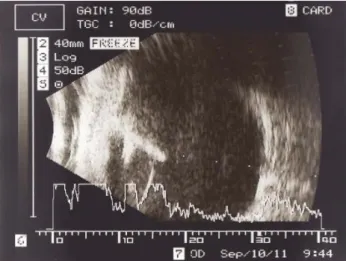
Ultrasonography examination showed an IOFB located in the limbus/pars plana that had penetrated into the vitreous cavity and posterior segment inflammation (Figure 2).
Figure 2: Ultrasonography showed an IOFB in the right eye
The surgery was performed 6 hours after administered.
The nail was lodged at the limbus (9 o’clock) and had penetrated into the vitreous cavity, contacting the edge of the lens. It was removed with forceps, and measured 9 mm in length. Anterior vitrectomy and vitreous tap were then performed around the wound and the vitreous con- tents were subjected to Gram staining and culture. The wound was sutured, and vancomycin (1 mg) and amikacin (0.2 mg) were injected intravitreally. The patient was also given oral paracetamol (500 mg) three times daily and prednisolone acetate eye drops six times daily for the right eye.
Figure 3: Anterior segment of the right eye after IOFB removal
On the first postoperative day, the uncorrected VA for the right eye was count fingers. Anterior segment examination showed a closed wound and two in-place sutures at the temporal limbus (Figure 3). Corneal edema, a hyperemic conjunctiva, subconjunctival bleeding, and a moderate depth anterior chamber with inflammation were noted.
As before, the pupil was round and no synechiae or lens opacities were observed. The posterior segment examin- ation revealed clear media, a normal optic disc, a flat retina, and macular edema with decreased foveal reflex.
Gram stain of the vitreous tap revealed the presence of Gram-positive cocci, while the KOH stain was negative.
The previous therapies were continued with the addition of 48 mg of methyl prednisolone orally once a day.
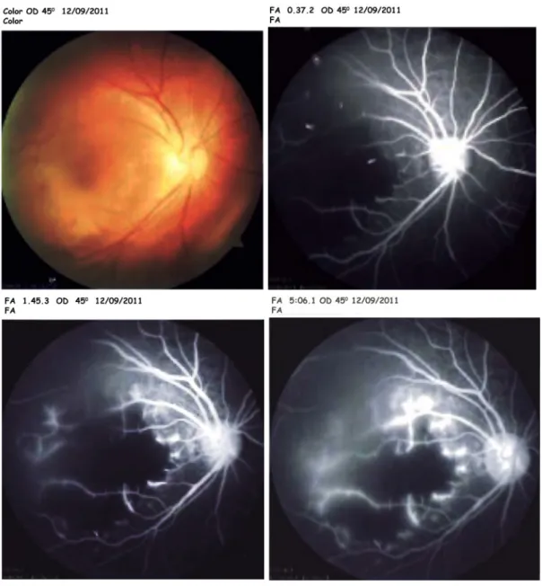
Angiographic examination of the right eye revealed hypofluorescence in the arteriovenous phase due to a vascular filling defect at the macula. Angiographic exam- ination of the left eye was within normal limits. Based on the angiogram results, we diagnosed macular ischemia of the right eye (Figure 4).
Optical coherence tomography (OCT) examination of the right eye revealed increased retinal thickness with re- duced intraretinal reflectivity, although the signal strength was ambiguous. OCT examination of the left eye was within normal limits (Figure 5).
Three days after surgery, the uncorrected VA of the right eye was still count fingers, although there was a decrease in subconjunctival bleeding, corneal edema, and anterior chamber inflammation. The posterior segment examin- ation continued to show macular edema. Previous ther- apies were continued with the addition of oral ciprofloxa- cin (750 mg) twice daily. The patient was discharged from the hospital with a review scheduled 1 week later.
Discussion
Endophthalmitis prevention using prophylactic intraocular antibiotic injection in patients with penetrating injuries remains controversial. However, in cases where there is a high suspicion of injury from a contaminated foreign body or indeed early signs of endophthalmitis are present, prophylactic treatment with IVABs should be considered.
Figure 4: Angiography of the right eye showed macular ischemia
Figure 5: OCT of the right eye
Intraocular antibiotics should target a broad range of Gram-positive and -negative organisms. Vancomycin re- mains the drug of choice for treatment of Gram-positive infections. The aminoglycoside class of antibiotics is se- lected particularly for their Gram-negative coverage.
However, caution should be exercised in their use be-
cause of reports of retinal vascular infarction following intravitreal injection [8]. IVABs were administrated in this patient because of concern that the IOFB may be contam- inated, given the patient’s history of employment as a furniture maker.
Cases of macular infarction secondary to administration of intraocular aminoglycosides have been observed after excessive intraocular doses while others have occurred after normal use [9]. There are several potentially toxic antibiotics in the aminoglycoside family including tobra- mycin and amikacin, with gentamicin having the greatest toxicity profile [10]. Following concerns about the risks of retinal toxicity caused by intravitreal gentamicin, many ophthalmologists now prefer intravitreal amikacin even though its use has also been associated with retinal tox- icity [11]. Massive doses may result in early superficial and intraretinal hemorrhages, retinal edema, cotton wool patches, arterial narrowing, and venous beading [9]. In our patient, on the first day following IOFB extraction and IVAB injection we noted sudden decreased VA with the posterior segment examination revealing macular ischemia. This was subsequently confirmed by OCT and FFA examinations.
In an animal study, aminoglycoside toxicity occurred be- cause this antibiotic affects primarily neurons and glia of the inner retina. This suggests that because this area has a high number of paramacular density cells, it is more susceptible to toxicity impairment. Other important factors include the gravitational forces of the antibiotic solution, which has a higher gravity than the vitreous or vitreous replacement compound. The latter was observed in studies using rabbits in which positioning of the eye on gentamicin injection caused toxic effects predominantly in the corresponding part of the retina [12].
Potential toxicity reactions of the retina are also subject to individual variation. Studies show that the risk of retinal toxicity is greater if drugs are injected towards the posterior pole with the needle pointed directly to the macula [8]. Moreover, higher injection frequencies may also increase the possibility of retinal toxicity [13]. Fur- thermore, intravitreal injections of aminoglycosides inten- ded for the subconjunctival space that inadvertently miss may direct a toxic dose into the retina [12].
In our case, another etiological possibility for the macular edema observed is vancomycin toxicity from the initial combined amikacin-vancomycin injection. Studies on rabbits demonstrated that intravitreal vancomycin injec- tion results in retinal toxicity and ERG abnormalities.
However, this effect only occurred with large doses (5 and 10 mg) of vancomycin [12]. The doses of vancomycin used in this case (1 mg) are considered safe, given that no retinal toxicity has been observed in experimental rabbit eyes and no cases of human macular toxicity attrib- uted to vancomycin at these doses have been reported.
Based on previous reports, the intravitreal amikacin injec- tion is strongly suspected as the main cause of ischemia in our patient.
The overall prognosis for this patient is good because there is no systemic disorder caused by the trauma. The visual prognosis is poor, because the macular ischemia is an irreversible condition.
Although still commonly used, especially in third world countries because of its availability and efficacy, we caution the use of amikacin as an intravitreal therapeutic
agent given its potential for toxicity to the retinal tissues.
Alternative and effective antibiotic agents are available with safer toxicity profiles.
Notes
Competing interests
The authors declare that they have no competing in- terests.
References
1. Al-Thowaibi A, Kumar M, Al-Matani I. An overview of penetrating ocular trauma with retained intraocular foreign body. Saudi J Ophthalmol. 2011 Apr;25(2):203-5. DOI:
10.1016/j.sjopt.2011.01.001
2. Öztas Z, Nalçaci S, Afrashi F, Erakgün T, Mentes J, Degirmenci C, Akkin C. Posterior segment intraocular foreign bodies: the effect of weight and size, early versus late vitrectomy and outcomes. Ulus Travma Acil Cerrahi Derg. 2015 Dec;21(6):496- 502. DOI: 10.5505/tjtes.2015.03608
3. Yang CS, Lu CK, Lee FL, Hsu WM, Lee YF, Lee SM. Treatment and outcome of traumatic endophthalmitis in open globe injury with retained intraocular foreign body. Ophthalmologica.
2010;224(2):79-85. DOI: 10.1159/000235725
4. Pathengay A, Miller DM, Flynn HW Jr, Dubovy SR. Curvularia endophthalmitis following open globe injuries. Arch Ophthalmol.
2012 May;130(5):652-4. DOI:
10.1001/archophthalmol.2011.1701
5. Ahmed Y, Schimel AM, Pathengay A, Colyer MH, Flynn HW Jr.
Endophthalmitis following open-globe injuries. Eye (Lond). 2012 Feb;26(2):212-7. DOI: 10.1038/eye.2011.313
6. Slean GR, Shorstein NH, Liu L, Paschal JF, Winthrop KL, Herrinton LJ. Pathogens and antibiotic sensitivities in endophthalmitis. Clin Exp Ophthalmol. 2016 Dec 25. DOI: 10.1111/ceo.12910 7. Lartey S, Armah P, Ampong A. A sudden total loss of vision after
routine cataract surgery. Ghana Med J. 2013 Jun;47(2):96-9.
8. Galloway G, Ramsay A, Jordan K, Vivian A. Macular infarction after intravitreal amikacin: mounting evidence against amikacin.
Br J Ophthalmol. 2002 Mar;86(3):359-60. DOI:
10.1136/bjo.86.3.359
9. Dille B. Toxicity of intraocular gentamicin. Arch Ophthalmol. 2007 Oct;125(10):1442; author reply 1442-3. DOI:
10.1001/archopht.125.10.1442-a
10. Penha FM, Rodrigues EB, Maia M, Furlani BA, Regatieri C, Melo GB, Magalhães O Jr, Manzano R, Farah ME. Retinal and ocular toxicity in ocular application of drugs and chemicals part II: retinal toxicity of current and new drugs. Ophthalmic Res.
2010;44(4):205-24. DOI: 10.1159/000316695 11. Jackson TL, Williamson TH. Amikacin retinal toxicity. Br J
Ophthalmol. 1999 Oct;83(10):1199-200.
12. Seawright AA, Bourke RD, Cooling RJ. Macula toxicity after intravitreal amikacin. Aust N Z J Ophthalmol. 1996
May;24(2):143-6. DOI: 10.1111/j.1442-9071.1996.tb01569.x 13. Campochiaro PA, Conway BP. Aminoglycoside toxicity a survey
of retinal specialists. Implications for ocular use. Arch Ophthalmol. 1991 Jul;109(7):946-50. DOI:
10.1001/archopht.1991.01080070058035
Corresponding author:
Arief Kartasasmita
Faculty of Medicine, Universitas Padjadjaran/Cicendo National Eye Hospital, Jl. Cicendo No 4, Bandung 40171, Indonesia, Phone: +62 22 4231280
a.kartasasmita@unpad.ac.id
Please cite as
Kartasasmita A, Mona S, Iskandar E, Sovani I, Panggabean D. Macular ischemia after intravitreal amikacin on patient with intraocular foreign body. GMS Ophthalmol Cases. 2017;7:Doc10.
DOI: 10.3205/oc000061, URN: urn:nbn:de:0183-oc0000612
This article is freely available from
http://www.egms.de/en/journals/oc/2017-7/oc000061.shtml Published:2017-03-24
Copyright
©2017 Kartasasmita et al. This is an Open Access article distributed under the terms of the Creative Commons Attribution 4.0 License. See license information at http://creativecommons.org/licenses/by/4.0/.