Methods for the measurement of cell and tissue
compatibility including tissue regeneration processes
Methoden zur Ermittlung der Zell- und Gewebekompatibilität einschließlich der Geweberegeneration
Abstract
Biocompatibility is one of the main requirements for the safe use of medical devices. Determination of cytotoxicity is part of the initial
Cornelia Wiegand
1Uta-Christina Hipler
1evaluation stipulated by ISO standards for the biological evaluation of medical devices. The use of cell cultures to test the biocompatibility of
drugs, biomaterials or treatment techniques used in various disciplines 1 Department of Dermatology, Friedrich Schiller University, Jena, Germany
is gaining in importance. A wide variety of self-initiated and commercially available cell lines has been evaluated and used: cultured fibroblasts from human skin, buccal mucosa, periodontal membrane, embryonic lung, epithelial and HeLa cells; cultures of human keratinocytes and HaCaT cells; different murine cell lines (C3H-L, Balb/c 3T3, L929 and others) as well as murine cells cultured from liver and spleen; T- lymphocytes from lymph nodes and macrophages obtained by lavage.
All of the above cells are suitable for use in biocompatibility tests.
Nevertheless, the general opinion is that toxicity testsin vitrowill be more convincing when performed with cells that are homologous with the human tissue concerned. In accordance, appropriate cell lines for use in cytotoxicity and tolerance tests concerning the skin would be human dermal fibroblasts and human epidermal keratinocytes, as they take an active part in the immune response, inflammatory processes, and wound healing.
The evaluation of thein vitro cytotoxicity of a biomaterial is often a qualitative analysis based on the morphological examination of cell damage and growth after direct or indirect contact with the material.
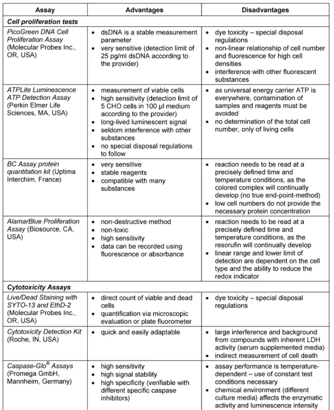
Different commercial assays based on the determination of nucleic acids, metabolic activity, protein content or membrane integrity are available to measure cell proliferation and cell viability. A small selection – Pico Green® DNA Cell Proliferation Assay, ATPLite™ Luminescence ATP Detection Assay, BC Assay: protein quantitation kit, AlamarBlue™
Proliferation Assay and Live/Dead Staining with SYTO-13 and EthD-2 – are discussed concerning sensitivity, reliability and applicability.
Keywords:biocompatibility, cell proliferation, cytotoxicity, fibroblasts, keratinocytes, skin
Zusammenfassung
Biokompatibilität ist eine der Hauptforderungen für den sicheren Einsatz von Medizinprodukten. Die Bestimmung der Zytotoxizität gehört daher auch zu den Evaluierungsprozessen, die in den ISO Standards für die biologische Beurteilung von medizinischen Produkten festgelegt sind.
Die Anwendung von Zellkulturen für die Testung auf Biokompatibilität von Pharmazeutika, Biomaterialien oder Behandlungsmethoden gewinnt dafür zunehmend an Bedeutung. Eine Vielzahl von selbst gewonnenen oder kommerziell erhältlichen Zelllinien kommen dabei zum Einsatz:
Fibroblasten isoliert aus humaner Haut, der Mundschleimhaut, der Wurzelhaut oder aus embryonalen Lungengewebe; epitheliale Zellen und HeLA-Zellen; Kulturen von humanen Keratinozyten und HaCaT-
Zellen; verschiedene murine Zelllinien (C3H-L, Balb/c 3T3, L929 u. a.) sowie Zellen isoliert aus der Milz oder der Leber der Maus und T-Lym- phozyten aus Lymphknoten.
Die genannten Zellen sind alle für Biokompatibilitätsuntersuchungen geeignet. Die generelle Meinung ist jedoch, dassin vitrodurchgeführte Toxizitätstest überzeugender sind, wenn sie mit Zellen durchgeführt werden, die im Zusammenhang mit dem humanen Gewebe stehen an dem die Applikation des Medizinproduktes erfolgt. Dementsprechend eignen sich am besten humane dermale Fibroblasten und humane epidermale Keratinozyten für die Anwendung in Zytotoxizitäts- und To- leranztests die die Haut betreffen, da sie eine wichtige Rolle bei der Immunantwort, inflammatorischen Prozessen und der Wundheilung spielen.
Die Evaluierung derin vitro-Zytotoxizität von Biomaterialien ist oft eine qualitative Analyse basierend auf der morphologischen Untersuchung von Zellschäden und Zellwachstum nach direktem oder indirektem Kontakt mit dem Material. Verschiedene kommerzielle Testsysteme sind erhältlich, um die Zellproliferation und Zellviabilität zu messen. Sie beruhen auf der Bestimmung von Nukleinsäure, der metabolischen Aktivität, dem Proteingehalt oder der Membranintegrität. Eine kleine Auswahl, wie Pico Green®DNA Cell Proliferation Assay, ATPLite™ Lumi- nescence ATP Detection Assay, BC Assay: protein quantitation kit, Ala- marBlue™ Proliferation Assay und Lebend/Tot-Färbung mit SYTO-13 und EthD-2, soll hier bezüglich ihrer Sensitivität, Verlässlichkeit und Anwendbarkeit vorgestellt werden.
Schlüsselwörter:Biokompatibilität, Fibroblasten, Haut, Keratinozyten, Zellproliferation, Zytotoxizität
Introduction
Biocompatibility has become the central requirement for the medical application of materials and devices. The determination of cytotoxicity is part of the initial evaluation process stipulated by ISO standards. However, it is not only the absence of a toxic effect that describes the term biocompatibility, but also the presence of a positive influ- ence in terms of biofunctionality (e.g., promotion of wound healing). Cell culture systems may be of value in testing the biocompatibility of drugs, biomaterials or treatment techniques, as they allow the direct measurement of cytotoxicity and effect on cellular growth or the determin- ation of tissue/material interactions. At present, a wide variety of self-initiated and commercially available test methods are used. These include different types of cell lines, such as cultured fibroblasts from human skin, buccal mucosa, periodontal membrane, embryonic lung, epithelial and HeLa cells, cultures of human keratinocytes and HaCaT cells, different murine cell lines (C3H-L, Balb/c 3T3, L929, liver and spleen cell lines, others), T-lympho- cytes from lymph nodes and macrophages obtained by lavage [1]. All of these cells are suitable for biocompati- bility tests. Nevertheless, the general opinion is that tox- icity testsin vitroare more convincing when performed with cells that are homologous with the human tissue concerned [1]. In accordance, appropriate cell lines for use in cytotoxicity and tolerance tests concerning the skin would be human dermal fibroblasts and human epidermal keratinocytes.
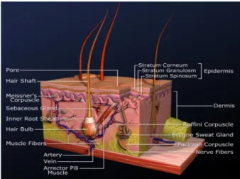
Fibroblasts are located in the dermis (Figure 1). Their main function is the maintenance of the structural integ- rity through continuous secretion of macromolecules of the extra-cellular matrix (collagen, glycosamine glycanes and glycoproteins). Tissue damage stimulates fibroblast proliferation and induces the release of cytokines. For experimental purposes, fibroblasts can be isolated from skin biopsies or phimoses through enzymatic digestion of the dermis or by explant method. They are available as the primary cell line NHDF (normal human dermal fibroblasts) from PromoCell (Heidelberg, Germany).
Figure 1: Structure of the skin
Keratinocytes are the most common cell type of the epi- dermis (Figure 1). They are named after the protein keratin, which they produce. They develop from epidermal stem cells located in the stratum basale (lowest layer of the epidermis) and differentiate into corneocytes during their movement to the stratum corneum (upper layer of the epidermis). There, they form a barrier which protects the body from foreign bodies and germs as well as dehyd- ration. Keratinocytes take an active part in the immune response, inflammatory processes, and wound healing as they are able to synthesize a broad range of cytokines, growth factors, and complement factors. Like fibroblasts, they can be isolated from skin biopsies and phimoses through enzymatic treatment of the epidermis. PromoCell (Heidelberg, Germany) also offers a primary keratinocyte cell line (NHEK, normal human epidermal keratinocytes).
Primary cell lines cannot be maintained in culture indef- initely. After a few passages, the cells lose their prolifera- tion ability, probably due to chromosomal alterations.
Those developing changes can influence experiment outcomes, which means there is a relatively small time frame for repeating tests. HaCaT cells are spontaneously immortalized keratinocytes. They originate from the isol- ate of the distal periphery of a melanoma located on the upper half of the back of a 62-year-old male patient and have been described in detail by Boukamp et al. [2].
HaCaT keratinocytes are practically immortal. It is possible to cultivate them for more than 140 passages. While they demonstrate a transformed phenotype in vitro, they are not tumorogenic, exhibit a steady expression of specific keratins and other identification markers, and show nor- mal differentiation.
Measurement of cell and tissue compatibility
Human HaCaT keratinocytes (kindly provided by Prof. N.E.
Fusenig, DKFZ, Heidelberg, Germany) are cultured in Dulbecco’s modified Eagle’s medium (DMEM, PromoCell, Heidelberg, Germany), supplemented with 10% fetal calf serum (FCS, PromoCell, Heidelberg, Germany) and 1%
antibiotic-antimycotic mix (PSF, PromoCell, Heidelberg, Germany) at 37°C in a humidified atmosphere containing 5% CO2. Primary fibroblasts (NHDF, PromoCell, Heidelberg, Germany) and keratinocytes (NHEK, PromoCell, Heidel- berg, Germany) can be maintained in appropriate basal media supplemented with growth factors but without antibiotics under the same conditions. For experiments, the cells are seeded at the required density into 96-well plates, 12-well plates, object slides etc. After allowing the cells to attach to their new surface, they are incubated with the test substances. Therefore, the cell culture me- dium is replaced by either fresh culture medium (negative control/medium control) or the test substance dissolved in culture medium or the material extract prepared ac- cording to the DIN standard EN ISO 10993-12.
The subsequent assessment of thein vitrocytotoxicity of the substance is often a qualitative analysis based on
the morphological examination of cell damage and growth after direct or indirect contact with the material. In gener- al, methods involving the measurement of cellular growth (cell counting or confluency evaluation) are more sensitive and discriminating than those in which the test materials are placed directly in the cell cultures to determine the zone of growth inhibition [3].
Cell counting with a haemocytometer is the most direct way of quantifying cell numbers. It is often done in con- junction with trypan blue exclusion staining. Trypan blue is not able to penetrate viable cells because of the membrane potential. However, dead cells lose their membrane integrity and are stained blue. This method is inexpensive and easy to perform, but unsuitable for high-throughput screening; it is only recommendable for suspension cells, as adherent cells need to be trypsinized prior to counting.
Determination of cell proliferation and cell viability has become the key technology to assess the growth behavior in a cell culture. A wide variety of commercial assays are available to this end. They are based on the determination of nucleic acids, metabolic activity, and protein content, or membrane integrity. A small selection (Table 1), such as Pico Green® DNA Cell Proliferation Assay, ATPLite™
Luminescence ATP Detection Assay, BC Assay: protein quantitation kit, AlamarBlue™ Proliferation Assay and Live/Dead Staining with SYTO-13 and EthD-2, is discussed here.
PicoGreen (Molecular Probes Inc, OR, USA) is a reagent for quantifying dsDNA, which provides a stable measure- ment parameter. The measurement is based on fluores- cence enhancement of the dye upon binding to dsDNA.
The relative fluorescence units measured correlate with the number of cells present (Figure 2). Cell number and PicoGreen fluorescence exhibit a linear relationship for low and medium cell densities, but which is lost for high cell densities. On the other hand, the assay shows a high sensitivity for low cell numbers, as it is able to detect dsDNA concentrations as low as 25 pg/ml.
Equally sensitive is ATPLite (Perkin Elmer Life Sciences, MA, USA), an ATP monitoring system based on the detec- tion of light generation by firefly luciferase. In an ATP-de- pendent reaction, the enzyme converts D-luciferin to oxyluciferin. The amount of emitted light is directly propor- tional to the ATP concentration (Figure 2) and can be measured with a luminometer. ATP is present in all metabolically active cells and declines rapidly when cells undergo necrosis and apoptosis. Therefore, it constitutes an equally stable parameter as dsDNA. However, it allows only the measurement of viable cells and not the quanti- fication of the total cell number.
Table 1: Cell quantification and cytotoxicity assays
Figure 2: Correlation of cell number and fluorescence intensity (blue), luminescence (red) and absorbance (black)
Another possibility is the determination of the protein content, which naturally corresponds directly to the cell number (Figure 2). The BC Assay (Uptima Interchim, France) is derived from the Biuret reaction (Cu2++ e-→ Cu+) using bicinchoninic acid. It is based on the measure- ment of the formation of a water soluble, purple-colored complex of bicinchoninic acid and Cu+. The test is sensi- tive, but not as sensitive as the PicoGreen or ATP assay, and low cell numbers do not provide the necessary protein concentration.
In contrast to the first three methods, the AlamarBlue Proliferation Assay (Biosource, CA, USA) is a non-destruc- tive method. The test uses the indicator dye resazurin, which is dark blue in color and possesses little intrinsic fluorescence until it is reduced by the metabolic activity of viable cells to resorufin, which is pink and highly fluorescent. Non-viable cells rapidly lose their metabolic capacity, do not reduce the dye, and thus do not generate a fluorescent signal. The assay allows simultaneous in- cubation and measurement but requires precisely defined time and temperature conditions, as the resorufin will continually develop. This presents a serious problem for assay handling. Figure 3 shows a cell standard curve in- cubated with AlamarBlue measured after 3 h (Figure 3a) and 5 h (Figure 3b).
Figure 3: Measurement of the same cell standard curve after 3h (a) and 5h (b) incubation with AlamarBlue
In conclusion, all of the cell quantification methods dis- cussed have advantages and disadvantages (summarized in Table 1), and the most suitable test method for a given study must be chosen. Besides, it is often necessary and always recommendable to verify the test results with at least two different assays. In that way, it is possible to exclude outside influences or false interactions of the components.
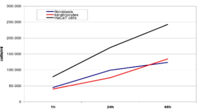
The assays can be used to determine the cell proliferation over a particular time period (Figure 4). So far, we have been able to demonstrate proliferative as well as cytotoxic effects of various test substances on keratinocytes and fibroblasts. Figure 5 and Figure 6 present examples of our results.
Figure 4: Proliferation of HaCaT cells (black), primary keratinocytes (red) and fibroblasts (blue) over a cultivation
period of 48 h determined by ATP quantification with the ATPLite Luminescence ATP Detection Assay
Figure 5: Proliferation of HaCaT keratinocytes depending on the melatonin concentrations after 24 and 48 h incubation.
The ATP bioluminescence assay was performed using melatonin in different concentrations [9].
Cell proliferation tests are able to express toxic effects via loss of proliferation ability or reduction of living cells.
These negative effects can be quantified by methods that determine cytotoxicity through the measurement of ne- crosis or apoptosis (Table 1).
Staining with SYTO-13 and Ethidiumhomodimer-2 enables the direct count of viable and dead cells. SYTO-13 and Ethidiumhomodimer-2 (Molecular Probes Inc, OR, USA) are green and red fluorescent dyes that bind to nucleic acids. While SYTO-13 can permeate membranes and is therefore able to stain living cells, Ethidiumhomodimer- 2 cannot, and can only enter dead cells, but possesses a higher binding affinity to DNA than SYTO-13. Treatment of the cells with a toxic substance leads to a reduction in the ratio of green to red cells (Figure 7).
Figure 6: ß-cyclodextrin in concentrations of 0.5 and 1% (w/v), respectively, exert a significant negative effect on HaCaT keratinocyte proliferation [10].
Figure 7: HaCaT keratinocytes not treated (left) and treated (right) with an antiseptic solution (Lavasept®Fresenius, Bad
Homburg, Germany)
Lactate dehydrogenase (LDH) in the supernatant is a sign for necrotic degradation of the cells depending on the release of LDH from the cytosol after cell membrane damage. The Cytotoxicity Detection Kit (Roche, IN, USA) is a colorimetric assay which measures the activity of re- leased LDH. Cytotoxic substances which affect membrane integrity cause a rapid release of LDH compared to the medium control (Figure 8). This process is accompanied by a rapid decline of cell viability (Figure 8). This method is quick and easily adaptable for different types of cells and test conditions, but the assay is sensitive to interfer- ence from compounds with inherent LDH activity, such as serum-supplemented culture media.
Frequently, the treatment with a substance does not kill the cells directly, but induces cellular damage that sub- sequently leads to pathological events. Apoptosis is the term for the induction of the programmed cell death. The members of the caspase family play a key effector role in mammalian cell apoptosis. The Caspase-Glo 3/7, 8 and 9 Assays (Promega GmbH, Mannheim, Germany) provide specific luminogenic substrates that form a lumin- escence signal proportional to the amount of caspase activity present (Figure 9).
In most cases, the release of inflammatory cytokines ac- companies the reaction of the cells to the test substance (Figure 10). Cells do not function independently from each other, they communicate through the secretion of interleukins such as IL-6 and IL-8. Interleukin 6 is a cy- tokine that provokes a broad range of cellular and physiological responses, e.g. inflammation, haemato- poiesis and neuronal differentiation. Interleukin 8 is a member of the superfamily of chemokines and acts as a chemo-attractant for neutrophils; it plays a crucial role in many inflammatory events. The cytokine concentration in the supernatant can be easily measured by ELISAs specific for IL-6 and IL-8 (Milenia biotec, Bad Nauheim, Germany).
Figure 8: The release of LDH under the influence of toxic ß-cyclodextrin concentrations correlates with the reduction of cell viability measured by ATP content [10].
Figure 9: β–cyclodextrin induces apoptosis in human keratinocytes via caspase-8 activation. The caspase-8 activity was determined using the bioluminescent Caspase-Glo™ 8 Assay kit (Promega GmbH, Mannheim, Germany) [11].
Figure 10: Cultivation of HaCaT keratinocytes with textile extracts (cotton) has a negative effect on cell proliferation determined by dsDNA content and triggers the release of the cytokine IL-8.
Determination of regeneration processes
Materials and substances used, for example, in the treatment of wounds do not only influence cell prolifera- tion and viability, but also affect their migration capacity.
Fibroblasts and keratinocytes enable the maintenance of the barrier function of normal skin. Upon wounding, this shield is disrupted and fibroblasts and keratinocytes migrate into the wound area to regenerate the skin.
Therefore, the cells must dissociate from one another and break the cell-matrix contacts to detach from the basal lamina. Migration into the wounded area entails remodelling of the extra-cellular matrix by its proteolytic
degradation as well as byde novosynthesis and depos- ition of newly formed matrix components. Both degrad- ation and reformation of the matrix are functions per- formed by dermal fibroblasts and epidermal keratinocytes.
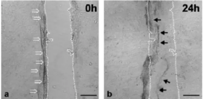
The mechanical scratch wounding of confluent mono- layers (Figure 11) serves as a model to study cell migra- tion at the wound margins [4]. The method allows the direct measurement of cell migration and layer regenera- tion. Using the scratch apparatus described by Büth et al. [4], the scratches can be inflicted with high precision and in reproducible fashion.
Figure 11: Conventional scratch wounding. Phase-contrast micrographs of HaCaT keratinocytes were taken directly (a) and 1 day (b) after scratch wounding. Keratinocytes were grown
on non-coated cover glasses. Scratch rims are denoted by dotted lines. Bars: 100 µm [4]
So far, all featured test methods utilize cell monolayers which are an exclusivelyin vitrocondition. Evidence sug- gests that cells behave differently within these two dimen- sions than in their familiar 3-dimensionalin vivoenviron- ment. For instance, fibroblasts reach confluency in monolayer cultures with a high proliferation rate and display protein biosynthesis levels that do not resemble thein vivosituation. On the other hand, fibroblasts em- bedded in 3-D collagen gels exhibit lower biosynthetic activity, regulated growth, and stellate shape; they also have protruding dendritic extensions and a state of differ- entiation close to thein vivosituation.
To mimic the conditions which cells experiencein vivo, the use of 3-D cell cultures is becoming increasingly popular. There are two types of matrices used for cell culture. “Ordered collagen matrices” (ORC) use the long range assembly properties of type I collagen monomers, and the cells are seeded afterwards into the collagen gel formed [5]. The “dermal equivalent” (Figure 12) is formed as a tissue-like structure from a collagen gel contracted by fibroblasts [6]. With 3-D cultures it is possible to study collagen degradation as well as polymerization and de- position of newly synthesized collagen. They allow the study of tissue remodellingin vitroand provide an assay system for wound contraction. It is even possible to pre- pare skin-equivalent tissue by co-culturing with keratino- cytes. Of course, these are time-consuming and elaborate preparations that require operator experience and the appropriate evaluation methods.
Figure 12: A contracted dermal equivalent formed from a gel containing 7.2x10⁴fibroblasts cast in square mold after 5 days.
Collagen concentration was 1.8 mg/ml [6].
Assessment of the potential of testing physical wound treatment techniques
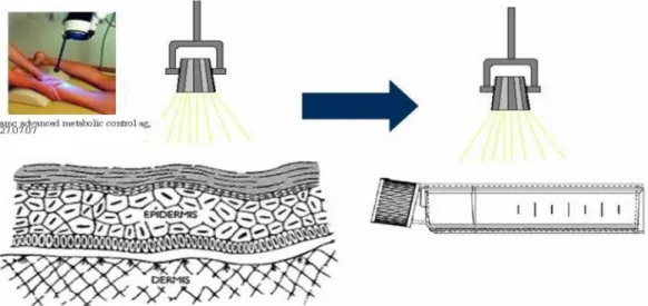
It has been reported that the use of physical wound treatment techniques, such as wIRA (water filtered in- frared A) and HVPC (high-voltage pulsed current), have a positive effect in terms of accelerated wound healing [7]
or additional antibacterial activity [8]. For the measure- ment of the effect of these physical methodsin vitroor for the application of a new plasma device on cells, sev- eral considerations must be taken into account to design an appropriate study. For one, it is necessary to scale the in vivoapplication parameters to thein vitrouse on cell monolayers (Figure 13).
For assessment of the bactericidal activity of HVPC, cotton pieces were soaked with bacterial suspension, placed on a steel plate, and covered with the electrode (42 mA, 128 Hz, 30 min) [8]. Cells need stable culture conditions that guarantee the establishment of an appropriate cell envir- onment during the experiment. Therefore,in vitrostudies that have been performed to assess the bactericidal activity of HVPC cannot be applied one-to-one on human cell cultures.
In conclusion, for testing physical treatment methods, the choice will probably tend to the use of a 3-D culture system which more closely resembles thein vivosituation, although cellular effects are far easier to detect and quantify in cell monolayers than in 3-D cultures. Hopefully, this summary of methods for measuring cell and tissue compatibility will provide ideas for the design of conclu- sivein vitrostudies.
Figure 13: For the investigation of the effects of wIRA, the scaling of the light intensity and application distance from the clinical use to the in vitro system is necessary.
References
1. van Wyk CW, Olivier A, Maritz JS. Cultured pulp fibroblasts: are they suitable for in vitro cytotoxicity testing? J Oral Pathol Med.
2001;30:168-77.
2. Boukamp P, Petrussevska RT, Breitkreutz D, Hornung J, Markham A, Fusenig NE. Normal keratinization in a spontaneously immortalized aneuploid human keratinocyte cell line. J Cell Biol.
1988;106:761-71.
3. Johnson HJ, Northup SJ, Seagraves PA, Garvin PJ, Wallin RF.
Biocompatibility test procedures for materials evaluation in vitro.
I. Comparative test system sensitivity. J Biomed Mater Res.
1983;17:571-86.
4. Büth H, Buttigieg PL, Ostafe R, Rehders M, Dannenmann SR, Schaschke N, Stark HJ, Boukamp P, Brix K. Cathepsin B is essential for regeneration of scratch-wounded normal human epidermal keratinocytes. Eur J Cell Biol. 2007,
doi:10.1016/j.ejcb.2007.03.009.
5. Besseau L, Coulomb B, Lebreton-Decoster C, Giraud-Guille MM.
Production of ordered collagen matrices for three-dimensional cell culture. Biomaterials. 2002;23:27-36.
6. Bell E, Sher S, Hull B, Merrill C, Rosen S, Chamson A, Asselineau D, Dubertret L, Coulomb B, Lapiere C, Nusgens B, Neveux Y. The reconstitution of living skin. J Invest Dermatol. 1983;81:2s-10s.
7. Hoffmann G. Wassergefiltertes Infrarot A (wIRA) zur Verbesserung der Wundheilung. GMS Krankenhaushyg Interdiszip.
2006;1:doc20. Verfügbar unter:
http://www.egms.de/en/journals/dgkh/2006- 1/dgkh000020.shtml.
8. Daeschlein G, Assadian O, Kloth LC, Meinl C, Ney F, Kramer A.
Antibacterial activity of positive and negative polarity low-voltage pulsed current (LVPC) on six typical Gram-positive and Gram- negative bacterial pathogens of chronic wounds. Wound Repair Regen. 2007;15(3):399-403.
9. Hipler UC, Fischer TW, Elsner P. HaCAT cell proliferation influenced by melatonin. Skin Pharmacol Appl Skin Physiol.
2003;16:379-85.
10. Hipler UC, Schönfelder U, Hipler C, Elsner P. Influence of cyclodextrins on the proliferation of HaCaT keratinocytes in vitro.
J Biomed Mater Res A. 2007;83:70-9.
11. Schönfelder U, Radestock A, Elsner P, Hipler UC. Cyclodextrin- induced apoptosis in human keratinocytes is caspase-8 dependent and accompanied by mitochondrial cytochrome c release. Exp Dermatol. 2006;15:883-90.
Corresponding author:
Dipl. Biochem. Cornelia Wiegand
Department of Dermatology, University Medical Center, Erfurter Straße 35, 07740 Jena, Germany, phone: +49 (0) 3641 937331, Fax: +49 (0) 3641 937437
C.Wiegand@med.uni-jena.de
Please cite as
Wiegand C, Hipler UC. Methods for the measurement of cell and tissue compatibility including tissue regeneration processes. GMS
Krankenhaushyg Interdiszip. 2008;3(1):Doc12.
This article is freely available from
http://www.egms.de/en/journals/dgkh/2008-3/dgkh000110.shtml
Copyright
©2008 Wiegand et al. This is an Open Access article distributed under the terms of the Creative Commons Attribution License
(http://creativecommons.org/licenses/by-nc-nd/3.0/deed.en). You are free: to Share — to copy, distribute and transmit the work, provided the original author and source are credited.
![Figure 6: ß-cyclodextrin in concentrations of 0.5 and 1% (w/v), respectively, exert a significant negative effect on HaCaT keratinocyte proliferation [10].](https://thumb-eu.123doks.com/thumbv2/1library_info/4841532.1629046/6.892.114.787.112.466/figure-cyclodextrin-concentrations-respectively-significant-negative-keratinocyte-proliferation.webp)
![Figure 8: The release of LDH under the influence of toxic ß-cyclodextrin concentrations correlates with the reduction of cell viability measured by ATP content [10].](https://thumb-eu.123doks.com/thumbv2/1library_info/4841532.1629046/7.892.110.786.118.322/figure-influence-cyclodextrin-concentrations-correlates-reduction-viability-measured.webp)