Cellular and Molecular Analysis of Fracture Healing in a Neurofibromatosis Type 1 Conditional Knockout Mice Model
D i s s e r t a t i o n
zur Erlangung des akademischen Grades d o c t o r r e r u m n a t u r a l i u m
(Dr. rer. nat.) im Fach Biologie eingereicht an der
Mathematisch-Naturwissenschaftlichen Fakultät I der Humboldt-Universität zu Berlin
von
M.Sc. Thaqif El Khassawna
Präsident der Humboldt-Universität zu Berlin Prof. Dr. Jan-Hendrik Olbertz
Dekan der Mathematisch-Naturwissenschaftlichen Fakultät I Prof. Stefan Hecht, PhD
Gutachter: 1. Prof. Dr. Georg Duda Gutachter: 2. Prof. Dr. Stefan Mundlos Gutachter: 3. Prof. Dr. Britt Wildemann Tag der mündlichen Prüfung:
11.04.2013
ABSTRACT
ABSTRACT
Neurofibromatosis type 1 (NF1) is an autosomal dominant genetic disease resulting from inactivating mutations in the gene encoding the protein neurofibromin. NF1 manifests itself as a heritable susceptibility to tumors of neural tissue, which are mainly located in the skin such as neurofibromas, and Café-au-lait spots (pigmented skin lesions). Besides these more common clinical manifestations, many NF1 patients – around 50% – have abnormalities of the skeleton. Long bones - usually the tibia - are often affected, and the clinical signs range from tibial bowing to spontaneous fractures and even non-unions. Therefore, further analysis of cellular and molecular events throughout the healing process would eventually contribute to therapeutic attempts in the clinic. Moreover, NF1 mice models could provide the understanding of the cell types involved in the resulting non-union and their behavior.
This study analyzed the healing progress of femur fractures in a model of NF1 long bone dysplasia - an Nf1Prx1 knockout mice model. This established mice model used a Prx1 -Cre- flox system to inactivate conditionally both copies of the Nf1 gene in the limb bud mesenchyme. Fracture callus in 8-week old mice, was assessed at days 7, 10, 14, and 21 by µCT, histology, biomechanics, and molecular analyses. In accordance with the previously reported results of cortical bone injury on the same mice model (Kolanczyk, et al., 2008);
bone healing was impaired in Nf1Prx1 mice femoral fracture. Results revealed increased periosteal bone deposition at the early stages of healing, decreased but persistent cartilage formation concomitant with fibrous tissue accumulation within the fracture site, decreased torsional stiffness, decreased bone mineral density, and increased fibrous tissue infiltration in the callus of mutant mice. This fibrous tissue accumulation hindered bone fracture healing, and was deposited by alpha smooth muscle actin-positive myofibroblasts, which were derived from a yet unidentified muscle fascia. This is further supported by the microarray analysis of callus tissues showing that genes crucial to muscle cells physiology, proliferation and differentiation were affected. In addition, extracellular matrix related genes were up-regulated in the mutants. In summary, this study shows a resemblance in the healing progression to the Nf1Prx1 mice model and NF1 patients, thereby, confirming the suitability of this mice model to explore the mechanism by which mutations in NF1 lead to non-unions. Moreover, in vitro and in vivo pilot assessments of MEK inhibitor treatment demonstrated a potential remedy for the lack of neurofibromin in bone healing.
ZUSAMMENFASSUNG
ZUSAMMENFASSUNG
Neurofibromatose Typ 1 (NF1) ist eine autosomal dominante Erbkrankheit, die durch inaktivierende Mutationen im Neurofibromin-Gen verursacht wird. NF1 manifestiert sich durch eine erhöhte Tumor-Inzidenz des neuralen Gewebes in der Haut (Neurofibroma) und pigmentierten Hautflecken. Neben diesen häufigeren klinischen Manifestationen haben rund 50% der NF1-Patienten Skelett-Anomalien. Häufiger sind Röhrenknochen betroffen, in der Regel die Tibia, und die klinischen Symptome reichen von Tibia-Krümmung über Spontanfrakturen bis hin zu Nonunions. Diese Studie analysiert den Heilungsverlauf von Femurfrakturen in Nf1Prx1-Knock-out-Mäusen, einem Mausmodell der NF1- Röhrenknochendysplasie. In diesem etablierten Mausmodell werden beide Kopien des NF1- Gens spezifisch mit Hillfe eines Prx1-abhängigen Cre-flox-Systems innerhalb der mesenchymalen Gliedmaßenknospen inaktiviert. Der Frakturkallus von acht Wochen alten Mäusen wurde an den Tagen 7, 10, 14 und 21 durch µCT, Histologie und molekulare Analysen evaluiert. Ähnlich wie in einer früheren Studie im gleichen Mausmodell (Kolanzcyk, et al., 2008), haben µCT und histologische Analysen eine beeinträchtigte Knochenheilung in Nf1Prx1-Mäusen gezeigt. Eine erhöhte periostale Knochenbildung in den frühen Stadien der Heilung war zu beobachten, sowie eine reduzierte, aber anhaltende Knorpelbildung und Bindegewebs-Akkumulation innerhalb der Fraktur. Desweiteren war die Torsionssteifigkeit und die Knochendichte verringert, wohingegen die Bindegewebs- Infiltration im Kallus von Nf1Prx1-Mäusen erhöht war. Wir konnten zeigen, dass der normalen Heilungsprozess durch dieses Bindegewebe behindert wird, welches durch alpha smooth muscle actin-positive Myofibroblasten gebildet wird, die ihrerseits aus einer bisher noch nicht identifizierten Muskelfaszie abgeleitet sind. Dieser Zusammenhang wird durch eine Microarray-Analyse der Kallus-Gewebe bestätigt, die ergab, dass durch den Knock-Out Gene reguliert wurden, die in Physiologie, Proliferation und Differenzierung von Muskelzellen involviert sind. Darüber hinaus waren extrazelluläre-Matrix-Gene in den Mutanten hoch regeuliert. Zusammenfassend konnten wir zeigen, dass eine Ähnlichkeit des Heilungsverlauf zwischen dem Nf1Prx1-Mausmodell und NF1-Patienten besteht. Folglich kann an diesem Mausmodell untersucht werden, durch welche Mechanismen die Mutationen im NF1 zu Knochenheilungsstörungen führen. Außerdem konnte in einer Pilotstudie der Effekt des Neurofibromin-Mangels auf die Knochenheilung durch Behandlung mit MEK-Inhibitoren in vitro und in vivo weitestgehend behoben werden.
DEDICATION
To Maha and Elia,
My wife and son
TABLE OF CONTENTS
TABLE OF CONTENTS
ABSTRACT ... I ZUSAMMENFASSUNG... II TABLE OF CONTENTS ... IV
INTRODUCTION ... 1
Structure and function of bone ... 2
Bone remodeling ... 3
Bone healing ... 6
Inflammatory phase ... 6
Reparative phase ... 6
Remodeling phase ... 7
Delayed and non-union healing ... 8
Neurofibromatosis type 1 (NF1) ... 9
Signal transduction in RAS / MAPK signaling cascade ... 11
Mesenchymal stroma cells (MSC) ... 12
Nf1Prx1 mice model ... 13
Neurofibromatosis type 1 - treatment options ... 15
OBJECTIVES OF THIS STUDY ... 16
MATERIALS AND METHODS ... 17
Experimental design ... 17
Animal model ... 17
Breeding of Nf1flox+/- Prx1cre+ mice ... 18
Genotyping ... 18
Surgical procedure ... 18
Quantification and evaluation of µCT of fracture callus ... 19
Histological analysis ... 21
Preparation of tissues for histological examination ... 22
Movat pentachrome staining ... 23
Tri-chrome (gallocyanin – chromotrop 2 R – aniline blue) staining ... 23
Tartrate-resistant acid phosphatase (TRAP) staining ... 23
Van Gieson / Von Kossa staining ... 23
Histomorphometry ... 24
Immunohistochemical staining ... 24
In situ hybridization ... 26
Preparation of mRNA and expression analysis... 26
Gene expression analysis by qPCR ... 28
Microarray hybridization ... 28
TABLE OF CONTENTS
Functional annotation ... 30
Network analysis using cytoscape ... 30
Cell culture ... 31
Aspiration of mesenchymal stroma cells ... 31
Osteogenic differentiation assay ... 31
Trails to compensate the lack of neurofibromin ... 32
In vitro assessment of MSCs viability and osteogenic differentiation under MEK inhibitors ... 33
In vivo assessment of AZD6244 MEK inhibitor ... 33
Statistical analyses ... 34
RESULTS ... 35
Breeding and genotyping ... 35
Decreased mechanical competence of fracture callus in Nf1Prx1 mice ... 35
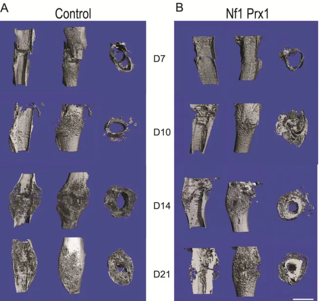
Affected formation and progression of Nf1Prx1 mice callus in µCT ... 36
Visualization of callus ... 36
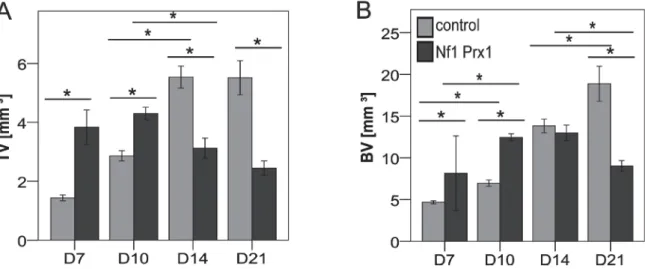
Quantitative analysis of fracture callus ... 37
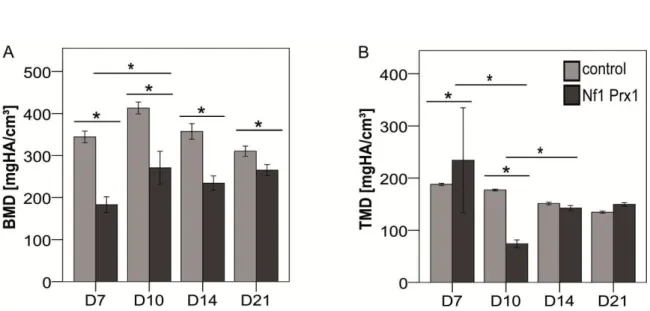
Decreased mineralization and persistent cartilage formation ... 41
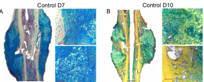
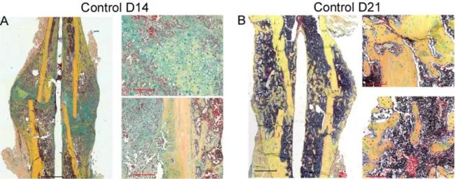
Descriptive histology ... 41
Histomorphometry: a quantitative analysis of fracture callus ... 44
Increased count of TRAP positive osteoclasts in the callus of Nf1Prx1 mice ... 48
Increased osteoid volume in Nf1Prx1 mice callus ... 49
Immunohistochemistry ... 50
In situ hybridization indicates periosteal surface bone formation and delayed cartilage formation in Nf1Prx1 mice ... 53
a- SMA staining characterizes abundant cell type in the fibrous tissue of Nf1Prx1 mice callus ... 56
Muscle fascia and the fracture site in Nf1Prx1 mice ... 58
Quantitative real time polymerase chain reaction (qPCR) ... 59
Microarray analysis ... 62
Time-point D0 ... 63
Time-point – 7 days post fracture ... 64
Time-point – 10 days post fracture ... 65
Time-point – 14 days post fracture ... 65
Time-point – 21 days post fracture ... 65
Functional annotation ... 67
Network analysis ... 68
In vitro testing of dual specificity mitogen-activated protein kinase kinase inhibitor ... 71
AZD6244 MEK inhibitor REMEDY cartilage formation in Nf1prx1 mice... 72
Results summary ... 74
DISCUSSION ... 77
Diminished cartilage formation and myofibroblasts invasion in the Nf1Prx1 callus ... 77
Lack of bony bridging after 21 days of healing in the Nf1Prx1 mice ... 79
Deformed morphology and bone structure during healing in Nf1Prx1 mice ... 80
Osteoblasts, mineralization and bone formation ... 81
Vascularization ... 83
Osteoclasts and bone resorption ... 85
Muscle fascia and the fracture site in Nf1Prx1 ... 86
Gene expression and finger printing of altered healing in Nf1Prx1 ... 87
MEK inhibitor influences cell survival despite aiding differentiation ... 88
TABLE OF CONTENTS
CONCLUSION ... 91
IMPLICATIONS AND FUTURE DIRECTIONS... 92
APPENDICES ... 93
Genotyping for transgenic mice strain breeding... 93
Microcomputer tomography ... 95
Preparation of bone samples for paraffin embedding ... 97
Preparation of bone samples for plastic embedding ... 98
Preparation of bone samples for cryostat embedding ... 99
Movat pentachrome staining ... 100
Tri-chrome (gallocyanin – chromotrop 2 R – aniline blue) staining ... 101
Tartrate-resistant acid phosphatase staining ... 102
Van-Gieson/Von Kossa staining ... 103
Immunohistochemical staining ... 104
In situ hybridization ... 106
TRIZOL RNA isolation and RNA quality control... 110
cDNA synthesis and qPCR protocol... 112
Database for annotation, visualization and integrated discovery (DAVID) v6.7 ... 113
Network analysis using enrichment map plug-in for cytoscape V2.8 ... 118
Aspiration of mesenchymal stroma cells from bone marrow and cultivation ... 119
Osteogenic differentiation of MSCs ... 120
Mechanical testing of mice femur... 121
ABBREVIATIONS LIST ... 126
ACKNOWLEDGMENTS ... 129
THESIS DECLARATION ... 130
REFERENCES ... 131
INTRODUCTION
INTRODUCTION
Bone fracture healing is a complex physiological process involving a series of timed steps leading to the regeneration of an injured bone's structure. Bone regeneration involves cellular processes, which require interplay between different cell types and regulatory factors. Processes of bone regeneration resemble processes observed in the embryonic skeletal development. Generally, in the endochondral ossification type of bone healing, the process relies on cartilaginous callus formation, which subsequently hardens by the “bone matrix mineralization” process. Callus is then reshaped through an osteoclast-mediated remodeling.
Neurofibromatosis type 1 (NF1) or von Recklinghausen’s disease [1] is a genetic disorder with an incidence of 1:3000 [2]. The NF1 gene (Nf1) encodes for a tumor suppressor protein called neurofibromin (Nf1) [3]. Mutations in the gene are causal to NF1, which primarily manifests itself with neurocutaneous symptoms [4]. Furthermore, skeletal lesions are also frequent in NF1 patients [5]. Among these, there can be a decreased bone mineral density due to higher bone resorption; dystrophic and none-dystrophic scoliosis are also common [6]. Bony dysplasia is found as well in about fifty percent of all NF1 patients [7]. Moreover, tibial bowing is a familiar symptom in NF1 patients, and this frequently results in spontaneous fractures followed by pseudarthrosis [8,9,10,11].
Neurofibromin is essential for successful fracture healing [12]. However, a profound understanding of fracture healing in the absence of neurofibromin is still required.
Therefore, fracture healing studies in animal models of NF1 are crucial for understanding the cellular and molecular interactions correlated with the lack of Nf1.
Various models of NF1 are available. Some of these have reported a minor discrepancy from the wild type, for example, the heterozygous Nf1 knockout mice generated and reported by Schindeler et al. 2007 who have shown no major complication of fracture healing [13]. Recently two new NF1 mice models were described. Although none of these showed a deviant phenotype from the wild-type mice, both showed a complete or at least partial healing impairment. The first one depended on ablation of Nf1 in osteoblasts [14];
the other one used local delivery of engineered adenovirus at the fracture site in a heterozygous Nf1 mice model to reach complete inactivation of Nf1 [15]. In contrast to these models, the Nf1 flox Prx1 Cre (Nf1Prx1) mice model used here [16] exhibits recapitulation of NF1 patients skeletal symptoms. The Nf1Prx1 mice model was established
INTRODUCTION
as a conditional homozygous inactivation of Nf1 in undifferentiated mesenchyme, where activation of the homeobox transcription factor (Prx1 promoter) led to the expression of Cre recombinase (Type I topoisomerase), which in turn excised Nf1 gene in an Nf1 floxed mice [16]. Thus, in the limb cells of mesenchymal origin such as endothelial cells, chondrocytes, and osteoblasts would have Nf1 knocked out. However, hematopoietic origin cells such as osteoclasts would have a normal Nf1 gene.
Structure and function of bone
Bone is a specialized form of connective tissue which forms the skeleton, and it has both protective and mechanical roles [17].
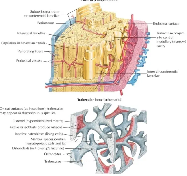
Morphologically seen, bone consists of two different types. One is the cortical bone; it has densely packed layers, which are concentric and diversely oriented to increase mechanical strength (Fig. 1, upper part). Trabecular bone, the second type, is more vascular [17], less dense than the cortical bone, and has a trabecular structure which allows a better adaptation to stress (Fig. 1, lower part). This structure-related adaptation is referred to as Wolff’s law [18]. Cavities around the trabeculae are filled with bone marrow, which is a habitat for hematopoietic stroma cells (HSCs) and mesenchymal stroma cells (MSCs) [19].
Bone is mostly made of the bone matrix, which consists of approximately 95% type I collagen, whereas proteoglycans, the non-collagenous proteins and various cell types form the remaining 5%. Among bone cells, there are osteoblasts, osteoclasts, osteocytes, chondrocytes, and bone-lining cells. The main biology of bone cells has to do with bone remodeling which involves orchestrated resorption and deposition of bone. In bone remodeling, osteoblasts form, deposit and mineralize bone tissue [20,21,22,23] and osteoclasts mediate bone resorption [24], where osteocytes are found within the concentric lamellae and play a role in calcium and phosphorus homoeostasis [25,26]. Other important components of bone are the periosteum and endosteum. The first covers the diaphyseal external surface, and the second covers the inner surface. The periosteum has a fibrous outer layer, which consists mainly of fibroblasts and collagen. However, the inner cambial layer is highly cellular and contains MSCs, osteoprogenitor cells, and osteoblasts, fibroblasts, microvasculature, and sympathetic nerves. Therefore, the inner cambial layer is involved in bone regeneration [27,28].
INTRODUCTION
In this thesis, a three-point femoral fracture model was used. The femur is a long bone and suitable for examining bone dysplasia. The femur diaphysis is mainly compact bone where the trabecular bone is primarily found in the epiphysis and metaphysis regions.
Figure 1: Bone histology, general overview. Compact bone is the dense part of bone, and accounts for 80%
of the total bone mass. Trabecular bone adapts to stress more than compact bone does because of its trabecular structure. Periosteum lines the outer surface of bone; it provides bone with progenitor cells after a bone fracture. (Modified from Netter’s Orthopedics, fourth edition)
Bone remodeling
Bone is a specialized form of metabolically active connective tissue. Every year, 10% of total bone tissue in humans is remodeled. Two balanced processes are crucial for this remodeling: bone formation and bone resorption. Regulation of these processes is essential for bone homeostasis. Imbalance can lead to pathological conditions as osteoporosis or
INTRODUCTION
osteopetrosis. Different cell types are required to achieve remodeling: osteoblasts, osteocytes and osteoclasts [29]. Osteoblasts are cells of mesenchymal origin; they produce the new bone matrix and aid its mineralization [30]. To go further into detail, bone formation takes place as mesenchymal cells proliferate and then differentiate into osteoblast precursors and ultimately into mature osteoblasts. Osteoblasts synthesize a matrix of osteoid composed mainly of type 1 collagen [31]. At a later stage, mature osteoblasts mineralize the osteoid matrix. Osteoblasts' proliferation and differentiation are subject to many solubility factors such as Runt-related transcription factor 2 (Runx2) [32] and a zinc finger-containing transcription factor (Osterix) [33]. Osteoclasts are multinucleated cells with foamy cytoplasm appearance. When actively resorbing bone, osteoclasts are also characterized by their ruffled border [34] which increases the surface-area interface for bone resorption.
Integrin receptors facilitate attachment of osteoclasts to the bone matrix. Subsequently osteoclasts produce hydrogen ions through the ruffled border to acidify bone matrix. In addition, hydrolytic enzymes as cathepsin K and metalloproteases are released to digest the organic matrix.
The basic multicellular unit (BMU) is the core of bone cellular physiology. BMU is composed of various cells responsible for dissolving and refilling an area of bone surface.
Osteoclast-mediated bone resorption (dissolving) takes place in 3 weeks, while osteoblast- mediated bone formation requires 3-4 months. Furthermore, bone type is also relevant;
trabecular bone remodeling takes place faster than cortical bone remodeling does. The emergence of a BMU increases as bone gets older [35]. However, the initiation of the process takes place via mechanical stress, tissue destruction, or cytokine signaling at the targeted locus [36,37]. Moreover, osteoblasts can initiate BMU through the expression of RANKL (receptor activator of nuclear factor kappa b ligand) [38]. Termination of BMU function, on the other hand, depends on inhibiting osteoclast activity. An in vitro study previously suggested that osteoclasts are inhibited upon engulfing osteocytes during bone resorption [39]. However, it has been established that the presence of either TGF-β (transforming growth factor - beta) or estrogens induce apoptosis in osteoclasts [40,41]. In addition, other cell types such as macrophages (i.e. mononuclear cells) prepare the bone lacuna for osteoblasts right after the resorption is terminated. Macrophages synthesize a thin collagen layer and releases osteopontin, which facilitates the attachment of osteoblasts [42].
INTRODUCTION
The RANK / RANKL system is one of the main systems regulating osteoblasts / osteoclasts interaction. RANKL is produced by osteoblasts and expressed on the pre-osteoblasts' surface, while pre-osteoclasts have the RANK receptor on their surface.
RANK is a member of the tumor necrosis factor (TNF)-receptor family; its activation results in translocation of nuclear factor kappa-light-chain-enhancer of activated B cells (NF-κB) to the nucleus, and this causes an increase in the transcription of genes involved in osteoclastogenesis (Fig. 2) [43,44].
This interaction and activation could be inhibited solely by the decoy receptor osteoprotegerin (OPG), which eventually terminates resorption. Support for the role of RANK / RANKL in osteoclastogenesis comes from in vitro studies which showed a prevention of osteoclastogenesis when RANK is blocked. Furthermore, absence of RANK in knockout mice models prevented the formation of osteoclasts; thus mice models lacking either RANK or RANKL exhibited severe osteopetrosis [45].
Figure 2: The RANK / RANKL system. RANKL produced by osteoblasts binds to RANK on the surface of osteoclast precursors and recruits the adaptor protein TRAF6, leading to NF-κB activation and translocation to the nucleus. NF-κB increases the expression of c-Fos, and c-Fos interacts with Nuclear Factor of Activated T-cells, (NFATc1) to trigger the transcription factors of osteoclastogenic genes. OPG inhibits the initiation of the process by binding to RANKL. NFAT, nuclear factor of activated T cells; c-Fos FBJ murine osteosarcoma viral oncogene homolog NF-κB, nuclear factor-κB; OPG, osteoprotegerin;
RANKL, receptor activator of nuclear factor-κB ligand; TRAF, tumor necrosis factor receptor associated factor. (Modified from Boyce and Xing Arthritis Research & Therapy 2007)
INTRODUCTION
Bone healing
Fracture healing involves complex processes of cell and tissue proliferation and differentiation. Many players are involved, including growth factors, inflammatory cytokines, antioxidants, bone breakdown cells (osteoclast) and bone building cells (osteoblast), hormones, amino acids, and uncounted nutrients.
Upon bone injury, blood supply is disrupted, connective tissue is damaged and mechanical stability is lost. Subsequently bone regeneration takes place (Fig. 3). Thereby crucial and extensive matrix synthesis and re-establishment of vascular supply is required. Woven bone is then formed which is finally remodeled into lamellar bone [46,47]. The detailed process is spatially and temporally regulated, which recapitulates aspects of embryonic skeletal development [48]. The healing process has three partially overlapping phases as mentioned below.
Inflammatory phase
Bone injury causes a disruption of bone matrix, blood vessels, and surrounding soft tissues.
This disruption results in a bleeding into the fracture gap and the release of bone marrow.
Bleeding consequently forms the initial hematoma [46,47]. The disrupted blood supply creates a hypoxic state around the fracture gap [49]. Hematoma is followed and accompanied by inflammation [50]. Cytokines, growth factors and extracellular matrix (ECM) proteins are present due to inflammation. These in turn stimulate, recruit, and aid the proliferation of cells essential to bone healing [47,51,52]. For example Interleukin-1 (IL-1), an inflammation producer which is mainly secreted by macrophages in the innate immune response; Interleukin-6 (IL-6) an immune response stimulant which is secreted by T cells in the adaptive immune response; both are known to recruit mesenchymal cells [52]. Thereby, MSCs are stimulated to differentiate into chondrocytes and osteoblasts, among other cell types.
Reparative phase
The reparative phase of fracture healing is highlighted by the development of new blood vessels and the onset of cartilage formation. The surrounding soft tissue provides vascular ingrowth initially to the periosteal area and subsequently to the endosteal area. Prior to fracture, the cortical blood supply is primarily from endosteal bone and branches out in a
INTRODUCTION
blood supply to the cortex arises from outside the bone rather than the inside of it. Mice models of fracture healing reveal that the intramembranous and endochondral bone formation are initiated during the first 10 days. Inflammatory mediators in the fracture hematoma recruit chondrocytes capable of producing fracture callus. The hematoma initiates – and is eventually replaced by – the ingrowth of fibrovascular tissue. This developing construct provides structural support to stabilize the bone ends.
In general at this stage, proteins produced by the osteoblasts and chondroblasts begin to consolidate into what is known as a soft callus. This soft, new bone substance eventually hardens into a hard callus as the bone weaves together. The closed fracture model examined in this thesis on the mice femur reflects two types of ossification:
Endochondral ossification
In the absence of rigid fixation, endochondral bone formation occurs [53]. Differentiation of progenitor cells into chondrocytes, and subsequent proliferation and secretion of biological factors by these chondrocytes results in the production of an abundant cartilaginous matrix, including collagen II [51]. This soft callus spans the fracture gap [27,54]. Later – in a processes similar to the one known to take place in the growth plate during development - chondrocytes undergo hypertrophy, and chondrocyte-mediated mineralization [46]. As vasculature begins to invade, the hypertrophic chondrocytes are removed and woven bone formation occurs after recruitment of osteo-progenitor cells [51].
Intramembranous ossification
A few millimeters proximal and distal from the fracture site, a hard callus of fully mineralized woven bone is formed [46,51]. To accomplish this, osteoblasts from the cortical bone and osteo-progenitors derived from the periosteum proliferate and deposit new bone matrix onto existing bone surfaces [51].
Remodeling phase
During this stage, newly formed woven bone is converted into lamellar bone. First, osteoclasts begin to erode a cavity referred to as the cutting cone. Osteoblasts migrate into this cone and deposit a layer of bone matrix in opposition to the existing surface. These mechanisms restore the original structure and biomechanical competence of the injured bone [26].
INTRODUCTION
Delayed and non-union healing
Bone growth and bone healing are complex physiological processes of a concerted interplay between different cell types and regulatory factors. Bone repair processes lead to bone union, and when they cease without bone union a pseudarthrosis (non-union healing) is usually formed; whereas delayed healing (also delayed union) refers to incomplete restorative process within the expected time interval. Despite bone’s natural healing capacity and the extensive amount of research conducted in this area, delayed healing and non-union of bones are frequently encountered. For example in the United States 5-10 % of the over 6 million fractures occurring annually develop into delayed or non-unions [46].
Figure 3: Course of bone healing of standard closed transverse femoral fracture in the rat. Day 1) Bone matrix and blood vessels are disrupted (arrow); formation of hematoma also takes place. Day 7) Chondrogenesis (thick arrow) and bone formation from the periosteum (thin arrow) are initiated. Day 14) Calcification of cartilage starts, (arrow indicates interface between cartilaginous callus and periosteal bone formation), also remodeling starts. Day 21) callus is mainly calcified cartilage, cortical bone is remodeling and partially bridged. Day 28) newly formed woven bone and late stage remodeling. (Figure modified from Einhorn T et. al 1998, clin Orthop Relat Res: suppl: 355:7-21).
Although advanced methods in trauma surgery are conducted, delayed and non-unions are still severe complications following a fracture [55]. In general, bone fractures also have a socio-economic impact. Large annual budgets cover not only primary treatments and follow-up operations due to delayed or non-unions but also the cost of lost employment resulting from such procedures. Furthermore, in industrial nations, the ageing of populations is increasing. This, in turn increases the expectation of bone fracture prevalence and possibly pseudarthrosis due to osteoporosis; for instance, in the year 2000, 3.8 million osteoporotic related non-unions were reported in the European Union. The direct costs resulting in the health care services for those non-union osteoporotic related fractures was about € 32 billion [56], Furthermore, it has been predicted that 40% of all postmenopausal women will suffer
INTRODUCTION
one or more fractures during their remaining lifetimes [56,57] with the highly associated risk of a non-union fracture.
However, pseudarthrosis occurrence is governed by many different factors. In normal patients, a mechanical parameter e.g. fracture instability is the main factor of pseudarthrosis.
Inadequate immobilization has been one of the first factors implicated in delayed healing due to the disruption of newly formed blood vessels [58,59]. 50% of long bone fractures in neurofibromatosis patients results in pseudarthrosis [7]. Hence, understanding, prevention, and effective treatment of such complications are desirable.
Neurofibromatosis type 1 (NF1)
Neurofibromatosis type 1 (NF1), also called von Recklinghausen’s neurofibromatosis, and peripheral neurofibromatosis is a term applied to a dominantly inherited neurocutaneous disease caused by an alteration of the NF1 gene mapping to chromosome 17q11.2.
NF1 is one of the most common autosomal dominantly inherited disorders with an estimated birth incidence of 1:2,500 and a prevalence of 1:3,500 individuals [60]. NF1 has a penetrance of almost 100%, although an extreme variability even within the members of the same family is possible.
The commonest form of NF is caused by mutations in the NF1 gene [61]. The cardinal features of NF1 are café-au-lait macules, neurofibromas and Lisch nodules present in 95%
of the patients. Café au lait macules may be present at birth, but are not noticed in the first few months of life. There is no relationship between the number and localization of café au lait spots and NF1 severity. Both axillary and inguinal freckling develops in 70% of patients during their puberty and are highly specific for NF1. Lisch nodules are multiple melanocytic hamartomas of the iris (clumps of pigment cells). They usually appear in late childhood and do not impair vision. Their detection often confirms NF1 diagnosis in individuals having multiple café au lait spots. However, a clinical hallmark of the NF1 is the development of dermal neurofibromas (dNFs) and / or plexiform neurofibromas (pNFs). dNFs most commonly appear in late childhood or adolescence as small spongy subcutaneous nodules.
In contrast, pNFs usually are congenital and characterized by a large soft subcutaneous mass following the course of diffusely thickened peripheral nerves. pNFs may affect the skin and superficial muscle causing hypertrophy or deformation of the nearby tissue and diffuse
INTRODUCTION
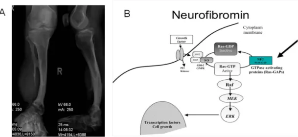
hyperpigmentation of the overlying skin. They can transform into malignant peripheral nerve sheath tumors (MPNST). Optic glioma and some osseous lesions are included as NF1 diagnostic criteria because their occurrence should suggest NF1. Optic gliomas are tumors made up of glial cells and may occur at any point of the optic nerve. Though optic gliomas are rarely malignant, they may cause painless proptosis, decreased visual acuity or neurological trouble. Bony abnormalities such as sharp scoliosis, dysplasia of a long bone or the sphenoid wing may provide useful diagnostic clues. A very sharp focal deformity of the spine, due to vertebral dysplasia, is almost exclusive to NF1 [62,63]. Furthermore, bowing of the tibia is a common skeletal diagnostic symptom (Fig. 4 A). Other NF1 manifestations include abnormalities in the development of the central nervous system (CNS) such as megalencephaly or seizures. Evaluation of the brain is required to exclude a tumor as the cause of seizures. Hydrocephalus caused by aqueductal stenosis is one of the most frequent complications and produces symptoms related to abnormal intracranial pressure. Cognitive and learning disabilities have also been shown to be present in up to 80% of the children with NF1 and can have significant effects on their learning performance and everyday life.
The most common cognitive problems involve perception, attention, language and motor deficit. Pulmonary manifestations such as interstitial fibrosis and bullae occur in 5 to 10% of NF1 patients. Interstitial fibrosis characteristically involves both lungs symmetrically with some basal predominance, whereas bullae usually are asymmetric and tend to develop in the upper lobes. Endocrine disorders have been reported in 1 to 3% of NF1 patients.
Pheochromocytoma is the most common disorder present in 1 to 6% of adults. In contrast, central precocious puberty is seen in 2.5 to 6% of children, almost invariably caused by a tumor in the hypothalamus. Although short stature is observed in 13% of NF1 patients, growth hormone deficiency was found in only 2.5% of children with NF1. The best characterized function of the NF1 gene is to act as a down-regulator of Ras proto-oncogene signaling by accelerating the switch of active Ras-GTP (Rat Sarcoma-oncogene-Guanosine Triphosphate) into inactive Ras-GDP (Rat Sarcoma-oncogene-Guanosine Diphosphate) [64]. The NF1 gene is considered to be a tumor suppressor as loss of heterozygosity (LOH) i.e. homozygote inactivation of the NF1 gene [65] is displayed in some cancer types such as malignant peripheral nerve sheath tumor, leukemia and melanomas.
INTRODUCTION
Signal transduction in RAS / MAPK signaling cascade
Ras proteins transmit extracellular signals to stimulate a receptor and phosphorylate its intracellular parts leading to an activation of Ras [66]. The Ras subfamily (Rat Sarcoma) is a protein subfamily of small guanosine triphosphatases (GTPases). A G-protein is active when bound to GTP, and inactive when bound to guanosine diphosphate GDP (i.e. GTP is hydrolyzed to GDP). Small GTPases are a family of hydrolase enzymes that can bind to and hydrolyze guanosine triphosphate (GTP). The GDP can then be replaced by free GTP.
Therefore, a G-protein can be either switched on or switched off. The major downstream target of Ras-GTP is Mitogen-Activated Protein Kinase (MAPK). The MAPK pathway is a signal transduction cascade. The cascade activates in a multistage process protein kinases through phosphorylation of serine or tyrosine residues, which affects transcription.
Mitogenic activation occurs via an enzyme encoded by the RAF1 gene (RAF), Dual- specificity mitogen activation protein kinase kinase 1 and 2 (MAP2K 1 and 2, better known as MEK1 / 2), and extracellular-signal-regulated kinases 1 and 2 (ERK 1 / 2) regulate cell growth, cell proliferation, and cell differentiation [67,68]. Briefly, RAF (also known as c- Raf) is a MAP Kinase Kinase Kinase (MAP3K) that functions downstream of the Ras subfamily of membrane associated GTPases to which it binds directly. Once activated, RAF can phosphorylate to activate MEK1 and MEK2, which, in turn, can phosphorylate to activate the serine / threonine-specific protein kinases ERK1 and ERK2. This results in the regulation of various processes such as embryogenesis, cell differentiation, cell growth, and apoptosis. Among the affected transcription factors are cAMP Response Element-binding Protein (CREB), cellular Myelocytomatosis oncogene(c-Myc) and Nuclear factor Kappa- light-chain-enhancer of activated B-cells (NF-kB). Furthermore, MAPK pathway is constitutively activated in the absence of neurofibromin (Fig. 4 B) as in NF1 patients [69,70]. In the last decade, studies suggest that MAPK activation inhibits osteogenic differentiation and promotes proliferation. In vitro experiments of Nf1 +/- osteoblasts show functional alterations of osteoblasts, which presented increased concentrations of osteopontin [71]. The same study showed that Nf1 +/- osteoblast conditioned media promoted osteoclasts migration and bone resorption.
INTRODUCTION
Figure 4: Neurofibromatosis type 1 lesions are caused by lack of neurofibromin. (A) X-ray film of an NF1 patient tibia; bowing of the tibia is a common symptom, which may lead to a spontaneous fracture. (B) Ras / MAPK signaling; growth factors bind to a receptor tyrosine kinase leading to its dimerization, and adaptor molecules direct the nucleotide exchange factor son of sevenless homolog 1(SO) to interact with activated RAS. Thus, three consequent phosphorylation cascades (Raf, MEK, and/ ERK) take place resulting in regulation of specific transcription factors of proliferation and differentiation. (A, Alwan, S et al. Clin Genet 2005: 67: 378-390, B, Schindeler, A., and Little, D. G. J Bone Miner Res 2006: 21(9): 1331-8)
Mesenchymal stroma cells (MSC)
Bone marrow stroma has been described to contain cells able to form bone. Nonetheless, minor subpopulations had the osteogenic potential; these were distinguished by their rapid adherence to cell culture plates and the fibroblast-like shape of their progeny in culture [72].
The cells gained their name based on the developmental origin in the mesenchymal [73].
MSCs are not only able to differentiate into mesenchymal cells, such as osteoblasts, adipocytes and chondrocytes, but also into non-mesenchymal cells including endothelial cells and neural cells [74]. For in vitro cultivation, MSCs are typically aspirated from bone marrow by centrifugation and subsequently by adherence to cell culture plastic [75].
However, other sources may be used to obtain MSCs such as muscle, fat, umbilical cord blood, liver, and spleen [76,77]. MSCs cell surface marker expression pattern is investigated [76,78,79]. It includes several cluster of differentiation (CD) markers such as CD105 also known as Src homology 2 (SH 2), CD73 also known as Src homology 3 / 4 (SH 3 / 4), CD44, CD90, thymocyte-1 (Thy-1), as well as the adhesion molecule CD106 also known as vascular cell adhesion molecule 1 (VCAM-1) [80]. None of these is alone specific for MSCs. However, absence of the hematopoietic markers CD45 and CD34 is needed to
INTRODUCTION
ECM (extracellular matrix) influences MSCs [81]. The ECM plays a role in cellular behavior; it maintains tissue architecture and acts as a ligand for cellular adhesion receptors such as integrin. Moreover, ECM provides signaling molecules, including growth factors and growth factor-binding proteins [81,82]. ECM - cell interaction modulates signaling cascades that control cell growth, differentiation, survival, and morphogenesis. Therefore, changes in the microenvironment affect these processes [83]. For example, MSC differentiation can be initiated by growth factors, such as TGF-β binding to trans- membranous serine / threonine kinase receptors. This activates the TGF-β / Smad pathway, which consequently regulates gene expression in the nucleus [84]. Several transcription factors are also known to be essential to MSC differentiation such as Runx2 that mediates osteogenesis. MSCs migration is necessary for bone healing and their sources of healing that may include: the bone marrow [85,86,87], periosteum [88,89,90], vessel walls [91], muscle [92,93,94] and circulation [95]. Due to their integral role during embryonic developmental stages and in fracture healing, MSCs were targeted – in the model described here –to achieve a conditional knockout of neurofibromin in the developing limb. Loss of neurofibromin is known to hyperactivate the Ras-MAPK pathway and accordingly, this affects cellular proliferation, differentiation and survival. In the situation described here, Ras-MAPK hyperactivation affects the MSCs themselves, as well as all cells to which they give rise.
Nf1
Prx1mice model
Several NF1 mice models have been generated. However, the goal was to develop an Nf1Prx1 mice, which recapitulates the patient's symptoms. NF1 patients show mosaicism in cells lacking neurofibromin. Homozygous Nf1 mice (Nf1 -/-) die in their fetal stages due to cardiovascular defects [96,97,98]. Heterozygous Nf1 mice (Nf1 +/-) are phenotypically quite variable and show only a few symptoms of neurofibromatosis type 1 disease patients [13,71,99,100]. Therefore, these mice models are not suitable for a close recapitulation of the NF1 patient’s musculoskeletal phenotype (Fig. 5). On the other hand, a conditional knockout system (Cre / loxP) works through targeted gene inactivation either time specifically, tissue specifically or both together[101]. In the Cre / loxP recombination system, a combination of genetic modification and recombination take place. The bacteriophage (P1) of E. coli codes for this site-specific recombinase, which achieves a targeted recombination at the loxP, sequences i.e. the recombination sites [102,103]. To
INTRODUCTION
conceive conditional knockout mice, both alleles of the Nf1 gene must be inactivated in mesenchymal precursor cells. Therefore, two mice strains were crossed: first, a strain of mice carrying the recombination site (the loxP site).It is important that the loxP sites flank the NF1 gene, especially exon 31, which is essential for the NF1 gene function [96].
Therefore, the target vector created to generate the Nf1flox / flox mice, which was utilized in this thesis, had loxP sites flanking exon 31. The second strain – required for the conditional knockout generation here – was a transgenic mice line bearing Cre recombinase under a specific promoter (Prx1 promoter). Consequently, Cre recombinase was expressed in cells with the active tissue-specific promoter Prx1. Taken together, in the Nf1Prx1 mice, Prx1 promoter was implemented to inactivate NF1 gene in the early mesenchymal precursor cells of the developing limb [104]. This will provide a heterozygous genotype at all body locations except those arising from MSC, which will have a homozygous ablation of NF1 gene. This approach overcomes the fatal consequences of a homozygous ablation of the NF1 gene in the whole body; it did accomplish a complete knockout in the limb, and that is where the focus of this thesis is. Moreover, no local ablation via viral inoculation and no restriction of one knocked out cell type at a time is required. Most importantly, this conditional knockout of the Nf1 gene resembles the LOH case seen in patients.
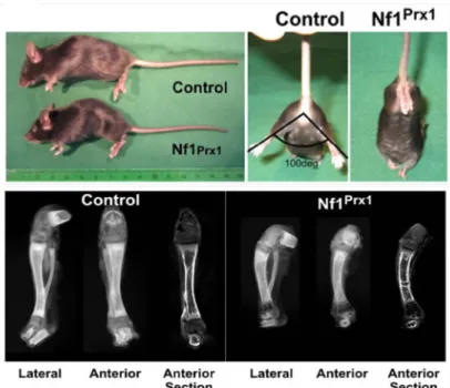
Figure 5: An Nf1Prx1 mice model recapitulates the symptoms of NF1 patients. The upper panel shows the smaller size and lack of ability of hind limb dispersion. The lower panel shows bowing of the tibia because of neurofibromin absence in the developing limb. (Modified from Kolanczyk, M. et al.: Hum Mol Genet, 16(8): 874-86)
INTRODUCTION
Neurofibromatosis type 1 - treatment options
Surgical removal of neurofibromas for both medical and cosmetic purposes is up to date one of the most common procedures on individuals suffering from NF1. Recently, laser technology has permitted a nonsurgical removal of small, cutaneous neurofibromas.
Moreover, chemotherapy was implemented to treat malignant peripheral nerve sheath tumors (MPNSTs) that are metastatic. Although varieties of combinations of chemotherapeutic agents were used, less than optimal outcomes were achieved. NF1 patients with severe bony defects must endure an orthopedic intervention such as limb sparing to avoid amputation. In trails to inhibit the MAPK pathway, lovastatin was the focus of many studies in the past few years and promising outcomes were reported. Recently, a phase I study examined the safety and tolerability of lovastatin in children with neurofibromatosis type 1. The study reported an improvement in areas of verbal and nonverbal memory [105]. However, the use of lovastatin in children with neurofibromatosis type 1 or in children with normal cholesterol levels is doubtful and was not reported to be free of side effects. More recently, in vitro studies looking at a more specific agents targeting a specific step within the Ras and/or MAPK pathways have shown some promising results in cancer research [106]. Some of these small molecules, which inhibit MEK, have reached phase I others reached phase II of clinical trials [106,107]. Inhibitors such as AZD6244 and AS703026 are financially feasible, commercially available and approved by the food and drug administration, those advantages are good motives to try these inhibitors in treating NF1.
OBJECTIVES
OBJECTIVES OF THIS STUDY
Neurofibromatosis type 1 disease is either inherited or presented through de novo mutations of the NF1 gene, resulting in a loss of neurofibromin function.
Neurofibromin is essential for fracture healing as it negatively regulates Ras-MAPK signaling.
Several mice models were generated and implemented in a fracture healing study. However, these mice models did not offer a close enough recapitulation of the patient's symptoms.
Therefore, in this study a conditional knockout mice system was used. The model was established and characterized to resemble lesions observed in NF1 patients. The knockout mice model has Nf1 ablation in mesenchymal precursor cells of the developing limb (Nf1Prx1).
This study hypothesizes that the Nf1Prx1 mice model would show temporal, morphological, and molecular differences in bone regeneration that would lead to healing deterioration when compared with the wild-type control mice.
The study aims at analyzing bone healing and its impairment in the Nf1Prx1 mice model at different time-points in a standard, closed femoral fracture model. Milestones are characterizing the fracture callus using micro-computed tomography to illustrate mineralization differences. Histology, enzymo-histochemical, immunohistochemical and immunofluorescence labeling methods were conducted to discriminate cells and tissues contributing in healing and their variances from the control. Furthermore, differential gene expression analysis and qPCR shall mark the molecular mechanisms throughout the healing process. The findings are to elucidate the pathogenesis of neurofibromatosis and its effect on fracture healing.
Hypotheses:
1. Delayed bone healing in the Nf1Prx1 mice is caused by temporal, morphological, and molecular differences when compared with the wild-type control mice.
2. MEK inhibitors treatment affects positively proliferation and differentiation of MSCs derived from Nf1Prx1 mice in vitro, and improves fracture healing in Nf1Prx1 mice in vivo.
MATERIALS AND METHODS
MATERIALS AND METHODS
Detailed protocols are described in the appendices; the annex has complete general lists of materials used in this thesis.
Experimental design
A closed fracture model [108] was used to study the effect of Nf1 ablated MSCs in bone healing. An Nf1Prx1 mice group was examined in comparison with a control group at four time-points after a femoral fracture (‘day is D’: D7, D10, D14, and D21). Resulting callus was investigated by micro-computed tomography (µCT), histology, and molecular analysis.
However, a biomechanical testing was done under my supervision by Mark Heyland as a part of his practical training at the Julius Wolff Institute.
Table 1 Experimental design (animal groups and planned tests)
Animal model
Male wild-type C57BL/6N mice (Charles River Laboratories, Wilmington, MA) served as control. Nf1flox+/- Prx1 cre+ mice were obtained from Dr. Mateusz Kolanczyk (Max- Planck institute for molecular genetics, Berlin, Germany). Mice were backcrossed onto the C57BL/6N background. This provided a genetic background for the mutants similar to that of the control mice. The local legal animal welfare representative approved all working protocols according to the guidelines for the care and use of research animals. (Landesamt
Procedure
Femoral fracture with intramedullary pin fixation
Control mice
(N = 95) Nf1Prx1 mice
(N = 98)
Sacrifice and harvest D0 D7 D10 D14 D21 D0 D7 D10 D14 D21
Biomechanics 10 10 10 10
Histomorphometry /
µCT 10 10 10 10 10 10 10 10
Undecalcified histology 10 10
Molecular biology 5 5 5 5 5 5 5 5 5 5
MEK inhibitor treated
mice /Cryostat histology 3
MATERIALS AND METHODS
für Arbeitsschutz, Gesundheitsschutz und Technische Sicherheit, Berlin-Germany - Registration number: G 0282/07).
Breeding of Nf1flox+/- Prx1cre+ mice
Five breeding cages were used. One Nf1flox+/- Prx1 cre+ male mice was mated with three wild-type female mice in each cage. Mendelian inheritance suggests for those two independent traits a 9:3:3:1 ratio. One out of every sixteen animal carries the NF1flox+/- Prx1Cre+ trait, from which only males were used for crossbreeding (hereafter denoted genotype A). Out of every sixteen animals, only three carried the NF1flox+/- trait (no Cre);
males and females were inbred. Nf1flox+/+ shall be passed onto 25% of their offspring, these are homozygous floxed animals (hereafter genotype B). To reduce the number of kept animals, only one male for every three females was kept. Inbreeding of homozygous mice is crucial to have new females to produce the knockout mice. Genotype B females were then backcrossed with the Genotype A males. The limitation is that Cre will pass onto the offspring only through males. This breeding setting gave rise to the experimental animals which were recognized phenotypically (smaller body size, limited hip joint movement, and brown fur) [16]. Nf1flox+/- Prx1 cre+ animals were generated in a 25% ratio, from which only males were used.
Genotyping
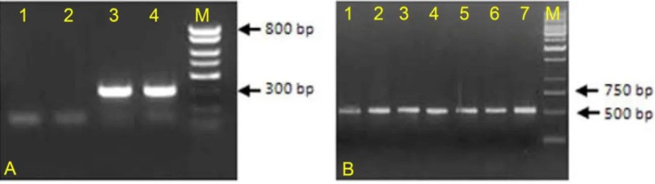
Mice were genotyped at all breeding stages. Only genotype A, inbred from the second generation onwards were trusted as they yielded only homozygous floxed mice. A tail tip biopsy was taken after marking the animal with a binary system using ear puncture. Tails were incubated in a dissolvent overnight, proteins were then salt precipitated; followed by an alcohol precipitation of DNA. The pellet was then washed, dissolved and kept at 4°C.
Genomic DNA was analyzed using standard PCR (see appendix A). According to the results, mice were sorted into control or mutant, heterozygous or homozygous types.
Surgical procedure
Eight-week old control and Nf1Prx1 male mice were used to generate unilateral standard closed fractures in the left femur using the three-points bending method [108]. The mice were anesthetized using a 2.5% isoflurane inhalation anesthesia mixed with oxygen. Before surgery, mice received subcutaneously buprenorphine (1 mg / kg body weight) to help relief
MATERIALS AND METHODS
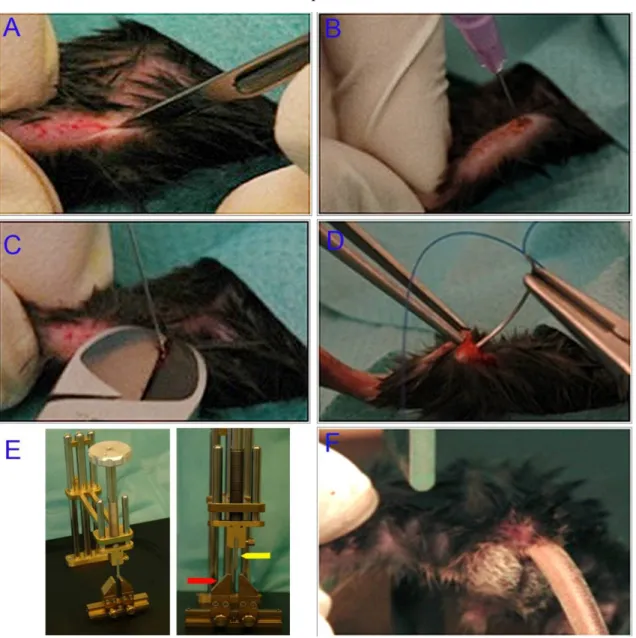
Braunoderm®, and then shaved. An incision of 2-3 mm was made through the skin, from the medial side to the patella. The patella was gently displaced to expose the intercondylar fossa. A needle (0.55 x 25 mm) was used to drill a channel along the femur axis. After removal of the needle, the mandrin (inner part) of a spinal needle was used as an intramedullary pin. This stainless steel intramedullary pin (17 G) was then inserted towards the great trochanter in the pre-drilled bone marrow space. Pin length (0.6-0.8 mm) was adapted to the mice femur using a cutter. Finally, the patella was repositioned and the wound was sutured with a biodegradable surgical suture (Fig. 6 A-D).
For the following femoral fracture, a three-point bending custom made fracture apparatus was used (Fig. 6 E). The two lower bending points of the gadget were fabricated as an adjustable grid (Fig. 6 E, red arrow). The third upper bending point was manufactured as a metal plate on the lower end of a bolt (Fig. 6 E, blue arrow). The bolt has a handle on its upper end. The loading mechanism was achieved by turning the handle in a clockwise direction. This caused the metal plate to move downwards. The mutant mice have a smaller body size; therefore, the fracture apparatus was adopted with smaller accessories. A mice was then held with its left femur positioned over the grid. An assisting person rotated down the bolt causing enough load to break the femur (Fig. 6 F). Fractures and position of the intramedullary pin were controlled by X-ray. Mice weight was measured right after the operation. The control mice registered body weight ranged between 22-32 grams where mutant mice had a body weight ranged between 15-24 grams.
On D7, D10, D14, and D21 after fracture, mice were again anesthetized as described above, and then placed into a CO2 flow airtight cage until they stopped breathing. The position of the nail was controlled again by X-ray. Then the thigh skin was cut. The broken femur was carefully removed with its tissue by freeing the knee and hip joints. The intramedullary pin was removed using a cutter. Bones for histology were fixed in PFA. Bones for µCT were then swaddled in gauze submerged in a sterile phosphate buffer solution (PBS) and stored at -20°C until further processing. The contralateral intact femur of each mice was also removed and processed in parallel to the broken ones. For molecular analysis, animals were sacrificed by cervical dislocation. Bones were flash frozen and kept at -80°C until used.
MATERIALS AND METHODS
Quantification and evaluation of µCT of fracture callus
Micro-computed tomography (µCT) was implemented to determine bone callus formation and its structural progression in both Nf1Prx1 and control mice. The intramedullary pins were removed from the sacrificed mice femora to prevent attenuations failure.
Figure 6: Surgical procedure of unilateral standard closed fractures in the left femur using the three-points bending method. (A) Under anesthesia, a medial incision was made to expose and displace the patella. (B) A channel was drilled along the femur using a standard needle. (C) A Mandrin was inserted in the predrilled channel and adapted to femur length using a cutter. (D) The incision was sutured after repositioning the patella. (E) The custom-made three-points bending fracture apparatus (red arrow grid, yellow arrow bolt).
(F) Fracture was made by loading the bone through lowering the bolt.
Scanning and image evaluation was done using an ex vivo µCT imaging device. Callus size, geometry, structure, and mineralization were characterized. Fractured and contralateral femora were batch scanned. A fixed isotropic voxel size of 10.5 μm was set with a 70,000
MATERIALS AND METHODS
enclosing the whole fracture callus (VOI). Cortical bone was manually deducted and only newly formed tissue with a preset global threshold of ≥ 190 mg HA/cm3 was included. This threshold was set by scanning manufacturer-delivered hydroxyapatite samples and calibration of the µCT scanner accordingly. All analyses were performed on the digitally extracted callus tissue using 3D distance techniques. Bone volume (BV) and total volume (TV) percentages were normalized to the number of slices, which covered the whole fracture callus. Bone mineral density (BMD) and tissue mineral density (TMD) percentages were normalized to the contralateral bones. For a detailed protocol, see appendix B.
Histological analysis
Histological analysis serves in visualization and quantification of not only various cell types involved in fracture healing, but also matrix proteins and mineralization. To achieve that, two embedding methods were used in this study. The paraffin embedding requires decalcification and is more suitable for immunohistochemical staining [109]. Plastic embedding of undecalcified bone is preferred in mineralization following up. All histological analyses of fractured femora at D7, D10, D14, and D21 post-fracture were conducted as previously described [109,110]. Several analyses were performed on paraffin sections as follows: i) Histomorphometry and descriptive histology by means of Movat pentachrome stain [111]. ii) Tartrate-resistant acid phosphatase (TRAP) enzymo- histochemical stain for osteoclasts [112]. iii) Osteocalcin immunohistochemical stain for osteoblasts. iv) Factor VIII immunohistochemical stain for blood vessels [113] v) Alpha smooth muscle actin, Myosin and DAPI immunofluorescence labeling (aSMA) for endothelial cells and myofibroblasts. vi) In situ hybridization (type I collagen, type II collagen, and osteopontin) vii) Desmin Immunohistochemical stain of myoblasts Viii) Tri- chrome (gallocyanin – Chromotrop 2 R – Aniline blue) stain for muscle, collagen and connective tissue.
Undecalcified femora with callus at D21 were processed, embedded in Polymethyl Methacrylate (PMMA) according to standard protocols [114]. Saggital 6-um sections of fracture callus were stained with Van Gieson / Von Kossa to differentiate osteoid and Safranin O / Von Kossa to describe mineralized tissue.
Cryostat embedding followed by Movat pentachrome staining was performed on bone specimens of Nf1prx1 mice, at D10 post fracture, from animals treated with MEK inhibitor.
MATERIALS AND METHODS
Preparation of tissues for histological examination Paraffin embedding
Paraffin embedding requires decalcifying bone samples, which are then used for immunohistochemical staining and in situ hybridization. Bones were fixed in 4% high- purity Paraformaldehyde solution (PFA) for 48 hours. Bones were decalcified at 4°C in a 1:2 mixture of 4% PFA and ethylenediaminetetraacetic acid (EDTA). The latter is a chelating agent, which adsorbs the calcium ions from the bone. After decalcification, bones were placed in embedding cassettes and then dehydrated in the dehydration machine.
Samples are then embedded in ‘Paraplast Plus’ tissue-embedding medium using the Heated Paraffin Embedding Module. Paraffin blocks were then solidified for 15 minutes on a cold plate. The solidified blocks could be stored at room temperature (RT). Before sectioning, the blocks were pre-cooled on ice. A rotary microtome was used to cut 6-µm-thick sections.
Sections were immediately placed in a warm water bath (42°C) to allow flattening of the sections. Floating sections were mounted on a glass slide and dried for 24 hours in an incubator at 37°C. For a detailed protocol, see appendix C.
Plastic embedding
This embedding method involves undecalcified bone samples, which are more convenient to evaluate bone mineralization and osteoid formation. Fixed bones were dehydrated manually.
Samples were soaked in xylene to remove all fat traces followed by 24 hours of incubation in a destabilized pre-infiltration solution, and then subsequently in the infiltration solution.
Five ml of embedding mixture were used to embed the samples in a plastic cylinder. After sealing the cylinder, polymerization was carried out for 48 hours at 4°C. Blocks were prepared for sectioning using a micro-grinding system and smoothened with silicon carbide papers with a grain size of 80. The samples were then placed in a hard-tissue microtome.
Sections of 6-µm thickness were prepared using a carbide blade. A lubricant fluid was used to grease the blocks during cutting. Sections were then placed on the slide using expanding fluid and pressed to dry for 48 h at 60°C. See appendix D
Cryostat embedding
Embedding bone samples in cryostat allows a fast preparation and convenient handling to evaluate bone callus, its cell types by immunohistochemical staining and its ossification.
Bones were fixed in 4% PFA for 2 hours. Afterwards, bones are soaked in three ascending
MATERIALS AND METHODS
were placed in stainless steel molds and mounted with embedding medium. The medium filled molds were dipped in an n-hexane beaker, which was already placed in a container with a dry ice - acetone mixture. After hardening, the formed block was removed from molds and immediately stored at – 80°C; see appendix E.
Movat pentachrome staining
Movat pentachrome staining differentiates the main events in bone callus as cartilage formation and hypertrophy, and matrix mineralization. Therefore, it was used to get a general overview of the different tissues that are involved in fracture healing [109]. The stain differentiates tissues so that mineralized bone appears bright yellow, non-mineralized cartilage and mineralized cartilage appear blue-green to yellow. The staining served for both the descriptive histology and the histomorphometric analysis, see appendix F.
Tri-chrome (gallocyanin – chromotrop 2 R – aniline blue) staining
Tri-chrome (gallocyanin – Chromotrop 2 R – Aniline blue) staining, also known as GRA stain, differentiates muscle tissue, bone tissue, and collagen. Bone appeared blue, muscle deep red, collagens appear light red. Therefore, the stain was used in a trail to identify a tissue that appeared to emerge from the muscle tissue before interring the fracture gap. See appendix G.
Tartrate-resistant acid phosphatase (TRAP) staining
Osteoclasts' count and location in a bone callus indicate the resorption level of trabecular bone to be substituted with compact bone during remodeling. The TRAP staining was used in order to visualize osteoclasts in paraffin sections. TRAP is an enzyme, which is synthesized by active osteoclasts, and it cleaves phosphate in the phosphorylated naphthol dye. Thus, the liberated daphthol binds to a diazonium salt and forms a red dye that colorizes the osteoclasts. Other cell types are able to cleave the phosphate in the dye as well.
Therefore, only cells located on the bone surface, TRAP positive and multi nucleated were taken into consideration. See appendix H.
Van Gieson / Von Kossa staining
Osteoid formation implies the matrix produced by osteoblasts and its calcification. Both processes shall be balanced to achieve normal healing; an increased osteoid area at the later time-points infers troubled calcification. To detect osteoid tissue in PMMA-embedded callus
MATERIALS AND METHODS
sections, Van Gieson's Stain was used. The stain is a mixture of picric acid and acid fuchsine. It is the simplest method for differential staining of collagen and other connective tissue. Here, the collagens (osteoid) appeared bright red or pink while the connective tissue red, mineralized cartilage and bone tissue turns black; see appendix I.
Histomorphometry
Despite the fact that Movat pentachrome is very informative, quantitative evaluation of the distinguished tissue types is required. Thereby, Movat pentachrome is a standard stain for a semi-automated histomorphometric evaluation of bone callus. Mainly newly formed mineralized tissue and cartilage tissue are quantified. The region of interest (ROI) enclosed the entire fracture callus. In principle, the analysis depended on measuring the pixels of the same color, which were then scaled as area (unit mm2). This semi-automated process was aided by the RUN software. Digital images were acquired using a Leica light microscope with preinstalled software. Subsequently, image size was reduced to 2000 pixels using Adobe Photoshop SC3 to enable RUN evaluation. During evaluation, images were rotated so that the proximal end of the femur was on the right-hand side and the medial side of the femur was facing upwards. Then, each tissue type was contoured manually and measured as one unit. To avoid color misconception, a live control under the microscope was essential.
The cortices' shift due to the fracture mechanics was also considered. The greater the shift, the larger the callus, and this is thus a measurement indicator of the newly formed tissue.
Immunohistochemical staining
Immunohistochemical staining specifically identifies antigens in tissues and on cell surfaces;
this allows a quantitative evaluation of either a specific cell types or matrix proteins. A color reaction visualizes the antigen-antibody complex. Principally, sections were pretreated with a digestive enzyme (pepsin or trypsin) to break bonds causing protein cross-linking resulted from fixation. Sections were then incubated with a normal serum originating from the same animal species as the secondary antibody. This was used to avoid unspecific binding of the secondary antibody. Generally, this technique employs an unlabeled primary antibody, in which sections are incubated for 1 hour, after which incubation with a biotinylated secondary antibody is followed for 30 minutes. Then incubation with avidin DH and biotinylated alkaline phophatase H. was performed. An alkaline phosphatase substrate solution was then utilized. Alkaline phosphatase catalyzes the hydrolysis of a variety of
MATERIALS AND METHODS
phosphate-containing substances in the alkaline pH range producing a colored insoluble precipitate thus visualizing the antigen presence. For a better representation, tissue was counter-stained with either hematoxylin or methyl green. Detailed protocols of all immunohistochemical stains are presented in appendix J.
Osteocalcin
Osteoblasts form woven bone during the reparative phase and compact bone in the remodeling phase of bone healing. Discrepancies in the count and location of osteoblasts affect bone healing. Therefore, an osteoblast specific stain is important to illustrate such discrepancies and their role in affecting bone anabolism. Osteocalcin is a protein secreted by osteoblasts and belongs to the non-mineralized bone extracellular matrix; it is often used as a marker for the bone formation process. Sections were pre-treated with trypsin, and then incubated with normal goat serum. Sections were then incubated with osteocalcin primary antibody (1:100). Osteoblasts were then visualized using the ABC-AP kit; all washing steps for osteocalcin staining were done in tris-buffered saline (TBS).
Factor VIII
Neovascularization is associated with osteogenesis induction. Factor VIII is a protein produced by vascular endothelium and enables vessel detection in histological sections. The sections were pretreated with pepsin, incubated in goat normal serum, and then bound with factor VIII primary antibody (1:50) and finally analyzed.
Desmin
Muscle tissue surrounding the fracture gap plays a non-negligible role. Desmin is a protein forming type III intermediate filament found near the Z line in sarcomeres. Desmin is one of the earliest protein markers for muscle tissue in embryogenesis [115]. It is also expressed at low levels and increases as the muscle cell nears a terminal differentiation. Muscle cells mature only in the presence of desmin. Sections were treated with trypsin, incubated in goat normal serum, and detected using a desmin antibody (1:50).
Alpha smooth muscle actin, myosin and DAPI
For immunofluorescence labeling of myofibroblasts, paraffin sections were pre-treated with the heat antigen-retrieval method using citrate buffer. Labeling was performed using a rabbit anti- mice alpha smooth muscle actin a-SMA1 antibody (1:200) visualized with a green secondary antibody-fluorophore conjugate, and a goat anti-mice myosin antibody (1:1000)