Analysis of matrilin function in knockout mice and
knockdown zebrafish
Inaugural-Dissertation zur Erlangung des Doktorgrades
der Mathematisch-Naturwissenschaftlichen Fakultät der Universität zu Köln
vorgelegt von
Ya-Ping Ko
aus Ping-Tung, Taiwan
Mai 2005
Berichterstatter: Prof. Dr. Mats Paulsson
Prof. Dr. Thomas Langer
Tag der mündlichen Prüfung: July 11, 2005
Table of contents
Abstract...1
Zusammenfassung ...3
1. Introduction ...5
1.1. Extracellular matrix proteins ... 5
1.1.1. Proteoglycans... 5
1.1.2. Collagens ... 7
1.1.3. Non-collagenous proteins ... 10
1.2. Matrilins... 11
1.2.1. VWA domains ... 12
1.2.2. EGF-like domains ... 13
1.2.3. Coiled-coil domains... 14
1.2.4. Matrilin-1 ... 15
1.2.5. Matrilin-2 ... 17
1.2.6. Matrilin-3 ... 19
1.2.7. Matrilin-4 ... 21
1.3. Model organisms... 23
1.4. Zebrafish matrilins... 23
1.4.1. Matrilin-1 ... 27
1.4.2. Matrilin-3a ... 27
1.4.3. Matrilin-3b ... 27
1.4.4. Matrilin-4 ... 28
1.4.5. Sequence analysis ... 28
1.5. Gene silencing methods ... 29
1.5.1. RNAi... 30
1.5.2. Morpholino antisense oligonucleotides (morpholinos) ... 31
1.6. The aims of the dissertation ... 36
2. Materials and Methods ...37
2.1. Characterization of matrilin-3 deficient mice... 37
2.1.1. Genotyping by PCR ... 37
2.1.2. Genotyping by Southern blot... 39
2.1.2.1. Dig-labeled probe for Southern blot ... 39
2.1.2.2. Genomic DNA extraction ... 40
2.1.2.3. Restriction enzyme digestion... 40
2.1.2.4. Southern blot... 40
2.1.3. Whole mount skeletal staining... 41
2.1.4. Immunohistochemistry ... 42
2.1.5. Northern blot... 43
2.1.6. In situ hybridization ... 44
2.1.7. Hematoxylin-eosin staining ... 44
2.1.8. Van Kossa staining... 45
2.1.9. Safranin orange staining ... 45
2.1.10. TRAP staining... 46
2.1.11. Glycosaminoglycan assay... 47
2.1.12. Cartilage extraction... 48
2.1.13. Western blot ... 48
2.2. Characterization of matrilin-1/-3 double null mice ... 49
2.2.1. Double fluorescence analysis for matrilin-4 and –1 in western blots of cartilage extracts ... 49
2.3. Characterization of zebrafish matrilins... 50
2.3.1. Expression and purification of recombinant matrilin-1, -3a, -3b and –4 VWA1 domains... 50
2.3.2. Preparation of antibodies against matrilin-1, -3a and -4... 52
2.3.3. Determination of cross-reactivity of antibodies against each matrilin with the other matrilins ... 52
2.3.4. Determination of cross-reactivity of the antibody against matrilin-3a with matrilin-3b ... 53
2.3.5. Whole mount immunostaining... 53
2.3.6. Immunostaining on sections ... 54
2.3.7. Temporal expression analysis by RT-PCR... 55
2.3.8. Whole mount skeletal staining... 56
2.3.9. Morpholino microinjection ... 57
3. Results...58
3.1. Matrilin-3 deficient mice ... 58
3.1.1. Generation of matrilin-3 deficient mice... 58
3.1.1.1. Genotyping of offspring... 60
3.1.2. Gross morphology of the skeleton is normal in matrilin-3 deficient
mice... 61
3.1.3. Matrilin-3-deficient mice show normal endochondral bone formation and intervertebral disk development... 62
3.1.4. Normal expression of other members of the matrilin family in matrilin-3-deficient skeletal tissues ... 68
3.1.5. Biochemical analyses reveal no difference in matrix protein content in matrilin-3 null mice ... 72
3.1.6. Summary... 76
3.2. Matrilin-1/matrilin-3 double deficient mice ... 76
3.2.1. Summary... 80
3.3. Matrilins in zebrafish ... 80
3.3.1. Generation of zebrafish-matrilin-specific antisera ... 81
3.3.2. Matrilin expression during development ... 86
3.3.3. Matrilins are differentially expressed ... 88
3.3.4. Morpholino knockdowns of matrilins... 92
3.3.4.1. Specificity of morpholinos... 92
3.3.4.2. Matrilin knockdown phenotypes ... 93
3.3.4.3. Matrilin-1 knockdown phenotype... 95
3.3.4.4. First characterization of matrilin-3a and matrilin-4 knockdown embryos……….….98
3.3.4.5. Phenotype frequency and survival rate... 98
4. Discussion ...100
4.1. Mouse matrilins ... 100
4.1.1. Matrilin-3 is dispensable for mouse skeletal development... 101
4.1.2. A biochemical phenotype in matrilin-1/matrilin-3 double deficient mice... 101
4.1.3. Matrilins in disease ... 102
4.2. Zebrafish matrilins... 104
4.3. Morpholino knockdowns of zebrafish matrilins... 106
4.3.1. Matrilin knockdown phenotypes ... 106
4.3.2. Dose dependency of morpholino knockdown effects... 107
4.3.3. Strength and limitations of morpholinos ... 108
4.3.4. Proper controls in morpholino experiments... 109
4.4. The difference in phenotype between knockout mice and knockdown zebrafish... 110
5. Perspectives ... 112
6. Bibliography... 113
Abbreviations ...125
Erklärung ...127
Acknowledgements ...128
Lebenslauf...130
Abstract
The matrilins are non-collagenous extracellular matrix proteins that form a subbranch of the superfamily of proteins containing VWA domains. Four matrilins are present in mammals, matrilin-1, -2, -3 and –4. The matrilins contain one or two VWA domains which are connected by a varying number of EGF-like domains, followed by a C-terminal α-helical coiled-coil domain. Matrilins serve as adaptors in the assembly of supramolecular structures in the extracellular matrix, but it is not known if this role is static or dynamic in nature. The in vivo functions of matrilins remain unclear and need to be elucidated in detail, in particular to understand the role of matrilins in inherited disease.
Mutations in the gene encoding human matrilin-3 lead to autosomal dominant skeletal disorders, such as multiple epiphyseal dysplasia (MED), which is characterized by short stature and early onset osteoarthritis, and bilateral hereditary microepiphyseal dysplasia, a variant form of MED characterized by pain in the hip and knee joints. In addition, a mutation in the first EGF-like domain of matrilin-3 has been linked to hand osteoarthritis in the Icelandic population.
Matrilin-3 null mice and matrilin-1/-3 double deficient mice were characterized.
Homozygous matrilin-3 mutant mice appear normal, are fertile, and show no obvious
skeletal malformations. Histological and ultrastructural analyses reveal an endochondral
bone formation indistinguishable from that of wildtype animals. Northern blot,
immunohistochemical, and biochemical analyses showed no compensatory upregulation
of any other member of the matrilin family. In matrilin-1/-3 double null mice,
biochemical analyses revealed a molecular phenotype in which the amount of matrilin-4
protein is increased and the band patterns of matrilin-3 and -4 are altered. The
upregulation of matrilin-4 is likely to represent a compensatory mechanism. Altogether,
the findings suggest functional redundancy among matrilins in mammals and
demonstrate that the phenotypes of MED-like disorders are not caused by the absence of
matrilin-3, but are likely to be due to dominant negative effects of the mutant proteins.
The zebrafish is a well established model organism for the study of vertebrate development. The matrilins are present in neither Drosophila nor in C. elegans and the zebrafish is therefore among the simplest organisms which express matrilins. Highly conserved orthologues, matrilin-1, -3a, -3b and –4, are present in zebrafish, while the matrilin-2 gene is missing. The temporal and spatial expression of zebrafish matrilins was characterized. Zebrafish matrilin-1 was found not only in skeletal tissue but also in notochord and intestine. Matrilin-3a expression is restricted to skeletal tissues, while the expression pattern of matrilin-3b has not yet been elucidated due to the lack of a specific antibody. Nevertheless, RT-PCR analysis reveals that matrilin-3b is expressed at 24 hpf and, interestingly, splice variants of matrilin-3b containing a proline- and serine/threonine-rich domain are found only in embryos but not in adult fish, indicating that this new domain probably has an important function during zebrafish development.
Similar to in mammals, matrilin-4 is the earliest and most widely expressed matrilin in zebrafish. Matrilin-4 is strongly expressed already at 24 hpf and is present in the skeletal tissues, soft connective tissues and nervous tissues.
Morpholino antisense oligonucleotides were used to knockdown matrilins expressed in
zebrafish. Malformations were seen at all the doses used and the phenotypes matched to
the tissue distribution of the respective matrilin. Injection of matrilin-1 or matrilin-4
morpholinos give curled body shape, smaller eyes or a truncated body axis depending
on dosage. The matrilin-3a knockdown embryos showed a serious skeletal phenotype.
Zusammenfassung
Die Matriline gehören zu den nicht-kollagenen Proteinen der extrazellulären Matrix, die einen Zweig der von Willebrandfaktor A Domänen (VWA) enthaltenden Proteinfamilie bilden. Bei Säugetieren gibt es insgesamt vier Matriline, Matrilin-1, -2, -3 und –4. Die Matriline enthalten ein oder zwei VWA Domänen, die durch eine unterschiedliche An- zahl von epidermalem Wachstumsfaktor ähnlichen Domänen (EGF) verbunden sind, ge- folgt von einer C-terminalen α-helikalen Coiled-Coil Domäne. Die Matriline dienen als Adaptorproteine bei der Verbindung von supramolekularen Strukturen in der extra- zellulären Matrix, es ist aber noch unbekannt, ob die Matriline dabei eine statische oder dynamische Rolle spielen. Die genauen in vivo Funktionen der Matriline verbleiben un- klar und müssen noch aufgeklärt werden, insbesondere die Rolle, die die Matriline bei der Entstehung von bestimmten Erbkrankheiten spielen.
Mutationen im menschlichen Matrilin-3 Gen führen zu autosomal dominanten Erb- krankheiten des Skelettsystems, wie z. B. der multiplen epiphysären Dysplasie (MED), die sich durch eine geringe Körpergröße und eine frühzeitig einsetzende Arthrose aus- zeichnen oder der bilateralen erblichen mikroepiphysären Dysplasie, einer Variante der MED, die durch Schmerzen in Hüft- und Kniegelenken charakterisiert ist. Außerdem wurde in der isländischen Bevölkerung eine Mutation in der ersten EGF Domäne von Matrilin-3 mit dem Vorkommen von Handarthrose in Verbindung gebracht.
Matrilin-3 defiziente und Matrilin-1/-3 doppeldefiziente Mäuse wurden untersucht. Die
homozygoten, mutierten Matrilin-3 Mäuse sehen normal aus, sind fruchtbar und haben
keinen offensichtlichen skeletalen Phänotyp. Histologische und ultrastrukturelle Unter-
suchungen zeigten eine endochondrale Knochenentwicklung, die sich von der vom
Wildtyp nicht unterschied. Northernblot, Immunhistochemie und biochemische Analy-
sen ergaben keine Hinweise auf eine Hochregulation der anderen Matriline, die das Feh-
len von Matrilin-3 hätten kompensieren können. Bei der biochemischen Analyse der
Matrilin-1/-3 doppeldefizienten Mäuse dagegen wurde ein molekularer Phänotyp ent-
deckt, in dem die Menge an Matrilin-4 erhöht war und sich das Bandenmuster von Ma-
trilin-3 und –4 verändert hatte. Die Hochregulation von Matrilin-4 beruht wahrschein-
lich auf einem Kompensationsmechanismus. Insgesamt deuten die Ergebnisse auf eine funktionelle Redundanz der Matriline bei Säugetieren hin. Die Phänotypen der skeleta- len Erkrankungen sind wahrscheinlich nicht durch das Fehlen von Matrilin-3 in der Ma- trix, sondern eher durch einen dominant negativen Effekt der mutierten Proteine zu er- klären.
Der Zebrafish ist ein gut eingeführter Modellorganismus bei der Untersuchung der Entwicklung von Wirbeltieren. Da die Matriline weder in Drosophila noch in C. elegans vorkommen, gehört der Zebrafisch zu den einfachsten Lebewesen, die Matriline exprimieren. Im Zebrafisch gibt es hoch konservierte orthologe Gene, die für Matrilin-1, -3a, 3b und –4 kodieren, während ein Matrilin-2 Gen fehlt. Die zeitliche und räumliche Expression von Matrilinen im Zebrafisch wurde untersucht. Zebrafisch Matrilin-1 konnte nicht nur in skeletalen Geweben, sondern auch im Notochord und Dünndarm nachgewiesen werden. Die Matrilin-3a Expression beschränkt sich auf skeletale Gewebe, während die Matrilin-3b Expression nicht untersucht werden konnte, da ein geeigneter Antikörper nicht zur Verfügung stand. Dennoch konnte mit Hilfe von RT-PCR bereits nachgewiesen werden, dass Matrilin-3b bereits 24 Stunden nach der Befruchtung exprimiert wird. Interessanterweise gibt es Spleißvarianten von Matrilin-3b, die eine an Prolin und Serin/Threonin reiche Domäne enthalten, welche nur in Embryos, aber nicht in erwachsenen Tieren vorkommt, was darauf hinweist, dass die Domäne möglicherweise eine Rolle während der Zebrafischentwicklung spielt. Wie bei Säugetieren ist Matrilin-4 beim Zebrafisch das am frühesten und breitesten exprimierte Matrilin. Matrilin-4 ist bereits 24 Stunden nach der Befruchtung stark exprimiert und kommt in skeletalen Geweben, weichen Bindegeweben und in Nervengeweben vor.
Morpholino Gegenstrangoligonukleotide wurden eingesetzt, um die Expression der Ma-
triline im Zebrafisch zeitweilig zu unterbinden. Fehlbildungen wurden bei allen ange-
wandten Dosierungen bobachtet und die Phänotypen passen zu der Gewebeverteilung
der entsprechenden Matriline. Abhängig von der Dosis ergaben die Injektionen von Ma-
trilin-1 bzw. Matrilin-4 spezifischen Morpholinos Tiere mit verdrehtem Körper, klei-
neren Augen oder gar einer verkürzten Körperachse. Die mit einem Matrilin-3a spe-
zifischen Morpholino behandelten Tiere zeigten einen schweren skeletalen Phänotyp.
1. Introduction
1.1. Extracellular matrix proteins
The extracellular matrix (ECM) is a complex structural entity surrounding and supporting cells within tissues. The ECM has received considerable attention due to its importance in cell-cell interactions, signalling, wound repair, cell adhesion and tissue function.
The major constituents of the ECM are proteoglycans and collagens, which form a tissue-specific network providing the tensile strength and resilience required. A number of non-collagenous proteins are also found in the ECM and serve to regulate matrix assembly and cell adhesion.
1.1.1. Proteoglycans
Proteoglycans are complex macromolecules consisting of a central protein core to which a variable number of glycosaminoglycan (GAG) chains are covalently attached.
Glycosaminoglycans are long, unbranched polyanionic carbohydrate chains consisting of repeating disaccharide units. There are four main classes of glycosaminoglycans:
hyaluronic acid, the chondroitin sulphates (chondroitin 4-sulphate, chondroitin 6-sulphate, dermatan sulphate), keratan sulphate and the heparin-heparan sulphate class.
As the glycosaminoglycans carry a large number of sulphate and carboxyl groups, the
proteoglycans have a high negative net charge, which in turn results in an extended
conformation due to electrostatic repulsion. Proteoglycans contribute about a third of
the dry mass of a hyaline cartilage. At this high concentration, they are presumably kept
in a compressed state by physical entrapment in the network of collagen fibers. The
electrostatic repulsion between the fixed charged groups will be strong and any further
compression will be resisted. Therefore, the proteoglycans contribute compressive
stiffness and elasticity to the cartilage.
Aggrecan is the most abundant proteoglycan in articular cartilage (Doege et al., 1994).
The protein core is about 230 kDa in size (Watanabe and Yamada, 2002) and is heavily substituted with GAG chains. Cartilage matrix deficiency (cmd) in mice (Rittenhouse et al., 1978) is a natural functional knockout of the aggrecan gene, in which 7 bp in exon 5 were deleted resulting in severely truncated molecules (Watanabe et al., 1994). The homozygotes (cmd/cmd) are characterized by dwarfism, short limbs, a short trunk, tail and snout, as well as a protruding tongue and cleft palate and mice die shortly after birth due to respiratory failure (Rittenhouse et al., 1978; Watanabe and Yamada, 2002). Even though heterozygous mice appear normal at birth, dwarfism and age-associated spinal degeneration are observed while aging (Watanabe et al., 1997), indicating that aggrecan plays an important role in cartilage development and maintenance.
One subgroup of proteoglycans are the small leucine-rich proteoglycans (SLRPs) that share a multiple leucine-rich repeat (LRR) structural motif flanked by cysteine residues.
The LRR domain is composed of ~10 repeats of 24 amino acid residues each, preferentially containing asparagin (N) and leucine (L) in conserved positions (LX
2LXLX
2NX(L/I)) (Iozzo, 1999). This motif is involved in many molecular recognition processes including cell adhesion, signal transduction, DNA repair and RNA processing (Kobe and Deisenhofer, 1994). SLRPs often interact with collagen, modifying the deposition and arrangement of collagen fibers in the extracellular matrix, but also with cells and with soluble growth factors (Ameye and Young, 2002). The interaction of SLRPs with cells and with growth factors like TGF-β may affect the cell proliferation in addition to modifying the extracellular environment (Ameye and Young, 2002).
The SLRP family is rapidly growing and more than 13 members are known including decorin, biglycan, asporin, fibromodulin, lumican, PRELP, keratocan, osteoadherin epiphican, mimican, opticin, chondroadherin and myctalopin (Ameye and Young, 2002).
Decorin is expressed throughout the body, stabilizes collagen fibrils and plays a
significant role in tissue development and assembly, as well as being involved in direct and indirect signaling (Reed and Iozzo, 2002). Mice harboring a targeted disruption of the decorin gene are viable but have fragile skin with markedly reduced tensile strength and irregular collagen fibril shape (Danielson et al., 1997; Reed and Iozzo, 2002).
Biglycan consists of a 45-kDa core protein made up almost entirely of leucine-rich repeats and is widely distributed in the extracellular matrices of bone and specialized, non-skeletal connective tissues. It was shown that biglycan-deficient mice develop age-related osteoporosis due to defects in bone marrow stromal cells (Chen et al., 2002).
Both decorin and biglycan bind to VWA domains in the N-terminal region of collagen VI and matrilins are in turn bound to these small leucine-rich proteoglycans (Wiberg et al., 2003).
1.1.2. Collagens
Collagens are the major proteins of the extracellular matrix, constituting 30% of the total protein mass. They play a dominant role in maintaining structures of various tissues and have been proven to have functions in cell adhesion, chemotaxis, migration and dynamic interplay between cells. In addition, collagens regulate tissue remodelling during growth, differentiation, morphogenesis and wound healing (Myllyharju and Kivirikko, 2004).
The primary feature of a typical collagen molecule is its long, stiff, triple-stranded
helical structure, in which three collagen polypeptide α chains are wound around one
another in a rope-like superhelix. An α chain is composed of a series of triplet Gly-X-Y
sequences, in which X is commonly proline and Y often hydroxyproline. Therefore,
collagens are extremely rich in proline and glycine and both amino acids are important
in the formation of the triple-stranded helix. Proline stabilizes the helical conformation
in each α chain because of its ring structure, while glycine is regularly spaced at every
third residue throughout the central region of the α chain. The hydroxyl groups of
hydroxyproline and hydroxylysine are thought to form interchain hydrogen bonds thus
helping to stabilize the triple helix.
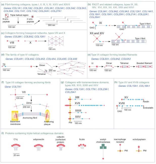
Up to date, 28 members of the collagen family have been found (R. Wagener, personal
communication, Myllyharju and Kivirikko, 2004). According to their assemblies,
collagens can be divided into the following subgroups (Fig. 1-1): fibril-forming (type I,
II, III, V, XI, XXIV and XXVII), fibril-associated collagens with interrupted triple
helices (FACIT) (types IX, XII, XIV, XVI, XIX, XX, XXI, XXII and XXVI),
hexagonal network forming (types VIII and X), beaded filament-forming (type VI),
anchoring fibril-forming (type VII), collagens with transmembrane domains (types XIII,
XVII, XXIII and XXV), nonfibril-forming (type IV), and multiplexin collagens (type
XV and XVIII) (Myllyharju and Kivirikko, 2004).
Fig. 1-1 Members of the collagen superfamily and their known supramolecular assemblies. The figure is from (Myllyharju and Kivirikko, 2004).
Among the collagens, collagen type II is cartilage-specific and the predominant
collagen in cartilage, representing about 90% of the collagen content. It forms a firm
network that provides tensile strength to the tissue. The fibres also contain type XI
collagen which is a minor, cartilage-specific collagen representing only a few percent of
the collagen content. Collagen type IX is associated with the collagen II/XI fibrils and
has a positively charged domain protruding out from the fibrils that has been suggested
to interact with proteoglycans. Collagen II/IX fibres form a fine network in the
superficial layer of adult articular cartilage. Collagen type X is mainly restricted to hypertrophic cartilage, but has also been shown to be present in the superficially layer of articular cartilage (Rucklidge et al., 1996). It has been suggested to have a role in endochondral ossification.
Collagens are known to mediate cell adhesion via integrin receptors. Previous studies have indicated the presence of a number of integrin recognition sites in collagens (Morton et al., 1994; Staatz et al., 1991). In addition, discoidin domain receptors (DDR), a subfamily of receptor tyrosine kinases, also act as receptors for collagens (Vogel et al., 1997) and the collagen binding sites in DDR2 have been identified (Leitinger, 2003).
The activation of DDRs mediates cell proliferation, migration and motility. Moreover, it was demonstrated that DDRs influence the expression and activity of metalloproteinases (Leitinger et al., 2004).
A great many mutations in collagens have been identified and shown cause diseases including osteogenesis imperfecta, many chondrodysplasias, several subtypes of the Ehlers-Danlos syndrome, Alport syndrome, Bethlem myopathy, certain subtypes of epidermolysis bullosa, Knobloch syndrome and also some cases of osteoporosis, arterial aneurysms, osteoarthrosis, and intervertebral disc degeneration (Myllyharju and Kivirikko, 2001).
1.1.3. Non-collagenous proteins
In addition to collagens and proteoglycans, many non-collagenous proteins are present in the ECM. However, in contrast to collagens and proteoglycans, which have been studied in detail since many years, the functions of many of the non-collagenous proteins are rather unclear.
Non-collagenous proteins typically contain multiple domains, each harbouring specific
binding sites for other matrix macromolecules and for cell surface receptors. The first
well characterized example was fibronectin, a large glycoprotein found both in blood
plasma and in the extracellular matrix (Hynes and Yamada, 1982). Fibronectin was
shown to have multiple functions, affecting cellular adhesion, cell migration, cellular morphology and spreading, cytoskeletal organization, oncogenic transformation, phagocytosis, embryonic differentiation and wound healing (Hynes and Yamada, 1982).
Fibronectin null mutant mice die early in embryogenesis because their endothelial cells fail to form proper blood vessels (George et al., 1993).
In addition to fibronectin, other proteins like osteonectin, tetranectin, tenascins, thrombospondins, laminins and matrilins also belong to the non-collagenous proteins.
The matrilins are widespread, but except for some information on their assembly with collagens and proteoglycans, their overall contribution to tissue function is not known.
1.2. Matrilins
The matrilins are a family of non-collagenous extracellular matrix proteins that form a subbranch of the superfamily of proteins containing VWA domains (for review, see (Whittaker and Hynes, 2002)).
The matrilin family consists of four members with a closely similar domain structure (Fig. 1-2). Two VWA domains are connected by a varying number of EGF-like domains.
These are followed by a C-terminal α-helical coiled-coil domain, which allows the oligomerization of the single subunits in a bouquet-like fashion. Only matrilin-3 lacks the second VWA domain and here the EGF-like domains are directly connected to the coiled-coil domain. In addition, matrilin-2 and -3 contain a stretch of amino acid residues at the N-terminus with a high frequency of positively charged side chains.
Uniquely, matrilin-2 contains a module between the second VWA domain and the
oligomerization domain that has no homology to any other known protein sequence.
Fig. 1-2 Domain structure of mouse matrilins (modified from Wagener et al., 2005).
The matrilins are differentially expressed. Matrilin-1 and –3 are mainly present in skeletal tissues (Aszodi et al., 1996; Klatt et al., 2000), whereas matrilin-2 and –4 (Klatt et al., 2001; Piecha et al., 1999) are more widely distributed. It is thought that matrilins have an adapter function in the extracellular matrix, connecting macromolecular networks (Hauser et al., 1996). This role for matrilins was confirmed by recent results showing that matrilins-1, -3 and –4 are associated with collagen VI microfibrils extracted from rat chondrosarcoma tissue. The matrilins are here bound to the small leucine-rich repeat proteins biglycan and decorin, which in turn interact with the N-terminal globular domains of the collagen VI molecules (Wiberg et al., 2003).
1.2.1. VWA domains
The VWA domains of matrilins consist of about 200 amino acid residues in a classical
Rossman fold with a central β-sheet surrounded by α-helices. A MIDAS (metal ion
dependent adhesion site) motif (DXSXSXnTXnD), which may be involved in ligand
binding, is perfectly conserved in nearly all matrilin VWA domains. The VWA domains
of matrilins represent an own subgroup of the VWA domains of extracellular matrix
proteins (http://www.ncbi.nlm.nih.gov/Structure/cdd/cdd.shtml) (Marchler-Bauer et al., 2005). All matrilin VWA domains have higher identity to the members of the subgroup than to any other VWA domain, indicating that they originate from a common ancestor VWA domain (Deak et al., 1999). Phylogenetic studies assigned the first and the second VWA domains into two different groups, of which each splits into a matrilin-1/3 and a matrilin-2/4 subbranch (Deak et al., 1999). Among other VWA domain family members, the VWA domains of different collagens, vitrin, α1 integrin, WARP and AMACO have the highest identity (approx. 30%). It is probable that the VWA domains are the principal interaction modules of matrilins, since matrilin-1 and -3 splice variants that lack all EGF domains exist in zebrafish (Ko et al., 2005).
VWA domains are found not only in the matrilins, but also in a large number of other extracellular proteins (for review see (Whittaker and Hynes, 2002)) such as von Willebrand factor, collagens type VI, VII, XII, XIV, XXI and XXII, complement factors B and C2, the H2 and H3 subunits of the inter- α-trypsin inhibitor, the α and β-chains of integrins and putative transmembrane proteins of lower eukaryotes (Whittaker and Hynes, 2002). Recently, also intracellular proteins like copines were identified as members of the family (Tomsig and Creutz, 2002; Whittaker and Hynes, 2002).
1.2.2. EGF-like domains
Except for some splice variants in zebrafish which lack EGF-like domains, all other matrilins contain at least one such domain, the highest number being in a splice variant of zebrafish matrilin-4 where 12 EGF domains are present (Ko et al., 2005). EGF domains contain 40-50 amino acid residues, including six conserved disulphide bonds.
The structure shows a two stranded β-sheet and often EGF domains are present in
multiple copies (Rao et al., 1995). The function of the EGF domains of matrilins is still
not clear. A comparison of the matrilin EGF domains with the key residues of the
calcium-binding motifs in calcium binding EGF domains (Handford et al., 1991; Rao et
al., 1995) showed no match with the consensus sequence. Neither was a non-canonical
calcium binding site (Malby et al., 2001) detected in matrilin EGF domains.
Moreover, even though matrilin EGF domains show an overall structural similarity to epidermal growth factor, there is no evidence that they retain growth factor activity. It is more likely that they serve as spacers between VWA domains, which in many other proteins show ligand binding activities.
Database searches show that EGF domains of scube proteins, fibulins and fibrillins have the highest homologies to matrilin EGF domains (Wagener et al., 2005).
1.2.3. Coiled-coil domains
The matrilins contain a C-terminal α-helical coiled-coil domain that allows oligomerization of the subunits in a bouquet-like fashion. Coiled-coil domains are characterised by heptad repeats (a-g) of amino acid residues that typically have non-polar amino acids at position a and d. The coiled-coil domains of matrilins contain 4.5 heptad repeats, showing the least perfect match with the consensus sequence for matrilin-3. Nevertheless, by SDS-PAGE and electron microscopy of full-length proteins, it has been shown that matrilin-1 and -4 form homo-trimers (Hauser and Paulsson, 1994;
Klatt et al., 2001), whereas matrilin-2 and -3 form homo-tetramers (Klatt et al., 2000;
Piecha et al., 1999). The oligomerization was also studied by using recombinantly
expressed coiled-coil domains (Frank et al., 2002). The analysis of oligomerization in
mixtures of isolated coiled-coil domains showed a broad range of interactions and the
only hetero-oligomers not found were those containing matrilin-2 and -3 or matrilin-3
and -4 (Frank et al., 2002). In contrast, here the coiled-coil domain of matrilin-2 formed
only trimers. It could well be that the oligomerization is influenced by the unique
domain adjacent to the coiled-coil domain of matrilin-2. Hetero-oligomeric forms of
matrilin-1 and -3 have been isolated from fetal human and calf cartilage (Klatt et al.,
2000; Kleemann-Fischer et al., 2001; Wu and Eyre, 1998). There is no conclusive
evidence for natural occurrence of other hetero-oligomers, but a SDS-PAGE band from
an extract of newborn mouse epiphyseal cartilage was shown by peptide mass
fingerprinting to contain both matrilin-1 and matrilin-4 (G. Sengle, unpublished results).
1.2.4. Matrilin-1
Matrilin-1, which was first purified from bovine cartilage, is the prototype member of the family and was originally called "cartilage matrix protein" or CMP (Paulsson and Heinegard, 1979). It is an abundant protein in many forms of cartilage. The single subunit has a molecular weight of 52,000 Da and contains 3.9% carbohydrate, probably in the form of N-linked glycans (Paulsson and Heinegard, 1981). In electron microscopy matrilin-1 shows the typical bouquet-like structure where the single VWA domains of the subunits are not resolved, indicating an interaction between VWA1 and VWA2 (Hauser and Paulsson, 1994) (Fig. 1-3). The solution structure of the oligomerization domain of matrilin-1 has been determined by heteronuclear NMR spectroscopy. As predicted, the domain folds into a parallel, disulfide-linked, three-stranded, α-helical coiled coil, spanning five heptad repeats in the amino acid sequence (Dames et al., 1998).
Fig. 1-3 Oligomerization of matrilin-1 visualized by electron microscopy. The figure is modified from (Hauser and Paulsson, 1994). In the schematic drawing the VWA domains are in dark blue, the EGF-like domains are in green and the coiled-coil domain is in light blue.
In the mouse, matrilin-1 can be detected by antibody labeling in myocardium at 9.5
days p.c. (Segat et al., 2000). This expression is however transient and of unknown
physiological significance. More lasting expression is seen for both matrilin-1 and –3 in
the condensing mesenchyme at day 12.5 p.c.. Matrilin-1 expression is restricted to
certain types of cartilage. In the mouse tibial growth plate, matrilin-1 is abundant in resting, proliferating and hypertrophic cartilage (Klatt et al., 2002), but it is not present in articular and intervertebral disc cartilages (Aszodi et al., 1996; Aszodi et al., 1994).
Moreover, in situ hybridization analysis of matrilin-1 mRNA showed a downregulation concomitant with the progress of hypertrophy at later stages of chondrogenesis (Aszodi et al., 1996).
Matrilin-1 is abundantly expressed in tracheal, nasal septum, auricular, and epiphyseal cartilages (Paulsson and Heinegard, 1982), and continued expression of matrilin-1 is seen in tissues that remain cartilaginous during the whole lifespan, e.g. in costal cartilage and in the nasal septum (Klatt et al., 2002).
The genetic basis for matrilin-1 gene expression has been studied in some detail in the chicken system. A minimal promoter has been defined that functions both in chondrocytes and fibroblasts (Goetinck et al., 1990). An enhancer exerts a chondrocyte-specific stimulation on the promoter activity and a silencer inhibits activity both in chondrocytes and fibroblasts. The enhancer is independent of the developmental stage of the chondrocytes, while promoter upstream control regions appear to restrict the promoter activity to certain chondrocyte developmental stages (Muratoglu et al., 1995). Transgenic experiments with the chicken matrilin-1 promoter in mouse have indicated that the tissue specific control elements are divided between the promoter upstream and intronic regions in a manner similar to that of the Col11a2 gene (Karcagi et al., 2004).
Matrilin-1 was first recognized as a protein tightly bound to aggrecan and which copurified with aggrecan in a variety of separation methods (Paulsson and Heinegard, 1979). The bound matrilin-1 molecules with time become covalently crosslinked to the aggrecan core protein, with at least some of the crosslinks not being sensitive to reduction (Hauser et al., 1996). In addition, an interaction was found between matrilin-1 and cartilage collagen fibrils (Winterbottom et al., 1992) and between matrilins and other non-collagenous molecules, in particular COMP and decorin (Mann et al., 2004).
Furthermore, matrilin-1 has the ability to enhance the adhesion of chondrocytes via
integrin α
1β
1(Makihira et al., 1999). However, two different strains of mice lacking
matrilin-1 show normal skeletal development (Aszodi et al., 1999; Huang et al., 1999).
Nevertheless, in one of the two strains the mice have alterations in type II collagen fibrillogenesis and fibril organization (Huang et al., 1999).
Matrilin-1 is a potential autoantigen able to trigger the tissue-specific immune response, as seen both in patients and in animals, resulting in relapsing polychondritis and related autoimmune diseases (Buckner et al., 2000; Hansson et al., 1999; Hansson et al., 2001;
Hansson and Holmdahl, 2002). It has also been reported that the expression of matrilin-1 is enhanced in knee osteoarthritic cartilage and in knee or hip rheumatoid arthritic cartilage (Okimura et al., 1997). In addition, elevated serum levels of matrilin-1 were detected in patients with active rheumatoid arthritis (Saxne and Heinegard, 1989).
Matrilin-1 also showed an increased expression in specimens from arthritic condylar cartilage of temporomandibular joints (Ohno et al., 2003).
1.2.5. Matrilin-2
Matrilin-2 is the largest matrilin with a calculated molecular weight of 104,300 Da and
also carries N-linked glycans (Piecha et al., 1999). A 3.9-kilobase matrilin-2 mRNA was
detected in a variety of mouse organs, including calvaria, uterus, heart, and brain, as
well as fibroblast and osteoblast cell lines (Deak et al., 1997). Similar to matrilin-1, in
electron microscopy the two VWA domains appear to bind to each other causing the
subunit to form a loop extending from the central coiled-coil (Fig. 1-4). Both
recombinant matrilin-2 and the protein detected by immunoblot in tissue extracts are
often degraded and actually seen as a ladder of bands in SDS-PAGE. This is most likely
due to proteolytic processing.
Fig. 1-4 Oligomerization of matrilin-2 visualized by electron microscopy. The figure is modified from (Piecha et al., 1999). In the schematic drawing the VWA domains are in dark blue, the EGF-like domains are in green, the coiled-coil domain is in light blue and the unique sequence is in gray.
The expression pattern of matrilin-2 is much broader than those of matrilin-1 and -3 and, despite being present also in cartilage, matrilin-2 is found mainly in loose connective tissue. The first matrilin-2 expression in the mouse embryo is in heart, just like for matrilin-1, but matrilin-2 is expressed later, at day 10.5 p.c., and the expression continues (Segat et al., 2000). Later in development it is produced by a wide variety of connective tissue cells, but also by smooth muscle cells and some epithelia (Piecha et al., 1999). Matrilin-2 protein is deposited by these cells into their pericellular matrix. In some cases it becomes associated with basement membranes, even though it is uncertain if it is an integral basement membrane protein. In other cases matrilin-2 is found as a component of a filamentous network of unknown overall composition (Piecha et al., 1999). In general, matrilin-2 has a complementary expression pattern to matrilin-1 and -3, even though there is some matrilin-2 present also in cartilage.
Matrilin-2 null mice have been produced and show no gross abnormalities during
embryonic or adult development, are fertile, and have a normal lifespan (Mates et al.,
2004).
1.2.6. Matrilin-3
The matrilin-3 subunit is the simplest in the matrilin family consisting of only one VWA domain followed by four EGF-like domains and a C-terminal coiled-coil domain (Wagener et al., 1997). By MALDI-TOF mass spectrometry the molecular weight was determined to 49,300 Da. This value closely matches the calculated mass, indicating the lack of glycosylation. Due to the absence of the second VWA domain there is no self-interaction in the subunits and in electron microscopy the tetrameric matrilin-3 appeared to have more extended and flexible arms (Fig. 1-5).
Fig. 1-5 Oligomerization of matrilin-3 visualized by electron microscopy. The figure is modified from (Klatt et al., 2000). In the schematic drawing the VWA domains are in dark blue, the EGF-like domains are in green and the coiled-coil domain is in light blue.
Matrilin-3 can form homotetramers via the coiled-coil domain (Klatt et al., 2000), and, in addition, mixed trimers and tetramers of matrilin-3 and matrilin-1 have been described for man (Kleemann-Fischer et al., 2001) and calf (Klatt et al., 2000; Wu and Eyre, 1998), whereas these heterooligomers could not be identified in mouse (Aszodi et al., 1999).
In mouse, matrilin-3 is expressed in dense connective tissue during growth and
remodeling and can be detected earliest at day 12.5 p.c. in the cartilage anlagen of the
developing bones. In newborn mice matrilin-3 is abundant in the developing occipital
bones and the bones of the nasal cavity, the cartilage primordium of the vertebral
bodies, the ribs and the long bones, as well as in sternum and trachea (Klatt et al., 2000;
Klatt et al., 2002), while in 6-week-old mice the expression is restricted to the growth plates of long bones, sternum and vertebrae (Klatt et al., 2002) and the tracheal perichondrium (Klatt et al., 2000). Matrilin-3 is mostly co-localized with matrilin-1, except for the most superficial cell layer in the articular cartilage, where only matrilin-3 can be detected. The matrilin-3 expression gradually ceases after birth while matrilin-1 remains in cartilages throughout life (Klatt et al., 2002).
Mutations in matrilin-3 were found to be linked to autosomal dominant forms of
multiple epiphyseal dysplasia (MED), a relatively mild and clinically variable
osteochondrodysplasia, primarily characterized by delayed and irregular ossification of
the epiphyses and early onset osteoarthritis (Chapman et al., 2001). The mutations
mostly affect residues within the conserved β strands of the single VWA domain of
matrilin-3 (Jackson et al., 2004; Mabuchi et al., 2004). In bilateral hereditary
micro-epiphyseal dysplasia (BHMED), which gives a skeletal phenotype similar to but
still distinct from common MED, a site close to the β-strands of matrilin-3 is affected
(Mostert et al., 2003). MED is also caused by autosomal dominant mutations in the
genes encoding COMP (Briggs et al., 1995) and the α1, α2, and α3 chains of type IX
collagen (COL9A1, COL9A2, and COL9A3) (Czarny-Ratajczak et al., 2001; Muragaki
et al., 1996; Paassilta et al., 1999) and it is of interest that mutations in the functionally
related protein matrilin-3 causes similar phenotypes. In addition, in an autosomal
recessive form of another osteochondrodysplasia, spondylo-epi-metaphyseal dysplasia
(SEMD), that is associated with vertebral, epiphyseal, and metaphyseal anomalies,
again the matrilin-3 gene is affected (Borochowitz et al., 2004). The disease is caused
by a change of a cysteine into a serine in the first EGF domain of matrilin-3, which
could lead to disturbance in the disulphide bond formation. In a genomic screen of the
Icelandic population, a mutation in the first EGF domain of matrilin-3 was linked to the
occurrence of hand osteoarthritis. Slightly more than 2% of patients with hand
osteoarthritis carry the mutation (Stefansson et al., 2003), but it was also identified in
unrelated controls (Jackson et al., 2004; Stefansson et al., 2003). In a recent study, the
influence of those MED, SEMD and hand osteoarthritis mutations on the secretion of
matrilin-3 was studied (Otten et al., 2005). Whereas matrilin-3 carrying the hand
osteoarthritis mutation could be secreted by chondrocytes at a similar rate as wildtype
matrilin-3, the matrilin-3 mutants causing MED and SMED, respectively, were retained in the endoplasmic reticulum. It is likely that this retention causes a chondrocyte dysfunction by which MED and SMED phenotypes could be explained. Similar observations were earlier made for COMP mutations leading to MED (for review see (Briggs and Chapman, 2002; Posey et al., 2004)). In contrast, the mutant matrilin-3 that has been linked to hand osteoarthritis is synthesized, processed, secreted and deposited in a way indistinguishable from the wildtype protein, suggesting, at the most, subtle effects of this mutation on the structure and function of the protein (Otten et al., 2005).
1.2.7. Matrilin-4
Matrilin-4 contains in mouse four EGF domains between the two VWA domains (Wagener et al., 1998). The recombinant protein has a molecular weight of 72,900 Da indicating 7 % posttranslational modifications (Klatt et al., 2001). Although matrilin-4 carries two VWA domains, in electron microscopy these show no obvious interaction (Fig. 1-6). The images show the three C-terminal VWA domains at the center, presumably held together by the coiled-coil domain. The subunits extend from this central structure and end in globular domains representing the N-terminal VWA domain.
The structure of matrilin-4 is reminiscent of that of matrilin-3, except for the latter protein forming tetramers.
Fig. 1-6 Oligomerization of matrilin-4 visualized by electron microscopy. The figure is modified from (Klatt et al., 2001). In the schematic drawing the VWA domains are in dark blue, the EGF-like domains are in green and the coiled-coil domain is in light blue.
Matrilin-4 is the most ubiquitous of all matrilins and appears to be present wherever another matrilin is found (Klatt et al., 2001). It can be detected by immunohistochemistry in the ectoplacental cone already at day 7.5 p.c. (Klatt et al., 2001). Affinity-purified antibodies detect a broad expression in dense and loose connective tissue, bone, cartilage, central and peripheral nervous system and in association with basement membranes. The expression in nervous tissue is more pronounced than for other matrilins, and indeed the brain appears to be the most abundant tissue source for matrilin-4.
When matrilin-4 expression was studied in the mouse by northern hybridization, mRNA could be detected in lung, sternum, brain, kidney and heart (Wagener et al., 1998). The broad tissue distribution is reminiscent of that of matrilin-2, and phylogenetic analyses show (Deak et al., 1999) that matrilin-4 and matrilin-2 descend from a common ancestor, further indicating a close relationship.
Matrilin-4 is often degraded and is actually seen as a ladder of bands in SDS-PAGE when isolated from tissue or cell culture (Klatt et al., 2001). This processing was studied in some detail. Recombinantly expressed matrilin-4 from human embryonic kidney-derived 293 (HEK-293) cells is found as a mixture of monomers, dimers and trimers (Klatt et al., 2001). Analysis of fragments by MALDI-TOF mass spectrometry and Edman degradation showed that the cleavage occurs at a distinct site in the short linker region which resides between the C-terminal VWA domain and the coiled-coil domain. The processing results in an almost complete subunit being released from the major part of the molecule consisting of the coiled coil together with remaining subunits.
Similar linker regions occur also in the other matrilins, but it is noteworthy that in
matrilin-1, which is the least sensitive to proteolysis, this linker is the shortest. At least
for matrilin-4 it has been shown that fragments corresponding to those characterised for
the recombinant protein occur also in tissue extracts (Klatt et al., 2001). It appears that
this depolymerization is a physiological process and it may serve the purpose of
decreasing the avidity of matrilins for their ligands and thereby cause a disassembly of
supramolecular structures held together by matrilins.
1.3. Model organisms
Several model organisms, including mouse, xenopus, zebrafish, chicken, Drosophila and C. elegans, are widely employed in the search for gene function since each organism has its own advantages and disadvantages. Mouse is the animal model most often used because it is the species closest to human and therefore the gene function in mouse is most likely to mimic that in human. However, the complexity in gene regulation and the occurrence of compensation of one gene product for another often makes it difficult to elucidate the function of a certain protein. This may account for the lack of obvious phenotypes in some knockout mice. Hence, use of different model organisms might be required to unveil protein function.
As matrilins were found only in vertebrates, but not in Drosophila or in C. elegans, the zebrafish (Danio rerio), which is a powerful model organism for the study of vertebrate development, is the lowest animal in which matrilin function can be studied. The embryos develop rapidly, with all organs having been formed by 72 hpf (hours post fertilization). The externally developing embryos are optically clear and are produced in large numbers, therefore large-scale mutagenesis programs can be monitored by simple microscopic observation of the embryos (Haffter et al., 1996). A genome-sequencing project will be completed soon and human diseases that resemble mutations in zebrafish have been extensively analyzed (Shin and Fishman, 2002).
1.4. Zebrafish matrilins
In a screen of the zebrafish databases (NCBI and Ensembl) with sequences of mammalian matrilin VWA domains as query, we identified single orthologue genes for matrilin-1 and -4 and two orthologue genes for matrilin-3. Part of the work performed in my dissertation builds on this identification and both studies were published together (Ko et al., 2005).
In contrast to in mammals, no orthologue of matrilin-2 was found in zebrafish, either by
RT (reverse-transcriptase) PCR using degenerated primers or by screening the
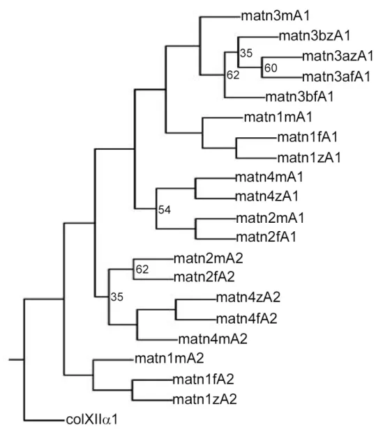
databases (Ensembl and NCBI); however, two forms of matrilin-3, matrilin-3a and -3b, are present. Phylogenetic trees show that the VWA domains of zebrafish are located in the same branches as the respective mouse domains, clearly showing that they are orthologues (Fig. 1-8). In apparent contrast to zebrafish, pufferfish (Fugu rubripes) contains a matrilin-2 gene.
The identity with the mammalian matrilins is more than 70% for the VWA domains and
only 28% for the coiled-coil domains of matrilin-3a and -3b. All zebrafish matrilins
show a greater variety of splice forms than in mammals, with splicing mainly affecting
the number of EGF-like repeats.
Fig. 1-7 Domain structure of zebrafish matrilins. matn, matrilin; P S/T –rich, proline- and threonine/serine-rich The figure is modified from (Ko et al., 2005).
Fig. 1-8 Phylogenetic analyses of matrilin VWA domains. VWA domain amino acid sequences of zebrafish (z), pufferfish (Fugu rubripes) (f) and mouse (m) were aligned with the PILEUP program of the GCG package, using the default parameters. The VWA4 domain of human collagen XII α1 (colXII α1) was taken as an outgroup. The aligned sequences were used for the construction of a tree by the PROTPARS program of the PHYLIP package, version 3.5. Bootstrap support values were obtained with 100 replicates and are given at the respective nodes when the values are below 70%. The VWA domains of pufferfish (Fugu rubripes) were derived from the draft sequence of pufferfish (Fugu rubripes) genome, available as pufferfish (Fugu rubripes) assembly release 3. The genomic sequence of the VWA1 domain of pufferfish (Fugu rubripes) matrilin-4 is not yet available. matn, matrilin. The figure is from (Ko et al., 2005).
1.4.1. Matrilin-1
The mature secreted protein has a calculated M
rof 51,555. It comprises two VWA domains that are connected by a single EGF-like domain followed by an α-helical coiled-coil oligomerization domain, and therefore completely resembles the mammalian matrilin-1 (Fig. 1-7). Furthermore, by RT-PCR an alternatively spliced mRNA that lacks the EGF-like domain was also detected (Fig. 1-7).
1.4.2. Matrilin-3a
For matrilin-3a alternatively spliced cDNAs occur that contain sequences coding for three or four EGF-like domains (Fig. 1-7). In addition, an isoform exists that lacks the four EGF-like domains that in mammals connect the VWA domain and the C-terminal coiled-coil domain (Fig. 1-7). The longest cDNA encodes a protein of 480 amino acid residues with a calculated M
rof 53,006, the shortest protein of 295 amino acid with a calculated M
rof 32,811.The stretch of amino acid residues N-terminal to the VWA domain is conserved between mammalian matrilin-3 and zebrafish matrilin-3a, but with fewer positively charged amino acid residues in zebrafish.
1.4.3. Matrilin-3b
In a later screen of the genomic database, a second matrilin-3 gene was identified.
RT-PCR yielded four alternatively spliced matrilin-3b cDNAs. The longest splice variant of matrilin-3b has an open reading frame of 1,437 bp that codes for a protein comprising 478 amino acid residues. After cleavage of a predicted signal peptide of 22 amino acid residues, the mature secreted protein has a calculated M
rof 50,136. The VWA domains of matrilin-3a and -3b are 80% identical at the amino acid level.
Uniquely in a matrilin, a proline- and threonine/serine-rich sequence (Fig. 1-9) precedes
the N-terminal VWA domain in matrilin-3b, which itself is followed by a single
EGF-like domain and the C-terminal coiled-coil domain. The long unique N-terminal
stretch of amino acid residues also contains a cluster of positively charged amino acid
residues (Fig. 1-7) similar to that in matrilin-3a. The matrilin-3b variant that lacks the proline- and threonine/serine-rich sequence and the EGF-like domain has the same domain structure as the shortest form of matrilin-3a, containing only the N-terminal positively charged stretch, a single VWA-domain and the coiled-coil domain. In addition, two isoforms exist that lack either the proline- and threonine/serine-rich sequence or the EGF-like domain (Fig. 1-7). The NetOGlyc server (http://www.cbs.dtu.dk/services/NetOGlyc/) predicted that the proline- and threonine/serine-rich sequence contains 33 potential mucin-type N-acetylgalactosamine O-glycosylation sites.
Fig. 1-9 Amino acid sequence of the proline- and serine/threonine-rich domain in zebrafish matrilin-3b.
Predicted mucin-type N-acetylgalactosamine O-glycosylation sites are shaded black.
1.4.4. Matrilin-4
As in the mammalian matrilin-4, the two VWA domains are connected by EGF-like domains followed by the C-terminal coiled-coil domain (Fig. 1-7). Alternatively spliced mRNAs exist with different numbers of EGF-like domains, ranging from four to twelve (Fig. 1-7). The longest form has a calculated M
rof 102,576 and thereby has nearly the same M
ras mammalian matrilin-2.
1.4.5. Sequence analysis
The identity of zebrafish VWA domains with their mouse counterparts is 71–72% and
the lengths of the VWA domains are strongly conserved. The matrilin-3 A1 domains
perfectly fit to the MIDAS (metal ion-dependent adhesion site) motif consensus
sequence (DXSXSXnTXnD), which is in contrast to human and mouse matrilin-3
where the threonine in the MIDAS motif has been exchanged for a serine residue.
Phylogenetic analysis did not allow construction of a tree of the zebrafish EGF-like domains with reasonable bootstrap values. Nevertheless, the zebrafish matrilin-4 EGF-like domains 7 and 8 are identical on the protein level and the domains 3, 4, 5, 6, 9, 10 and 11 are nearly identical, and are probably the products of recent duplication events. The identity of the orthologue EGF-like domains is lower than for the VWA domains with highest values of 66.7% for the EGF-like domain of zebrafish and mouse matrilin-1, 65% for the EGF-like domain 11 of zebrafish matrilin-4 and EGF-like domain 3 of mouse matrilin-4 and 55.8% for EGF-like domain 4 of zebrafish matrilin-3a and the EGF-like domains 1 and 4 of mouse matrilin-3.
All zebrafish matrilins contain a coiled-coil α-helix at the C-terminus, as predicted by the COILS program (Lupas et al., 1991). As for mouse matrilin-3, the agreement with the consensus is the lowest for zebrafish matrilin-3b, whereas matrilin-3a has a higher match. The coiled-coil domains of zebrafish and mouse matrilin-1 show an identity of 67%, whereas it is 48% for matrilin-4 and only 28% for each of matrilin-3a and -3b.
1.5. Gene silencing methods
Gene silencing by antisense oligonucleotides is increasingly used to achieve
loss-of-function or knockdown of genes of interest and forms an attractive alternative to
knockouts. Several antisense oligonucleotides with modified backbones have over the
past decade been designed to improve specificity and efficacy (Braasch and Corey,
2002). However, their ability to provide unambiguous phenotypes has been debated and,
in some instances, they have proven seriously flawed regarding specificity, cell toxicity,
efficiency and efficacy (Braasch and Corey, 2002; Summerton and Weller, 1997). The
discovery of RNAi was a breakthrough and, indeed, more than 3000 publications have
used RNAi since 2002. In addition to RNAi, morpholino antisense oligonucleotides
(short: morpholinos) have been in use since 2000 and the two techniques are considered
as the most powerful antisense approaches.
1.5.1. RNAi
RNA interference (RNAi) is a gene silencing technique in which exogenous double-stranded RNA (dsRNA) that is complimentary to known targeted mRNA is introduced into a cell and triggers the degradation of that particular mRNA, thereby diminishing or abolishing gene expression (Hannon, 2002).
The specificity component of the RNAi machinery is small-interference RNA (siRNA).
dsRNA is cleaved into ~23 bp siRNAs by dicer (Denli and Hannon, 2003), an enzyme that belongs to the RNase III family. Then the siRNA-dicer complex recruits additional components to form an RNA-Induced Silencing Complex (RISC) in which the unwound siRNA base pairs with complementary mRNA, thus guiding the RNAi machinery to the target mRNA resulting in the effective cleavage and subsequent degradation of the mRNA (Denli and Hannon, 2003; Hammond et al., 2000; Zamore et al., 2000) (Fig.
1-10).
The technique has proven effectively in Drosophila (Schwarz et al., 2002) , C. elegans (Fire et al., 1998), plants (Hamilton and Baulcombe, 1999) and, recently, in mammalian cell culture (Chiu and Rana, 2002). The usefulness of RNAi in animal experiments and preclinical drug development remains to be established (Paroo and Corey, 2004).
Even though RNAi is considered as a powerful technique to elucidate the function of a gene in respect of efficiency, efficacy and cost, the real mechanism has been rather mysterious with regard to specificity. It has been observed that two classes of siRNA (21-22nt and 24-26nt) existing in plants differ not only in size but also in their mechanism of gene-silencing (Hamilton et al., 2002). The short siRNA (21-22 nt) is correlated directly with mRNA degradation whereas the long siRNA (24-26 nt) is involved in systemic silencing and DNA methylation (Denli and Hannon, 2003;
Hamilton et al., 2002). In addition, micro RNAs (miRNAs) are also found to be
processed by dicer from 70 nt pre-miRNA into a 22 nt mature form that can regulate
gene expression either through mRNA degradation or translational repression which
depends on the degree of the complementation to mRNA sequence (Bartel, 2004; Denli
and Hannon, 2003). Nevertheless, siRNA and miRNA induced gene silencing through
mRNA degradation pathways are both mediated by the RNA-induced silencing complex (RISC) (Bartel, 2004). Therefore, it is difficult to distinguish which component accounts for the final gene silencing. The propensity for nonspecific effects is always the main concern in the antisense field. The frequent lack of proper and sufficient controls in papers describing the use of RNAi has been noticed (Anonymous, 2003).
Fig. 1-10 Present model for the RNAi mRNA degradation pathway. Anti-parallel dicer dimers cleave long dsRNAs to form small-interfering RNAs (siRNAs) in an ATP-dependent manner. siRNAs are incorporated in the RNA-Induced Silencing Complex (RISC) and ATP-dependent unwinding of siRNAs activates RISC. Active RISC is thus guided to degrade the specific target mRNAs. The figure is modified from (Denli and Hannon, 2003).
1.5.2. Morpholino antisense oligonucleotides (morpholinos)
Morpholinos are non-ionic DNA analogs comprised of a nucleic acid base, a
morpholine ring and a non-ionic phosphorodiamidate intersubunit linkage. (Fig. 1-11)
Morpholinos were first developed for clinical therapeutic applications, where previous antisense approaches had proven seriously flawed (Summerton and Weller, 1997). They were introduced into developmental biology as a tool to inhibit gene function in 2000 (Heasman et al., 2000), and since then they have been used by researchers in various model organisms, including see urchin (Howard et al., 2001), xenopus (Heasman et al., 2000), zebrafish (Nasevicius and Ekker, 2000), chicken (Kos et al., 2001) and mouse (Coonrod et al., 2001). In addition, a successful and efficient delivery of morpholinos in adherent and nonadherent cultured cells has been reported (Morcos, 2001). Moreover, an entire issue of the journal “Genesis” (volume 30, issue 3, 2001) was dedicated to articles studying gene function in development using the morpholino approach.
Fig. 1-11 Structure of DNA and morpholino oligonucleotides. The figure is from (Corey and Abrams, 2001).
In contrast to traditional antisense oligonucleotide approaches that utilize RNase H
based degradation of mRNA as a mechanism of action, morpholinos do not recruit
RNase H and thus the efficacy is achieved through nonclassical antisense approaches
(Summerton, 1999; Summerton and Weller, 1997). It has been demonstrated that
morpholinos are not subject to a wide range of nucleases (Summerton and Weller, 1997)
and that morpholinos are not degraded in the organism. As a consequence, there is no risk that modified nucleosides or nucleotides resulting from degradation of an antisense oligonucleotide might be toxic or might be incorporated into cellular genetic material and thereby lead to mutations and/or other undesired biological effects.
Moreover, the phosphorodiamidate linkage in morpholinos gives an excellent water solubility and provides a neutrally charged backbone which is less likely to interact with cellular proteins thus reducing the risk for non-specific side effect (Corey and Abrams, 2001; Ekker, 2000).
In summary, morpholinos have greater efficacy, specificity, solubility and stability than other antisense oligonucleotides (Heasman, 2002).
Morpholinos can function either through altering pre-mRNA splicing or inhibiting translation (Ekker and Larson, 2001). Binding of morpholinos to exon/intron junctions will lead to that an entire exon is left out, resulting in the formation of a non-functional mRNA (Fig. 1-12) (Ekker and Larson, 2001). These splice-blocking morpholinos have the advantage that the efficacy of the knockdown can be easily quantified using RT-PCR or standard RNA analysis techniques without the use of antibodies (Draper et al., 2001).
It was observed that targeting of the splice donor boundaries gives a better knockdown
than blocking at an splice acceptor site but the reason is unknown. Nevertheless,
morpholinos targeting splice donor sites have produced pronounced phenotypes in
zebrafish embryos (Draper et al., 2001; Yan et al., 2002).
Fig. 1-12 The use of morpholinos in altering RNA processing. (a) Cartoon of RNA splicing events for an arbitrary gene. (b) One example of morpholino induced alterations in RNA splicing, exon skipping. The figure is from (Ekker and Larson, 2001).
Fig. 1-13 The use of morpholinos (MO) for translational inhibition. (a) Cartoon of the translation of an arbitrary mRNA. The 40S ribosomal subunit scans the leader sequence, identifies the start codon, and then recruits the 60S subunit for polypeptide synthesis initiation. (b) Binding of an MO to the 5’ end of the gene inhibits the scanning process and translation. (c) Effective target selection for morpholino. The figure is from (Ekker and Larson, 2001).
Translational inhibition is the other morpholino targeting strategy. Morpholinos with a
sequence selected to target the leader sequence or nearby bases can sterically inhibit
scanning of the mRNA by the 40S ribosomal subunit and subsequently result in
translational inhibition (Fig. 1-13). Efficacy appears restricted to target sites within the
leader sequence and sequences surrounding the start codon (Fig. 1-13 C) (Ekker and
Larson, 2001; Summerton, 1999). Once translation has been initiated, morpholinos are
not capable of altering the activity of ribosome complex. Binding of morpholinos to
mRNA does not appear to facilitate or retard mRNA degradation (Nasevicius and Ekker,
2000). Hence, the efficacy of translational-inhibition morpholinos should be evaluated
at the protein level instead of the mRNA level.
1.6. The aims of the dissertation
Despite the increased biochemical information on matrilin interactions, the detailed in vivo functions are not known. It is clear that matrilins serve as adaptors in the assembly of supramolecular structures in the extracellular matrix, but it is not known if this role is static or dynamic in nature. Matrilins therefore need to be studied in genetic models.
Matrilin-3 deficient mice were generated in collaboration with Dr. A. Aszodi, Martinsried, and one aim of this dissertation was to characterize the matrilin-3 gene function in these null mutation mice.
However, the redundancy within the family has caused problems in this regard and the two single gene inactivations performed so far for matrilin-1 and -2 have not yielded any change in phenotype. These studies are at present being continued through the establishment of double knockouts. Matrilin-1/-3 double knockout mice were generated and the biochemical analysis of these mice was the second aim of this dissertation.
Since matrilins are neither found in Drosophila nor in C. elegans but are present in
vertebrates, the zebrafish was chosen as a second model organism and gene silencing by
use of morpholinos employed as an alternative way to elucidate matrilin function.
2. Materials and Methods
2.1. Characterization of matrilin-3 deficient mice
2.1.1. Genotyping by PCR
The genotype of offspring from heterozygous mice was screened by PCR. Mouse tails (0.5 cm) were digested with 1 mg/ml proteinase K in 1xPCR buffer at 55
oC for 3-5 hours till all tissues were lysed. 0.5 ul of clear supernatant, which contains genomic DNA, was obtained by centrifugation and used for the PCR reaction. DNA polymerase and 10x PCR-buffer were from the Expand High Fidelity PCR kit (Roche).
Fig. 2-1 Primer location.