AUS DEM LEHRSTUHL FÜR INNERE MEDIZIN II PROF. DR. MED. L. MAIER DER FAKULTÄT FÜR MEDIZIN DER UNIVERSITÄT REGENSBURG
Association between human papillomavirus and primary lung cancer – a systematic review and pilot study
Inaugural – Dissertation zur Erlangung des Doktorgrades
der Medizin
der
Fakultät für Medizin der Universität Regensburg
vorgelegt von Julia Karnosky
2020
„ - F r e i s e i t e - „
AUS DEM LEHRSTUHL FÜR INNERE MEDIZIN II PROF. DR. MED. L. MAIER DER FAKULTÄT FÜR MEDIZIN DER UNIVERSITÄT REGENSBURG
Association between human papillomavirus and primary lung cancer – a systematic review and pilot study
Inaugural – Dissertation zur Erlangung des Doktorgrades
der Medizin
der
Fakultät für Medizin der Universität Regensburg
vorgelegt von Julia Karnosky
2020
Dekan: Prof. Dr. Dr. Torsten E. Reichert 1. Berichterstatter: Prof. Dr. med. Christian Schulz 2. Berichterstatter: Prof. Dr. med. Matthias Evert Tag der mündlichen Prüfung: 13.07.2020
- 1 -
1. Table of Contents
1. Table of Contents ... - 1 -
2. Abbrevations ... - 3 -
3. Summary (Zusammenfassung) ... - 5 -
3.1 Fragestellung ... - 5 -
3.2 Methoden ... - 5 -
3.3 Ergebnisse ... - 7 -
3.4 Schlussfolgerung ... - 9 -
4. Introduction ... - 10 -
4.1 Epidemiology ... - 10 -
4.2 Lung Cancer ... - 11 -
4.3 Etiology ... - 13 -
4.4 Human Papillomavirus ... - 14 -
4.5 HPV and Cervical Cancer ... - 16 -
4.6 HPV and other Organ Sites ... - 17 -
5. Materials and Methods ... - 18 -
5.1. Meta-analysis ... - 18 -
5.1.1 Literature Research ... - 18 -
5.1.2 Acquisition of Publications ... - 19 -
5.1.3 Data Selection ... - 20 -
5.1.3.1 Inclusion Criteria ... - 20 -
5.1.3.2 Exclusion Criteria ... - 21 -
5.1.4 Data Extraction ... - 21 -
5.2 Experimental Part ... - 22 -
5.2.1 Biopsy Material ... - 22 -
5.2.2 HPV Detection ... - 22 -
5.3 Statistical Analysis ... - 23 -
- 2 -
6. Results ... - 24 -
6.1 Meta-analysis ... - 24 -
6.1.1 Databank Research ... - 24 -
6.1.2 General Literature Research ... - 24 -
6.1.3 Overview of the Literature ... - 26 -
6.1.4 Patients Characteristics ... - 27 -
6.1.5 Continents ... - 29 -
6.1.5.1 Europe ... - 29 -
6.1.5.2 Asia ... - 34 -
6.1.5.3 The Americas ... - 39 -
6.1.5.4 Continents compared ... - 43 -
6.1.6 Squamous Cell Carcinoma vs. Adenocarcinomas ... - 44 -
6.1.7 Cases vs. Controls ... - 45 -
6.1.8 FFPE vs. fresh frozen ... - 46 -
6.1.9 1990s vs 21st century ... - 47 -
6.2 Experimental Part ... - 48 -
6.2.1 Patient Characteristics ... - 48 -
6.2.2 HPV Detection ... - 50 -
7. Discussion ... - 51 -
8. Conclusion ... - 68 -
9. Table of illustrations ... - 71 -
9.1. Pictures ... - 71 -
9.2 Tables ... - 72 -
10. Appendix ... - 73 -
11. References ... - 89 -
- 3 -
2. Abbreviations
- AC = adenocarcinoma
- ALK = anaplastic lymphoma kinase
- BRAF = v-Raf murine sarcoma viral oncogene homolog B1 - CDKN2A = TP53 and cyclin-dependent kinase inhibitor 2A - CIN = cervical intraepithelial neoplasia
- COPD = chronic obstructive pulmonary disease - DDR2 = discoidin domain receptor 2
- E = early region of HPV genome - EBC = exhaled breath condensate - EBUS = endobronchial ultrasound
- EGFR = epidermal growth factor receptor
- EML4-ALK = echinoderm microtubule associated protein-like protein 4 fused with Anaplastic Lymphoma Kinase
- FFPE = formalin-fixed paraffin-embedded - FGR1 = fibroblast growth factor receptor 1
- HNSCC = head and neck squamous cell carcinoma - HCC = hepatocellular carcinoma
- HPV = Human Papillomavirus - ISH = In situ hybridization
- KRAS = Kirsten rat sarcoma viral oncogene homolog - L = long region of HPV genome
- LC = lung cancer
- LCR = long control region of HPV genome - MET = mesenchymal-epithelial transition factor
- MOOSE = Meta-analysis of Observational Studies in Epidemiology - NSCLC = non-small cell lung cancer
- OS = overall survival
- PIK3CA = phosphoinositide-3-kinase catalytic subunit alpha isoform - PTEN = phosphatase and tensin homolog
- PCR = Polymerase chain reaction
- PRISMA = Preferred Reporting Items for Systematic Reviews and Meta-Analysis
- 4 -
- PROSPERO = International prospective register of systematic reviews - RET = rearranged during transfection
- ROS1 = c-ros oncogene 1 - SCLC = small cell lung cancer - SCC = squamous cell carcinoma - SOX2 = SRY related HMG box gene 2 - TP53 = tumor suppressor protein 53
- 5 -
3. Summary (Zusammenfassung)
3.1 Fragestellung
Im Jahr 1979 haben Syrjänen et al. erstmals über einen möglichen Zusammenhang zwischen einer HPV Infektion und der Entwicklung von Bronchialkarzinomen berichtet. Im weiteren Verlauf wurden zahlreiche Studien mit unterschiedlichsten Detektionsmethoden durchgeführt.
In diesen zeigten sich sowohl geographische Unterschiede, als auch Unterschiede, welche auf die verwendete Analysemethode zurückgeführt wurden.
Ziel dieser Arbeit ist es, zum einen den aktuellen Stand in der Literatur bezüglich HPV Detektion mittels PCR in Lungenkrebsgewebe zu erfassen und zum anderen, dies mit den Ergebnissen unserer eigenen Kohorte zu vergleichen.
3.2 Methoden
Mittels einer strukturierten Literatursuche wurde zunächst versucht alle Publikationen zu erfassen, welche sich dem beschriebenen Thema widmen. Eine Einschränkung bezüglich des Publikationszeitraums oder der Sprache, in welcher die Publikation verfasst wurde, erfolgte nicht. Die erfassten Studien wurden anhand ihres Titels und, soweit verfügbar, Abstracts von zwei unabhängigen Personen begutachtet. Bei Uneinigkeit wurde die Entscheidung durch eine dritte Person getroffen. Anschließend wurde von allen eingeschlossenen Publikationen die Volltextversion beschafft und hinsichtlich der Ein- und Ausschlusskriterien geprüft.
Von allen eingeschlossenen Studien wurden die Fallzahlen sowie die Anzahl der HPV positiven und negativen Fälle und Kontrollen erfasst. Außerdem wurden allgemeine bibliographische Daten sowie Details bezüglich der analysierten Fälle dokumentiert und ausgewertet.
Aus der Gewebebank des Instituts für Pathologie der Universität Regensburg wurden 16 Fälle von Plattenepithelkarzinomen der Lunge, welche im Zeitraum von Mai 2017 bis März 2018 in der Klinik für Thoraxchirurgie operativ reseziert wurden, herausgesucht. Von 14 Patienten war ausreichend Gewebe vorhanden, sodass eine nested PCR nach der Methode von Sotlar et al. (1) durchgeführt werden konnte. Von zwei Patienten standen zusätzlich separate bioptische
- 6 -
Gewebeproben zur Verfügung, welche beide analysiert und in die Gesamtuntersuchung einbezogen wurden.
Die statistische Auswertung wurde mittels Microsoft Excel (2013) durchgeführt. Die Anzahl der HPV positiven und negativen Fälle und Kontrollen wurde aus den selektierten Arbeiten extrahiert und nachfolgend die HPV Prävalenzen anhand der einzelnen Patientendaten berechnet. Die Berechnung von Mittelwerten und Standardabweichungen erfolgte ebenfalls mittels Excel 2013. Die statistische Signifikanz der HPV Prävalenzunterschiede wurde mittels Chi-Quadrat-Test berechnet. Ein p-Wert < 0.05 wurde als statistisch signifikant eingestuft.
- 7 -
3.3 Ergebnisse
In der durchgeführten Literatursuche im Mai 2018 wurden 3884 Publikationen mit einem möglichen Themenbezug gefunden. Nach Deduplikation wurden 2624 Publikationen zum weiteren Screening mittels Titel und Abstract in die Literaturverwaltungssoftware Covidence eingeschlossen. Es wurden 340 Publikationen in die Volltextanalyse eingeschlossen. Nach Anwendung der Ein- und Ausschlusskriterien wurden 73 Veröffentlichungen in diesen Systematic Review eingeschlossen.
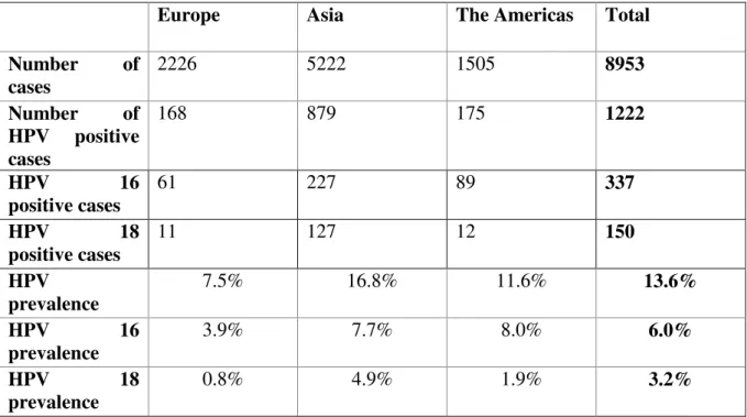
Die eingeschlossenen 73 Studien enthalten 8953 Lungenkrebspatienten und 754 Kontrollpatienten. Die HPV Prävalenz lag bei 13.6 % für alle HPV Arten kombiniert. Die HPV 16 Prävalenz war höher als die HPV 18 Prävalenz (6.0 % gegenüber 3.2 %, p<0.01).
Studien aus Europa, Asien und Amerika erfüllten die Einschlusskriterien. Auf allen Kontinenten war die HPV 16 Prävalenz signifikant höher als die HPV 18 Prävalenz (p<0.01).
Beim Vergleich der einzelnen Kontinente untereinander zeigten sich statisch signifikante Unterschiede (p<0.01) mit der geringsten HPV-Prävalenz in Europa (7.5%) gegenüber 16.6%
in Asien und 11.6% in Amerika. Die HPV Prävalenz in Plattenepithelkarzinomen war höher als in Adenokarzinomen (18.6 % gegenüber 9.6 %). Dieser Unterschied war statistisch hochsignifikant (p<0.01). Die höchste HPV Prävalenz zeigte sich in Plattenepithelkarzinomen bei asiatischen Patienten (29.5 %).
Um eine zufällige HPV Detektion einschätzen zu können, wurden die eingeschlossenen Fall- Kontroll-Studien gesondert analysiert. In den 15 eingeschlossenen Fall-Kontroll-Studien wurden 1750 Lungenkrebspatienten und 754 gesunde Kontrollen analysiert. Es wurden nur Kontrollen in diese Subgruppenanalyse eingeschlossen, von welchen Lungenbiopsien analysiert wurden. Die HPV Prävalenz in den Lungenkrebsproben war statistisch signifikant höher als in der Proben der Kontrollgruppe (31.3 % gegenüber 5.4 %, p<0.01).
Um Unterschiede in der Prävalenz zu minimieren, die durch mögliche Einflussfaktoren zustande kommen können, wurden zum einen nur Veröffentlichungen eingeschlossen, welche PCR als Nachweismethode nutzen und zum anderen weitere Subgruppenanalysen durchgeführt.
Um einen möglichen Einfluss der Methode, mit welcher das Tumorgewebe behandelt wurde, zu untersuchen, wurden die Prävalenzen in den Studien, welche Formalin-fixierte und in Paraffin eingebettete Proben verwendeten, mit jenen verglichen, welche frische, gefrorene
- 8 -
Proben enthielten. Hierbei ergab sich kein statistisch signifikanter Unterschied der HPV Prävalenz (13.4 % gegenüber 13.5 %; p=0.9).
Da es in den letzten 30 Jahren zu großen Verbesserungen der PCR Methodik gekommen ist, wurden die Studien des 21. Jahrhunderts mit den in den 1990er Jahren durchgeführten Untersuchungen verglichen. Hier zeigte sich ein statistisch hoch signifikanter Unterschied zwischen den beiden Untersuchungszeiträumen (p<0.01) mit einer HPV-Prävalenz von 37.9%
in den Studien aus den 90er Jahren gegenüber einer HPV-Prävalenz von 8.5 % in den Studien, welche im 21. Jahrhundert durchgeführt wurden
Das Durchschnittsalter der 14 Patienten, welche in Zusammenarbeit mit dem Institut für Pathologie der Universität Regensburg untersucht wurden, lag bei 69.6 Jahren. Acht Patienten waren männlich und alle Patienten waren aktuelle oder frühere Raucher. (Für einen Patienten war keine Raucheranamnese verfügbar.) Bei allen Patienten wurden primäre Plattenepithelkarzinome der Lunge diagnostiziert. Die mittels nested-PCR bestimmte HPV Prävalenz war 0 %.
- 9 -
3.4 Schlussfolgerung
Obwohl in den letzten Jahrzehnten große Fortschritte in der Lungenkrebstherapie gemacht wurden, ist Lungenkrebs immer noch mit einer sehr schlechten Prognose vergesellschaftet.
Die Identifikation von vermeidbaren Risikofaktoren ist daher von besonderer Bedeutung, insbesondere bei Patienten ohne Raucheranamnese.
In vielen Studien konnte HPV DNA in Lungenkrebsgewebe nachgewiesen werden.
Allerdings konnte bisher kein eindeutiger kausaler Zusammenhang belegt werden.
Prävalenzunterschiede in Fall-Kontroll Studien deuten jedoch auf einen potentiellen Zusammenhang hin. Um dies kausal belegen zu können, sind jedoch weitere Studien notwendig, welche ergänzend die Expression der HPV Onkogene und eine mögliche Integration von HPV in das Hostgenom untersuchen.
Geographische Unterschiede der HPV Prävalenz wurden festgestellt mit einer höheren HPV Prävalenz in Asien gegenüber Europa und Nordamerika. Allerdings gibt es bisher noch keine Erklärungsansätze für diese Beobachtung.
Der Infektionsweg von HPV im Zusammenhang mit Lungenkrebs ist bisher ungeklärt. Nach aktuellem Stand der Forschung verursacht HPV keine Virämie, weshalb eine Blutstrominfektion unwahrscheinlich erscheint. Eine direkte Infektion wie beim Zervixkarzinom erscheint aber aufgrund der anatomischen Gegebenheiten ebenfalls unwahrscheinlich.
Zusammenfassend werden weitere Studien durchgeführt werden müssen, welche insbesondere versuchen, einen kausalen Zusammenhang zwischen HPV-Infektion und Lungenkrebs nachzuweisen und einige der obigen Fragen zu beantworten.
- 10 -
4. Introduction
4.1 Epidemiology
Lung cancer is estimated to be the leading cause of cancer related mortality worldwide with 2.1 million new lung cancer cases and 1.8 million predicted deaths worldwide. (1) This represents almost one in five cancer deaths worldwide in 2018 (18.4%).
This is the case not only in smokers. Even in never-smokers lung cancer is the most common cause of cancer death worldwide.
According to the GLOBOCAN 2018 report there will be an estimated 18.1 million new cancer cases and 9.6 million cancer deaths in 2018 worldwide. In this statistic all cancer subtypes except nonmelanoma skin cancer are included. In both sexes combined lung cancer is the cancer type most commonly diagnosed and the leading cause of cancer mortality. Lung cancer is the second most frequent type of cancer by incidence and the leading cause of cancer mortality in males. In females breast cancer is the most frequent type of cancer and the leading cause of mortality followed for incidence by colorectal and lung cancer. Concerning cancer mortality lung cancer is the second and colorectal cancer the third most common cause.
The most commonly diagnosed types of cancer underlie geographic variation depending on ethnicity, life style factors, virus infections and other still unknown factors.
Western Europe is one of the regions worldwide where cancer is the number one reason of premature death according to the WHO.
The incidence rate for lung cancer in Western Europe in 2018 is estimated to be 43.3 per 100.000 in males and 25.7 per 100.000 in females. The lung cancer incidence in males has been stable due to a lowering in smoking prevalence. But the lung cancer incidence in females is rising in most parts of the world. In some countries, like in the USA for example, the lung cancer incidence is now higher in young women then in young men. (2) This is mainly attributed to changes in smoking behavior.
In 2018 a group of Swiss and Canadian scientists estimated the lifetime and 10 year risk of developing lung cancer in a Swiss population between 1995 and 2003. (3) During that time the estimated lifetime risk decreased in men while it increased in women (7.1% to 6.9% and
- 11 -
2.5% to 4.1% respectively). The collected Data also showed that current and former smokers have an increased risk of developing lung cancer when compared to never smokers.
4.2 Lung Cancer
Lung cancer was historically divided into two major groups according to morphology: small cell lung cancer (SCLC) and non-small cell lung cancer (NSCLC). NSCLC can be further divided into adeno-, squamous cell carcinoma, large cell carcinoma and more rare subtypes.
Over time the distinction between in squamous cell carcinoma and adenocarcinoma has been shown to be important as they show differences in response to specific therapies. (4) In the last decade lung cancer is increasingly classified by its genetic alterations and oncogenic driver mutations.
Even though imaging techniques play an important role to determine the tumor stadium, in contrast to hepatocellular carcinoma (HCC) diagnosis of lung cancer to this day has to be verified by histology.
Approximately 30% of all non-small cell lung cancers are classified as squamous cell carcinomas (SCC). (5) SCC is defined as a malignant epithelial tumor expressing squamous differentiation in the form of keratinization or bridging. It can be further divided by the degree of keratinization into poorly, mildly or highly differentiated SCC. Smokers have a 10-20 fold higher risk of SCC of the lung than non-smokers.
The most commonly altered genes in SCC are: FGR1 (fibroblast growth factor receptor 1), PIK3CA (phosphoinositide-3-kinase catalytic subunit alpha isoform), DDR2 (discoidin domain receptor 2), MET, SOX2 (SRY related HMG box gene 2), PTEN (phosphatase and tensin homolog) and CDKN2A (TP53 and cyclin-dependent kinase inhibitor 2A). Some of these have therapeutic consequences already while others are still subject of ongoing research.
Adenocarcinomas (AC) represent approximately 50% of all NSCLC and approximately 38%
of all lung cancers in the Western world. (6) Distribution of histological subtypes does underlie some geographic differences. Anatomically they are more often located in the periphery of the lung. Usually diagnosis is achieved by light microscopy where glandular morphology can be seen. This morphology can be further divided into acinar, leptic, papillary or solid with mucin patterns. Further evaluation of AC is done by immunohistochemistry
- 12 -
using adenocarcinoma marker. Apart from that adenocarcinomas are tested for oncogenic mutations which allow for targeted therapies.
The most commonly tested for altered genes in adenocarcinomas are:
- EGFR (epidermal growth factor receptor)
- EML4-ALK (echinoderm microtubule associated protein-like protein 4 fused with anaplastic lymphoma kinase)
- KRAS (Kirsten rat sarcoma viral oncogene homolog) - MET (mesenchymal-epithelial transition factor) - ROS1 (c-ros oncogene 1)
- RET (rearranged during transfection)
- BRAF (v-Raf murine sarcoma viral oncogene homolog B1) - TP53 (tumor suppressor protein 53)
Some of these gene alterations are aims for targeted therapies (e.g. EGFR = Gefitinib, Erlotinib; ALK = crizotinib, alectinib) and are therefore routinely tested for in newly diagnosed lung cancers.
The symptoms of lung cancer are unspecific such as coughing or hemoptysis. In many cases B-symptomatic is the first sign of a malignant disease which usually only occurs in a more advanced tumor stage. This and the lack of a reliable screening method or tumor marker is the reason why lung cancer is most often diagnosed in an advanced tumor stage.
In a British study using the data from the National Cancer Registration the results showed that with an increased tumor stage at diagnosis the one year survival decreased. (7) While in some tumor entities a major decrease in survival rates is associated with stage 4 cancer (e.g. breast, colorectal cancer) in lung cancer a decrease in survival rates can be seen in every tumor stage.
To this day lung cancer prognosis can best be determined according to the TNM stage at primary diagnosis. (6) According to the International Association for the Staging of Lung Cancer (IASLC) the current 5 year survival rate based on TNM classification decreased from 73% in stage IA cancer to 13% in stage IV.
Outcome in patients with SCLC is even worse with a 2 year survival rate of only 4.6%. (8)
- 13 -
4.3 Etiology
Tobacco smoking is still the main cause of lung cancer worldwide. (9) Usually the amount of smoking patients have done in their life is related to the amount of tobacco consumed over time (“pack-years”). (10) Even though there are lung cancer cases in never-smokers there is a strong correlation between smoking and lung cancer incidence, most prevalent in small cell lung cancer and squamous cell carcinoma. On the other hand adenocarcinoma is the most common lung cancer subtype in never-smokers. (4)
Apart from smoking different risk factors have been suggested especially in never-smokers such as environmental tobacco smoke, air pollution, cooking oil fumes. (11) Different pulmonary diseases have been identified that are associated with a higher risk for lung cancer e.g. former infection with tuberculosis, COPD, emphysema. (12) Up to this point the causal association between these diseases and the development of lung cancer has not been fully established. On the other hand there are risk factors specific to certain occupations (e.g. coal miners) or certain geographic regions (e.g. areas with domestic radon exposure).
A growing number of lung cancer incidence takes place in never-smokers. This group makes up between 15 and 25% of the lung cancer patients and accounts for up to 300.000 deaths every year. (13) If lung cancer in never smokers would be accounted for as an original entity among cancer types it would still be the seventh leading cause of mortality among solid tumors. Lung cancer in never-smokers is more frequent in women and there are distinct oncogenic mutational patterns. ALK and EGFR activating mutations are frequent and with consequences both for therapy and prognosis. Today’s data on chemotherapy response in smokers as compared to never-smokers is not conclusive but an area of ongoing research.
That is why some scientists suggest to treat lung cancer in never-smokers as an independent tumor entity.
In recent years cancer prevention has been a topic of increased interest in scientific research.
It is undisputable that smoking cessation is the most important factor in prevention of lung cancer development. Smoking cessation reduces the risk for all subtypes of lung cancer, especially for SCC and SCLC.
To this day no reliable tumor marker for lung cancer screening has been established. In 2011 a large multi-center study on lung cancer screening in high risk patients was conducted by The National Lung Screening Trial Research Team in the United States. (14) From 2002
- 14 -
through 2004 approximately 53.000 patients in 33 US medical centers were included and randomly assigned into a group that was screened with low dose CT-scan or conventional chest X-ray. Results have shown that low dose CT scan was able to reduce lung cancer mortality in a high risk population. No lung cancer screening programs for high risk patients or for the general public have yet been established.
4.4 Human Papillomavirus
Since the first identification of human papillomavirus more than 200 different subtypes have been identified. They are classified into high-risk HPV types (16,18,31,33,39,45,51,52,58) and low-risk HPV types (6,11,42,43,44). (15) In some other publications a differentiation between high-, intermediate- and low-risk HPV types is made.
Human papillomaviruses are small non-enveloped viruses which belong to the Papillomaviridae family. They are between 50 and 60 nanometer in diameter and contain one circular, double-stranded DNA genome consisting of approximately 7000 to 8000 bp. The genome is divided into three regions: the long control region (LCR), the early region (E) and the late region (L). (16)
The long control region controls viral gene expression and replication while the late region encodes structural proteins. The early region encodes genes needed for viral gene expression, replication and survival. The names of the different regions refer to the phase in the viral life cycle in which they are expressed.
Those main regions can be further divided by the function of their gene products. According to today’s research the early region encodes for six different proteins. Of those E5, E6 and E7 are the three oncogenes encoded by the virus. E5 is involved in disturbing growth-factor signaling pathways and avoiding the host immune system. E6 and E7 work synergistically to provide an environment that allows the virus to replicate. E6 of high-risk HPV-types also targets the tumor-suppressor protein p53 and inactivates it. E7 as the third encoded oncoprotein targets cell-cycle regulating proteins such as retinoblastoma protein (Rb).
As for the other early proteins: E1 possesses DNA helicase activity and is thereby the only enzyme encoded by a HPV virus, E2 controls the expression of the other proteins and it
- 15 -
recruits E1 to the viral origin. It thereby increases viral DNA replication. As a third function it plays a critical role in transferring the viral genome during division of the host cells. The function of E4 is still unknown. (17)
There are two late genes (L1 and L2) and one long control region (LCR) which regulate the expression of the early genes. L1 and L2 are the major and minor components of the viral capsid. The L1 region can form virus-like particles (VLPs) which are the basis for prophylactic vaccination.
Many different regions of the HPV genome can be detected by PCR: some, for example the L1 regions, can be used as consensus primers to detect many different types of HPV and some like the E6 and E7 as type specific primers (e.g. E6 HPV16 and E7 HPV18).
HPVs are epitheliotropic and their life cycle takes place in squamous epithelia. According to current knowledge HPV enters through microtrauma in the epithelia and reaches the basal cell which is believed to be the target cell for HPC infection. The infection method of HPV is not yet fully understood. After initial infection there seems to be an unproductive state in which only the early viral oncogenes are expressed. One of the specific features of HPV infection is its ability to persist over years. To achieve this the viral genome has to be preserved over multiple cell division. The method by which this is achieved is still unknown. When one of the daughter cells starts to differentiate the virus starts its productive stage. The virus redirects the host cells DNA replicative system and amplification and expression of late viral genes starts. Those are necessary to subsequently produce progeny virus and release them.
Almost all studies on HPV are done on cervical cancer cells. It has not yet been established whether all of the before mentioned stages and means of infection are also valid in other infection sites.
The transmission route of HPV into lung tissue has not been explained. One of the possible infection routes is oral sexual activity as a possible pathway to lung cancer cells. Another possible route that has been discussed is hematogenous. Neither of that could be proven by now. (18)
Some studies have tried to detect HPV DNA in blood cells. But even though HPV 16/18 prevalence has been shown to exist in some studies (19), these results are still very controversial because to this day it is believed that HPV infection does not cause viremia.
- 16 -
4.5 HPV and Cervical Cancer
In 1979 Kari J. Syrjänen first published a case report describing histological changes in bronchial epithelium resembling a squamous cell carcinoma of the lung similar to changes of condylomatous nature in the genital tract. (20) Even though an association of HPV with carcinogenesis was not established at that time it was proposed to observe such lesions closely until proven otherwise.
The Nobel Prize in Physiology or Medicine 2008 was awarded to Harald zur Hausen for establishing the connection between HPV infection and cervical cancer. He first drew attention to a possible association between HPV infection and carcinogenesis in 1976. (21) In the 1960s there were first reports about double stranded DNA of the human papillomavirus which in the 1970s was further distinguished to be different types of HPV. (16) But it wasn’t until 1974 when the human wart virus was isolated from plantar warts by zur Hausen et al.
and used for hybridization of cutaneous, genital warts and cervical cancer. (HPV was still referred to as Human Wart Virus then).While high positive rates were found in cutaneous warts, no positive signal was seen in cervical cancer specimens. (22) The authors concluded that more research had to be done on the possible existence of different types of HPV virus.
This hypothesis was later proven in different studies. (23)
The first experiments to establish a relationship between HPV and cervical cancer were initiated in 1972 by zur Hausen in Heidelberg. (24) In 1976 Meisels and Fortin first suggested that there might be an association between koilocytotic cells that were found in cervical cancer and HPV infection. (25)
In 1982 the first three publications on HPV DNA sequences in human tumors were published.
They all referred to cervical cancer.
In the following years research about the association of HPV and cervical cancer increased proving for example the interaction of E6 with p53 and its resulting protein degradation. (26) Today the carcinogenetic potential of HPV is well established and it is known that more than 95% of cervical cancer biopsies contain high-risk HPV subtypes.
Three different types of HPV vaccination are currently available (e.g. Gardasil, Gardasil 9, Cervarix). (27) Since 2015 the WHO recommends a two dose program. And vaccination with Gardasil has been proven to be effective to reduce the incidence of HPV 16/18 infection. It
- 17 -
has not yet been fully established whether vaccination is also effective in cervical intraepithelial neoplasia (CIN) or adenocarcinoma in situ.
4.6 HPV and other Organ Sites
Since the oncogenic potential of HPV has been established there has been a lot of research on a possible association of HPV with cancers of other organ sites.
Shortly after HPV DNA was detected in cervical cancer some of the same as well as new HPV subtypes were detected in other anogenital cancers. But since many of the risk factors of cervical cancer are the same as for other anogenital cancers a causal relationship between HPV infection and those kinds of cancer is still subject to ongoing research. (28) Even though HPV DNA has been detected in vaginal, penil and anal cancer.
Recurrent respiratory papillomatosis is a rare disease where benign tumors (papilloma) form a long the aerodigestive tract. Although these papillomas usually cause a mechanical problem (e.g. obstruction of the airways), some cases of transformation of these papillomas into squamous cell carcinomas have been described. (29) Today it is well established that recurrent respiratory papillomatosis is associated with HPV infection.
So one of the organ sites that have been examined early are tumors of the upper aerodigestive tract and head and neck cancer. (30)
In recent years research was done on most other organ sites. There are still many questions concerning HPV infection: e.g. how long and where do transient infections take place, does HPV cause viremia, does HPV present an individual risk factor in other organ sites like it does in cervical cancer or does it present as an co-carcinogen and does every high-risk HPV infection impose a risk of developing cancer in every organ site.
HPV DNA has not only been detected in neoplastic tissue, but also in normal tissue in several organ sites. Its meaning has yet to be determined.
Since the first suggestion of a possible association between HPV and lung cancer numerous studies have been executed all over the world. Those include not only different types of detection but also different sampling methods. In this meta-analysis and the included analyzed tumor tissue we took a closer look at HPV detection by PCR in bronchoscopic biopsy specimens or resected lung tissue in primary squamous cell carcinoma of the lung.
- 18 -
5. Materials and Methods
5.1. Meta-analysis
The intention behind conduction of this systematic review was to get an idea of the prevalence of HPV infection in lung cancer both internationally and on the different continents as basis for further research and to detect the possibility of an association in our own patient collective.
Reporting of this meta-analysis is done according to the recommendation of Stroup et.al (31) for reporting observational studies published in 2000. They held a workshop 1997 in Atlanta with twenty-seven participants who were selected by a committee based on their clinical, statistical and other expertise. A systematic review of the published literature on reporting meta-analysis of observational studies was conducted. Based on the workshop results a checklist of recommendations on what to report was developed. These are referred to as
“MOOSE” (Meta-analysis of Observational Studies in Epidemiology)-criteria.
5.1.1 Literature Research
Before starting the literature research we registered the concept for our meta-analysis on the international prospective register of systematic reviews (PROSPERO) in March 2018. At that point of time no other systematic reviews on the association of HPV infection and lung cancer were registered.
In cooperation with Dr. Helge Knüttel a librarian at the medical library of the University Clinic Regensburg we searched the digital databases EMBASE (via Ovid, 1974–present), MEDLINE (via Ovid, 1946–present), Cochrane Library (Cochrane Database of Systematic Reviews, Database of Abstracts of Reviews of Effect, Cochrane Central Register of Controlled Trials, Health Technology Assessment Database, NHS Economic Evaluation Database; from inception to present) and Science Citation Index Expanded (Web of Science, 1965–present) as well as the search engine Google Scholar (no date limit).
- 19 -
Using the default sort order by relevance only the first 200 records from Google Scholar were assessed. Records from Google Scholar were downloaded with Anne-Wil Harzing’s “Publish or Perish” program (https://harzing.com/resources/publish-or-perish).
In addition to the bibliographic databases we searched the following registers of clinical trials for completed studies: WHO's International Clinical Trials Registry Platform, ClinicalTrials.gov, EU Clinical Trials Register and the German Clinical Trials Register.
We employed highly sensitive search strategies in order to identify all possibly relevant studies. We searched for the concepts “lung cancer” AND “HPV”. Controlled terms from the databases’ thesauri and a broad range of synonyms were used. Within each concept search terms were combined using the Boolean operator OR. No limits such as for study type, publication type, publication date or language were applied. We adjusted the search strategy according to the databases/search engines.
A draft search strategy for EMBASE (Ovid syntax) was published and is available from:
http://doi.org/10.5283/epub.36830.
We screened the reference lists of included studies and of relevant systematic reviews for additional studies.
Records from searching were uploaded to a reference management software for deduplication.
Screening and eligibility assessment was done using the COVIDENCE online program for managing systematic reviews.
5.1.2 Acquisition of Publications
The full texts of the publications identified by the literature research were found via internet research or if the full text was not available online, by borrowing paper copies from our own library or by contacting other libraries through different services offered by our medical library (“Fernleihe”, “Subito”).
The full texts were obtained in the original language. If that was neither German nor English a translation was made using an online translation program and checked with a college or translator fluent in that particular language.
- 20 - 5.1.3 Data Selection
After deduplication the titles and abstracts of the remaining publications were analyzed by two independent reviewers (Dr. Franziska Koll, Julia Karnosky) for relevance and matching inclusion criteria. If there was no concordance between the two reviewers the final decision was made by an independent third reviewer (Prof. Christian Schulz). Those publications where no abstract was available were included into the full text review as well.
All studies that were recognized as possibly eligible for inclusion were analyzed using EndNote Citation Software (Version X9).
For all the included full texts the same analysis system with the same reviewers was used.
Analysis of the publications was done according to the following inclusion and exclusion criteria:
5.1.3.1 Inclusion Criteria
All studies reporting HPV prevalence in primary lung cancer cases in adults were included.
Case reports were excluded. As detection method only PCR from fresh frozen and/or paraffin- embedded tissue were included. HPV detection in blood samples or any other means of detection was excluded. All types of tissue sampling method were included. HPV detection in archival tumor tissue was included as well.
No exclusion was done depending on whether HPV low-risk or high-risk were analyzed.
Only studies that provide data specific to HPV prevalence in lung cancer tissue were included.
No exclusions were made based on language. We included journal articles as well as abstracts and conference reports if they met the inclusion criteria. If an abstract and a journal article on the same patient population was available the journal article was included. Journal articles that reported about not only cases of HPV detection in primary lung cancer but e.g. in head and neck cancer as well, were included but only the data of the primary lung cancer group were extracted.
- 21 - 5.1.3.2 Exclusion Criteria
Exclusion Criteria were as following:
- Case Reports - Reviews
- HPV detection in minors
- Any data on HPV prevalence that was done by a detection method other than PCR - HPV detection in blood samples or any other biomaterial then tumor tissue
- HPV infection in non-primary lung cancer or metastatic disease of a different primary origin
- Publications for which neither abstract nor full text were available online or by borrowing a paper copy through a library
The excluded full text publications were documented stating the reason for exclusion.
5.1.4 Data Extraction
For all included studies title, first author, year of publication, country of research, journal in which the study was published and language of publication were collected. Additionally the study size, the detection method for HPV, the tissue sampling and processing method and the types of HPV detected were collected to be able to compare the overall prevalence of HPV infection in lung cancer to our own experiments. Not only the calculated HPV prevalence in the included studies but also the case numbers as well as the number of cases detected to be positive and negative were collected.
Data for a subgroup analysis for Squamous Cell Carcinoma and HPV types 16 and 18 were gathered as well.
In order to evaluate other possible factors influencing the results a subgroup analysis was done on basis of the used tissue processing method.
To exclude that any detected difference is influenced by improvement in PCR technology the studies were stratified by the time in which they were done and analyzed separately in another subgroup analysis.
- 22 -
5.2 Experimental Part
5.2.1 Biopsy Material
All the included materials were obtained from the tissue bank at the department of pathology at the University of Regensburg. Initially a list of 16 patients with primary SCC of the lung diagnosed in 2017 or 2018 was created. After obtaining the biopsy specimens it was determined that enough tissue for PCR was still available for 14 patients. In addition two separate biopsy specimens were available for two patients. Both were included in the final analysis. From all the included patients paraffin-embedded tissue from resected lung cancer was available. All of them were operated on by the Department of Thoracic Surgery at the University Clinic Regensburg between May 2017 and March 2018.
5.2.2 HPV Detection
HPV detection was done according to the method reported by Sotlar et al. in 2004. (32) They evaluated a nested multiplex PCR assay. It incorporates consensus primers and type-specific primers. The viral E6/E7 oncogenes are used as primer targets. In the first round of amplification a broad spectrum of HPV subtypes is analyzed. 18 different HPV subtypes including all known high-risk HPV types are included. They were combined with type- specific primers used for nested PCR in following rounds of amplification. This novel PCR assay was initially tested on cervical scrapes. In their study the detected sensitivity of this new multiplex PCR was similar to the sensitivity of the nested PCRs done with consensus primers MY09/MY11 and GP5+/GP6+ combined. The sensitivity was higher than in a conventional PCR with either of the two. The advantage of this way of HPV detection is that it allows for exact HPV typing and detection of multiple HPV types at once.
All experimental analyses were carried out in cooperation with the Department of Pathology of the University Hospital Regensburg.The in this institute already established methodology of HPV-analytics was employed to the subject of this study. The evaluation of the multiplex- PCR-data was performed under the supervision and in cooperation with Prof. Dr. Wolfgang Dietmaier, chief of molecular pathological diagnostics in the Department of Pathology.
- 23 -
5.3 Statistical Analysis
All publications that matched the inclusion criteria were added to an Excel chart. Data was analyzed using Microsoft Excel (2013). The total number of cases as well as the number of positive and negative HPV detection were collected and the overall HPV prevalence was calculated. All tests for statistical significance were done using the Chi-squared-test. A p- value < 0.5 was determined to be statistically significant. Afterwards all the calculation were repeated using SPSS Version 22.
Other information e.g. on smoking behavior, gender etc. was collected from the individual publication. Mean value of age as well as standard deviation were calculated by means of Excel 2013.
If the information was not available in all of the included studies its information was collected for the studies that provide it and the number of studies which do so is acknowledged.
- 24 -
6. Results
6.1 Meta-analysis
6.1.1 Databank Research
An overview of all analyzed studies is documented in the PRISMA flowchart (Appendix Figure 3: Prisma flowchart).
6.1.2 General Literature Research
In the literature research done in May 2018 3884 publications with a possible connection to the research question were found in the following databases respectively:
• Medline (n=705),
• Embase (n=1376),
• Cochrane (n=12),
• Science Citation Index Expanded (n=1508),
• Google Scholar (n=200),
• WHO's International Clinical Trials Registry Platform (n=5),
• ClinicalTrials.gov (n=28)
• EU Clinical Trials Register (n=10).
After deduplication the remaining 2624 publications were imported into Covidence (management software for systematic reviews run by the Cochrane library).
They were evaluated by both reviewers on relevance for the research question. 2268 of the screened titles and abstracts did not relate to the current research and were excluded. For 16 publications no abstract was available, so it was not possible to evaluate whether the publication might be relevant. The decision was made to include these 16 publications into the full text review as well. In summary 340 publications were entered into the full text review.
- 25 -
The full text version of three of these publications was not available neither online nor by borrowing the paper version from a library (33-35).
The remaining 337 full text versions were screened by both investigators. After applying the inclusion and exclusion criteria 73 publications were included in this systematic review.
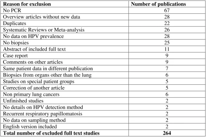
Reason for exclusion Number of publications
No PCR 67
Overview articles without new data 28
Duplicates 22
Systematic Reviews or Meta-analysis 26
No data on HPV prevalence 28
No biopsies 25
Abstract of included full text 11
Case report 9
Comments on other articles 9
Same patient data in different publication 7
Biopsies from organs other than the lung 6
Studies on special patient groups 5
Correction of another article 5
Non primary lung cancers 6
Unfinished studies 2
No details on HPV detection method 2
Recurrent respiratory papillomatosis 2
No data on sampling method 2
English version included 2
Total number of excluded full text studies 264
Table 1: Excluded full text studies
The most common reason for exclusion was that no PCR was done to detect HPV. This was the case in 67 publications. In two studies the HPV detection method was not detailed. While manually screening the included title and abstracts 22 more duplicates were found and excluded. Nine publications were case reports. There were five corrections and nine comments on publications that were screened. Since none of the corrections changed the content of the publications they were excluded.
There were 26 systematic reviews and meta-analysis which were analyzed separately for additional studies but were not included. An additional 28 overview articles were excluded because they did not specifically provide data on HPV prevalence in lung cancer.
- 26 -
In 25 studies no lung biopsies were analyzed but HPV detection was done in other materials such as blood samples or cell lines. Six studies analyzed HPV prevalence in cancers other than lung cancer or on metastasis.
While 28 studies did give detailed information on different aspects of lung cancer no data on HPV prevalence was included.
We included study registers into our general literature research. After screening the titles and abstracts of the registered studies two were included into the full text review. The aspired date of conclusion for these two studies is set after this systematic review is finished.
The same patients were analyzed in two separate publications in seven cases. The newer or the one providing more detail was included if they fit the inclusion criteria. Four studies were published twice with the same information but in different languages. In these cases the English version was included.
In 11 cases an abstract was found published in a different journal than the full text. In these cases only the full text was included since the abstract did not provide additional information.
Five studies reported on HPV prevalence in lung cancer in special patient groups e.g. patients after lung transplantation, immunocompromised patients and butchers. Those were excluded because the spectrum of infections known to occur in such patient group differ from those in other patients. For the same reason two publications on HPV prevalence in patients with recurrent respiratory papillomatosis were excluded.
6.1.3 Overview of the Literature
After application of inclusion and exclusion criteria 73 publication were eligible for inclusion.
A list of all included studies is recorded in Table 19: List of publications.
15 of the publications were case-control studies in which lung tissue was used as a control. In the case that studies did use other tumors as controls (e.g. cervical cancer tissue) or the control was not done on tissue but for example blood samples, only the data of the analyzed lung tumor tissue were included and the study was not counted as a case-control study.
The studies were stratified according to the geographical region in which the patients lived.
No studies from Africa or Australia were found. There were 35 studies on patients from Asia, 23 studies on European patients and 15 studies carried out on the American continent.
- 27 -
The countries most represented were: Japan (n=11), China (n=10), USA (n=9) and Italy (n=5). Only three studies from Germany met the inclusion criteria. Six studies were done in multiple countries with the information summarized in one publication.
We did not exclude publications because of the language in which they were published. Most of the publications were written in English (n=68). The other publications were published in Chinese (n=3), French (n=1) and German (n=1). If a paper was published in more than one language but with the same content the more recent edition was included.
In order to get information on as many cases as possible not only journal articles but every type of available publication was included. Of the 73 included publications 62 were journal articles. Of the remaining publications 6 were abstracts, 3 were poster presentations and 2 were meeting abstracts.
The research included in this systematic review was published in more than 20 journals worldwide. The most common being: Lung Cancer (n=5), British Journal of Cancer (n=4), Oncology Reports (n=4) and Oncology Letters (n=4).
Of all the included studies 26 were performed in the 21st century. Of the remaining studies six were done in the 1990s. In 29 cases no data on the time of research was available. The time of research in the remaining 12 studies spanned both the 1990s and 21st century.
6.1.4 Patients Characteristics
A total number of 8953 lung cancer patients was included into this systematic review.
Of all the included studies 25 provided data on the patients age. The average age of the included patients was 62.2 years (SD: 4.9 years).
Information on patients’ gender was available in 48 out of the 73 included studies. Those studies included 5957 patient. Of them 3756 were male and 2201 were female, 63.1 % and 36.9 % respectively.
Smoking behavior was detailed in 31 of the studies (42.5 %). There were 3409 current or former smokers, 1767 never smokers and in 3777 cases no information on smoking status was available. The rate of smokers was 38.1 %. If only the rate of smokers in the studies that
- 28 -
provide such information is calculated the rate of smokers is 62.7 %. This rate has to be seen very critical since both studies only including non-smokers as well as only smokers were included. It also has to be taken into consideration that there is no generally accepted rule to when a patient is considered to be a smoker. Definitions vary largely.
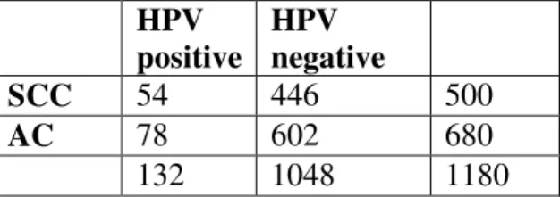
Only the information on primary SCC and primary AC of the lung was collected. There were 2629 cases of SCC and 2669 cases of AC. In the remaining cases it was neither one of them or the histological subtype was not detailed. In total 29.4 % of the included cases were squamous cell carcinomas and 29.8 % were adenocarcinomas.
- 29 - 6.1.5 Continents
6.1.5.1 Europe
Reference Country No.
of cases
Year HPV prevalence
in %
Specimen type used
Histologíc subtypes tested
HPV types detected Anantharaman
et al. (36)
multiple European countries
290 2014 9.7 FFPE,
fresh frozen
SCC/AC/others 11,16, 51,58 Argyri et al.
(37)
Greece 67 2017 3.0 SCC/AC/others 16,53
Carpagnano et al. (38)
Italy 89 2011 16.4 FFPE SCC/AC/others 16,30,
31,39 Ciotti et al.
(39)
Italy 38 2006 8.0 FFPE,
fresh
SCC/AC/others 16,18 Coissard et al.
(40)
France 218 2005 1.8 fresh
frozen
SCC/AC/others 16 Eberlein-
Gonska et al.
(41)
Germany 55 1992 5.5 fresh SCC/AC/others 16
Galvan et al.
(42)
Italy, United Kingdom
100 2012 0 fresh
frozen
SCC/AC/others none
Gatta et al.
(43)
Italy 50 2012 4.0 FFPE SCC
Guliani et al.
(44)
Italy 78 2007 12.8 fresh
frozen
SCC/AC/others 16,18, 31,53 Hennig et al.
(45)
Norway 22 1999 13.6 FFPE SCC/AC/others 6
Miasko et al.
(46)
Poland 94 2004 12.7 SCC/AC/others
Miasko et al.
(47)
Poland 40 2001 10.0 FFPE SCC/AC/others
Papadopoulou et al. (48)
Greece 52 1998 40.0 fresh
frozen, FFPE
SCC 6,11,
16,18 Podsiadlo et
al. (49)
Poland 33 2012 3.0 fresh NSCLC/SCLC 120
Sagerup et al.
(50)
Norway 334 2014 3.9 fresh
frozen
SCC/AC/others 11,16, 33,66 Sarchianaki et
al. (51)
Greece 100 2014 19.0 FFPE SCC/AC/others 6,11,16, 18,31, 33,59 Shamanin et
al. (52)
Germany 85 1994 0 fresh
frozen
SCC/AC/others none
- 30 - Spandidos et
al. (53)
Greece 99 1996 15.0 FFPE SCC/AC/others 11,16, 18,33 Syrjanen et al.
(54)
Finland 77 2012 5.2 FFPE,
archival tissue
SCC/AC/others 6,16
Van Boerdonk et al. (55)
Netherlands 211 2013 0 FFPE, archival tissue
SCC/AC/others none
Thomas et al (56)
France 31 1995 16.0 fresh
frozen
SCC/AC/others 6, 11
Welt et al (57) Germany 38 1997 0 FFPE SCC/SCLC none
Zafer et al (58) Turkey 40 2004 5.0 fresh frozen
SCC/AC/others 18
Total 2226
Table 2: European studies
From Europe 23 studies were included containing 2226 patients. The most common countries of origin were Italy (n=4) and Greece (n=4), followed by Germany (n=3) and Poland (n=3).
HPV prevalence was tested in multiple countries and reported in one publication in two studies (36, 42).
21 publications were in English, one in French (56) and one in German (41).
Of all the European studies 19 publications were journal articles, two poster presentations, one meeting abstract and one was an abstract.
All of the included studies from Europe incorporated NSCLC tumor biopsies.
Two of the included studies only contained SCC samples.(43, 48) There were no studies only analyzing adenocarcinomas found in Europe.
The processing method of the analyzed tissue was not detailed in two of the included studies (37, 46).
Of all the included studies 13 used formalin-fixed paraffin-embedded tissue (56.5 %), nine studies used fresh frozen tissue, two studies used fresh tumor specimens (41, 49) and one study also included archival tissue(55). Four studies analyzed tissue that was processed by more than one of the methods.
- 31 -
Information on the used primers was available in seven studies (30.4 %). The primer used most often in the European subgroup was the consensus primer MY09/MY11 (n=5).
Four of the included studies were case-control studies (38, 41, 42, 51).
The total number of cases included in these studies was 351, the number of controls was 222.
The number of HPV positive cases and controls was 37 and one case respectively. The overall HPV prevalence was calculated to be 10.5 % in the lung cancer cases compared to 0.45 % in the control cases from benign lung disease.
Only in nine studies information on the patients’ age was provided. The average age was 64.7 years with a standard deviation of 2.3 years.
Information on the patients’ gender was available in 13 of the included studies. 66.5 % of the patients were male and 33.5 % were female (935/472 cases).
Smoking behavior was detailed in 11 studies. 44.3 % were current or former smoker, 4.2 % were never smokers and in 51.5 % the smoking behavior was unknown.
All but five studies contained information on the tissue sampling method. It was resected lung tissue in nine studies and bronchoscopic biopsy in three. In six studies both surgically resected lung tissue and bronchoscopic biopsies were analyzed.
The total number of cases included from Europe was 2226. The maximum number of cases in one of the studies was 334, the minimal 22. On average there were 96.8 patients per study.
The most prevalent HPV subtype detected was HPV 16 (n=21), followed by HPV 18 (n=7).
No data on the detected HPV subtype were available in three studies (43, 46, 47).
Of the included 2226 cases 168 were detected to be HPV positive by PCR. The HPV prevalence in the European subgroup was 7.5 %. The highest detected HPV prevalence in one of the European studies was 40 % (48). No HPV DNA was detected in four of the studies (42, 52, 55, 57).
Data on HPV 16 prevalence was included in 15 of the European studies. In total 1556 cases were tested of which 61 were found to be HPV 16 positive (3.9 %). Information on HPV 18 prevalence was available in 13 studies. HPV 18 prevalence was detected to be 0.8 %, meaning 11 out of the 1436 therefore analyzed cases.
- 32 - Positive Negative Total
HPV 16 61 1495 1556 HPV 18 11 1425 1436
72 2920 2992
Table 3: HPV 16 and 18 prevalence in Europe compared
The difference between the detected HPV prevalence for HPV 16 and HPV 18 in the included European studies was statistically highly significant (p<0.01).
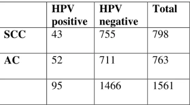
The number of included studies from Europe that contained information on HPV prevalence in squamous cell carcinoma of the lung was 19. Of the included 798 cases 43 were HPV positive (5.4 %). Four studies did not detect HPV DNA in SCC (42, 52, 55, 57).
Ten studies incorporate information on HPV 16 prevalence in SCC; it was calculated to be 2.8
%. Of the studies on SCC nine provided data on HPV 18 prevalence. Only three of the analyzed 408 lung cancer cases were detected to be HPV 18 positive (0.7 %).
Samples of adenocarcinomas of European patients were analyzed in 17 studies. In total 763 cases were examined. 52 cases were HPV positive (6.8 %). The prevalence for HPV 16 (n=9) and HPV 18 (n=8) in adenocarcinomas was 2.9 %. In eight studies AC samples were analyzed for HPV 18 DNA. None of the analyzed 365 cases were tested to be positive for HPV 18 DNA.
HPV positive
HPV negative
Total
SCC 43 755 798
AC 52 711 763
95 1466 1561
Table 4:HPV prevalence in SCC and AC in Europe compared
- 33 -
There was no statistically significant difference between the HPV prevalence in SCC and AC analyzed in the included studies from Europe (p=0.24).
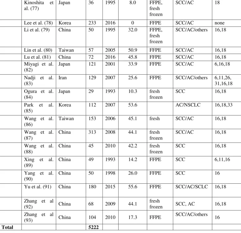
- 34 - 6.1.5.2 Asia
Reference Country
No.
of cases
Year
HPV prevalence
in %
Specimen type used
Histologic subtypes tested
HPV types detected Aguayo et al.
(59)
Pakistan, China
60 2010 13.0 FFPE SCC/AC/others 16
Baba et al.
(60)
Japan 57 2010 19.3 FFPE SCC/AC 6,16,
18,33 Cheng et al.
(61)
Taiwan 141 2004 38.3 SCC/AC 6,11
Cheng et al.
(62)
Taiwan 141 2001 54.6 FFPE,
fresh frozen
SCC/AC 16,18
Fan et al. (63) China 262 2015 8.4 FFPE SCC/AC 16,18,
31,58 Goto et al.
(64)
Japan/
Singapore/
China/
Korea
304 2011 7.9 FFPE SCC/AC 6,11,
16,18
Halimi et al.
(65)
Iran 30 2011 10.0 FFPE SCC
Hartley et al.
(66)
Lebanon 20 2015 0 FFPE SCLC none
Hirayasu et al. (67)
Japan 73 1996 60.3 FFPE SCC 6,16,18
Hiroshima et al. (68)
Japan 22 1999 4.5 FFPE AC 16
Ilahi et al.
(69)
Pakistan 9 2016 11.1 FFPE SCC/AC/others 16
Isa et al. (70) Japan 96 2015 1.0 FFPE SCC/AC/others 6
Ito et al. (71) Japan 901 2014 0.9 SCC/AC/others
Iwakawa et al. (72)
Japan 297 2010 0 fresh
frozen
AC none
Jafari et al.
(73)
Iran 50 2013 18.0 FFPE SCC/AC/others 6,18
Jain et al. (74) India 40 2005 5.0 fresh frozen
SCC/AC/others 18 Kato et al.
(75)
Japan 42 2012 16.7 FFPE SCC/AC/others 16,58
Kawaguchi et al. (76)
Japan 876 2016 0.3 FFPE SCC/AC 16,62,66