Cologne, September 2009
Suey van Baarle
Division site selection in B. subtilis and
Characterization of YpbR, a bacterial
dynamin
Division site selection in B. subtilis and characterization of YpbR, a bacterial dynamin
Inaugural-Dissertation zur
Erlangung des Doktorgrades
der Mathematisch-Naturwissenschaftlichen Fakultät der Universität zu Köln
vorgelegt von Suey van Baarle
aus Dordrecht, Niederlande
Köln, September 2009
Berichtstatter: Prof. Dr. Reinhard Krämer
Institut für Biochemie der Universität zu Köln Prof. Dr. Günter Schwarz
Institut für Biochemie der Universität zu Köln
Tag der mündlichen Prüfung: 23 November 2009
Index
I. Abstract... VII II. Zusammenfassung... IX III. Abbreviations………... XI
1. Introduction……….. 1
1.1 The cytoskeleton……… 1
The eukaryotic cytoskeleton……… 1
The bacterial cytoskeleton……….. 2
1.2 Bacterial cell division………... 3
Cytosolic factors……… 4
Attachment to the membrane……….. 7
Membrane components of the divisome……… 8
The final steps of septation………. 9
Asymmetric cell division……… 10
1.3 Division site selection……… 11
Timing of cell division………. 11
Spatial control of cell division……… 11
Nucleoid occlusion……….. 11
The Min system………... 12
A new component of the B. subtilis Min system, MinJ……….. 15
1.4 Dynamin………. 18
A bacterial dynamin………. 22
1.5 Aim of the research………. 23
MinJ……… 23
YpbR……….. 24
2. Material and Methods……… 26
2.1 Bacterial strains, plasmids and oligonucleotides……… 26
Table 2.1: bacterial strains………. 26
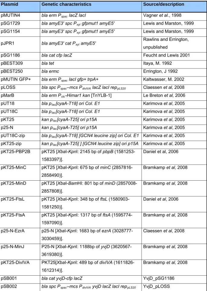
Table 2.2: bacterial plasmids………. 31
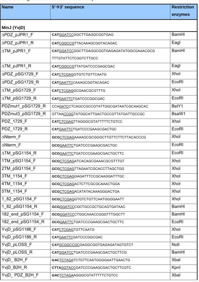
Table 2.3: oligonucleotides……… 33
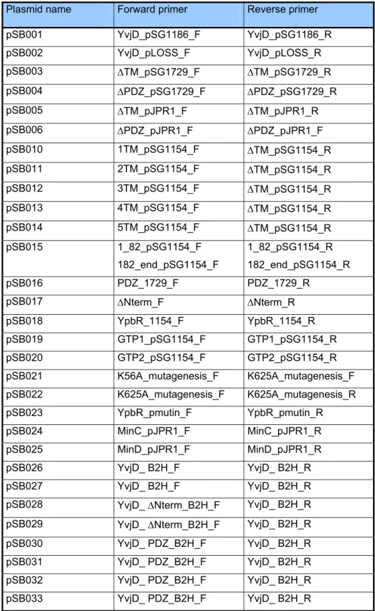
Table 2.4: oligonucleotide combinations………. 35
Strain construction……….. 36
2.2 Media and growth conditions………. 37
Stock solutions………. 37
Index
B. subtilis media……….. 38
Growth experiments……… 40
E. coli growth media……… 40
2.3 Molecular biological techniques……… 40
Preparation of competent E. coli DH5 cells ……….. 40
Preparation of competent BTH101………. 41
Transformation of E. coli DH5 cells………. 41
Transformation of B. subtilis cells……….. 42
General cloning techniques………. 42
DNA amplification………. 42
Agarose gel electrophoresis and gel extraction……….. 43
Sequencing……… 43
Site-directed mutagenesis……….. 43
2.4 Microscopy methods……… 45
Fluorescence microscopy……… 45
Time lapse microscopy……… 45
Transmission electron microscopy………. 46
2.5 Protein analysis………. 47
Polyacrylamide-gel electrophoresis……… 47
Immunoblotting……….. 47
Membrane preparation………. 48
Protein estimation according to Lowry……….. 48
Co-immunoprecipitation……… 49
-galactosidase assay……….. 49
Sporulation assay………. 50
Alkaline phosphatase assay……… 50
Bacterial two-hybrid………. 51
2.6 Genetic analysis………. 51
Synthetic lethal screen………. 51
3. Results……….. 53
3.1 MinJ……… 53
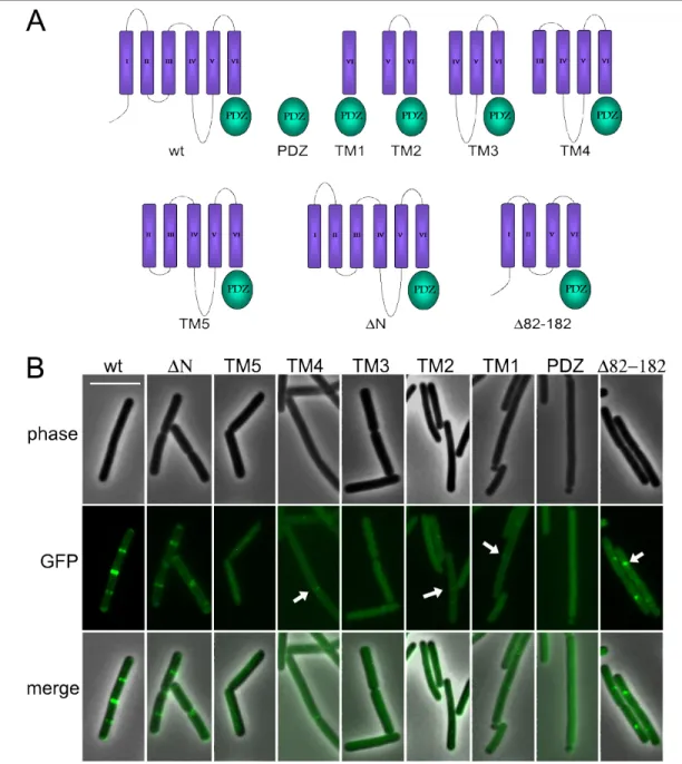
Topology of MinJ……….. 53
MinJ mutants………. 55
The PDZ domain is sensitive to mutations……… 57
MinJ mutants interact diferentially with divisome components………. 58
MinJ localizes preferentially to late septa……….. 59
Localization of MinJ-CFP is dependent on MinD and DivIVA………. 61
MinD localizes to midcell prior to MinJ……… 63
Cell division efficiency is decreased in minJ and minCD ……….. 64
FtsA-YFP remains associated with the poles in Min-deficient cells………. 67
Late division proteins are retained at the poles in Min-deficient cells…………. 67
Index
The failure of GFP-PBP-2B and GFP-FtsL to localize to the Z ring
is due to uncontrolled MinCD………. 69
Overexpression of MinD n the absence of MinJ leads to filamentation……….. 70
The effect of MinD is dependent on MinC……… 73
MinJ localizes to asymmetric septa……….. 74
Synthetic lethal screen………. 75
3.2 YpbR……….. 77
Function of YpbR……… 77
Localization of YpbR………. 81
Effect of salt on the localization of YpbR and YpbR mutants………. 83
Protein stability of YpbR……… 84
Protein stability of YpbR under salt stress………. 85
Localization of YpbR during sporulation ……… 87
4. Discussion……… 88
4.1 MinJ……….. 88
Localization and dynamics of MinJ……….. 88
The Min system is required for efficient cell division………. 90
The Min system contributes to disassembly of the divisome……….. 92
MinCD can inhibit formation of the divisiome………. 93
MinJ acts as a sensor and regulates MinCD……….. 95
Rethinking the Min system……… 98
A new model for the function of the Min system……… 100
Division site selection………. 101
4.2 YpbR………. 105
Bacterial GTPases………. 105
YpbR is membrane-associated………. 106
Role of GTPase activity in localization of YpbR………. 108
Role of YpbR in sporulation………... 108
A possible role in cell division?………. 109
YpbR function……….. 111
A model for YpbR function………. 112
A new member of the bacterial cytoskeleton………. 112
5. References ………. 113
6. Acknowledgements……….. 126
7. Affirmation……… 128
8. Scientific activities………. 129
9. Curriculum Vitae …..………. 130
Abstract
I. Abstract
Cell division in Bacillus subtilis takes place precisely at midcell through the action of Noc, which prevents division from occurring over the nucleoids, and the Min system, which prevents cell division from taking place at the poles. Originally it was thought that the Min system acts directly on FtsZ, preventing the formation of a Z ring, and therefore the formation of a complete cytokinetic ring at the poles. Recently, a new component of the B. subtilis Min system was identified, MinJ, which acts as a bridge between DivIVA and MinCD. By using time lapse microscopy, it is shown here that MinJ moves from the poles to midcell prior to septation, and after this process is completed, moves back to the poles. This indicates that its main site of action is at midcell. Additionally, in the absence of a functional Min system, FtsA remains at the poles instead of being disassembled, which subsequently forms a double ring leading to minicell formation. Late divisome proteins FtsL and PBP-2B are also not disassembled from completed division sites. Previously it was described that FtsL and PBP-2B fail to localize in the absence of MinJ, which seems to lead to the filamentous phenotype of minJ cells. In this thesis it was shown that this is due to dispersed MinCD, since in a minCDJ mutant GFP-PBP-2B and GFP- FtsL localize to midcell again, although they are also found at the poles. MinJ mutants were isolated that are able to complement the cell length phenotype of minJ cells, but lead to an increased production of minicells, showing that in cells expressing these mutants cytokinesis is not impaired but division site selection is. Additionally, it was shown that overexpression of MinD in the absence of MinJ leads to lethal filamentation in B. subtilis, although cytoplasmic components of the cytokinetic ring are able to localize in these cells. In the absence of a functional Min system, cells contain multiple FtsA rings, indicating an impairment in cell division efficiency. Taken together, these results show that i) the Min system is involved in the disassembly of the divisome, rather than preventing its assembly at the poles, which also explains its preferential localization to midcell prior to septation, ii) the failure to disassemble the divisome leads to minicell formation, iii) the Min system is needed for efficient cell division, and iv) MinJ is able to regulate MinCD activity, possibly by restricting its activity to certain sites.
Using a bacterial two-hybrid screen, it was found that MinJ interacts with YpbR. YpbR is a large protein containing two GTPase domains and shows homology to the dynamin-like proteins.
Although YpbR does not appear to be important for growth, morphology, or sporulation, it was shown that cells deficient in YpbR are less susceptible to antibiotics than wild type. Also, under salt stress, ypbR cells show an altered morphology of septa. This indicates that YpbR probably
Abstract
plays a role in stabilizing the membrane under conditions that impart stress to the lipid membrane. YpbR-GFP is localized to the membrane and forms foci, but when cells are exposed to high concentrations of NaCl, YpbR-GFP forms a uniform structure on the membrane. Also, the first GTPase domain alone (GTPase1) is able to localize in a similar pattern as wild type and forms a uniform structure on the membrane under salt stress, but the second GTPase domain is diffuse throughout the cell. Using mutants that are unable to hydrolyze GTPase with the first GTPase domain (K56A) or the second (K625A), it was shown that the localization to the membrane and subsequent change of localization under salt stress is not dependent on the GTP binding. Under normal conditions, YpbR-GFP and YpbR mutant proteins are found mostly in the membrane fraction but also somewhat in the cytoplasmic fraction, but under salt stress all of the proteins are found mostly in the membrane fraction. It was also shown that YpbR-GFP localizes to spores early in sporulation and its overexpression increases sporulation efficiency. It is postulated that YpbR is mostly involved in stabilizing the membrane or involved in membrane dynamics, but is not essential in B. subtilis.
Zusammenfassung II. Zusammenfassung
Bacillus subtilis Zellen teilen sich exakt in der Zellmitte. Dass die Zellteilung nicht im Bereich des Chromosoms erfolgt, wird dabei von Noc gewährleistet, während das Min-System verhindert, dass Teilung nicht an den Polen stattfindet. Dabei wurde zunächst angenommen, dass das Min- System direkt die Polymerisation von FtsZ inhibiert und so die Entstehung des Z-Rings und die Bildung des kompletten zytokinetischen Rings an den Polen inhibiert. Es konnte allerdings eine neue Komponente des Min-Systems identifiziert werden, MinJ, welche als Verbindung zwischen DivIVA und MinCD fungiert. Unter Verwendung von Zeitreihen-Mikroskopie konnte gezeigt werden, dass MinJ bereits vor Beginn der Zellteilung in der Zellmitte lokalisiert und nach Abschluss der Teilung wieder an den Polen akkumuliert. Dies weist darauf hin, dass der Hauptwirkungsort für MinJ die Zellmitte ist. Des weiteren konnte hier gezeigt werden, dass ohne ein funktionales Min-System FtsA an den Polen verbleibt, was die Entstehung eines Doppelrings und somit die Entstehung von Minizellen zur Folge hat. Auch die Komplexe aus den späten Zellteilungsproteinen FtsL und PBP-2B werden ohne ein funktionales Min-System nicht an der Zellteilungsebene aufgelöst. Unter Verwendung einer MinJ-Deletionsmutante wurde bereits herausgefunden, dass FtsL und PBP-2B nicht korrekt lokalisieren können, was zur Entstehung von filamentösen Zellen führt. In dieser Arbeit wurde zusätzlich gezeigt, dass dies auf die unspezifische Lokalisation von MinCD zurückzuführen ist, da sich in einer ∆minCDJ-Mutante PBP-2B-GFP und FtsL-GFP sowohl in der Zellmitte als auch an den Polen wiederfanden. Es wurden MinJ-Mutanten isoliert, die in der Lage waren, den Zelllängen-Phänotyp von ∆minCDJ- Mutanten zu komplementieren, dabei allerdings eine erhöhte Produktion von Minizellen zeigten.
Diese Mutanten sind also in der toplogischen Regulation der der Zellteilung beeinträchtigt, nicht aber in der Zytokinese per se. Zusätzlich führt die Überexpression von MinD in Abwesenheit von MinJ zu einem letalen, filamentösen Phänotyp in B. subtilis, obwohl die zytoplasmatischen Komponenten, wie FtsA, des zytokinetischen Rings korrekt lokalisieren. Zusammenfassend bedeutet dies, dass das Min-System für die effektive Durchführung der Zellteilung benötigt wird.
Es ist eher an der Auflösung des Zellteilungs-Divisoms beteiligt, als dass es ihre Lokalisation an den Polen verhindert, was durch die bevorzugte Konzentration in der Zellmitte kurz vor der Zellteilung gezeigt wurde. Wird das Divisom nach erfolgter Zellteilung allerdings nicht aufgelöst, entstehen Minizellen. Letztlich wird die Aktivität von MinCD, wahrscheinlich durch dessen lokale Beschränkung, durch MinJ reguliert.
Zusammenfassung
Unter Verwendung eines bakteriellen Zwei-Hybrid-Systems konnte eine Interaktion von MinJ mit YpbR gezeigt werden. YpbR ist ein großes Protein mit zwei GTPase-Domänen, das Ähnlichkeit zu Dynamin aufweist. Obwohl YpbR nicht an Wachstum, Morphologie oder Sporulation beteiligt ist, zeigten sich YpbR-Deletionsmutanten weniger empfindlich gegenüber bestimmten Antibiotika im Vergleich zum Wildtyp. Unter Salzstress zeigten diese Mutanten eine veränderte Morphologie der Teilungssepten. Dies ist ein Indiz dafür, dass YpbR möglicherweise unter Stressbedingungen stabilisierend auf die Membran wirkt. Während YpbR-GFP unter physiologischen Bedingungen punktuell an der Membran lokalisiert, bildet es unter hohem Salzstress eine uniforme Struktur an der Membran aus. Die erste GTPase-Domäne (GTPase 1) folgt unter Salzstress diesem Muster, im Gegensatz dazu bleibt die zweite GTPase-Domäne über die Zelle verteilt. Untersuchungen mit Mutanten, die nicht mehr in der Lage waren, GTP mit der ersten (K56A) oder der zweiten GTPase-Domäne (K625A) zu binden, zeigten, dass die Lokalisation an die Membran und die veränderte Lokalisation unter Salzstress nicht von der GTPase-Aktivität abhängig ist. Während die YpbR-GFP und YpbR-Mutanten unter physiologischen Bedingungen überwiegend in der Membranfraktion aber auch in der zytoplasmatischen Fraktion zu finden waren, akkumulierten diese unter Salzstress vorwiegend in der Membranfraktion. Zusätzlich konnte gezeigt werden, dass YpbR-GFP im frühen Sporulationsstadium an den Sporen lokalisiert und eine Überexpression zu verstärkter Sporulation führt. Es wird daher postuliert, dass YpbR vorwiegend in die Stabilisierung der Membran bzw. Membrandynamik involviert ist, allerdings nicht essentiell für B. subtilis ist.
Abbreviations III. Abbreviations
ADP adenosine-5'-diphosphate ATP adenosine-5'-triphosphate DAPI 4'-6-diamidino-2-phenylindole
BCIP 5-bromo-4-chloro-3-indolyl phosphate BDLP bacterial dynamin-like protein
CAA casamino acids
CFP cyan fluorescent protein ddH2Odd double deionized water DMSO dimethyl sulfoxide DNA deoxyribonucleic acid dNTP’s deoxyribonucleotides EDTA ethylenediaminetetraacetic acid GFP green fluorescent protein GDP guanosine-5'-diphosphate GTP guanosine-5'-triphosphate
IPTG Isopropyl β-D-1-thiogalactopyranoside kb kilo base pairs
kDa kilo Dalton
LB Luria Bertani-Medium MD mitochondrial dividing ring NBT nitro blue tetrazolium chloride OD600 optical density at 600nm
PAGE polyacrylamide-gel electrophoresis PCR polymerase chain reaction
PD plastid dividing ring
PDF plastid dividing dynamin FtsZ ring RT room temperature
TAE tris-Acetat/EDTA-Buffer SDS sodium dodecylsulfate TM transmembrane helix
Tris tris-(hydroxymethyl)-aminomethane
V Volt
X-Gal 5-bromo-4-chloro-3-indolyl-β-D-galactopyranoside YFP yellow fluorescent protein
Introduction
1. Introduction
Cell biology in eukaryotes has been well studied for decades, but only recently has there been interest in exploring cell biology in bacteria. This is probably due to the fact that in general, prokaryotes do not contain internal membrane structures except for a few notable exceptions and have therefore been considered to be nothing more than a bag of enzymes lacking any kind of internal organization. The past decade has seen a boom in bacterial cell biology which has shown that the bacterial cell is in fact highly organized. This insight is mostly due to the advance in microscopy technology that has allowed visualization within even the smallest cells.
1.1 The cytoskeleton The eukaryotic cytoskeleton
The cytoskeleton is a remarkable system of filaments that allow cells to move around, organize internal structures, maintain their shape, engage in whole-cell locomotion, and provide mechanical strength and resistance to shear strength. The cytoskeleton is involved in a number of intracellular processes, including chromosome segregation, cell division, intracellular trafficking of organelles, and supporting the plasma membrane. All of these tasks are accomplished by three types of protein families: tubulin, actin, and intermediate filaments (IFs).
Although they have different functions within cells, all three types of proteins are able to self- associate and polymerize to form long, helical filaments.
Tubulin and actin can bind and hydrolyze GTP and ATP respectively, which plays an important role in their dynamics. Each tubulin monomer is bound to a GTP molecule that is hydrolyzed to GDP soon after its assembly into the polymer (Heald and Nogales, 2002). Likewise, actin monomers are bound to ATP which are hydrolyzed to ADP after assembly. GDP-bound tubulin and ADP-bound actin have less affinity for their neighboring subunits, causing them to dissociate from the polymer (Pollard et al, 2004). While the subunits of actin and tubulin filaments are globular, IF monomers are elongated molecules and form a coiled-coil with another monomer.
Dimers then line up together and form a rope-like structure due to strong lateral hydrophobic interactions (Hermann and Aebi, 1998).
Because these three elements are highly dynamic, they are perfectly suited to direct movement of the cell and within the cell. Actin filaments can attach to the membrane and form a cortex and
Introduction
are largely involved in forming protrusive structures, such as microvilli, which are structures found on intestinal epithelial cells and pseudopodia, allowing the cell to move across a surface (Alberts et al, 2002). Also, actin filaments, together with myosin, can make up molecular motors which can mediate the intracellular transport of organelles. Actin is also necessary for cell division in eukaryotic cells, forming a contractile ring after chromosome segregation, which cleaves the cell in two to create two daughter cells (Alberts et al, 2002). Tubulin also forms structures that impart locomotion to cells. Tubulin filaments, together with dynein and other accessory proteins, are components of eukaryotic cilia and flagella. Importantly, tubulin is responsible for segregating replicated chromosomes. Intermediate filaments are able to withstand large strain and therefore play an important role in maintaining cell integrity.
The bacterial cytoskeleton
The discovery of a bacterial tubulin, FtsZ, which plays an important role in cell division, along with refined techniques such as fluorescence microscopy have opened the door to the field of bacterial cell biology. Over the past few decades, work on the model organisms Escherichia coli, Bacillus subtilis, and Caulobacter crescentus has shown that, in addition to tubulin, bacteria contain homologues of all three major eukaryotic cytoskeletal elements. Moreover, these discoveries have challenged the old paradigm and shown that despite their formidable size, bacteria are actually highly organized.
Three basic actin homologues have been discovered in bacteria, including MreB and MreB homologues, ParM, and FtsA, which all carry out a variety of different functions (Shi and Rothfield, 2006). MreB proteins have been assigned a variety of functions within the cell, but are mostly implicated in cell shape. In recent years MreB proteins have been shown to form cytoskeletal structures which determine cell morphology. MreB proteins form helical filamentous structures that coil around the rod-shaped cell, located on the undersurface of the cytoplasmic membrane and extending along the length of the cell (Jones et al, 2001; Shih and Rothfield, 2006). The main role of MreB proteins appears to be to direct peptidoglycan synthesis in a helical pattern, allowing cells to elongate while maintaining their cell diameter (Shih and Rothfield, 2006, Carbadillo-Lopez, 2007). It has also been suggested that MreB plays a role in chromosome segregation in E. coli and C. cescentus (Kruse et al, 2003; Gitai et al, 2005), although this is somewhat debated (Defeu Soufo and Graumann, 2004; Formstone and Errington, 2005). Another type of actin homologue, ParM, is involved in segregating plasmids. It is part of the Par system, which consists of parC, centromere-like sites encoded on the plasmid,
Introduction
and ParR, which binds to parC sequences and which links ParM to the plasmid DNA (Shih and Rothfield, 2006). The Par system leads to segregation of plasmids though polymerization of ParM, whereby ParM filaments bound to two plasmids polymerize and push the plasmids to opposite ends of the cell, away from each other (Shih and Rothfield, 2006). FtsA is the only actin homologue in bacteria which does not self-associate and form filaments (Gueiros-Filho, 2007).
A bacterial IF, crescentin, has also been identified, although thus far, it has only been found in Caulobacter crescentus. C. crescentus has a unique, comma-like cell shape, and crescentin is responsible for this curvature (Ausmees et al, 2003). There are also some bacterial cytoskeletal components for which no homologue exists in eukaryotes, which include MinD and ParA. MinD is considered part of the cytoskeleton because it, together with MinC and MinE, is organized into helical structures that coil around E. coli cells through MinD polymerization (Shih and Rothfield, 2006). ParA proteins act in a similar manner to ParM, playing a role in plasmid partitioning through polymerization and thus pushing plasmids apart.
The fact that many so-called eukaryotic cytoskeletal elements are found in bacteria suggests that they probably originated in prokaryotes and have been highly conserved since. However, it is interesting that although the overall structure of the monomeric and polymerized elements have been conserved, their functions have not been. While tubulin plays a role in bacterial cell division, eukaryotes require it, among other things, for chromosome segregation, while in turn, bacteria rely on actin-like proteins to segregate plasmids. It has been suggested that the differences in the roles of these proteins is due to differences in polymerization properties (Shih and Rothfield, 2006).
1.2. Bacterial cell division
Cell division of bacteria is an interesting aspect regarding the cytoskeleton and cell biology, as this was the first process described in which a number of cytoskeletal elements play an important role. In rod-shaped bacteria, cell division allows for the generation of two equally sized daughter cells from a parent cell, each containing one copy of the genetic material. This task is carried out by a complex machinery termed the divisome. It is composed of variety of proteins, including cytosolic components that act as a scaffold and recruit a number of membrane-integral proteins which are then involved in synthesizing new cell wall material. Currently, the divisome is thought to encompass ~18 proteins (Gueiros-Filho, 2007). See table 1 for a list of cell division
Introduction
proteins involved in B. subtilis and E. coli cell division. The steps leading up to successful cell division are as follows: 1. selection of the division site, 2. assembly of the cytoplasmic apparatus, 3. interaction with membrane proteins that anchor the cytoplasmic apparatus to the membrane, and 4. assembly of proteins with extracellular domains. The proteins involved will be named by their B. subtilis name, unless otherwise stated.
Cytosolic factors
Cell division begins with recruitment of the prinicipal player, FtsZ, to midcell. As mentioned before, FtsZ is a tubulin homologue and like tubulin, it binds and hydrolyzes GTP. In the presence of GTP, purified FtsZ readily assembles into filaments (Erickson, 1996; Mukherjee and Lutkenhaus, 1998). Again, like tubulin, hydrolysis of GTP, leading to GDP-bound FtsZ, leads to instability and thus disassembly of the filaments (Mukherjee and Lutkenhaus, 1998). FtsZ can polymerize into a variety of structures including protofilaments, one-subunit thick strings of FtsZ monomers, and these protofilaments can in turn form two dimensional sheets and 3D bundles through lateral interactions (Gonzalez et al, 2003). Lateral interactions between filaments are greatly enhanced by FtsZ-binding proteins. When cells are prepared to divide after segregation of the nucleoids, FtsZ polymerizes to form a ring, termed the Z ring, at midcell. The exact structure of the Z ring, such as the thickness and the amount of protofilaments, is not yet known.
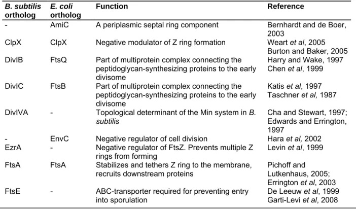
Table 1: Summary of B. subtilis and E. coli division proteins (adapted from Gueiros-Filho, 2007) B. subtilis
ortholog
E. coli ortholog
Function Reference
- AmiC A periplasmic septal ring component Bernhardt and de Boer, 2003
ClpX ClpX Negative modulator of Z ring formation Weart et al, 2005 Burton and Baker, 2005 DivIB FtsQ Part of multiprotein complex connecting the
peptidoglycan-synthesizing proteins to the early divisome
Harry and Wake, 1997 Chen et al, 1999 DivIC FtsB Part of multiprotein complex connecting the
peptidoglycan-synthesizing proteins to the early divisome
Katis et al, 1997 Taschner et al, 1987 DivIVA - Topological determinant of the Min system in B.
subtilis Cha and Stewart, 1997;
Edwards and Errington, 1997
- EnvC Negative regulator of cell division Hara et al, 2002 EzrA - Negative regulator of FtsZ. Prevents multiple Z
rings from forming Levin et al, 1999 FtsA FtsA Stabilizes and tethers Z ring to the membrane,
recruits downstream proteins Pichoff and Lutkenhaus, 2005;
Errington et al, 2003 FtsE - ABC-transporter required for preventing entry
into sporulation De Leeuw et al, 1999
Garti-Levi et al, 2008
Introduction
FtsL FtsL Part of multiprotein complex connecting the peptidoglycan-synthesizing proteins to the early divisome
Daniel and Errington, 2000
Guzman et al, 1992 - FtsN Part of multiprotein complex connecting the
peptidoglycan-synthesizing proteins to the early divisome
Dai et al, 1993
FtsW FtsW Putative peptidoglycan transporter Holtje, 1998
Henriques et al, 1992 FtsX - ABC-transporter required for preventing entry
into sporulation De Leeuw et al, 1999
Garti-Levi et al, 2008 FtsZ FtsZ Self-assembles to form the Z ring required for
divisome assembly Erickson, 1996;
Mukherjee and Lutkenhaus, 1998 MinC MinC Part of the Min system. Negative regulator of
FtsZ, prevents Z rings from forming at the cell poles
de Boer et al, 1992 Levin et al, 1992 MinD MinD Part of the Min system. Interacts with MinC and
brings it to the membrane. de Boer et al, 1991 Levin et al, 1992 - MinE Part of the Min system. Topological determinant
of MinCD in E. coli de Boer et al, 1988, 1989
MinJ - Part of the Min system. Topological determinant
of MinCD in B. subtilis Bramkamp et al, 2008;
Patrick and Kearns, 2008
Noc - Effector of nucleoid occlusion in B. subtilis.
Negative regulator of FtsZ
Wu and Errington, 2004 Pbp2b FtsI or
Pbp3b Transpeptidase involved in septal peptidoglycan
biosynthesis Ghuysen, 1991;
Nguyen-Disteche, 1998 SepF - Positive regulator of FtsZ Hamoen et al, 2006;
Ishikawa et al, 2006 - SlmA Effector of nucleoid occlusion in E. coli. Negative
regulator of FtsZ Bernhardt and de Boer, 2005
SpoIIE - B. subtilis sporulation protein needed for efficient polar septum formation and binds to FtsZ.
Lucet et al, 2000 SpoIIIE FtsK DNA pump involved in chromosome segregation
and dimer resolution. Required for septum formation in E. coli
Iyer et al, 2004; Begg et al, 1995
SulA SOS-induced septation inhibitor Huisman et al, 1984 YneA SOS-induced septation inhibitor Kawai et al, 2003 ZapA ZapA Positive modulator of FtsZ. Promotes polymer
bundling and stabilizes Z ring Gueiros-Filho and Losick, 2002
Aarsman et al, 2005 - ZapB Stimulates Z ring assembly and cell division Ebersbach et al, 2008 - ZipA Tethering of the Z ring to the plasma membrane Hale and de Boer, 1997
Following polymerization of FtsZ at midcell, a number of cytosolic proteins are recruited to the Z ring. In B. subtilis these include FtsA, ZapA, which are conserved and present in many bacterial groups, and SepF, which is only found in a subset of Gram positive cells (Gueiros-Filho, 2007).
FtsA, as mentioned before, is an actin homologue and although it does not self-associate, its structure is quite similar to actin (van den Ent and Lowe, 2000). FtsA is absolutely required for septum formation; in E. coli, deletion of ftsA is lethal, while in B. subtilis, deletion of ftsA results in highly filamentous cells (Beall and Lutkenhaus, 1992). FtsA associates with the membrane
Introduction
through a C-terminal amphipathic alpha-helix (Pichoff and Lutkenhaus, 2005). FtsA directly interacts with the extreme C terminus of FtsZ (Din et al, 1998; Ma and Margolin, 1999; Yan et al, 2000). It promotes Z ring assembly by bringing FtsZ polymers to the membrane (Pichoff and Lutkenhaus, 2005). The cellular ratio of FtsA to FtsZ is important for cell division; in E. coli, this ratio is 1:100, which means there is not sufficient FtsA to form a complete ring. Thus, it probably only makes widely interspersed contacts with the Z ring. In B. subtilis, the ratio of FtsA to FtsZ is 1:5. A second, crucial function of FtsA is to recruit downstream proteins to the Z ring (Errington et al, 2003; Adams and Errington, 2009).
ZapA is a positive modulator of FtsZ. It also binds directly to FtsZ, which promotes the formation of higher order assemblies of FtsZ (Gueiros-Filho and Losick, 2002; Low et al, 2004). ZapA is normally not required for Z ring formation; however, if secondary mutations are generated which lower the chance for FtsZ to form, then ZapA becomes essential (Gueiros-Filho and Losick, 2002). Like ZapA, SepF is also a positive, but non-essential factor of Z ring formation. In the absence of SepF, cells show a slight division inhibition phenotype and a variation of the septum morphology (Hamoen et al, 2006). SepF overexpression can restore the division phenotype of an ftsA mutant, indicating that SepF and FtsA have overlapping roles (Ishikawa et al, 2006). In
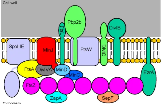
Figure 1.1. Divisome components of B. subtilis. The divisome is made up of a variety of proteins, many of which are cytosolic. FtsZ is the first protein to localize to the division site, forming the Z ring.
The Z ring then recruits other cytosolic proteins such as ZapA, SepF, and FtsA, which tethers the Z ring to the membrane. The other components of the divisome are membrane-spanning proteins, many of which have an extracellular domain, although only Ppb2b has been assigned a specific function.
Also shown are components of the the Min system, including MinC, MinD, MinJ and DivIVA. The figure does not represent actual protein-protein interactions.
Introduction
vitro it was shown that SepF interacts with itself and FtsZ (Ishikawa et al, 2006; Hamoen et al, 2006).
Attachment to the membrane
For efficient cell division, it is crucial that the Z ring is tethered to the plasma membrane. In E.
coli, ZipA is the protein which binds the Z ring to the membrane. Its localization to the Z ring is dependent only on FtsZ (Hale and de Boer, 1999). ZipA is essential for for division in E. coli, although strangely, overexpression of zipA abolishes cell division (Hale and de Boer, 1997; Liu et al, 1999). Additionally, the protein is able to stabilize FtsZ polymers in vitro (RayChaudhuri, 1999), so that it not only acts as an anchor but also as a positive modulator of Z ring assembly.
ZipA is a membrane protein with an N-terminal membrane anchor that is followed by a large cytoplasmic portion that consists of two domains (Ohashi et al, 2002). It binds to the C terminus of FtsZ, similar to FtsA (Hale et al, 2000; Haney et al, 2001). The primary sequence of ZipA is poorly conserved, with a homologue only reported for Haemophilus influenza (RayChaudhuri, 1999).
In B. subtilis, one candidate for a membrane anchor is EzrA, although it has actually been identified as a negative regulator of FtsZ polymerization (Levin et al, 1999). Like ZipA, it is a membrane protein with a short N-terminal domain and a large cytoplasmic domain (Levin et al, 1999). Although EzrA has a similar topology to ZipA, it seems to play a different role.
Interestingly, EzrA is localized homogenously throughout the plasma membrane, but localizes to the Z ring once it is formed (Levin et al, 1999). It can bind to FtsZ and prevent the growth of polymers, but cannot disassemble formed polymers (Haeusser et al, 2004). In the absence of EzrA, cells often have more than one Z ring, which is often found at the poles (Levin et al, 1999).
It has been suggested that EzrA is in charge of controlling that the Z ring does not form prematurely, or, alternatively, that it is necessary for the complete disassembly of the Z ring at the end of division (Gueiros-Filho, 2007). Although it seems that EzrA is not the anchor of the Z ring to the membrane, no other candidate has been proposed to take over this function in B.
subtilis, except for FtsA, which has been suggested to tether the Z ring to the membrane (Adams and Errington, 2009).
Introduction
Membrane components of the divisome
After assembly of the Z ring along with cytosolic components such as FtsA and ZapA, the membrane components of the divisome are recruited. Most of these proteins have a substantial extracellular domain, suggesting they have functions outside of the cell (Gueiros-Filho, 2007).
None of these membrane components appear to interact directly with FtsZ, so that they are probably dependent on another FtsZ-binding protein for localization. Although these membrane components consist of a variety of proteins, their functions remain largely unknown. The overall divisome components at midcell are visualized in figure 1.1.
Interestingly, the order of assembly of these membrane-spanning components differs greatly between E. coli and B. subtilis. In E. coli, the assembly of the divisome follows a linear pattern.
After assembly of the cytosolic components and ZipA, the assembly is as follows: Ftsk [FtsB, FtsQ, FtsL] FtswFtsIFtsN (Errington et al, 2003; Goehring and Beckwith, 2005). However, in B. subtilis, the membrane-spanning proteins are all interdependent on one another for localization at midcell. Following assembly of the Z ring along with FtsA, all of them localize to the Z ring at roughly the same time. In B. subtilis, it appears that the assembly of the divisome occurs in two steps: first the cytosolic components, FtsZ, FtsA, ZapA and EzrA assemble at midcell, and then at roughly the same time DivIB, FtsL, PPB-2B and DivIVA assemble (Gamba et al, 2009).
One of the components of the divisome is SpoIIIE (FtsK in E. coli) which is a family of DNA translocases involved in chromosome segregation and conjugation of mobile elements (Iyer et al, 2004). SpoIIIE localizes as a focus at the center of a closing septum. Here, it represents a DNA translocation pore responsible for the correct distribution of chromosomes that occasionally fail to completely segregate and become trapped by the constricting septum, which is especially important during sporulation (Sharp and Pogliano, 2002; Britton and Grossman, 1999). E. coli FtsK is a remarkable protein since it appears to read the sequence of DNA while translocating (Pease et al, 2005). The sequence that imparts this directionality has the consensus 5′- GGGNAGGG-3′ and is termed FtsK Orienting Polarized Sequence (KOPS) (Bigot et al, 2005;
Levy et al, 2005), which is recognized by the FtsKγ domain (Ptacin et al, 2006). FtsK is, in addition to pumping DNA, also responsible for septum formation (Begg at al, 1995). It is possible that SpoIIIE connects cytokinesis to chromosome segregation.
Introduction
For proper septum formation, it is important that at the division site, new cell wall synthesis occurs. This is carried out by the penicillin binding proteins (PBPs), which are involved in the assembly of peptidoglycan from its precursors through polymerization of GlucNAc-MurNAc sugar subunits into glycan strands, and subsequent connection of glycan strands through peptide chain cross links. There are 16 PBPs known for B. subtilis and 11 for E. coli (Scheffers and Pinho, 2005). One PBP involved in septum formation is PBP-2B, one of the only proteins of the divisome for which the biochemical function has been determined, which is to catalyze the transpeptidation reaction during synthesis of new peptidoglycan (Ghuysen, 1991; Nguyen- Disteche, 1998).
FtsW is a member of the SEDS (shape, elongation, division and sporulation) family of proteins, which are frequently encoded by a gene that lies close to the gene for a class B PBP (Errington et al, 2003). Interestingly, when such an SEDS-encoding gene is inactivated, the phenotype is often identical to that of the inactivation of its cognate PBP-gene, suggesting that their functions are somehow linked (Errington et al, 2003). It has been suggested that the function for SEDS proteins, and therefore FtsW, is the targeting of their cognate PBPs to their sites of activity, as well as to translocate the lipid-linked precursor for peptidoglycan synthesis and delivery to the peptidoglycan-synthesis machinery (Errington et al, 2003).
Other components of the membrane-spanning part of the divisome include DivIB, DivIC and FtsL. All except for DivIB are essential for septum formation (Levin and Losick, 1994; Daniel et al, 1998) although cells deficient in DivIB are impaired in septation. These three proteins likely form a ternary complex, as it has been suggested that the E. coli equivalents, FtsB, FtsQ and FtsL, are preassembled as a complex in the membrane before being recruited to the cytokinetic ring (Buddelmeijer and Beckwith, 2004). It is not known what the function of this complex could be, although there is evidence which suggests that FtsL is involved in a rate-limiting step during in cell division (Bramkamp et al, 2006).
The final steps of septation
Following assembly of the divisome, the synthesis of septal peptidoglycan and invagination of the cell membrane is triggered. It is still not clear what exactly induces the signal for septation or what drives the process of constriction. There are two possibilities: first, constriction may be an indirect consequence of the ingrowth of the peptidoglycan wall. Second, it is possible that the force for constriction results from the dynamic behavior of the FtsZ cytoskeleton. This is
Introduction
supported by the fact that membrane invagination can be uncoupled from septal wall growth by inactivation of Ppb2B (Daniel et al, 2000), and by the fact that L-form bacteria, which lack a cell wall, are still able to divide (Sidiqui et al, 2006; Leaver et al, 2009). Also, membrane-targeted FtsZ incubated with lipid vesicles was able to produce visible constrictions in liposomes (Osawa et al, 2008). It is not yet known how the Z ring itself could lead to constriction; it is possible that there is a regulated disassembly of the FtsZ polymer (Gueiros-Filho, 2007).
After constriction of the cell membrane, two daughter cells can remain connected to each other through the peptidoglycan cell wall. In B. subtilis, hydrolysis of the inner portion of the peptidoglycan wall allows for the physical separation of the daughter cells. This is carried out by autolysins (also called murein hydrolases), of which many variants have been found in B. subtilis (Gueiros-Filho, 2007; Smith et al, 2000). These autolysins are probably targeted to the septum;
this has already been shown for LytF and LytE in B. subtilis (Yamamoto et al, 2003) and AmiC in E. coli (Bernhardt and de Boer, 2003).
The constriction of Gram-negative bacteria is further complicated by the outer membrane. In E.
coli, this is solved by the Tol-Pal system, a series of proteins well conserved in Gram-negative bacteria, which consists of at least five proteins. TolA, TolQ and TolR are inner membrane proteins, while TolB is periplasmic and Pal is an outer membrane protein. These proteins allow the outer membrane to constrict by presumably linking the outer membrane to the inner membrane. This probably occurs after lysis of the peptidoglycan wall by autolysins, creating meshes which allow the Tol proteins to reach through the meshes and grab hold of Pal, in the outer membrane (Gerding et al, 2007).
Asymmetric cell division
In addition to the process of cytokinesis as described above, a number of bacterial species can also undergo a highly specialized form of cell division, called sporulation. Sporulation has been well studied in B. subtilis, which can form endospores under certain conditions, such as starvation or a high population density. The spore is capable of resisting a number of environmental stresses and can stay dormant for an undetermined amount of time. In order for a spore to be formed, the cell must undergo asymmetric division. The first step to complete this asymmetric division is to switch the division site from midcell to polar. A cell that is ready to sporulate will form a Z ring at midcell, although it will not mature into a proper divisome. This medial Z ring then transforms into spirals, which move towards both poles (Ben-Yehuda and Losick, 2002). The exact molecular mechanism behind the switch from midcell to polar rings is
Introduction
not known, although the factors regulating asymmetric division are controlled by Spo0A and the alternative sigma factor H (Ben-Yehuda and Losick, 2002; Levin and Losick, 1996). Also, the protein SpoIIE, which is capable of directly binding to FtsZ, plays a role in changing the position of the Z ring (Lucet et al, 2000). Interestingly, only one of the polar ring will actually become a septum (Levin and Losick, 1996), which may be due to a lack in one or more divisome proteins at one ring (Pogliano et al, 1999).
1.3 Division site selection Timing of cell division
Cell division in all bacteria is subject to both spatial and temporal regulation. It is of vital importance that cells divide only at midcell after duplication and segregation of the genetic material, to ensure that each daughter cell receives at least one copy. The actual timing of Z ring formation is not well known in B. subtilis and E. coli. It has been shown, however, that Z ring formation depends on both replication initiation and cells replicating at least the origin-proximal region of their chromosomes (Harry, 2001). Z ring formation in E. coli coincides with termination of DNA replication (Den Blaauwen et al, 1999). However, the molecular mechanism which links DNA replication and Z ring formation in these two organisms is still unknown. There is clear evidence of a cell cycle in C. crescentus. This unusual bacterium is present in two forms: a sessile stalked cell, where chromosome replication and cell division take place, and a swarmer cell, which must differentiate back to a stalked cell in order to divide. In swarmer cells, FtsZ is degraded though proteolysis by the master regulator CtrA (Kelly et al, 1998). As cells differentiate back into stalked cells, the level of FtsZ rises.
Spatial control of cell division
The mechanism with which cells determine the division plane is better understood. In rod- shaped bacteria, many cells make use of nucleoid occlusion to prevent division across the nucleoid, and the Min system to prevent cell division from occurring at the poles.
Nucleoid occlusion
The term nucleoid occlusion was so named due to the observation that bacteria do not form a septum over their nucleoid under conditions that stop DNA replication and segregation (Woldringh et al, 1991). Originally it was thought that the molecular basis of nucleoid occlusion
Introduction
was DNA itself, since it was observed that division takes place where the DNA concentration is lowest (Harry, 2001b). However, it has now been shown that DNA-binding proteins are able to inhibit Z ring formation over the nucleoid. In B. subtilis this protein is Noc, while SlmA fulfills the same function in E. coli, although the two proteins are not homologous (Wu and Errington, 2004;
Bernhardt and de Boer, 2005). The noc mutation alone has no obvious phenotype, but together with a deletion of minD cells have a severe division defect and fail to form Z rings between segregated nucleoids. However, only by inhibiting DNA replication could Z rings be seen over the nucleoid in a noc mutant (Wu and Errington, 2004). Also, there has never been evidence of a direct interaction between Noc and FtsZ. Noc shows high homology to ParB, a DNA-binding protein which binds a consensus sequences on the chromosome. Recently it was shown that Noc also binds specific sites on the chromosome, with the exception of the terminus region (Wu et al, 2009).
SlmA acts in a similar manner to Noc. It binds to DNA and the absence of SlmA together with depletion of DnaA (resulting in an arrest of DNA replication) resulted in cytokinesis across the nucleoid (Bernhardt and de Boer, 2005). Interestingly, SlmA can directly interact with FtsZ and in vitro it can promote polymer assembly. This ability of promoting polymer assembly may be explained by SlmA competing with other positive division factors such as ZapA, and thereby reducing the ability of FtsZ polymers to develop into a functional cytokinetic ring.
The Min system
The Min system has been especially well characterized in E. coli. It was originally identified after mutation of a locus in E. coli resulted in the production of tiny, anucleate cells (minicells). This minicell locus was subsequently termed minB and implicated in division site selection (de Boer et al, 1988). The minicell locus was found to code for three gene products, MinC, MinD and MinE (de Boer et al, 1989). Without any one of these proteins, a significant portion of cells divide at the cell poles. Since MinC also plays a role in division inhibition that results from the expression of the dicB gene, it was suggested that MinC is the actual inhibiting factor of the Min system (de Boer, 1990). Meanwhile, MinD is a membrane-associated ATPase that sequesters MinC to the plasma membrane, and MinE acts as a topological factor, controlling the localization of MinD, and therefore MinC (de Boer et al, 1989;1991). In general, in the absence of MinCD cells often divide at the pole, generating minicells, while overexpression of MinCD leads to
Introduction
filamentation, indicating that the Min system can inhibit cell division (de Boer et al, 1992;
Marston and Errington, 1999).
Since the first step of cell division is the assembly of the Z ring, it seems logical that FtsZ is the target of the Min system, and that the main site of action of the Min system is at the cell poles. It has been shown that FtsZ rings are generally absent when MinCD is overproduced (Bi and Lutkenhaus, 1993; Hu et al, 1999). Sedimentation assays have been a widely used method to determine the effect on FtsZ polymerization. These make use of the fact that purified FtsZ, when polymerized, forms a pellet after centrifugation. Determining the relative amount of FtsZ in the pellet then allows one to determine the degree of polymerization. Using this assay, it was shown that purified MinC inhibits the polymerization of FtsZ. Interestingly, MinC does not inhibit the GTPase activity of FtsZ (Hu et al, 1999). Because the GTPase activity is linked to FtsZ assembly, this indicates that MinC must act after polymer assembly. There is quite a lot of evidence which supports this. It has been shown that MinC reduces the rigidity of FtsZ polymers, making them easy to break (Dajkovic et al, 2008). Also, in the presence of MinC, FtsZ polymers do form, but they are shorter than those incubated without MinC (Scheffers, 2008). Thirdly, expression of a mutant FtsZ where the mutation was predicted to stabilize the polymer could overcome the effects of MinCD overexpression (Levin et al, 2001). Thus, it seems that the main function of MinC is to inhibit lateral interactions between FtsZ. MinC functions as a dimer and, at least in E. coli, consists of two independent domains, a C and an N terminal domain (MinCC and MinCN) (Hu and Lutkenhaus, 2000). MinCC is necessary for interaction with itself and also for interaction with MinD, while MinCN is important for interaction and inhibition of FtsZ.
Interestingly, purified MinCN alone can inhibit FtsZ polymerization, while purified MinCC is only functional together with MinD (Shiomi and Margolin, 2007). It has recently been shown that the inhibitory activity of MinCC requires the C-terminal tail of FtsZ, which is also the binding site of ZipA and FtsA. It was also shown that MinCC together with MinD is able to displace FtsA from the Z ring, providing an alternative way in which MinC is able to inhibit Z ring formation (Shen and Lutkenhaus, 2009). One in vitro study of B. subtilis MinC showed that it also does not affect FtsZ GTPase activity, but rather, inhibits the lateral interactions between FtsZ polymers (Scheffers, 2008). The crystal structure of Thermotoga maritima MinC also revealed that it is a dimer. Since each N-terminal domain of MinC recognizes one FtsZ molecule, it seems logical that MinC only has an effect on FtsZ polymers, since MinC functions as a dimer and can therefore only recognize an FtsZ filament (Cordell et al, 2001).
Introduction
In vitro studies as well as genetic studies with the E. coli Min system have shown that MinC is only active in the presence of MinD. Expression of MinC in minD mutant cells have no effect on the division process (de Boer et al, 1989). It has also been shown that the MinC/MinD mediated division block is sensitive to MinE, whereas the DicB/MinC division block is not, suggesting that MinD is responsible for the sensitivity to MinE. Furthermore, yeast two hybrid analysis revealed that MinD interacts strongly with MinC and MinE, while there is no interaction between MinE and MinC (Huang et al, 1996). Purified MinD is able to bind ATP as well as hydrolyze it. ATP-binding of MinD is required for membrane binding (Hu and Lutkenhaus, 2001). Interestingly, mutations in the ATP-binding domain of MinD render the protein defective in its ability to activate the MinC- Figure 1.2. Organization and localization of the E. coli and B. subtilis Min system. In E. coli, MinD bound to ATP forms along the membrane and poles, forming a MinD zone, which recruits MinC. A MinE ring interacts with the outer edges of MinD zones. MinE stimulates the ATPase activity of MinD, which leads to dissociation from the membrane. MinD then assembles at the opposite poles, which then attracts MinE, again leading to dissociation from the membrane. In this manner, MinCD oscillates from pole to pole. The B. subtilis Min system is much more static. DivIVA recognizes and is retained at the poles, recruiting MinJ, which recruits MinCD. As cells elongate and the chromosomes are replicated and segregated, the divisome is assembled at midcell, which recruits the DivIVA-MinJ-MinCD complex, which, after disassembly of the divisome and separation of the daughter cells, remains at the poles. Adapted from Lutkenhaus, 2007.
Introduction
mediated division inhibition mechanism, suggesting that the ATPase activity of MinD is required for functioning of the Min system (de Boer et al, 1991).
MinE imparts topological specificity by ensuring that the concentration of MinCD is highest at the poles. Recent advances in fluorescence microscopy have provided evidence to show how MinE determines that MinCD activity is restricted to the poles. Using time-lapse microscopy, it was shown that MinD oscillates from pole to pole in a dividing cell, repeating this pattern every 10 to 20 seconds (Raskin and de Boer, 1999a). MinC itself cannot oscillate but due to its interaction with MinD is carried along (Raskin and de Boer, 1999b; Hu and Lutkenhaus, 1999). MinE oscillates as well, assembling at the edges of MinD zones as a ring prior to moving poleward (Fu et al, 2001; Hale et al, 2001). The oscillation of MinD is dependent on MinE: without MinE, MinD remains uniformly distributed along the membrane (Rowland et al, 2000). This is because MinE chases MinD and stimulates its ATP hydrolysis, which results in the release of MinD, and thus MinC, from the membrane (Hu and Lutkenhaus, 2001). This ensures that the concentration of MinC is highest at the poles and lowest at midcell, allowing cells to divide between segregated nucleoids (see figure 1.2).
The B. subtilis Min system also contains MinC and MinD, but no MinE. Instead, topological control over MinCD is exterted through DivIVA and MinJ. Mutations in divIVA lead to a dispersed MinCD localization, and minicell formation, and interestingly it also leads to cell filamentation (Marston et al, 1998). Fluoresence microscopy showed that DivIVA fused to GFP localized to the cell poles and late at the septa, where it is then retained as new cell poles are formed (Edwards and Errington, 1997). DivIVA is quite a remarkable protein, as it has the ability to find curved membranes, not only in B. subtilis, but also in unrelated cells such as E. coli (which doesn’t contain a DivIVA homologue) or yeast cells (Edwards et al, 2000). This indicates that DivIVA has the intrinsic ability to recognize curved membranes. However, it cannot impose curvature itself (Lenarcic et al, 2009). Since DivIVA is stably localized, the Min system in B. subtilis is much more static than that of E. coli.
A new component of the B. subtilis Min system, MinJ
For approximately ten years, the B. subtilis model of the Min system was that it consisted of three proteins, MinC, MinD and DivIVA, and was extremely static. Recently, however, a new component of the Min system was discovered, MinJ (Bramkamp et al, 2008; Patrick and Kearns, 2008). In the absence of MinJ cells form minicells and, as is the case with DivIVA, cells become
Introduction
long and filamentous. MinJ showed a similar localization pattern as DivIVA, localizing to the poles and to the septum. Two sets of data indicate that MinJ acts as the bridge between DivIVA and MinD. First of all, a bacterial two-hybrid showed that MinJ interacts with both MinD and DivIVA, while DivIVA did not interact with MinD. MinD interacted with MinC and MinJ (Bramkamp et al, 2008; Patrick and Kearns, 2008). Secondly, in the absence of DivIVA, MinJ fails to localize.
However, in the absence of MinJ, DivIVA still localizes, but MinD does not. In the absence of MinD, MinJ still localizes (Bramkamp et al, 2008). Therefore, the order of localization seems to be DivIVAMinJMinDMinC. Interestingly, the filamentous phenotype of minJ cells can be complemented by simultaneously inactivating MinCD, indicating that the filamentous phenotype is due to dispersed MinCD. Also, in the absence of MinJ, the divisome components Ppb2B and FtsL fail to localize, although FtsZ and FtsA still form rings (Bramkamp et al, 2008). This challenges the classical view of Min system function, since it was thought that the filamentous phenotype observed when MinCD is dispersed is due to the failure of FtsZ to form rings.
There has been a recent report which suggests that the B. subtilis Min system is not as static as previously thought. MinC-GFP expressed from its native promoter was shown to actually leave
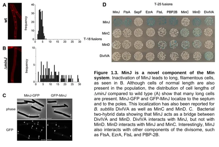
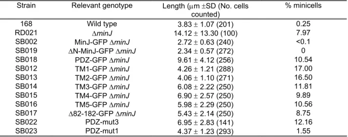
Figure 1.3. MinJ is a novel component of the Min system. Inactivation of MinJ leads to long, filamentous cells, as seen in B. Although cells of normal length are also present in the population, the distribution of cell lengths of
minJ compared to wild type (A) show that many long cells are present. MinJ-GFP and GFP-MinJ localize to the septum and to the poles. This localization has also been reported for B. subtilis DivIVA as well as MinC and MinD. C. Bacterial two-hybrid data showing that MinJ acts as a bridge between DivIVA and MinD. DivIVA interacts with MinJ, but not with MinD. MinD interacts with MinJ and MinC. Interestingly, MinJ also interacts with other components of the divisome, such as FtsA, EzrA, FtsL and PBP-2B.
Introduction
the poles and move towards the Z ring right before septation (Gregory et al, 2008). In fact, it appears that the main site of action of MinC is not necessarily at the poles, but at midcell. In the same paper, it was reported that 73% of minicells are actually formed near recently completed septa, which are new poles, and only a few minicells are the result of cell division at old poles. It was also observed that in the absence of MinCD, the coupling between FtsZ ring assembly and cell division was defective. Interestingly, there is also evidence that the Min system is not only involved in division site selection, but also the timing of cell division, which could be the result of the defect in coupling FtsZ assembly to septation. Taken together, these results allow for a refinement of the current model of the B. subtilis Min system.