The cell division protein FtsL of Bacillus subtilis and its proteolysis
Inaugural-Dissertation zur
Erlangung des Doktorgrades
der mathematisch-naturwissenschaftlichen Fakultät der Universität Köln
vorgelegt von Inga Wadenpohl
aus Leverkusen
Köln, August 2010
Berichterstatter: Prof. Dr. Reinhard Krämer Prof. Dr. Ulrich Baumann
Index
I Abstract IV
II Zusammenfassung V
III Abbreviations VI
1 Introduction
1.1 Cell division in Bacillus subtilis 1
1.2 The division protein FtsL 2
1.2.1 Localisation and stability depending on other cell division proteins 3
1.2.2 FtsL proteolysis by RasP 6
1.3 Intra-membrane proteolysis 7
1.3.1 Intra-membrane proteases 7
1.3.2 The S2P family 8
1.3.3 Substrate recognition by Rhomboid proteases 9
1.3.4 The protease RasP 10
1.4 Aim of research 11
2 Materials and Methods
2.1 Oligonucleotides, plasmids and bacterial strains 12
2.2 Bacterial growth conditions 21
2.2.1 Growth of Bacillus subtilis 21
2.2.2 Growth of E. coli 22
2.3 Molecular biology 23
2.3.1 Preparation of competent E. coli cells 23 2.3.2 Transformation of E. coli cells 24 2.3.3 Transformation of Bacillus subtilis cells 24 2.3.4 Preparation of genomic DNA 25 2.3.5 Amplification of DNA 26 2.3.6 Agarose gel electrophoresis 26
2.3.7 Plasmid construction 27
2.3.8 Slot-Lysis 28
2.3.9 Colony PCR 28
2.4 Protein biochemistry 29
2.4.1 Polyacrylamide gel electrophoresis (PAGE) 29 2.4.2 Preparation of protein samples for PAGE by precipitation 30 2.4.3 Staining of polyacrylamide gels 30
2.4.4 Immuno-blotting 32
2.4.5 Determination of protein concentrations 33 2.4.6 Purification of FtsL 34 2.4.7 Purification of MBP-FtsL 35
2.4.8 Purification of RasP 37
2.4.9 Purification of RsiW* 39 2.4.10 Preparation of liposomes 40 2.4.11 Reconstitution of membrane proteins into liposomes 41 2.4.12 MBP-FtsL proteolysis assay in liposomes 42
2.5 Fluorescence microscopy of Bacillus subtilis 42 2.5.1 General microscopy techniques 42
2.6 Other methods 43
2.6.1 Co-expression experiments in E. coli BL21 43 2.6.2 Bacterial Adenylate Cyclase Two-Hybrid Assay 43
3 Results
3.1 The putative N-terminal cleavage product of FtsL 45 3.1.1 Overexpression and of the putative cleavage product 45 3.1.2 Localisation and stability of the putative cleavage product 48 3.2 In vitro proteolysis of FtsL by RasP 50
3.3 Heterologous co-expression of RasP, FtsL and FtsL interaction partners 67 3.3.1 Co-expression of FtsL, DivIC and DivIB 68 3.3.2 Co-expression of FtsL, DivIC and RasP 71 3.3.3 Influence of the putative recognition motif on FtsL proteolysis 72 3.3.4 Influence of the N-terminal domains of FtsL and DivIC on the
protein-protein interactions 74 3.3.5 Co-expression of FtsL, DivIC∆Nand RasP 77
4 Discussion
4.1 FtsL provides a scaffold for cytokinesis 79 4.2 A possible function of FtsL oligomerisation during complex assembly 82 4.3 The role of FtsL protolysis by RasP during cell division 83 4.3.1 The intramembrane protease RasP is inactive in vitro 84 4.3.2 Substrate recognition is essential for FtsL cleavage by RasP 86 4.3.3 RasP is involved in preventing divisome re-assembly 86 4.3.4 RasP seems to degrade FtsL without prior site-1-cleavage 88 4.4 A new model for FtsL proteolysis by RasP 90
5 References 91
6 Acknowledgements 97
7 Addendum 99
8 Affirmation 103
9 Curriculum vitae 104
I Abstract
Cell division in Bacillus subtilis is a highly regulated process. Division takes place precisely at midcell resulting in two equally sized daughter cells. It is important that the divisome is disassembled after division is completed and does not directly re-assemble. Otherwise a new cycle of division is initiated close to the new formed cell pole, resulting in non viable, DNA- less mini cells. This study analyses the role of the intramembrane protease RasP in preventing divisome re-assembly.
RasP degrades the late cell division protein FtsL in vivo. We tried to establish an in vitro assay to investigate this proteolysis. Both proteins were purified, but RasP seems to be partly unfolded after solubilisation. Therefore a heterolous co-expression system in E. coli was established instead.
It is shown here that the division protein DivIC can protect FtsL against RasP cleavage. This stabilisation is achieved by inhibiting substrate recognition. It could be shown that a recognition motif within the cytosolic N-terminal domain of FtsL is essential for degradation by RasP. FtsL and DivIC tightly interact with each other. Direct interaction of the N-terminal domains blocks accessibility of the FtsL substrate recognition motif. Hence, as long as FtsL is incorporated in the divisome, RasP cleavage is impaired. After the division complex disassembles RasP is able to degrade FtsL. This cleavage removes FtsL from the membrane.
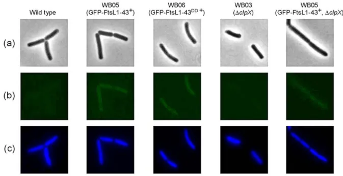
Using fluorescence microscopy it was shown that the cytosolic cleavage product is then rapidly degraded by general proteolysis.
A complex network of the late division proteins FtsL, DivIC and DivIB most likely provides a scaffold for cytokinesis. Since these proteins are strongly interdependent on each other for correct assembly, complete degradation of FtsL should efficiently prevent re-assembly of the divisome close to the new cell pole.
II Zusammenfassung
Zellteilung in Bacillus subtilis ist ein exakt regulierter Prozess. Die Teilung erfolgt präzise in der Zellmitte, so dass zwei gleich große Tochterzellen gebildet werden. Es ist wichtig, dass der Zellteilungapparat anschließend vollständig disassembliert und sich nicht direkt wieder zusammenlagert. Andernfalls wird eine erneute Zellteilung nahe des neu gebildeten Zellpols eingeleitet, die zu nicht lebensfähigen, DNS-freien Minizellen führt. In dieser Arbeit wurde untersucht, welche Rolle die Intramembran-Protease RasP bei der Verhinderung einer solchen Re-Assemblierung spielt.
In vivo kann RasP das Zellteilungsprotein FtsL abbauen. Zur näheren Untersuchung dieser Proteolyse sollte ein in vitro Assay etabliert werden. Beide Proteine konnten gereinigt werden, jedoch scheint RasP nach der Solubilisierung teilweise entfaltet vorzuliegen. Daher wurde stattdessen ein heterologes Co-Expressionssystem in E. coli etabliert.
Es wurde gezeigt, dass das Zellteilungsprotein DivIC FtsL vor diesem Abbau durch RasP schützen kann. Diese geschieht, indem die Substraterkennung durch die Protease verhindert wird. Es konnte gezeigt werden, dass ein Substraterkennungsmotif in der cytosolischen, N- terminalen Domäne von FtsL für die Proteolyse essentiell ist. FtsL und DivIC interagiren stark miteinander. Dabei führt direkte Interaktion der N-terminalen Domänen dazu, dass das Substraterkennungsmotif nicht zugänglich ist. Daher kann RasP FtsL nicht abbauen, solange das Protein noch in den Zellteilungsapparat eingebunden ist. Erst nachdem der Komplex nach der Zellteilung zerfällt, kann FtsL von RasP hydrolisiert werden. Dadurch wird FtsL aus der Membran entfernt. Mittels Fluoreszenzmikroskopie wurde gezeigt, dass das cytosolische Fragment danach sehr schnell durch generelle Proteolyse abgebaut wird.
Ein komplexes Netzwerk der Zellteilungsproteine FtsL, DivIC und DivIB bildet ein strukturelles Gerüst für die Cytokinese. Da die Komplexbildung dieser Proteine stark voneinander abhängig ist, sollte der vollständige Abbau von FtsL eine Re-Assemblierung des Zellteilungsapparates und die Bildung von Minizellen verhindern.
III Abbreviations
AAA-proteins ATPases associated with a various cellular activities ABC transporter ATP-binding cassette transporters
APS Ammoniumperoxodisulfate ATP Adenosine-5'-triphosphate
BACTH Bacterial Adenylate Cyclase Two-Hybrid Assay BCA Bicinchoninic acid (assay)
BCIP 5-Bromo-4-chloro-3-indolyl phosphate BSA Bovine serum albumin
CAA Casamino acids
DAPI 4'-6-diamidino-2-phenylindole DDM n-Dodecyl-β-D-maltoside
DMSO Dimethyl sulfoxide DNA Deoxyribonucleic acid dNTPs Deoxyribonucleotides DTT Dithiothreitol
E. coli Escherichia coli
EDTA Ethylenediaminetetraacetic acid GFP Green fluorescent protein GTP Guanosine-5'-triphosphate IPTG Isopropyl β-D-1-thiogalactopyranoside kb Kilo base pairs
kDa Kilo Dalton
LAPAO Laurylamidodimethylpropylaminoxide LB Luria Bertani-Medium
MBP Maltose binding protein MCS Multiple cloning site
MES 2-(N-morpholino)-ethanesulfonic acid NBT Nitro blue tetrazolium chloride
NEB New England Biolabs OD600 Optical density at 600nm
PAGE Polyacrylamide gel electrophoresis PCR Polymerase chain reaction
PVDF Polyvinylidene fluoride RT Room temperature
TAE Tris-Acetat/EDTA-Buffer S1P Site-1-protease
S2P Site-2-protease
SDS Sodium dodecylsulfate
SREBPs Sterol regulatory element binding proteins TCA Trichloroacetic acid
TEMED N,N,N′,N′-Tetramethylethan-1,2-diamin
1 Introduction
1.1 Cell division in Bacillus subtilis
Bacillus subtilis is a Gram-positive soil bacterium. It is often used as a model organism as it is naturally competent and its genome has been completely sequenced. Under normal growth conditions Bacillus subtilis cells grow along their axis. Reproduction is achieved by dividing a parental cell into two equally sized daughter cells both containing a copy of the bacterial chromosome. This mechanism requires a multi-protein machinery, termed the divisome, and is tightly regulated. The first step of divisome assembly is the localisation of FtsZ to the new division site at mid cell [Bi and Lutkenhaus, 1991], [Beall and Lutkenhaus, 1991]. FtsZ is a GTPase and a bacterial tubulin homologue. It polymerises into a ring like structure, the so called Z-ring. Other cytosolic proteins are recruited to this ring, including the actin homologue FtsA, ZapA and SepF [Bork et al., 1992; Din et al., 1998], [Gueiros-Filho and Losick, 2002], [Hamoen et al., 2006]. All of them can promote and stabilise the Z-ring assembly. For efficient cell division the cytosolic part of the divisome has to be tethered to the membrane. The membrane integrated protein EzrA has been suggested as an anchor, because of its large cytosolic domain interacting with FtsZ [Levin et al., 1999]. However, EzrA is a negative regulator of FtsZ polymerisation and is also localized throughout the whole cytoplasmatic membrane [Kawai and Ogaswara, 2006], [Levin et al., 1999]. It has been suggested, that instead FtsA might tether the ring to the membrane by a C-terminal amphipatic helix [Pichoff and Lutkenhaus, 2005].
The divisome is then completed by several membrane spanning proteins. Among these is the protein MinJ, which is part of the Min system [Bramkamp et al., 2008], [Patrick and Kearns, 2008]. The Min system is involved in division site selection and seems to play in role in divisome assembly and disassembly as well [van Baarle and Bramkamp, 2010]. In the later stages of assembly the proteins DivIB, DivIC, FtsL, FtsW and the penicillin binding protein Pbp2B localise to the division site. In E. coli these so called late division proteins are recruited in a linear dependence pathway [Goehring et al., 2006]. However, in Bacillus subtilis the proteins seem to be interdependent for correct localization at the division site [Errington and Daniel, 2001], [Daniel et al., 2006] or at least for correct and stable assembly [Foster and Popham, 2001]. The function of many membrane spanning division proteins is unknown. However, for Pbp2B a clear biochemical function has been shown. It is involved in synthesising peptidoglycan for the new cell wall by catalysing the transpeptidation, [Nguyen-
Distèche, 1998]. For FtsW a function has been suggested that is linked to Pbp2B. Studies of the E. coli FtsW show that it targets the transpeptidase FtsI to the division site [Mercer and Weiss, 2002]. FtsI is the E. coli homologue to Pbp2B. Also it was suggested, that FtsW could translocate lipid-linked precursors for peptidoglycan synthesis and delivery to peptidoglycan synthesis machinery [Lara et al., 2005]. In contrast FstL, DivIB and DivIC seem to play more of a structural and/or regulatory role. A schematic overview of divisome assembly is shown in Fig. 1.1. When the assembly is completed the Z-ring starts constricting and synchronously a new cross-wall is synthesised. Division results in a pair of sister cells that are joined by this layer of cell wall material. They are later separated through cell wall autolysis [Blackman et al., 1998].
Figure 1.1: Schematic view of divisome assembly in Bacillus subtilis. The tubulin homologue FtsZ polymerises into the so called FtsZ ring, which is stabilised by cytosolic factors. The actin homologue FtsA most likely tethers the FtsZ ring to the membrane. In the late stages of assembly the membrane spanning proteins FtsL, DivIC, DivIB and Pbp2B are recruited interdependently.
1.2 The division protein FtsL
et al., 2005]. The protein is essential for cell division, a ftsL knock out is lethal. A conditional mutant can be constructed with the only copy of ftsL under the control of a xylose inducible promoter. When these cells are brought into a medium without xylose they continue growing, but fail to divide, resulting in abnormally long cells [Daniel et al., 1998]. Overexpression of FtsL in Bacillus subtilis leads to shorter cell length compared to a wild type control.
Apparently the cells are able to divide more often when the FtsL concentration is increased, resulting in shorter length. FtsL seems to be a rate limiting factor for division.
1.2.1 Localisation and stability depending on other cell division proteins
The late cell division proteins seem to be all interdependent in their recruitment to the division site (see chapter 1.1). DivIC and DivIB have been suggested as primary interaction partners of FtsL. The predicted DivIC structure shows strong similarity to FtsL and the protein is essential for cell division as well [Daniel et al., 2006]. The predicted structure of DivIB shows a similiar N-terminal cytosolic domain and a transmembrane domain, but its extracellular domain is considerably bigger than the ones of FtsL and DivIC. The protein is not essential, though DivIB deficient cells are temperature sensitive [Rowland et al., 1997].
FtsL is intrinsically unstable. When transcription in the conditional mutant is shut down, the protein rapidly disappears. How FtsL stability is influenced by other cell division proteins has been previously studied by measuring protein levels in vivo [Daniel et al., 2006]. FtsL is dependent on DivIC in its stability and vice versa. DivIB overexpression has no effect on FtsL levels at 37°C. However, at higher temperatures a DivIB null mutant shows a division phenotype, which can be overcome by overexpression of FtsL. This suggests that DivIB might play a role in stabilising FtsL at high temperatures. Interestingly in the absence of FtsL, DivIC is destabilised by DivIB overexpression. Apparently DivIB is somehow involved in DivIC turnover when FtsL is absent.
Direct protein-protein interactions of FtsL, DivIC and DivIB have previously been discussed controversially [Sievers and Errington, 2000], [Robson et al., 2002], [Daniel et al., 2006].
Because of the strong interdependency of these proteins in vivo their direct interactions were mostly studied in heterologous systems. This enables analysis of only two possible interaction partners without other Bacillus subtilis division proteins present. Controls showed that none
of the Bacillus subtilis proteins is interfering with the E. coli division proteins or is recruited to the division site by them [Robichon et al., 2008]. The broadest approach to investigate the interaction network of the late division proteins of Bacillus subtilis was done by utilising an E. coli based bacterial two-hybrid and a yeast three-hybrid system [Daniel et al., 2006]. In the bacterial two-hybrid system FtsL showed direct interactions with all other late cell division proteins and self-interaction. The most prominent interaction was the FtsL-DivIC interaction.
DivIC itself only seems to interact with FtsL, while DivIB can interact with FtsL and Pbp2B.
This would hint towards an indirect effect of DivIB on DivIC turnover in the absence of FtsL as mentioned above. Further experiments with the yeast three-hybrid system showed a ternary complex of FtsL, DivIC and DivIB. This led to a model supposing that FtsL and DivIC form stable heterodimers. DivIB can then interact with these dimers for further stabilisation.
Apart from in vivo stabilisation studies and bacterial two-hybrid assays the interactions of FtsL, DivIC, DivIB and Pbp2B have also been analysed in E. coli by a method called artificial septal targeting [Robichon et al., 2008]. One of the proteins was fused to ZapA from E. coli and by that targeted to midcell. The second protein was fused to GFP and its localisation was checked using fluorescence microscopy. It was determined which interactions of were strong enough to recruit the GFP fused protein to midcell. Only two pairs of protein-protein interactions resulted in recruitment of the GFP-fused “prey” protein. Those were FtsL-DivIC interaction and DivIB-Pbp2B interaction. This was in accordance to the previous bacterial two-hybrid assays which revealed these pairs to be the strongest interactions. Interestingly other interactions, that were observed in the bacterial two-hybrid studies and indicated by the in vivo data (such as FtsL-Pbp2B or FtsL-DivIB), were not sufficient to result in protein recruitment. However, these findings cannot explain the actual recruitment mechanisms in Bacillus subtilis. As mentioned above all of the four proteins are interdependent for localisation. Obviously the interaction network between them is more complex and not only regulated by each proteins primary interaction partners.
While this interdependency makes it relatively difficult to study the late division proteins of
which is involved in DNA translocation during sporulation [Burton et al., 2007]. The protein FtsQ, the E. coli homologue of DivIB, is dependent on FtsK for its localisation [Chen and Beckwith , 2001]. It has been shown that a trimeric FtsQ/FtsL/FtsB complex can assemble in the absence of other cell division proteins [Buddelmeijer and Beckwith, 2004]. This led to the idea that such complexes pre-assemble and are then recruited to the division site by FtsB interaction with FtsQ. However this model cannot be applied to the situation in Bacillus subtilis, as there DivIB is not essential at normal growth conditions. For the E. coli proteins a lot of information has been gained, how different domains interact with each other and how those interactions influence recruitment of FtsL, FtsB and FtsQ themselves as well as other downstream division proteins. The N-terminal half of FtsB is necessary for interaction with FtsL and in complex with FtsL sufficient to recruit downstream proteins [Gonzales and Beckwith, 2009]. The C-terminal part of FtsB seems to be required for interaction with FtsQ [Gonzales and Beckwith, 2009]. In accordance to that the C-terminal part of FtsQ is important for interaction with FtsL and FtsB [Goehring et al., 2007], [van den Ent et al., 2008]. The direct interactions patterns for the FtsL domains were also analysed [Gonzales et al., 2010]. It seems the interaction of FtsQ and FtsL is not only mediated by FtsB. The C-terminal part of FtsL is necessary for interaction with FtsQ, but not for interaction with FtsB. The main function of the N-terminal part of FtsL appears to be the recruitment of downstream proteins.
In addition the extracellular domain of FtsL is not only important for interaction with FtsQ, but for self interaction of FtsL as well. In vitro E. coli FtsL can form SDS resistant dimers [Ghigo and Beckwith, 2000]. The reason for such stable folding is most likely the predicted coiled-coil conformation of the C-terminal domain. In accordance to that mutation of the leucine zipper like heptad repeat motif impairs FtsL dimerisation. The mutation also affects FtsL function and localisation. Probably the localisation defect is due to impaired interaction with FtsQ. Surprisingly, mutation of the heptad repeat motif had no effect on the function of FtsL in Bacillus subtilis.
It is very interesting that such a consistent interaction network of FtsL, FtsB and FtsQ could be shown for the E. coli proteins, but it is apparently not completely conserved in Bacillus subtilis. While some interactions seem to be similiar, the general influence of FtsL, DivIC and DivIB on each other and their localisation pattern differs in Bacillus subtilis.
1.2.2 FtsL proteolysis by RasP
FtsL has been shown to be a substrate of the intra-membrane protease RasP [Bramkamp et al., 2006]. In a RasP mutant strain FtsL is significantly stabilised and cells are shorter compared to a wild type control. This phenotype can be complemented by insertion of yluC (the gene encoding RasP) into the amyE locus under the control of a xylose inducible promoter. In an E.
coli BL21 co-expression system RasP was able to degrade FtsL, while the active site mutant RasP-E21A did not hydrolise FtsL.
Another known substrate of RasP is the anti-sigma factor RsiW of Bacillus subtilis [Schöbel et al., 2004]. Sequence alignment of FtsL and RsiW shows conserved two conserved boxes.
One putative substrate recognition motif in the N-terminal cytoplasmic domain (25-KKRAS- 29) and one putative cleavage site within the transmembrane domain (39-VLFAAAV-45).
When the N-terminus of FtsL is successively truncated by five amino acids from ∆10-FtsL to
∆30-FtsL protein stability is increasing, especially in the case of the 30 amino acid truncation [Bramkamp et al., 2006]. This truncation completely lacks the putative recognition motif. The same stabilising effect can be observed when altering the motif from 25-KKRAS-29 to 25- AVAVA-29 (FtsL-25A) or 25-KKAVA-29 (FtsL-25B). This shows that the cytoplasmic domain and the putative substrate recognition are indeed important for FtsL turnover.
RasP belongs to the site-2-protease family. These proteases usually cleave their substrates after an initial cut by a site-1-protease has been performed. However, for FtsL no such site-1- protease has been identified yet. The site-1-protease PrsW is involved in cleaving RsiW prior to RasP cleavage [Heinrich and Wiegert, 2006], [Ellermeier and Losick, 2006], but is not able to degrade FtsL [Wiegert, personal communication].
So far no cleavage products of FtsL have been identified. This might be due to the small size of the protein and unspecific degradation of the resulting peptides. In most cases an intra- membrane cleavage releases a biologically active protein or peptide into the cytosol. This is the case for RsiW, where the anti-sigma factor is released from the membrane by RasP cleavage and then further degraded to activate the sigma-factor σw. If the conserved motif within the transmembrane domain of FtsL is interpreted as the cleavage site, the predicted
mechanism for temporal control of cell division or to prevent re-assembly after division is completed. It has been shown, that spontaneous re-assembly of a division close to a new cell pole [Gregory et al., 2008] or reduced disassembly [van Baarle and Bramkamp, 2010] can lead to mini cell formation in Bacillus subtilis,. Removing essential division proteins from the membrane could provide an effective mechanism to prevent re-assembly.
1.3 Intra-membrane proteolysis
1.3.1 Intra-membrane proteases
Proteases catalyse the hydrolysis of amide bonds that link amino acids into peptides and proteins. There are four general types of proteases known so far. Serine/threonine proteases, cysteine proteases, aspartyl proteases and metalloproteases. Hundreds of examples from all kind of organisms have been identified for each class. Most of them are soluble proteins, which are either freely distributed within an aqueous environment or the soluble part of the protease is linked to a membrane via an anchor. Intra-membrane proteases or intra-membrane cleaving proteases (I-CLiPs) are membrane spanning proteases that are able to hydrolise their transmembrane substrates within the hydrophobic environment of a lipid bilayer [Wolfe et al., 1999]. The substrates are unusual, too. They are typically folded into an α-helix. In this conformation the backbone amide bonds are not accessible for a nucleophilic attack, because the amino side chains will provide a steric block. This means that intra-membrane proteases must be able to create a hydrophilic micro environment as well as partly bend or unfold their substrates. It is therefore not very surprising, that a lot of substrates contain helix-breaking residues near the cleavage site. Three types of intra-membrane proteases have been identified so far. The S2P family, the Rhomboid family and the intra-membrane proteases Presenilin and SPP, which all can be sorted into the general protease classes known from soluble proteases.
The S2P proteases are metalloproteases [Rawson et al., 1997], Rhomboid proteins are serine proteases [Urban et al., 2001] Presenilin and SPP belong to the aspartyl proteases [Wolfe et al., 1999], [Weihofen et al., 2002]. Apparently these enzymes are able to create a special environment and prepare their substrates for cleavage, but the hydrolysis of the amide bond itself follows the principles well known from soluble proteases.
1.3.2 The S2P family
The first discovery of an intra-membrane protease was linked to regulation of sterol and fatty acid metabolism [Brown and Goldstein, 1997]. Sterol regulatory element binding proteins (SREBPs) are proteins with two transmembrane domains and a cytosolic transcription factor domain. At reduced cholesterol levels SREBP is transported to the Golgi apparatus [Nohturfft et al., 1999]. There it is cleaved in two steps to release the transcription factor, which will be translocated to the nucleus. First the luminal loop between the two transmembrane domains of SREBP is cleaved by the membrane-tethered Site-1-protease (S1P) [Sakai et al., 1998]. The second step is the hydrolysis of a bond predicted to lie three residues within the transmembrane helix. This degradation step is carried out by the Site-2-protease (S2P) [Duncan et al., 1998]. Most intra-membrane proteases are part of proteolytic cascades like this and require initial cleavage of their substrates by other proteases.
Complementation studies have revealed that S2P contains a conserved HEXXH motif, characteristic for zinc metalloproteases [Rawson et al., 1997]. In agreement with data from soluble zinc metalloproteases both histidines and the glutamate are essential for proteolytic activity of S2P. The two histidines most likely coordinate the zinc, while the glutamate interacts with a water molecule. About 300 residues from the HEXXH motif another essential amino acid was identified. It is a conserved aspartate that in involved in the zinc coordination [Zelenski et al., 1999], [Feng et al., 2007].
Several members of the S2P family are known in different organisms. Among them are S2P in Methanocaldococcus jannaschii, SpoIVFB and RasP in Bacillus subtilis as well as the E.
coli. RasP homologue RseP (YaeL). SpoIVFB is involved in sporulation, processing the membrane bound transcription factor σk [Campo and Rudner, 2006]. After engulfment of the forespore σk is cleaved and released into the mother cell. Interestingly SREBP and σk have an opposite membrane orientation, which correlates with the opposite orientation of the proteases S2P and SpoIVFB [Rudner et al., 1999]. This suggests that the catalytic region must align with the substrate in a matching directionality. Some insights into the mechanism of S2P family proteases come from structural data. S2P from Methanocaldococcus jannaschii has
different conformations. The conformations of the core domains are identical, but TM1 and TM6 are 10-12 Å farer apart in one conformation. Only in this conformation the active site would be accessible, suggesting that this is an open state of the protease. In the closed state a hydrophilic channel is formed which opens into the cytoplasm. Water molecules could access the zinc through this channel. The sequence of the core domains is relatively similar in all S2P proteases, suggesting that its structure might be similar as well and the active site position would be conserved. However, different members of the family cleave their respective substrates at different positions of the transmembrane helices. If the active site position is conserved, this means that the protease must be able to recognise a certain motif of the substrate for appropriate positioning of the cleavage site. Upon changing from closed to open position a number of buried amino acids become exposed, that might be involved in substrate recognition. So far relatively little is known about specific substrate recognition by S2P proteases. However, recently substrate recognition by Rhomboid proteases has been investigated and the results support the idea of choosing the cleavage side by substrate recognition (see chapter 1.3.3).
1.3.3 Substrate recognition by Rhomboid proteases
The name giving member of the Rhomboid family is Rhomboid-1 found in Drosophila.
Rhomboid-1 is the protease required for cleavage of the protein Spitz [Lee et al., 2001]. Spitz is the Drosophila ortholog of the epidermal growth factor (EGF). Full-length Spitz is located in the ER until Star ushers it to the Golgi apparatus. There it is cleaved by Rhomboid-1 and the product is secreted for intercellular communication. Rhomboid-1 requires three conserved residues for proteolytic activity, a serine, a histidine and an asparagine [Urban et al., 2001]
which form a catalytic triad as known from soluble serine proteases. Rhomboid-1 can cleave Spitz without prior processing by other proteases. This is an exception among the intra- membrane proteases. Apparently regulation is achieved mainly by the Star-mediated translocation of Spitz. The yeast Rhomboid RBD1 cleaves two mitochondrial membrane proteins [Esser et al., 2002], [Herlan et al., 2003]. The human ortholog of RBD1 is PARL and it could restore proteolysis, growth rates and mitochondrial morphology in a RBD1 mutant [McQuibban et al., 2003]. Obviously the role of these Rhomboids in mitochondrial function has been evolutionary conserved. Rhomboids are also present in Bacteria and surprisingly bacterial Rhomboids are capable of cleaving Drosophila Rhomboid substrates [Urban et al.,
2002]. Obviously not only specific functions but also substrate recognition by Rhomboids is widely conserved. This led to an intensive search for specific recognition motifs. In the case of Spitz most of the transmembrane domain could be swapped with that of a non-substrate protein without affecting cleavage. Only the N-terminal quarter of this domain turned out to be sufficient for recognition [Urban and Freeman, 2003]. Inserting this motif into the Notch ligand Delta converted it into a Rhomboid-1 substrate. As two critical residues a gylcine and an alanine were identified. Apparently Rhomboid-1 requires helix-destabilising residues for substrate cleavage like S2P and SPP. However, recently it has been discovered that while Rhomboids do require helix-destabilisation they primary recognise their substrates by a specific sequence near the cleavage site [Strisovsky et al., 2009]. This sequence specificity seems to be widely conserved among Rhomboids. The recognition motif is found among many different Rhomboid substrates. Further studies with a model substrate showed that not only its natural protease, but also several bacterial Rhomboids react sensitive to mutation of the recognition motif. Moreover the position of this recognition motif determined the site of cleavage.
1.3.4 The protease RasP
RasP is an intra-membrane protease from Bacillus subtilis. It belongs to the S2P family. RasP contains the conserved HEXXH motif and a mutation of the glutamate to alanine abolishes activity [Bramkamp et al., 2006]. The predicted structure of RasP shows four transmembrane domains and a PDZ domain. Both termini are facing the outside of the cell.
So far two substrates of RasP have been identified. As mentioned before it cleaves the anti- sigma factor RsiW [Schöbel et al., 2006] and the cell division protein FtsL [Bramkamp et al., 2006]. While its role in cell division is unclear, its involvement in RsiW processing has been investigated in more detail. RsiW is a membrane integrated anti-sigma factor. The corresponding sigma factor σw belongs to the ECF sigma factors. ECF sigma factors regulate genes linked to extracytoplasmic functions [Lonetto et al., 1994]. In Bacillus subtilis σw is needed for a cellular response to alkaline shock. The first step in RsiW degradation is
process represents a typical proteolytic cascade as known from other S2P proteases.
Interestingly though, recent studies revealed that RsiW seems not to be degraded in a simple two-step fashion by PrsW and RasP. PrsW cleaves RsiW site specific, but it has been shown that other peptidases must be involved in further degradation before RasP cleavage occurs [Heinrich et al., 2009].
This is in contrast to the findings for the E. coli RasP homologue RseP. RseP cleaves the anti- sigma factor RseA [Kanehara et al., 2002]. Site-1-proteolysis of RseA is carried out by the protease DegS [Alba et al., 2002]. After DegS degradation the newly exposed C-terminal residue is a valine. If this position is mutated, RseP cleavage is impaired [Li et al., 2009].
Structural analysis of RseP showed that most likely its second PDZ domain binds this single hydrophobic acid. Site-specific cleavage of RseA by DegS therefore is sufficient to trigger further degradation by RseP.
1.4 Aim of research
The aim of research was to investigate a possible role of RasP in regulating cell division and to gain more insight about the mechanism of FtsL cleavage.
So far, regulation of cell division in Bacillus subtilis has mainly been analysed under the aspect of spatial control. The fact that the rate limiting factor FtsL is degraded by the intramembrane protease RasP lend support to the notion that proteolysis might be part of temporal control of cell division. The complex interaction network of the late division proteins suggests that degradation of FtsL might lead to divisome disassembly and termination of cytokinesis. However, this would only be possible if the protease is able to cleave FtsL within the assembled divisome. Therefore, an important goal of this study was to test if FtsL is degraded by RasP in the presence of its divisomal interaction partners DivIC and/or DivIB.
To investigate FtsL proteolysis by RasP on a mechanistic level we wanted to establish an in vitro assay. This would provide a tool to determine the FtsL cleavage site and study the influence of the putative substrate recognition motif. A functional in vitro assay could also be used to study site-1-cleavage of FtsL. Possible candidates for site-1-proteases could be purified and tested for their ability to directly cleave FtsL.
2 Materials and Methods
2.1 Oligonucleotides, plasmids and bacterial strains
Table 2.1: Oligonucleotides
Primer name Sequence (5’-3’)
ftsl-1-43-for CGCTCTAGACGCAAAATTAAAAGGAGG ftsl-1-43-rev GCTCGGCCGTCATTACGCAGCAAAGAGGAC
ftsl-1-43dd-rev GCTCGGCCGTCATTAGTCGTCAAAGAGGACAAGAAG gfpftsl-1-43-for CCCCTCGAGATGAGCAATTTAGCTTACC
gfpftsl-1-43-rev GCTGAATTCTCATTACGCAGCAAAGAGGAC
gfpftsl-1-43dd-rev GCTGAATTCTCATTAGTCGTCAAAGAGGACAAGAAG ftsl-61-117-for GGGGGGCATATGCAAACCAATATTGAGGTG
ftsl-61-117-rev CCCGGATCCTTCCTGTATGTTTTTCAC ybbm-1-107-for GGGCTCGAGATGAGCTGTCCTGAACAA
ybbm-1-107-rev GAGGGATCCGCTGTTAAAAAAACCCCCGCCCATC ylucduet-for GATCATATGTTCGTGAATAC
ftslduet-for CATGGATCCATGAGCAATTTAGC
ftslduet-rev CATGCGGCCGCTTCCTGTATGTTTTTCAC
∆N-ftslduet-for CGGGAATTCGACTCTCGGAGAAAAAGTG
∆C-ftslduet-rev CGGAAGCTTTCAATTGGTTTGATATGCCGC
divICduet-for GGGGGGCATATGTTGAATTTTTCCAGGGAACG divICduet-rev CCCCTCGAGCTTGCTCTTCTTCTCCAC
∆N-divICduet-for GGGGGGCATATGCGCAAAGGGCTGTACAGA
divIBduet-for CGGGAATTCGATGAACCCGGGTCAAGAC divIBduet-rev CGGAAGCTTTCAATTTTCATCTTCCTTTTTAGC ftslB2H-for GGGTCTAGAGATGAGCAATTTAGCTTAC
∆N-ftslB2H-for GGGTCTAGAGACTCTCGGAGAAAAAGTG
ftslB2H-rev GCGGGTACCCTATTCCTGTATGTTTTTCAC divICB2H-for GGGTCTAGAGTTGAATTTTTCCAGGGAACG
∆N-divICB2H-for GGGTCTAGAGCGCAAAGGGCTGTACAGA
divICB2H-rev GCGGGTACCCTACTTGCTCTTCTTCTCCAC
Table 2.2: Plasmids
Plasmid Characteristic trait Source
pJPR1 bla amyE3’ cat Pxyl amyE5’ Rawlings, Errington,
unpublished pWB20 bla amyE3’ cat Pxyl-ftsL1-43 amyE5’ this study pWB21 bla amyE3’ cat Pxyl-ftsL1-43dd amyE5’ this study
pSG1729 bla amyE3’ cat Pxyl-gfpmut1 amyE5’ Lewis and Marston, 1999
pWB22 bla amyE3’ cat Pxyl-gfpmut1-ftsL1-43 amyE5’ this study pWB22 bla amyE3’ cat Pxyl-gfpmut1-ftsL1-43dd amyE5’ this study
pET16b bla PT7lac-10his lacI Novagen
pWB23 bla PT7lac-10his-ftsL lacI Bramkamp,
unpublished pWB24 bla PT7lac-10his-ftsL61-117 lacI this study pWB25 bla PT7lac-10his-ybbM1-107 lacI this study
pOPTM-FtsL bla PT7lac-mbp-ftsL-10his lacI Löwe, unpublished pOPTM-FtsLCT bla PT7lac-mbp-ftsLCT-10his lacI Löwe, unpublished pHis17-RasP bla PT7lac-yluC-10his lacI Löwe, unpublished
pETDuet-1 bla PT7lac- PT7 lacI Novagen
pWB1 cat PT7lac-6his-ftsL lacI this study
pWB2 cat PT7lac-6his-ftsL PT7-yluC-S lacI this study pWB2 cat PT7lac-6his-ftsL PT7-yluC-E21A-S lacI this study
pWB5 cat PT7lac-6his-∆N-ftsL lacI this study
pWB6 cat PT7lac-6his-∆N-ftsL PT7-yluC-S lacI this study pWB7 cat PT7lac-6his-∆N-ftsL PT7-yluC-E21A-S lacI this study
pWB26 cat PT7lac-6his-∆C-ftsL lacI this study
pWB17 cat PT7lac-6his-ftsL25B lacI this study
pWB18 cat PT7lac-6his-ftsL25B PT7-yluC-S lacI this study pWB19 cat PT7lac-6his-ftsL25B PT7-yluC-E21A-S lacI this study
pWB4 bla PT7-divIC-S lacI this study
pWB8 bla PT7-∆N-divIC-S lacI this study
pWB27 bla PT7lac-6his-divIB lacI this study
pWB28 bla PT7lac-6his-divIB PT7-divIC-S lacI this study
pUT18C bla Plac-cya675-1197 Euromedex
pKT25 aphA Plac-cya1-675 Euromedex
pUT18C-zip bla Plac-cya675-1197-zip (construct encoding leucine
zipper from GCN4) Euromedex
pKT25-zip aphA Plac-cya1-675-zip (construct encoding leucine
zipper from GCN4) Euromedex
pWB9 bla Plac-cya675-1197-ftsL this study
pWB10 aphA Plac-cya1-675-ftsL this study
pWB11 bla Plac-cya675-1197-divIC this study
pWB12 aphA Plac-cya1-675-divIC this study
pWB13 bla Plac-cya675-1197-∆N-ftsL this study
pWB14 aphA Plac-cya1-675-∆N-ftsL this study
pWB15 bla Plac-cya675-1197-∆N-divIC this study
pWB16 aphA Plac-cya1-675-∆N-divIC this study
Table 2.3 Bacterial strains
Strain Relevant genotype/characteristic trait Source Bacillus subtilis
168 trypC2
WB01 trypC2, amyE::spec Pxyl-ftsL1-43 this study
WB02 trypC2, amyE::spec Pxyl-ftsL1-43dd this study
WB03 trypC2, clpX::XXX this study
WB04 trypC2, clpX::XXX, amyE::spec Pxyl-ftsL1-43 this study WB05 trypC2, amyE::spec Pxyl-gfpmut1-ftsL1-43 this study WB06 trypC2, amyE::spec Pxyl-gfpmut1-ftsL1-43dd this study
WB07 trypC2, clpX::XXX, amyE::spec
Pxyl-gfpmut1-ftsL1-43 this study
E. coli
DH5α supE44, ∆lacU169(φ80lacZ∆M15), hsdR17,
recA1, endA1, gyrA96, thi-1, relA1 Invitrogen BL21(DE3) F- ompT [lon] hsdSB (rB-m B-) λ(DE3) pol(T7) Novagen
BL21(DE3)/pWB23 FtsL+ this study
BL21(DE3)/pWB24 FtsL61-117+ this study
BL21(DE3)/pWB25 YbbM1-107+ this study
BL21(DE3)/pOPTM-FtsL MBP- FtsL+ this study BL21(DE3)/
pOPTM-FtsLCT MBP- FtsL∆C+ this study
BL21(DE3)/pHis17-RasP RasP+ this study
BL21(DE3)
/pHis17-RasPE21A RasP-E21A+ this study
BL21(DE3)/pWB1/
pETDuet-1 FtsL+ this study
BL21(DE3)/pWB2/
pETDuet-1 FtsL+, RasP+ this study
BL21(DE3)/pWB3/
pETDuet-1 FtsL+, RasP-E21A+ this study
BL21(DE3)/pWB5 ∆N-FtsL+ this study
BL21(DE3)/pWB6 ∆N-FtsL+, RasP+ this study
BL21(DE3)/pWB7 ∆N-FtsL+, RasP-E21A+ this study
BL21(DE3)/pWB26 ∆C-FtsL+ this study
BL21(DE3)/pWB17 FtsL25B+ this study
BL21(DE3)/pWB18 FtsL25B+, RasP+ this study
BL21(DE3)/pWB19 FtsL25B+, RasP-E21A+ this study
BL21(DE3)/pWB4 DivIC+ this study
BL21(DE3)/pWB2/pWB4 FtsL+, RasP+, DivIC+ this study
BL21(DE3)/pWB3/pWB4 FtsL+, RasP-E21A+, DivIC+ this study
BL21(DE3)/pWB1/pWB8 FtsL+, ∆N-DivIC+ this study
BL21(DE3)/pWB2/pWB8 FtsL+, RasP+, ∆N-DivIC+ this study
BL21(DE3)/pWB3/pWB8 FtsL+, RasP-E21A+, ∆N-DivIC+ this study
BL21(DE3)/pWB8/pWB17 FtsL25B+, ∆N-DivIC+ this study
BL21(DE3)/pWB8/pWB18 FtsL25B+, ∆N-DivIC+, RasP+ this study
BL21(DE3)/pWB8/pWB19 FtsL25B+, ∆N-DivIC+, RasP-E21A+ this study
BL21(DE3)/pWB5/pWB8 ∆N-FtsL+, ∆N-DivIC+ this study
BHT101 F-, cya-99, araD139, galE15, galK16, rpsL1
(Strr), hsdR2, mcrA1, mcrB1 Euromedex
BHT101/pKT25/pUT18C CyaA-T18+, CyaA-T25+ this study
BHT101/pKT25-zip/
pUT18C-zip CyaA-T18-zip+, CyaA-T25-zip+ this study
BHT101/pWB9/pWB10 CyaA-T18-FtsL+, CyaA-T25-FtsL+ this study BHT101/pWB9/pWB14 CyaA-T18-FtsL+, CyaA-T25-∆N-FtsL+ this study BHT101/pWB10/pWB13 CyaA-T18-∆N-FtsL+, CyaA-T25-FtsL+ this study BHT101/pWB13/pWB14 CyaA-T18-∆N-FtsL+, CyaA-T25-∆N-FtsL+ this study BHT101/pWB11/pWB12 CyaA-T18-DivIC+, CyaA-T25-DivIC+ this study
BHT101/pWB11/pWB16 CyaA-T18-DivIC+, CyaA-T25-∆N-DivIC+ this study BHT101/pWB12/pWB15 CyaA-T18-∆N-DivIC+, CyaA-T25-DivIC+ this study BHT101/pWB15/pWB16 CyaA-T18-∆N-DivIC+, CyaA-T25-∆N-DivIC+ this study
BHT101/pWB9/pWB12 CyaA-T18-FtsL+, CyaA-T25-DivIC+ this study BHT101/pWB10/pWB11 CyaA-T18-DivIC+, CyaA-T25-FtsL+ this study BHT101/pWB9/pWB16 CyaA-T18-FtsL+, CyaA-T25-∆N-DivIC+ this study BHT101/pWB10/pWB15 CyaA-T18-∆N-DivIC+, CyaA-T25-FtsL+ this study BHT101/pWB12/pWB13 CyaA-T18-∆N-FtsL+, CyaA-T25-DivIC+ this study BHT101/pWB11/pWB14 CyaA-T18-DivIC+, CyaA-T25-∆N-FtsL+ this study BHT101/pWB13/pWB16 CyaA-T18-∆N-FtsL+, CyaA-T25-∆N-DivIC+ this study BHT101/pWB14/pWB15 CyaA-T18-∆N-DivIC+, CyaA-T25-∆N-FtsL+ this study
2.2 Bacterial growth conditions 2.2.1 Growth of Bacillus subtilis
Bacillus subtilis was grown on nutrient agar plates made with 13 grams nutrient broth (Oxoid) and 15 grams agar per one litre of distilled water. The plate medium was sterilised for 20 minutes at 121°C.
If not otherwise noted liquid cultures of Bacillus subtilis were done in 10 ml CH medium and incubated in a 100 ml shaking flask at 37°C. Overnight cultures were grown in 5 ml CH medium in a test-tube at 37°C. When necessary media were supplemented with antibiotics and other additives according to table 2.4.
For growth experiments fresh CH medium was inoculated with an overnight liquid culture to result in a start OD600 of 0.1. Samples were taken every 30 minutes.
Solution G, 2.5 l
25 g Casein hydrolysate (Oxoid) 9.2 g L-glutamic acid
3.13 g L-alanine 3.48 g L-asparagine 3.4 g KH2PO4
1.34 g NH4Cl 0.27 g Na2SO4
0.24 g NH4NO3
2.45 mg FeCl3 · 6 H2O 2.35 l distilled H2O
Adjust pH to 7.0 with 10 N NaOH and autoclave.
Solution D
0.1 M CaCl2 · 2 H2O Autoclave
Solution F
1 M MgSO4 · 7 H2O Autoclave
Solution H, 500 ml 5.5 g MnSO4 · 5 H2O Add distilled H2O Autoclave
L-Tryptophan 2 mg/ml distilled H2O Sterile filter.
CH Medium, 1 l 1 l Solution G 1.0 ml Solution D 0.4 ml Solution F 2.0 ml Solution H 10 ml L-Tryptophan
Table 2.4: Concentrations of antibiotics and other medium additives for Bacillus subtilis Compound Final concentration
Chloramphenicol 5 µg/ml
Kanamycin 5 µg/ml
Spectinomycin 50 µg/ml
Erythromycin 1 µg/ml
Lincomycin 25 µg/ml
Tetracyclin 12 µg/ml
IPTG 1 mM
Starch 0.1 %
Xylose 0.5 %
2.2.2 Growth of E. coli
E. coli was grown on LB (Luria Bertani) agar plates made with 15 grams agar per one litre of LB medium. The plate medium was sterilised for 20 minutes at 121°C.
Liquid cultures were done in LB medium at 37°C. Overnight cultures were done in 5 ml LB medium in test-tubes. When necessary media were supplemented with antibiotics and other additives according to table 2.5.
LB Medium, 1 l 10 g Bacto-Trypton 5 g Bacto-Yeast extract 10 g NaCl
1 l distilled H2O Autoclave
Table 2.5: Concentrations of antibiotics and other medium additives for E. coli Compound Final concentration
Chloramphenicol 50-100 µg/ml
Kanamycin 50 µg/ml
Carbenicillin 50-100 µg/ml
IPTG 1 mM
X-gal 120-160 µg/ml
2.3 Molecular biology
2.3.1 Preparation of competent E. coli cells
E. coli cells were freshly streaked out on LB plates. An overnight culture of 10 ml was inoculated and incubated in a 100 ml shaking flask at room temperature. In the morning the main culture was done in SOB medium. The medium was inoculated resulting in a start OD600
of approximately 0.05. The cells were incubated at room temperature in a shaking flask. The volume of the shaking flask should be about ten times bigger than the culture volume. When an OD600 between 0.3-0.6 was reached, the cells were incubated on ice for 10 minutes and afterwards harvested by centrifugation (10 minutes, 3220 g, 4°C). The supernatant was discarded and the pellet was resuspended in 80 ml of ice-cold TB buffer per 250 ml of main culture. After incubation on ice for 10 minutes the cells were spun down again and resuspended in 20 ml of ice-cold TB buffer per 250 ml of main culture. 0.7 ml of DMSO per 10 ml were added drop by drop and the cells were again left on ice for 10 minutes. The suspension was divided into 100-200 µl aliquots using pre-cooled reaction tubes. The aliquots were flash frozen in liquid nitrogen and stored at -80°C.
SOB Medium, 250 ml 5 g Bacto-Trypton
1.25 g Bacto-Yeast extract 0.125 g NaCl
625 µl 1M KCl solution Add distilled H2O
Autoclave and add 1.25 ml of 2M MgCl2 solution afterwards.
TB buffer, 200 ml 605 mg Pipes 333 mg CaCl2
3.725 g KCl Add distilled H2O
Adjust pH to 6.7 (with KOH) and afterwards add 1.39 g MnCl2. Sterile filter.