• Keine Ergebnisse gefunden

Inactivation of Pleiotropic Regulator 1 reveals p53-dependent Control of Cell Proliferation and Apoptosis by the Pso4- complex

N/A
N/A
Protected

Academic year: 2021

Aktie "Inactivation of Pleiotropic Regulator 1 reveals p53-dependent Control of Cell Proliferation and Apoptosis by the Pso4- complex"

Copied!
132
0
0

Wird geladen.... (Jetzt Volltext ansehen)

Volltext

(1)

Inactivation of Pleiotropic Regulator 1 reveals p53-dependent Control of Cell Proliferation and Apoptosis by the Pso4-

complex

Inaugural-Dissertation zur

Erlangung des Doktorgrades

der Mathematisch-Naturwissenschaftlichen Fakultät der Universität zu Köln

vorgelegt von André Kleinridders

aus Krefeld

Köln 2007

(2)

Berichterstatter: Prof. Dr. Jens Brüning Prof. Dr. Thomas Langer

Tag der mündlichen Prüfung:_________09. Juli 2007 ________________

(3)

Für Tina

(4)

„Darin besteht das Wesen der Wissenschaft. Zuerst denkt man an etwas, das wahr sein könnte. Dann sieht man nach, ob es der Fall ist und im allgemeinen ist es nicht der Fall.“

Bertrand Russell (1872-1970), brit. Philosoph und Mathematiker, 1950

Nobelpreis für Literatur

(5)

Table of contents

Table of contents

Table of figures ... IV List of tables ... VI Abbreviations ... VII

1 Introduction ... 1

1.1 PLRG-1, an evolutionary conserved component of the spliceosome 1 1.2 Regulation of pre-mRNA splicing ... 2

1.3 Role of PLRG-1 in DNA repair ... 6

1.4 DNA damage leads to cell cycle arrest ... 9

1.5 Regulation of apoptosis in DNA damage and repair ... 14

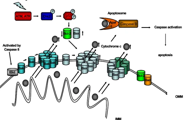
1.6 Involvement of the Bcl-2 family in the regulation of apoptosis... 16

1.7 Objectives ... 19

2 Materials and Methods ... 20

2.1 Chemicals and antibodies ... 20

2.2 Molecular biology ... 22

2.2.1 Competent E.coli and isolation of plasmid DNA ... 22

2.2.2 Construction of targeting vectors ... 22

2.2.3 TA-cloning ... 24

2.2.4 Generation of gene replacement vectors... 24

2.2.5 Isolation of genomic DNA ... 24

2.2.6 DNA electrophoresis... 25

2.2.7 Pulsed-field gel electrophoresis ... 25

2.2.8 DNA sequencing ... 26

2.2.9 Quantification of DNA... 26

2.2.10 PCR ... 26

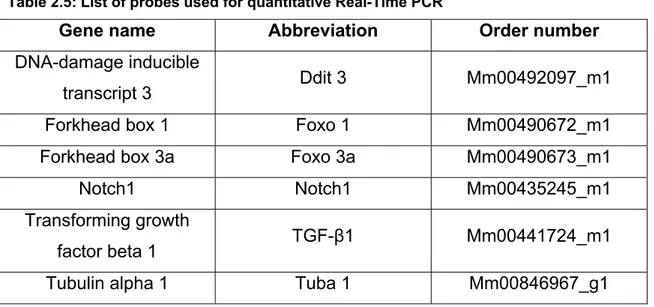
2.2.11 RT-PCR and quantitative Real-Time PCR ... 29

2.2.12 DNA hybridization ... 31

2.2.13 Northern Blot and RNA hybridization... 31

2.3 Cell culture ... 32

2.3.1 Primary embryonic fibroblast (EF) culture ... 32

2.3.2 Embryonic stem (ES) cell culture ... 33

2.3.3 HTN-Cre-mediated deletion in vitro ... 34

2.3.4 Cell cycle analysis ... 34

2.3.5 Cell cycle analysis using fluorescence-activated cell sorter (FACS) analysis... 34

3

(6)

2.3.7 Analysis of apoptosis ... 35

2.3.8 DNA double-strand breaks after UV-treatment ... 36

2.3.9 Immunofluorescence ... 36

2.3.10 RNA interference (RNAi) ... 37

2.4 Biochemistry ... 37

2.4.1 Protein extraction from tissue... 37

2.4.2 Protein extraction from cells ... 37

2.4.3 Nuclear and cytoplasmic protein extraction ... 38

2.4.4 Immunprecipitation ... 38

2.4.5 Western Blot ... 39

2.4.6 Immunohistochemistry ... 40

2.5 Statistical methods ... 40

2.6 Animal Care ... 41

2.6.1 Mouse experiments ... 41

2.6.2 Mice ... 41

3 Results... 42

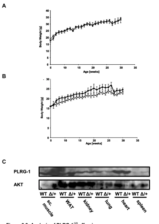
3.1 Murine expression pattern of PLRG-1... 43

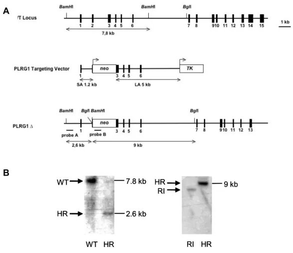
3.2 Generation of a conventional PLRG-1 gene replacement vector... 44

3.3 Analysis of PLRG-1 ∆/+ mice... 46

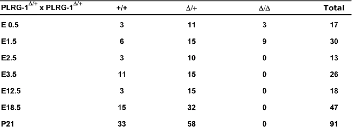
3.4 PLRG-1 ∆/∆ mice are embryonic lethal... 47

3.5 Generation of a conditional PLRG-1 gene replacement vector ... 50

3.6 Inactivation of PLRG-1 in mouse embryonic fibroblasts blocks cell proliferation ... 53

3.7 PLRG-1 deficiency prevents S-phase entry... 55

3.8 PLRG-1 deficiency increases p53 expression and induces apoptosis ... 57

3.9 No evidence for altered splicing in PLRG1-deficient MEFs ... 60

3.10 PLRG-1-deficient MEFs exhibit no spontaneously detectable DNA double-strand breaks... 62

3.11 PLRG-1 deficiency results in enhanced γ-H2AX phosphorylation . 63 3.12 Conditional inactivation of PLRG-1 in heart and skeletal muscle .. 64

3.13 Conditional inactivation of PLRG-1 in heart results in dilated cardiomyopathy due to increased cardiomyocyte apoptosis... 66

3.14 Conditional inactivation of PLRG-1 in the central nervous system of mice results in early postnatal lethality ... 68

3.15 Neuron-restricted PLRG-1 deficiency results in massive neuronal apoptosis ... 69

3.16 Interaction of the nuclear CDC5L-PLRG-1 complex with the p53 phosphatase WIP1 is disrupted in the absence of PLRG-1... 71

3.17 Rescue of the apoptotic PLRG-1 phenotype by knockdown of p5373

4 Discussion ... 77

(7)

Table of contents 4.1 Essential role for PLRG-1 in the development of the preimplantation

murine embryo... 77

4.2 Disruption of other splicing factors and their effects ... 78

4.3 Function of PLRG-1 orthologues ... 79

4.4 PLRG-1 in control of cell cycle and apoptosis... 80

4.5 Impaired DNA damage repair as a consequence of PLRG-1 deficiency ... 83

4.6 Proposed model for PLRG-1, linking DNA repair, control of cell cycle progression and apoptosis via the Pso4-complex ... 85

5 Summary... 89

6 Zusammenfassung ... 90

7 Kurzzusammenfassung... 91

8 References... 92

9 Acknowledgements ... 115

10 Versicherung ... 116

11 Lebenslauf ... 117

(8)

Table of figures

Figure 1.1: Schematic representaion of splicing reaction...5

Figure 1.2: Schematic representation of the Pso4-complex in ICL repair....8

Figure 1.3: Schematic representation of DNA damage checkpoint pathway...9

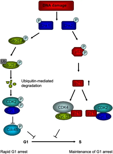
Figure 1.4: Schematic representation of the G 1 /S checkpoint...11

Figure 1.5: Apoptosis signalling in response to p53 activation...18

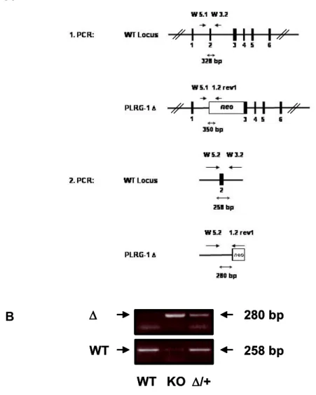
Figure 2.1: Schematic representation of semi-nested PCR method...28

Figure 3.1: Ubiquitous expression of PLRG-1...43

Figure 3.2: Conventional inactivation of the PLRG-1 gene...46

Figure 3.3: Analysis of PLRG-1 ∆/+ offspring...47

Figure 3.4: Identification of genotype using semi-nested PCR...49

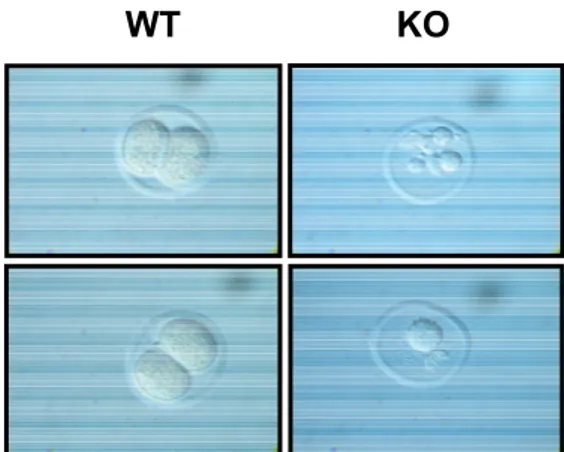
Figure 3.5: Representative morphology of wild type and PLRG-1-deficient embryos at ED1.5...50

Figure 3.6: Conditional inactivation of the PLRG-1 gene...52

Figure 3.7: Verification of PLRG-1 deletion in MEFs upon HTNC treatment...53

Figure 3.8: Growth rates and morphology of untreated or Cre-treated wild type and PLRG-1 flox/flox MEFs...55

Figure 3.9: FACS analysis of serum (FCS)-stimulated cell cycle progression in control and PLRG-1-deficient MEFs...56

Figure 3.10: 3 H-thymidine incorporation of wild type and PLRG-1-deficient MEFs...57

Figure 3.11: Western blot analysis of HTNC-treated wild type (WT) and PLRG-1 flox/flox MEF (KO)...59

Figure 3.12: TUNEL analysis of wild type and PLRG-1-deficient MEFs...60

Figure 3.13: Splicing in PLRG-1-deficient MEFs is unaltered...61

Figure 3.14: Pulse-field gel electrophoresis of PLRG-1 flox/flox (control) and PLRG-1-deficient MEFs (KO)...62

Figure 3.15: Analysis of γ-H2AX phosphorylation in untreated and HTNC-treated PLRG-1 flox/flox MEFs...63

Figure 3.16: Heart-restricted PLRG-1 deficiency in PLRG-1 mus mice...65

(9)

Table of figures Figure 3.17: Survival rate of control and PLRG-1 ∆mus mice...65 Figure 3.18: Heart-phenotype of PLRG-1 ∆mus mice...66 Figure 3.19: Histological analysis of control- and PLRG-1 ∆mus -hearts at the age of 24 days...67 Figure 3.20: Western blot analysis of control (C) and

PLRG-1 ∆mus -(KO) hearts...68 Figure 3.21: Survival rate of control and PLRG-1 ∆CNS mice...69 Figure 3.22: Analysis of neuron-restricted recombination of the

PLRG-1 flox/flox allele...69 Figure 3.23: Immunohistochemical analysis of brains dissected from 3-day-old control and PLRG-1 ∆CNS mice...70 Figure 3.24: Western blot analysis of control (C) and PLRG-1 ∆CNS (KO) hearts...71 Figure 3.25: CDC5L and WIP1 fail to form complexes in the nucleus of PLRG-1-deficient MEFs...73 Figure 3.26: Rescue of the apoptotic PLRG-1 phenotype by

knockdown of p53...74 Figure 4.1: Schematic representation of G1/S-phase progression

and block...81 Figure 4.2: Proposed model for novel integration of pre-mRNA

splicing components linking DNA repair, and control of cell cycle

progression and apoptosis via CDC5L-PLRG-1 interaction...87

(10)

List of tables

Table 2.1: Chemicals...20

Table 2.2: Oligonucleotides used for generation of gene replacement vectors and probes for Southern and Northern Blot...22

Table 2.3: List of primers used for genotyping...28

Table 2.4: List of oligonucleotides used for amplification of cDNA...30

Table 2.5: List of probes used for quantitative Real-Time PCR...30

Table 2.6: Antibodies used for biochemical studies...39

Table 3.1: Genotype analysis of PLRG-1 ∆/+ -intercross offspring based on a semi-nested PCR approach...48

Table 3.2: Genotype analysis of mice obtained from breedings of PLRG-1 flox/flox mice with PLRG-1 flox/+ MCKCre mice...64

Table 3.3: Genotype analysis of mice obtained from breedings of

PLRG-1 flox/flox mice with PLRG-1 flox/+ SynCre mice...68

(11)

Abbreviations

Abbreviations

A adenosine

aa amino acid

AP apurine / apyrimidine

BAC bacterial artificial chromosome

bp base pairs

BSA bovine serum albumin C control

°C temperature in degrees celsius

cDNA complementary DNA

CO 2 carbondioxide

cps counts per second

Cre causes recombination, recombinase from phage P1 C-terminal carboxy-terminal

d day/s

DEPC diethylpyrocarbonate

DNA desoxyribonucleic acid

dNTP desoxyribonucleotide-triphosphate DMEM Dulbecco’s modified Eagle medium

DMSO Dimethyl sulfoxide

DTT 1,4-Dithio- DL-threitol E. coli Escherichia coli

ECL enhanced cheminoluminescence

ED embryonic day

EDTA ethylene-diaminetetraacetic acid

EF embryonic fibroblasts

ES embryonic stem cells

EtBr ethidiumbromide EtOH ethanol

F farad

FACS fluorescence activated cell sorting

FCS foetal calf serum

(12)

flox lox P flanked

Flp site-specific recombinase, product of yeast FLP1-gene FRT Flp recombination target

g gram G guanosine GANC ganciclovir

G418 geneticin sulfate

h hour/s

HCl Hydrogenchlorid

HEPES N-2-hydroxyethylpiperazine-N’-2-ethanesulfonic acid

HR Homologous recombinant

HSV-tk Herpes simplex virus-thymidine kinase

ICL Interstrand crosslinks

LA long arm of homology LIF leukaemia inhibitory factor

Kb Kilobase pairs

KCl potassium chloride

kD/kDa kilodalton

loxP recognition sequence for Cre (locus of x-ing over of phage P1) M molar

MCK Muscle creatinine kinase MgCl 2 magnesium chloride

min minute

ml milliliter mM millimolar

MMC mitomycin C

µg microgram µl microliter µM micromolar

NaCl sodium chloride

NaF sodium fluoride

NaOH sodium hydroxide

Na 3 O 4 V sodium orthovanadate

(13)

Abbreviations neo neomycin resistance gene

N-terminal amino-terminal

OD optical density

OH hydroxyl PBS Phosphate buffered saline PCR polymerase chain reaction PFGE pulsed-field gel electrophoresis

RNA ribonucleic acid

rpm revolutions per minute

RS arginine/serine rich

RT room temperature

s second SA short arm of homology SDS sodium dodecyl sulfate

ss single stranded

SSC sodium chloride/ sodium citrate buffer SYN synapsin

T thymidine TAE Tris-acetic acid-EDTA buffer

Taq polymerase from Thermus aquaticus TBE Tris-acetic basic-EDTA buffer

T-DNA transfer-DNA

TE Tris-EDTA buffer

Tris 2-amino-2-(hydroxymethyl-)1,3-propandiole TWEEN polyoxyethylene-sorbitan-monolaureate U unit

UV ultraviolet V volt

WT wild type

Y pyrimidine

5´ five prime end of DNA sequences 3´ three prime end of DNA sequences

∆ knockout/PLRG-1-deleted allele

(14)
(15)

Introduction

1 Introduction

1.1 PLRG-1, an evolutionary conserved component of the spliceosome

Mammalian Pleiotropic Regulator (PLRG-) 1 is an essential component of the spliceosome. PLRG-1 belongs to a highly conserved family of seven WD40 domain containing proteins in eukaryotes (Ajuh et al., 2000; Ajuh et al., 2001). Founding members of this WD40-repeat protein family, PRL1 and PRL2, were first identified by T-DNA tagging in Arabidopsis thaliana (Nemeth et al., 1998). Mutation of Arabidopsis PRL1 confers hypersensitivity to glucose, sucrose and several plant hormones and results in transcriptional derepression of glucose and stress responsive genes (Nemeth et al., 1998). Whereas in plants the PRL genes are uniquely duplicated, in yeast, C. elegans and mammals there are only single orthologues of the Pleiotropic Regulator family which play important roles in the control of cellular homeostasis. siRNA- mediated knock-down of PLRG-1 (D1054.15) in C. elegans results in early embryonic lethality, whereas mutation of the yeast homologue YPL151c (Prp46) arrests cell proliferation causing a dual block in G 1 /S- and M-phase progression (Albers et al., 2003; Sonnichsen et al., 2005). Despite genetic studies of mutations, the exact regulatory function of PLRG-1 orthologues and the molecular mechanisms by which they integrate splicing and control cell proliferation so far remain elusive.

Human PLRG-1 has been initially identified as a subunit of spliceosomal

complexes purified from HeLa nuclear extracts by coimmunoprecipitation with

CDC5L (Ajuh et al., 2000). Later, it was observed that interaction between

PLRG-1 and CDC5L is essential for pre-mRNA processing as peptides

inhibiting complex formation of CDC5L with PLRG-1 efficiently block pre-mRNA

splicing (Ajuh and Lamond, 2003). These studies also revealed that other

proteins not directly implicated in regulation of pre-mRNA splicing, such as the

cell cycle phosphatase PPM1D/WIP1, PP1-α and the DNA-dependent kinase

(DDK)-α co-purified with the PLRG-1 containing CDC5L complex (Ajuh et al.,

(16)

and genome integrity. WIP1, a serine/threonine phosphatase, is a member of the PP2C family and was shown to be a negative regulator of p53 (Fiscella et al., 1997; Lu et al., 2004; Takekawa et al., 2000). PP1-α another serine/threonine phosphatase, plays an important role in the control of cell cycle progression and apoptosis by interacting with retinoblastoma protein (Liu et al., 1999) The other complex component, DNA-dependent kinase DDK-α, a serine/threonine kinase, is activated by DNA association and plays a role in processes such as DNA double-strand break repair, telomere maintenance and gene transcription (Boulton and Jackson, 1996; Boulton and Jackson, 1998;

Smith and Jackson, 1999; Taccioli et al., 1998).

Recently, the CDC5L-PLRG-1 complex was demonstrated to interact with the WRN protein, which is deficient in Werner syndrome and required for processing of DNA interstrand crosslinks (Zhang et al., 2005). In addition to pre- mRNA splicing, the CDC5L-PLRG-1 complex is thus possibly involved in DNA repair. Another known component of the nuclear CDC5L-PLRG-1 complex is the mammalian homologue Pso4/PRP19, which is essential both for pre-mRNA- splicing and UV-radiation mediated crosslink repair (Mahajan and Mitchell, 2003). These findings strongly suggest that the Pso4-CDC5L-PLRG-1 complex integrates pre-mRNA splicing and DNA-repair.

1.2 Regulation of pre-mRNA splicing

Most genes in eukaryotes are interrupted several times by intervening

sequences, known as introns, which are transcribed into primary messenger

RNA transcripts (pre-mRNA). For functional translation, these non-coding

sequences have to be precisely excised, generating functional mRNAs. This

task is performed by a ribonucleoprotein complex, called spliceosome. This

complex consists of five small nuclear ribonuclearprotein (SnRNP) complexes

U1, U2, U4, U5 and U6 and about 300 other non-snRNP proteins (Rappsilber et

al., 2002; Zhou et al., 2002). The spliceosome orchestrates the precise excision

of introns by several RNA-RNA, RNA-protein and protein-protein interactions. A

key feature of correct splicing is the presence of a conserved 5´ and 3´ splice

(17)

Introduction site, a branch point region usually 20-40 nucleotides upstream of the 3´ splice site and a polypyrimidine tract in the intron (Reed 1989; Smith et al., 1989;

Stephens and Schneider 1992).

For many years it was thought that the spliceosome assembles in a stepwise manner. First, U1 snRNP binds to the 5´ splice site, while the serine/arginine (SR) protein U2AF 65 binds the polypyrimidine tract and another SR protein U2AF 35 binds the 3´ splice site (Merendino et al., 1999; Wu et al., 1999; Zamore et al., 1992; Zorio and Blumenthal, 1999). This RNA-protein complex is known as the E-complex (Michaud and Reed, 1991). Then, U2 snRNP binds weakly to the pre-mRNA using the integral associated U2 snRNP protein SF3b (Hastings and Krainer, 2001). Next, U2 snRNP binds to the branchpoint region forming the A-complex, a process dependent on ATP (Furmen and Glitz, 1995; Kramer, 1996). Subsequently, the U4/U6 . U5 tri- snRNP complex adheres to the 5´ splice site by interacting with Prp8 and the pre-mRNA in the presence of ATP, thereby generating the B-complex (Brown and Beggs, 1992; Umen and Guthrie, 1995; Umen and Guthrie, 1995).

Exclusion of U1 and U4 snRNP leads to the active C-complex, which catalyzes the excision of introns (Fig. 1.1) (Jurica et al., 2004; Yean and Liu, 1991).

Recent findings suggest, that the spliceosome is partly preassembled (Nilsen 2002; Stevens et al., 2003). Upon complete assembly, the spliceosome undergoes dramatic conformational changes involving both its RNA and proteins prior to catalysis. These rearrangements result in the exclusion of two snRNPs, U1 and U4, and inclusion of new proteins, responsible for correct splicing reactions (Makarov et al., 2002; Staley and Guthrie, 1998) leading to the active complex.

The splicing reaction occurs in two steps by transesterification. First, the 2´OH group of the branchpoint adenosine performs a nucleophilic attack at the 5´ splice site, destroying the phosphodiesterbond and thereby generating a 2´5´phosphodiester link (lariat structure in intron) between the adenosine of the branchpoint sequence and the 5´ terminal intronic nucleotide. Second, the free 3´OH group of the 5´ exon performs a nucleophilic attack at the 3´ splice junction, thereby generating a new phosphodiesterbond between the 5´ and 3´

exon resulting in excision of the intronic sequence (Pasman and Garcia-Blanco,

(18)

PLRG-1 and CDC5L have both been shown to be involved in splicing reactions. These nuclear proteins are components of a large complex containing other splicing factors (Ajuh et al., 2000; Burns et al., 1999; Chen et al., 1999; McDonald et al., 1999; Neubauer et al., 1998). This conserved subspliceosomal complex is known as NTC (nineteen complex) in yeast and Prp19/CDC5L complex (Pso4-complex) in mammals (Ajuh et al., 2000;

Makarova et al., 2002). Depletion of this complex leads to a block in splicing before the first transesterfication step in splicing and lariat formation of the pre- mRNA (Makarova et al., 2002).

Although it was shown that both proteins are essential for splicing per se (Ajuh et al., 2001), mutation of CDC5L in yeast yielded in a G 2 /M block in part due to incorrect splicing of TUB1 α-tubulin gene (Burns et al., 2002). Mutation of Prp46p (PLRG-1 in mammals) in yeast also results in a G 2 /M block (Albers et al., 2003). These findings indicate that not only splicing factors are required for correct pre-mRNA splicing, but in addition they play an important role in cell cycle regulation.

Recent findings showed that the SR protein SF2/ASF is a proto- oncogene. Overexpression of SF2/ASF in vitro resulted in immortal rodent fibroblasts. In addition, SF2/ASF is upregulated in many tumors and knockdown of this SR protein inhibits tumor growth (Karni et al., 2007). Furthermore, it was shown that nuclear protein phosphatases, such as PP2Cγ, PP1, PP2A or WIP1, are components in spliceosomal assembly and catalysis (Fig 1.1) (Moorhead et al., 2007). These proteins also play functional roles in chromosome condensation (Trinkle-Mulcahy and Lamond, 2006; Vagnarelli et al., 2006), chromatid cohesion (Kitajima et al., 2006), TGFß signalling (Duan et al., 2006;

Knockaert et al., 2006; Lin et al., 2006) or in posttranslational control of p53 (Lu

et al., 2005).

(19)

Introduction

In summary, splicing factors assembled in the spliceosome are not only required for correct pre-mRNA splicing. They also play important roles in other pathways, such as cell cycle progression, cytokine signalling or DNA damage response.

A YYYYYY AG

GU

Exon1 Exon2

A YYYYYY AG

GU

Exon1 Exon2

U2AF65 U2AF35 U1 snRNP

A YYYYYY AG

GU

Exon1 Exon2

U2AF65 U2AF35 U1 snRNP U2 snRNP

A YYYYYY AG

GU

Exon1 Exon2

U2AF65 U2AF35 U1 snRNP U2 snRNP

U4 snRNP U5 snRNP

U6 snRNP

A YYYYYY AG

GU

Exon1 Exon2

U2AF65 U2AF35 U2 snRNP

U5 snRNP U6 snRNP

A YYYYYY AG

Exon1 Exon2

U2AF65 U2AF35 U2 snRNP

U5 snRNP U6 snRNP

Exon2 Exon1

A YYYYYY

PP1, PP2A WIP1, PP2Cγ, PP1 WIP1

E-complex

A-complex

B-complex

C-complex

A YYYYYY AG

GU

Exon1 GU A YYYYYY AG Exon2

Exon1 Exon2Exon2

A YYYYYY AG

GU

Exon1 Exon2

U2AF65 U2AF35 U1 snRNP

A YYYYYY AG

GU

Exon1 GU A YYYYYY AG Exon2

Exon1 Exon2Exon2

U2AF65 U2AF65 U2AFU2AF3535 U1 snRNP

U1 snRNP

A YYYYYY AG

GU

Exon1 Exon2Exon2

U2AF65 U2AF35 U1 snRNP

U1 snRNP U2 snRNPU2 snRNP

A YYYYYY AG

GU

Exon1 Exon2Exon2

U2AF65 U2AF35 U1 snRNP

U1 snRNP U2 snRNPU2 snRNP

U4 snRNP U4 snRNP

U5 snRNP U5 snRNP

U6 snRNP U6 snRNP

A YYYYYY AG

GU

Exon1 Exon2Exon2

U2AF65 U2AF35 U2 snRNP

U2 snRNP

U5 snRNP U5 snRNP

U6 snRNP U6 snRNP

A YYYYYY AG

Exon1

Exon1 Exon2Exon2

U2AF65 U2AF35 U2 snRNP

U2 snRNP

U5 snRNP U5 snRNP

U6 snRNP U6 snRNP

Exon2 Exon2 Exon1

Exon1 A YYYYYY

PP1, PP2A WIP1, PP2Cγ, PP1 WIP1

E-complex

A-complex

B-complex

C-complex

Figure 1.1: Schematic representaion of splicing reaction

Nuclear protein phosphatases, which are

involved in spliceosomal assembly and catalysis are depicted next to arrows. A represents the adenosine in the branchoint region,

YYYYYY the polypyrimidine tract, GU

the 5´ splice site and AG the 3´ splice site. This scheme was composed based on information from:

Hastings et al., 2001;

Moorhead et al., 2007

(20)

1.3 Role of PLRG-1 in DNA repair

DNA damage is a common cellular event that, if persistant, can lead to mutations, cancer, cell death or even the death of an entire organism. Several cellular responses enable the cell to either eliminate or to cope and survive with the damage or to activate programmed cell death, called apoptosis. These cellular responses include the removal of DNA damage, through repair and restoration of the integrity of the genome, activation of DNA damage checkpoints, leading to cell cycle arrest until damage is restored, transcriptional responses, which change the cellular profile, and apoptosis, when damage is not reparable and cells are severely dysregulated.

The term “DNA damage” comprises genome mutations, as for example in Down syndrome, and chromosomal mutations, such as translocation, deletion, insertion and inversion. Gene mutations are generated spontaneously or by external influences, such as ultraviolet (UV) radiation, inducing reactive oxygen species or leading to DNA base damages, here pyrimidine dimers or photoadducts. Other DNA damages are DNA backbone damages, including abasic sites and DNA single- and double-strand breaks. Abasic sites can develop spontaneously, due to base excision repair or formation of unstable base adducts (Memisoglu and Samson, 1998; Mol et al., 1999; Wilson, 1998).

DNA single-strand breaks are caused by DNA damaging agents or as intermediates in nucleotide excision repair (Sancar 1996; Wood, 1997). DNA double-strand breaks are natural intermediates in recombination, but can also be generated by damaging agents, like ionizing radiation (Bonura et al., 1993;

Natarajan et al., 1993; Priebe et al., 1994). Other agents, like mytomycin C or cisplatin, lead to interstrand or protein-DNA crosslinks (Jones and Yeung, 1990;

Matsumoto et al., 1989; Stevnsner et al., 1993).

The cell relies on various DNA repair mechanisms that are activated in response to DNA damage, including direct repair, base excision repair, nucleotide excision repair, DNA double-strand break repair and repair of interstrand crosslinks.

Direct repair reverses photoadducts resulting from UV-irridation by

photolyases or O 6 -alkyl guanine appearance in DNA by transfer of the alkyl

(21)

Introduction group from the DNA to a cysteine in O 6 -alkylguanine-DNA alkyltransferase (Baer and Sancar, 1989; Gerson et al., 1987).

Base excision repair removes a damaged base by generating an apurin or apyrimidine (AP) site and subsequently filling the gap with an undamaged base. Alternatively, a long-patch pathway is used, where a complex consisting of RFC/PCNA-Pol δ/ε enables repair synthesis and nick translation, displacing several nucleotides (Frosina et al., 1996). The flap structure is then cleaved and the long-patch repair is ligated. Nucleotide excision repair is the major pathway for removing bulky adducts and uses a multiprotein complex to resolve DNA damage (Aboussekhra et al., 1995; Evans et al., 1997).

DNA double-strand breaks are repaired either by homologous recombination or nonhomologous end-joining (Takata et al., 1998). The advantage of homologous recombination is the full restoration of the lesion, without losing information, except in cases where the two duplexes are not exactly homologous and gene conversion may take place.

Nonhomologous end-joining is essential for V(D)J recombination and is thought to be the major pathway for DNA double-strand break repair induced by ionizing radiation (DiBiase et al., 2000; Grawunder et al., 1998; Wang et al., 2001). Crosslinks induce DNA double-strand breaks during replication both in vivo and in vitro, presumably due to replication fork collapse and nuclease attack (Bessho, 2003; McHugh et al., 2000; Rothfuss and Grompe, 2004). The major error free pathway involves incision of the crosslinked DNA by the nucleotide excision repair enzymes, followed by gap filling via recombination and involvement of RecA protein (Cole, 1973; Sladek et al., 1989). Another error-prone pathway involved in ICL repair, consists of the NER pathway, subsequently followed by DNA polymerase II-dependent DNA synthesis (Berardini et al., 1999; Jachymczyk et al., 1981).

The mismatch repair factor mutSβ recognizes the ICL by stimulation of

PCNA. Subsequently, the WRN protein, RPA and the Pso4-complex are

recruited to the point of DNA damage by an unknown mechanism (Zhang et al.,

2005). This complex then unwinds the double-stranded DNA, forming single

strands near to the interstrand crosslink. The unwound DNA represents a

landmark for asymmetrical incisions that release the interstrand crosslink from

(22)

endonucleases produces the incision on both sides of the ICL (Kuraoka et al., 2000). The resulting gap is repaired by either translesion bypass or homologous recombination (Berardini et al., 1997; Berardini et al., 1999 McHugh et al., 2000;) (Fig. 1.2). The gap filling takes place in the following S-phase, where homologous recombination occurs, showing that replication and cell cycle progression are crucial for repairing ICL in mammals (Akkari et al., 2000).

Figure 1.2: Schematic representation of the Pso4-complex in ICL repair

ICL is represented by the connection of the complementary DNA strands. The Pso4

complex consists of tetrameric Pso4, also known as Prp19, CDC5L,PLRG-1 and

Spf27. This scheme was composed based on information from: Ohi et al., 2005 and

Zhang et al., 2005

(23)

Introduction However, until now, it is not clear, whether DNA repair proteins participate directly in DNA damage checkpoint responses.

1.4 DNA damage leads to cell cycle arrest

The G 1 /S, intra-S and G 2 /M DNA damage checkpoints are responsible for the delay or arrest of cell cycle progression in response to DNA damage. A common feature of these checkpoints is their signal transduction pathway (Fig.

1.3).

However, the functions of different components of the DNA damage checkpoint pathway are not rigidly defined, as for example the protein ataxia telengiectesia mutated (ATM) can act as a sensor and as a signal transducer (Sancar et al., 2004).

DNA damage is recognized by two groups of proteins: the two phosphoinositide 3-kinase-like kinase (PIKK) familiy members ATM and ATM and Rad3 related (ATR) and the RFC/PCNA (clamp loader/polymerase clamp)- related Rad17-RFC/9-1-1 complex. ATM and ATR are serine/threonine kinases, whose activation leads to phosphorylation of proteins, such as Chk1, Chk2,

DNA damage

Sensors:

e.g. ATM,ATR

Mediators:

e.g. BRCA1, 53BP

Transducers:

e.g. Chk1, Chk2

Effectors:

e.g. p53, Cdc25

G1 S G2 M

DNA damage

Sensors:

e.g. ATM,ATR

Mediators:

e.g. BRCA1, 53BP

Transducers:

e.g. Chk1, Chk2

Effectors:

e.g. p53, Cdc25

G1 S G2 M

Figure 1.3: Schematic representation of DNA damage checkpoint pathway

Components of human DNA checkpoint protein are depicted here. DNA damage is detected by sensors, which activate with or without the help of mediators, transducers and effectors.

The effectors themselves inhibit cell cycle progression until DNA damage is repaired or lead to apoptosis (for p53).

Adapted from Sancar et al.

2004

(24)

BRCA1 or p53 (Emili, 1998; Gardner et al., 1999; Sanchez et al., 1999; Vialard et al., 1998; Walworth et al., 1993). ATM is activated by DNA double-strand breaks, whereas ATR seems to be more important for DNA damage checkpoint response caused by UV irridation or stalled replication forks (Guo et al., 2000;

Liu et al., 2000; Matsuoka et al., 1998). Mediators like BRCA1 or 53 BP1 link the DNA damage sensors ATM, ATR with the transducers Chk1 and Chk2 (Schultz et al., 2000; Soulier and Lowndes, 1999; Sun et al., 1998; Wang et al., 2002). This signal cascade is required for correct DNA damage checkpoint response leading to a block in cell cycle transition. In mammals, DNA double- strand breaks are sensed by ATM transducing its signal to Chk2, whereas ATR activates Chk1 after UV irridation (Hirao et al., 2000; Matsuoka et al., 2000;

Zhao and Piwnica-Worms, 2001). ATM and ATR phosphorylate p53 at serine 15 leading to its stabilization and activation (Banin et al., 1998; Canman et al., 1998; Lakin et al., 1999). Chk1 and 2 themselves phosphorylate p53 and Cdc25 leading to a G 1 /S phase block (Lin et al., 1992; Mailand et al., 2000; Melchionna et al., 2000; Sanchez et al., 1997; Shieh et al., 2000; Zhao et al., 2002).

Phosphorylation of Cdc25 results in binding to the 14-3-3δ protein excluding

Cdc25 from the nucleus and targeting it for degradation by the 26S proteasome

(Mailand et al., 2000, Peng et al., 1997). Loss of nuclear Cdc25 results in

Cdk2/CyclinE phosphorylation by Wee1 and Myt1 kinases leading to its

inactivation (Coulonval et al., 2003; Mailand et al., 2000). This inactive complex

is not able to activate Cdc45, thereby inhibiting replication initiation (Costanzo et

al., 2003) (Fig. 1.4).

(25)

Introduction

DNA damage

Chk1 Chk2

P P

p53

p21 P

P

Cyc E CDK2 p21 CDK4

Cyc D1 p21

G1 S

Maintenance of G1 arrest Cdc25 A P

Cdc25 A P Ub

Ubiquitin-mediated degradation

Cyc E CDK2 P

P

Cdc45 P

Rapid G1 arrest

DNA damage

Chk1 Chk2

P P P

P

p53 p53

p21 P P P P

Cyc E CDK2 p21 Cyc E CDK2 p21 CDK4

Cyc D1 p21 CDK4 Cyc D1 p21

G1 S

Maintenance of G1 arrest Cdc25 A P P

Cdc25 A P Cdc25 A P P Ub

Ub

Ubiquitin-mediated degradation

Cyc E CDK2 Cyc E CDK2 P P

P P

Cdc45 P Cdc45 P P

Rapid G1 arrest

The tumour suppressor p53 acts as a major player in a broad range of cellular stress responses. The functional signal transduction circuit of p53 consists of upstream mediators involving DNA damage, hypoxia, nucleotide depletion, aberrant growth signals, and chemotherapeutic drugs (Levine, 1997), whereas the regulatory circuit consists of p53 itself, Mdm2, p14 Arf (in mouse p19 Arf ) and E2F-1, and downstream effectors including genes responsible for cell cycle arrest, such as p21 (el-Diary et al., 1994), inhibition of angiogenesis and metastasis (Kelly-Spratt et al., 2004; Zhang et al., 2000), apoptosis and DNA repair (Ford et al., 1997; Miyashita et al., 1995).

Two regulatory loops are essential for maintaining cellular p53 levels. On the one hand, Mdm2 forms a feedback loop with p53, in which p53 positively regulates Mdm2 by activating its transcription. Mdm2, in turn, negatively regulates p53 by promoting p53 ubiquitination and degradation (Honda et al.,

Figure 1.4: Schematic representation of the G

1

/S checkpoint

Due to DNA damage, Chk1 and 2 are phosphorylated and activated, leading to Cdc25A and p53 phosphorylation,

respectively. On the one

hand, Cdc25A phosphorylation leads to its

nuclear exclusion and degradation, enabling the phosphorylation and inactivation of CDK2/Cyc E

complex by Wee1 and Myt1

kinases. As a result, Cdc45

is not phosphorylated,

blocking replication

initiation. On the other hand

phosphorylation of p53

stabilizes itself and

enhances p21 expression, a

CDK inhibitor. p21 binds and

inhibits CDK4/Cyc D1

complex leading to

hypophosphorylation of

retinoblastoma protein (Rb,

not shown). Rb binds and

inhibits E2F, blocking

transcription of genes

responsible for G

1

/S-phase

progression. Adapted from

Sancar et al. 2004

(26)

1997). On the other hand, E2F-1 activates p14 Arf transcription, whereas p14 Arf facilitates proteolytic degradation of E2F-1 (Mason et al., 2002). These loops are interconnected by p53 and p14 Arf . p53 inhibits the transcription of p14 Arf , whereas the latter p14 Arf interacts with Mdm2, thereby inhibiting ubiquitination and degradation of p53. In turn, p53 inhibits the transcription of p14 Arf .(Sancar et al., 2004)

In response to DNA damage, upstream activators of p53, such as ATM, ATR, Chk1 and Chk2 are activated, leading to phosphorylation of p53 and/or Mdm2 (Banin et al., 1998; Shieh et al., 2000). Phosphorylation of these proteins activates p53 through three distinct mechanisms: first by stabilizing p53 by disrupting the binding of Mdm2 to p53; second by enhancing p53 transactivation activity and third by promoting p53 shuttling into the nucleus (Jabbur et al., 2000; Sakaguchi et al., 1997; Unger et al., 1999; Zhang and Xions, 2001).

Activated p53 itself activates or represses target genes (e.g. Mdm2) or interacts with other coactivator or transcrition factors, such as CBP and Ets1, thereby activating or repressing the transcription of target genes (Alarcon et al., 1999;

Lambert et al., 1998; Xu et al., 2002). In addition, p53 promotes apoptosis by activating pro-apoptotic genes, such as Bax, Bad or Apaf-1 (Jiang et al., 2006;

Miyishtia et al., 1994; Moroni et al., 2001).

Another target of p53 is the serine/threonine phosphatase WIP1, which dephosphorylates and destabilizes p53 (Lu et al., 2005). WIP1 was found to attenuate UV-induced phosphorylation of p53 at Ser 46 by inactivating p38MAPK and thereby inhibiting apoptosis (Takekawa et al., 2000).

Interestingly, over-expression of WIP1 is found in many tumours, including breast cancer (Bernards, 2004).

The intra-S-phase checkpoint is activated by DNA damage or stalled

replication machinery occurring in the S-phase leading to a block in DNA

replication. This checkpoint requires a large set of checkpoint proteins, such as

ATM, the M/R/N complex, MDC1 and BRCA1 (Howlett et al., 2002; Mirzoeva

and Petrini, 2001; Scully et al., 1997; Stewart et al., 2003). Due to spontaneous

occurrence of DNA double-strand breaks, ATM, ATR and DNA-PK

phosphorylate the histone variant H2AX at serine residue 139 (Burma et al.,

2001; Park et al., 2003; Ward and Chen, 2001). The phosphorylated H2AX,

called γ-H2AX, binds to MDC1, thereby establishing a large zone of modified

(27)

Introduction chromatin surrounding the DNA double-strand break (Goldberg et al., 2003).

The interaction of both proteins enables the recruitment of other factors, such as Nbs1, Rad51 or BRCA1 to the DNA lesion leading to DNA damage repair (Lou et al., 2003; Stewart et al., 2003). These supramolecular structures at DNA double-strand breaks are termed ´foci´.

Similar to the G 1 /S checkpoint, an ATM-regulated pathway is responsible for Cdc25A regulation (Xiao et al., 2003). A second pathway depends on the phosphorylation of SMC1 by ATM resulting in DNA synthesis inhibition (Kim et al., 2002; Yazdi et al., 2002).

Activation of the G 2 /M checkpoint prevents cells progressing into mitosis, a process triggered by p53-mediated regulation of the Cyclin-dependent kinase Cdc2 (Winters et al., 1998). Cdc2 is activated through phosphorylation at threonine 161 by the CDK-activating kinase (CAK) and is bound to Cyclin B (Gu et al., 1992; Larochelle et al., 2007). During the G 2 phase, the Cdc2/Cyclin B complex is inactive when Cdc2 is phosphorylated on tyrosine 15 and threonine 14 by the protein kinases Wee1 and Myt1, respectively (Den Haese et al., 1995;

Fattaey and Booker, 1997; Mueller et al., 1995). At the transition of G 2 into M phase, the phosphatase Cdc25 dephosphorylates Cdc2 (Lammer et al., 1998).

In turn, the Cdc2/Cyclin B complex phosphorylates and further activates Cdc25, initiating a positive feedback loop (Hoffmann et al., 1993; Margolis et al., 2006).

Activated Cdc2/Cyclin B complex leads to progression into mitosis (Li et al., 1997). p53 binds and inhibits CAK in vitro, thereby preventing the activation of Cdc2 at threonine 161 (Schneider et al., 1998).

The CDK inhibitor p21 inhibits CDK activity by binding directly to CDK/Cyclin complexes leading to a G 2 /M arrest (Dulic et al., 1998; Medema et al., 1998). Also, it interferes with the activating phosphorylation of Cdc2 by CAK, thus preventing phosphorylation at threonine 161 (Mandal et al., 1998). Another mechanism involving CDK2-mediated Cdc2 inhibition was shown in Xenopus.

There, p21 inhibits CDK2 causing loss of Cdc2 activity (Guadagno and Newport, 1996).

Additionally, the p53 target gene Gadd45 is able to dissociate the

Cdc2/Cyclin B1 complex by binding to Cdc2 with its N-terminal part (Zhan et al.,

1999).

(28)

Another mechanism for the G 2 /M arrest is the regulation of the subcellular localization of Cdc2. The p53 target gene 14-3-3δ is responsible for nuclear exclusion of Cdc2 (Chan et al., 1999). It binds directly to the Cdc2/Cyclin B1 complex and sequesters it to the cytoplasm.

Furthermore, p53 induces transcription of target genes, such as reprimo (Ohki et al., 2000), B99 (Utrera et al., 1998) and MCG10 (Zhu and Chen 2000).

All three proteins contribute to an arrest of cells in G 2 phase, but the precise mechanisms of the specific cell cycle arrest are still unknown.

Repression of topoisomerase II is another mechanism for the G 2 /M arrest by p53 (Sandri et al., 1996; Wang et al., 1997). During the G 2 /M transition, topoisomerase II is responsible for creating higher order compaction of chromatin and inhibition blocks cells in G 2 /M progression (Anderson and Roberge, 1996).

When cell cycle progression is impaired due to unreparable DNA damage, the cell induces programmed cell death.

1.5 Regulation of apoptosis in DNA damage and repair

Programmed cell death, called apoptosis, is a crucial and conserved pathway in multicellular organisms (Kerr et al., 1972). The genetically programmed cell death ensures the proper elimination of dysregulated cells and maintains tissue homeostasis. Important roles for apoptosis in many diseases have been revealed in recent years. Lack of apoptosis may result in cancer, while excessive cell death leads to neurodegeneration (Landesman-Bollag et al., 1998; Zuscik et al., 2000). In contrast to necrosis, apoptosis requires energy, concerted action of a cascade of genes and does not lead to inflammation (Fadok et al., 1992; Shiraishi et al., 2001; Slee et al., 1999).

Apoptotic cells shrink and condense, the cytoskeleton collapses, the nuclear

envelope dissambles and the DNA is fragmented. Furthermore, apoptotic cells

are characterized by cell membrane bledding and phosphatidylserine exposure

allowing macrophages to phagocytose them (Fadok et al., 1992).

(29)

Introduction The tight control of this pathway is essential for every metazoan, because dysregulated apoptosis results in the death of the organism. In mammalian cells, apoptosis is mediated by extrinsic (death-receptor-mediated) and intrinsic (mitochondria-mediated) signaling pathways (Du et al., 2000; Li et al., 2001; Liu et al., 1996; Scaffidi et al., 1998; Susin et al., 1999; Verhagen et al., 2000). However, damage or stress in many organelles such as nucleus or ER (besides mitochondria) may trigger apoptosis (Kaufman et al., 1999; Li et al., 2006; Patil and Walter, 2001; Rich et al., 2000; Zhou et al., 2001). These pathways converge onto a family of proteases, the caspases. Activation of caspase-family proteases is at the core of apoptotic cell death, representing a common point of intersection (Nunez et al., 1998). Common to these proteases is their expression as zymogens that possess a cysteine in their active site, which is responsible for cleaving their substrates after aspartate residues (Alnemri 1997). Caspases that participate in apoptosis can be divided into two major classes, the upstream `initiator` caspases group, consisting of caspases 2, 8, 9, 10 and 12 and the downstream `executor` caspases group including caspases 3, 6 and 7. Caspases collaborate in proteolytic cascades, whereas the activation of the caspases is dependent on the ability of certain procaspases to oligomerize and autoactivate themselves (Srinivasula et al., 1998; Van de Craen et al., 1999).

Procaspases either assemble at the plasma membrane (caspase 8) or

reside in aggregates in the cytoplasm (caspase 9) (Fan et al., 2005). In the first

case, binding of either FasL or TNFα to their respective `death` receptors leads

to recruitment of procaspase 8 (initiator caspase) in the vicinity of the receptor

through interaction with the adaptor molecule Fas-associated death domain

(FADD). This results in formation of a `death-inducing signal complex` (DISC)

and results in the dimerization and autoactivation of procaspase 8 (Kischkel et

al., 1995; Medema et al., 1997). In turn, the active caspase 8 leads to activation

of executor caspases such as caspase 3 (Woo et al., 1998). The mitochondria-

mediated pathway for apoptosis is activated by a myriad stimuli, including

growth factor deprivation, oxidant, DNA-damaging agents and others (Aoki et

al., 1997; Cook et al., 1999; Lee et al., 2000; Madeo et al., 1999; Maroto and

Perez-Polo, 1997; Sanz et al., 2000; Zhan et al., 1994; Zhan et al., 1999).

(30)

In the mitochondria-mediated pathway, procaspase 9 (initiator caspase) is recruited to a protein complex called the `apoptosome`, which consists of several Apaf-1 molecules. Oligomerization of Apaf-1 is induced by cytochrome c, that is released from the mitochondria (Srinivasula et al., 1998). The following recruitment of procaspase 9 leads to its activation (Srinivasula et al., 1998).

This in turn cleaves and activates procaspase 3 (Li et al., 1997). Active caspase 3 will cleave specific vital substrates, such as poly (ADP-ribose) polymerase (PARP), PAK, certain isoforms of PKC and ICAD, resulting in the complete destruction of the cell associated with a typical DNA degradation pattern (`ladder pattern`) (Frutos et al., 1999; Sakahira et al., 1998 Tewari et al., 1995;

Walter et al., 1998) .

Mitochondria can also participate in cell death pathways induced via TNF-family `death receptors`, through cross-talk mechanisms involving proteins, such as Bid, a pro-apoptotic member of the Bcl-2-family (Grinberg et al., 2005). In addition, caspase 8 cleaves and activates proteins from the Bcl-2 family, such as Bid, which catalyzes the permeabilization of the outer mitochondrial membrane, thus releasing cytochrome c and thereby facilitating the formation of the apoptosome (Kim et al., 2000; Korsmeyer et al., 2000 Li et al., 1998).

However, mitochondrial and death receptor pathway for caspase activation are fully capable of independent operation in most types of cells (Fulda et al., 2001).

1.6 Involvement of the Bcl-2 family in the regulation of apoptosis

Apoptosis and caspase activation can be modulated by members of the

Bcl-2 family. This protein family consists of anti- and pro-apoptotic members,

which have the ability to prevent or force cells into undergoing apoptosis,

through the modulation of mitochondrial release of cytochrome c and other

apoptogenic proteins from this organelle (Liu et al., 1996; Scarlett and Murphy,

1997, Susin et al., 1999). It has been shown that the relative amounts of pro-

and anti-apoptotic proteins determines the susceptibility of the cell to undergo

(31)

Introduction apoptosis (Oltvai et al., 1993; Wada et al., 1998). Members of the Bcl-2 family proteins are capable of physically interacting, forming homo- and heterodimers, and function as agonists or antagonists of each other (Gross et al., 1998;

Mikhailov et al., 2001; Mikhailov et al., 2003). Pro-apoptotic members of the Bcl- 2 family, such as Bax and Bak are capable of forming homo- and heterooligomers, thereby forming a pore, which lead to a limited permeabilization of the outer mitochondrial membrane and release of cytochrome c and apoptogenic factors, such as AIF, thereby leading to initiation of apoptosis (Jurgensmeyer et al., 1998; Kluck et al., 1999; Korsmeyer et al., 2000; Petit et al., 1998; Susin et al., 1998).

There exist at least 20 Bcl-2-related proteins in mammals and all of them possess at least one of four conserved motifs known as Bcl-2 homology domains (BH1 to BH4). Most anti-apoptotic Bcl-2 members contain BH1, BH2 and BH3 domains, whereas Bcl-2 and Bcl- XL possess all four domains (Kelekar and Thompson, 1998). BH1,2 and 3 domains are required for their dimerisation, being essential for pore formation (Mikhailov et al., 2003; Muchmore et al., 1996). The anti-apoptotic members located in the outer mitochondrial membrane inhibit apoptosis by preventing the opening of voltage-dependent anion channels (VDAC), which would lead to an influx of ions, followed by water influx and rupture of the outer mitochondrial membrane, releasing cytochrome c as a result (Narita et al., 1998; Shimizu et al., 1999).

The pro-apoptotic members can further be divided into two subfamilies, members which contain BH1 to BH3 domains, such as Bax, Bak and Bok, and members containing solely the BH3 domain, such as Bik, Hrk, Bim L , Noxa, Bad, Puma, Bmf and Bid (Adams and Cory, 1998; Reed et al., 1998). BH3-only proteins are expressed in many different cell types (Hsu et al., 1997; O´Reilly et al., 2000). They are primarily located in the cytoplasm and can relocalize to the outer mitochondrial membrane in response to an apoptotic stimulation, such as DNA damage, to induce cytochrome c release (Gross et al., 1998;

Jurgensmeyer et al., 1998).

Pro-apoptotic members, such as Bax and Bak, are able to form homo-

and oligomers, as well as heterodimers by binding through their BH 3 domain

(Chittenden et al., 1995). This leads to the release of cytochrome c and

(32)

activation of caspase 8, initiating the caspase cascade leading to apoptosis (Mikhailov et al., 2003).

The BH3-only protein Bid is cleaved and activated by caspase 8, resulting in a tBid (Li et al., 1998). tBid itself can oligomerize with Bax or Bak forming large pores, thus releasing apoptogenic proteins, such as cytochrome c (Wei et al., 2000).

In response to DNA damage, members of the PIKK family ATM and ATR are activated leading to phosphorylation serine 15 phosphorylation, following by enhanced expression of Bax or by Bax translocation to the mitochondria and reduced Bcl-2 expression (Miyashita et al., 1994; Miyashita and Reed, 1995;

Thornborrow et al., 2002). This shifts the ratio between pro- and apoptotic Bcl-2 family members, leading to a pro-apoptotic signal. Furthermore, p53 can activate caspase 8, which results in cleavage of Bid to tBid. tBid translocates to the mitochondria and promotes Bak and Bax assembly generating a transition permeability pore. This leads to apoptosome formation (Haupt et al., 2003), further activation of the caspase cascade and apotosis (Fig. 1.5).

Figure 1.5: Apoptosis signalling in response to p53 activation

In response to DNA damage, p53 is activated, promoting the activation of Bax and inhibiting the transcription of Bcl-2. This shifts the ratio between pro- and antiapoptotic Bcl-2 familiy members leading to a pro-apoptotic signal further promoting apoptosis. IMM and OMM stands for inner and outer mitochondrial membrane.

Bak DNA damage

p53 P ATM, ATR P

Bax

BaxBax Bax Bax Bax Bax

Bax Bax Bax Chk2

P

OMM Cytochrome c

Apoptosome

Caspase activation

apoptosis Bax Bax

Bax Bax Bax Caspase 9 Apaf

-1

IMM Bax

Bak tBid

Bak Bak Bak Bak

Bak Bak Bak Activated by

Caspase 8

Bcl-2

Bcl-2 Bad Bak

Bak DNA damage

p53 p53 P P P ATM, ATR P

ATM, ATR

Bax Bax

Bax BaxBax Bax Bax Bax Bax Bax Bax Bax Bax Bax Bax Bax Bax Bax Bax Bax Chk2

P P

OMM Cytochrome c

Apoptosome

Caspase activation

apoptosis Bax Bax

Bax Bax Bax Caspase 9 Apaf

-1 Caspase 9 Caspase 9 Apaf

-1 Apaf -1

IMM Bax

Bax Bak

Bak tBid tBid

Bak Bak Bak Bak

Bak Bak Bak Bak

Bak Bak Bak Bak Bak Bak Activated by

Caspase 8

Bcl-2 Bcl-2

Bcl-2 Bcl-2

Bad

Bad

(33)

Introduction 1.7 Objectives

To elucidate the function of mammalian PLRG-1 in vivo, a conventional

knockout of PLRG-1 in the mouse was generated using established gene

targeting techniques. The targeting strategy resulted in a translational stop after

12 aminoacids ensuring the ablation of the protein. Due to embryonic lethality of

the conventional knockout, another conditional gene targeting vector was

generated, in which exon 3 was flanked by loxP sites. This construct was

introduced into V6.5 ES cells and correctly targeted clones were injected into

blastocysts, leading to successful generation of chimeras and germline

transmission of the conditional PLRG-1 allele (PLRG-1 flox/+ ) onto following

generations. Establishing homozygous loxP flanked mouse embryonic

fibroblasts (MEF) enabled the functional characterization of PLRG-1 in vitro with

the use of cell permeable Cre protein. The physiological role of PLRG-1 was

defined by crossing mice carrying the conditional PLRG-1 allele with a muscle-

specific and a neuron-specific Cre reporter mouse strain to selectively ablate

PLRG-1 in these tissues.

(34)

2 Materials and Methods

2.1 Chemicals and antibodies

All chemicals used in this work are listed in table 2.1 and were mainly obtained from Sigma (Steinheim, Germany), Merck (Darmstadt, Germany) or Applichem (Darmstadt, Germany), if not otherwise stated.

Restriction enzymes were purchased from the following companies:

Invitrogen (Karlsruhe, Germany), MBI Fermentas (St Leon-Rot, Germany), NEB (Schwalbach, Germany), Roche (Mannheim, Germany), and Takara (over Boehringer, Ingelheim, Germany).

Size markers for agarose gel electrophoresis were delivered from MBI Fermentas (Gene Ruler DNA Ladder Mix and λ/HindIII Marker).

Size marker for Pulsed-field gel electrophoresis was delivered from NEB (Yeast chromosome PFG marker).

Size marker for SDS-PAGE gel electrophoresis was purchased from MBI Fermentas (Prestained Protein Ladder Mix).

Table 2.1: Chemicals

Chemicals Company

Agarose Peqlab, Erlangen, Germany

Agarose Ultra Pure Invitrogen, Karlsruhe, Germany Agarose Pulse Field Certified Bio-Rad, München, Germany β-Mercaptoethanol Merck, Darmstadt, Germany

Boric Acid Applichem, Darmstadt, Germany

Bovine serum albumin (BSA) Applichem, Darmstadt, Germany Calciumchloride (CaCl 2 ) Merck, Darmstadt, Germany

Chloroform Applichem, Darmstadt, Germany

Desoxy-ribonucleotid-triphosphates Amersham, Freiburg, Germany Dextran sulfate Amersham, Uppsala, Sweden

Dimethylformamide (DMF) Merck, Darmstadt, Germany

Dimethylsufoxide (DMSO) Sigma-Aldrich, Steinheim, Germany Disodium hydrogen phosphate Applichem, Darmstadt, Germany Dithiothreitol (DTT) Aplichem, Darmstadt, Germany

Ethanol, abs. Roth, Karlsruhe, Germany

Ethidium bromide Applichem, Darmstadt, Germany

Ethylendiamine tetraacetate (EDTA) Applichem, Darmstadt, Germany

(35)

Materials and Methods Chemicals Company Fetal calf serum (FCS) for ES cells PAA, Pasching, Austria

Fetal calf serum (FCS) for EF cells Invitrogen, Karlsruhe, Germany Gelatin, Type B Sigma, Steinheim, Germany

Glacial acetic acid Merck, Darmstadt, Germany

Glycerin Applichem, Darmstadt, Germany

Hepes Applichem, Darmstadt, Germany

Hydrochloric acid (37 %) KMF Laborchemie, Lohmar,

Germany

Imidazole Applichem, Darmstadt, Germany

Isopropyl-β-D-Thiogalacto-pyranoside (IPTG)

Biomol, Hamburg, Germany Isopropanol (2-Propanol) Roth, Karlsruhe, Germany L (+)-Tartaric acid Sigma, Steinheim, Germany

L-Arabinose Sigma. Steinheim, Germany

L-Glutamine Invitrogen, Karlsruhe, Germany

Luria-Bertani (LB) Agar Sigma, Steinheim, Germany Luria-Bertani (LB) Medium Applichem, Darmstadt, Germany Magnesiumchloride (MgCl2) Merck, Darmstadt, Germany Manganchlorid (MnCl2) Merck, Darmstadt, Germany

Ni-NTA-agarose Qiagen, Hilden, Germany

Nitrogen (liquid) (N 2 ) Linde, Pullach, Germany N-Lauroylsarcosine Sigma, Steinheim, Germany

Non essentiell aminoacids (NEAA) Invitrogen, Karlsruhe, Germany

α-P 32 -CTP Amersham, Freiburg, Germany

Orange G Chroma Gesellschaft Schmidt & Co, Stuttgart, Germany

Phenol/Chloroforme/Isoamylalkohol Applichem, Darmstadt, Germany Potassium chloride Merck, Darmstadt, Germany Potassium hydroxide Merck, Darmstadt, Germany

Proteinase K Roche, Switzerland

Roswell Park Memorial Institute medium (RPMI)

PAA, Pasching, Austria Salmon sperm DNA Biomol, Hamburg, Germany Sodiumacetate (NaOAc) Applichem, Darmstadt, Germany Sodiumchloride Applichem, Darmstadt, Germany Sodium-Citrate Merck, Darmstadt, Germany Sodiumdodecylsulfat (SDS) Applichem, Darmstadt, Germany Sodiumhydroxide Applichem, Darmstadt, Germany

Sodiumpyruvate Invitrogen, Karlsruhe, Germany

Spermidin Sigma, Steinheim, Germany

Trishydroxymethylaminomethan (Tris) Applichem, Darmstadt, Germany

(36)

2.2 Molecular biology

Standard methods in molecular biology were performed according to protocols published in Sambrook et al. (1989)

2.2.1 Competent E.coli and isolation of plasmid DNA

Competent Escherichia coli DH5α cells were prepared according to the protocol of Inoue et al. (1990) and used for heat shock transformations.

Plasmid DNA was isolated from transformed E.coli using an alkaline lysis method (E.Z.N.A. ® Plasmid Miniprep Kit 1, Peqlab, Erlangen, Germany) according to the protocol of Zhou et al. (1990).

2.2.2 Construction of targeting vectors

All constructs used for vector generation were confirmed by sequencing.

Primers used for the generation of targeting vectors are listed in table 2.2.

Name Sequence (5´-3´) Purpose

P1 GTA GAC TAA ACG GCG

GCG ACA TG

Used for amplification of the short arm of homology for the

conventional PLRG-1 targeting vector

P2 GTG TGT GTA CAG AAT

GCA TCT GTA CC

Used for amplification of the short arm of homology for the

conventional PLRG-1 targeting vector

P_long1_5

GGC CGG CCA GGT CTT AAA GGT GCA TAC TCA

CAG GAC

Used for amplification of the long arm of homology for the

conventional PLRG-1

targeting vector

Table 2.2: Oligonucleotides used for generation of gene replacement vectors and probes

for Southern and Northern Blot

(37)

Materials and Methods

Name Sequence (5´-3´) Purpose

P_long6_3

CTC GAG GCA TCA ATG TCA CCA AAC CTG TAG

CAC T

Used for amplification of the long arm of homology for the

conventional PLRG-1 targeting vector

KA5 CGG CCG CGT AGA CTA

AAC GGC GGC GAC ATG

Used for amplification of the short arm of homology for the conditional PLRG-1 targeting

vector

KA3 CCG CGG TCA AGG GTC

CAA GTG AAT TAA AGA C

Used for amplification of the short arm of homology for the conditional PLRG-1 targeting

vector

flExon3_5

GGC GCG CCG GTC TCA TCC AAA AAG GTT TTG

TGT

Used for amplification of the loxP flanked exon 3

flExon3_3

GGC CGG CCG AAT CAA CTT GAG TTT TCC CTG

TAG

Used for amplification of the loxP flanked exon 3

LA5 CTC GAG CTA GCC TGT

GGG GAG ACC ATC T

Used for amplification of the long arm of homology for the conditional PLRG-1 targeting

vector

LA3 GTT TAA ACA AAC ACC

CTC TCA CGA GTG GGG

Used for amplification of the long arm of homology for the conditional PLRG-1 targeting

vector

SB5A CAT TGC TGT ATC GGC

GGT ACG TTT

Used for amplification of probe A

SB3A CTT GGT GCT CCT TAC

TTG GAG GTT

Used for amplification of probe A

Neo5 TGA ATG AAC TGC AGG

ACG AGG CA

Used for amplification of neo probe

Neo3 GCC GCC AAG CTC TTC

AGC AAT AT

Used for amplification of neo probe

PLRGN5 CAT CAG TAC AGT GCG

TGG TGT GA

Used for amplification of PLRG-1 northern probe PLRGN3 CTA AAA TCG CTT TCT CTT

GAT AAT TTC

Used for amplification of

PLRG-1 northern probe

(38)

2.2.3 TA-cloning

All PCR products used for gene replacement vectors were first introduced into the TOPO cloning vector (Invitrogen, Germany) using the TA- overhangs generated by PCR following the manufacturer's protocols followed by sequencing.

2.2.4 Generation of gene replacement vectors

All PCR products used for gene replacement vectors were cut out of the TOPO cloning vector containing the short arm of homology, long arm of homology and loxP flanked exon3, respectively, using the appropriate restriction endonucleases. Cloning was performed according to protocols published in Sambrook et al. (1989) and ligation of DNA fragments using the NEB T4-ligase according to the manufacturer’s protocol.

2.2.5 Isolation of genomic DNA

Mouse tail biopsies or cultured cells were incubated overnight at 56°C in lysis buffer (10 mM Tris-HCl [pH 8]; 10 mM EDTA; 150 mM NaCl; 0.2% SDS;

400 mg/ml proteinase K) on a thermomixer. DNA was precipitated by adding an equal volume of isopropanol, mixed and pelleted by centrifugation. The pellet was washed in 70% EtOH, dried, and resuspended in TE-buffer (10 mM Tris- HCl [pH 8]; 1 mM EDTA) plus RNAseI (50 µg/ml). When used for Southern Blotting, DNA was resuspended in TE containing 50 µg/ml RNAse A.

Total DNA from ES cells grown in 96-well tissue culture dishes was extracted according to the protocol of Pasparakis and Kollias (1995).

For the preparation of DNA from mouse tissue, 100 mg tissue was

incubated overnight at 56°C in tissue lysis buffer (0.1 M Tris-HCl, [pH 8.5]; 5

mM EDTA; 0.2% SDS; 0.2 M NaCl; 1 g/ml proteinase K) on a thermomixer.

Abbildung

Figure 1.2: Schematic representation of the Pso4-complex in ICL repair
Figure 1.4: Schematic  representation of the G 1 /S  checkpoint
Figure 1.5: Apoptosis signalling in response to p53 activation
Table 2.5: List of probes used for quantitative Real-Time PCR
+7

Referenzen

ÄHNLICHE DOKUMENTE

To determine whether the T371I mutant rescued the infectivity defect present in HIV-1 K359A , we generated a proviral clone, HIV-1 K359A/T371I , encoding both mutations and

In ionic crystals that have been treated to obtain a relatively large number of lattice defects, small dielectric loss maxima have been ob­.. served in the

Indeed, when analysed in the context of heterologous promoter in yeast assay, most binding sites from cell cycle genes can support p53 dependent transactivation, whereas the most

DISKUSSION Obwohl Ref-1 und DTT bei der Steigerung der sequenzspezifischen DNA-Bindung additiv wirken (Kapitel 5.3, Abb. 5-6c), kann die additive Wirkung nicht darauf

Interestingly, although both mutations strongly affect transactivation of p53-dependent apoptotic genes and render cells apoptosis-deficient, our data show that this deficiency is

Durch die Zugabe von BSA, anstatt des gereinigten wtp53, konnte im zweiten Versuch ein unspezifischer „Protein-Effekt“ auf die Telomeraseaktivität ausgeschlossen werden (s. Hier

the theoretical data for Au only polycrystalline sam- ples were taken into account: the monocrystals of Au seem to make s av very much outside the interval (43) and this can

If one writes down the simplest relativistic forms of mass and energy-momentum balance for the case where there is only an electromagnetic force present, it is found that, if the