1
Microbial ‘gardening’ by a seaweed holobiont: surface metabolites attract
1
protective and deter pathogenic epibacterial settlement
2 3
Mahasweta Saha1,2,3 and Florian Weinberger1 4
1Benthic Ecology, GEOMAR Helmholtz Centre for Ocean Research, Düsternbrookerweg 20, 5
24105 Kiel, Germany.
6
2School of Biological Sciences, University of Essex, Wivenhoe Park, CO4 3SQ, Colchester, 7
United Kingdom.
8
3Current address: Marine Ecology and Biodiversity, Plymouth Marine Laboratory, Prospect 9
Place, PL1 3DH Plymouth, United Kingdom.
10
Author for correspondence:
11
Mahasweta Saha 12
Email: sahamahasweta@gmail.com 13
Phone: +44 1752633415 14
Running Head: Chemical ‘gardening’ of beneficial epibacteria by an invasive seaweed 15
Summary 16
1. Epimicrobial communities on seaweed surfaces usually contain not only potentially 17
pathogenic, but also potentially beneficial microorganisms. Capacity of terrestrial 18
plants for chemically mediated recruitment i.e. ‘gardening’ of bacterial communities 19
in the rhizosphere was recently demonstrated. Empirical evidence directly linking 20
such chemical ‘gardening’ with the beneficial role of gardened microbes in terrestrial 21
plants is rare and largely missing for aquatic macrophytes.
22
2. Here we demonstrate that our model invasive seaweed holobiont Agarophyton 23
vermiculophyllum possesses beneficial microbiota on its surface that provide 24
2
protection from bacterial pathogens. Metabolites from the algal holobiont’s surface 25
reduced settlement of opportunistic pathogens but attracted protective epibacterial 26
settlement.
27
3. We tested 58 different bacterial species (isolated from the surface of A.
28
vermiculophyllum) individually in tip bleaching assays. Kordia algicida was identified 29
as a ‘significant pathogen’ inducing a bleaching disease. In addition, 9 other species 30
significantly reduced the risk of algal bleaching and were thus ‘significantly 31
protective’. Additionally, 2 ‘potential pathogens’ and 10 ‘potential protectors’ were 32
identified. When 19 significant and potential protectors and 3 significant and 33
potential pathogens were tested together, the protective strains fully prevented 34
bleaching, suggesting that a component of A. vermiculophyllum’s epimicrobiome 35
provides an associational defence against pathogens. Chemically mediated selective 36
recruitment of microbes was demonstrated in bioassays, where A. vermiculophyllum 37
surface metabolites attracted the settlement of protective strains, but reduced 38
settlement of pathogens.
39
4. Synthesis: The capacity of an aquatic macrophyte to chemically ‘garden’ protective 40
microorganisms to the benefit of strengthened disease resistance is demonstrated 41
for the first time. Such a role of surface chemistry in ‘gardening’ of microbes as 42
found in the current study could also be applicable to other host plant – microbe 43
interactions. Our results may open new avenues towards manipulation of the 44
surface microbiome of seaweeds via chemical ‘gardening’, enhancing sustainable 45
production of healthy seaweeds.
46
3
Key words: Agarophyton vermiculophyllum, Macrophyte, Chemical Defence, Plant-microbe 47
interactions, Gracilaria vermiculophylla, Bleaching, Gardening, Invasive, Seaweed, 48
Holobiont.
49
50
Introduction 51
All eukaryotes including terrestrial plants and aquatic macrophytes are influenced by 52
complex interactions with microbial communities. The animal gut microbiome is very well 53
known to influence the health and nutritional status of its host (Hooper et al., 2002; Flint et 54
al., 2012), ultimately forming a metaorganism or holobiont that consists of the host and 55
associated microbiomes (Bordenstein & Theis, 2015). These microbes form an integral part 56
of a plant or animal phenotype, influencing the fitness and ecological traits of their hosts.
57
58
The outer body surface is the primary physiological and ecological interface of multicellular 59
aquatic organisms like water plants or seaweeds with the environment (Wahl, 2008). Apart 60
from exchange and uptake of nutrients, this interface is involved in the exchange of 61
chemical cues and signals that mediate the recognition of an organism by a partner, a 62
parasite, an epibiont or a predator. This surface is often colonized by complex microbial 63
communities, a biofilm-like epimicrobiome that has also been denoted as ‘second skin’
64
(Wahl et al., 2012). Marine macroalgae i.e. seaweeds have an additional diffusive boundary 65
layer (Hurd, 2000) along with their ‘second skin’ that serves as the micro-niche of chemically 66
mediated ecological interactions. This micro-niche is analogous to the rhizosphere of plant 67
roots (Hartmann et al., 2009) or the phycosphere of phytoplankton (Bell & Mitchell, 1972).
68
4
This niche is an ecological interface of seaweed-microbe relationships, modulates most of 69
the interactions between the seaweed host and the environment and is typically 70
characterized by a specific chemical fingerprint.
71
Seaweeds are omnipresent organisms in photic coastal zones, play key roles in carbon 72
fixation, biogeochemical cycling and food web formation. They can drive the biogeochemical 73
pump and release climate cooling gases like dimethyl sulphide (Van Alstyne & Houser, 74
2003). They act as nursery ground and protective shelters for many animals (Schiel & Foster, 75
2006; Pereira et al., 2017). Seaweeds also provide substrate for numerous sessile organisms, 76
ranging from bacteria to macro-invertebrates (Wahl, 1989). Epibacteria that colonize the 77
surfaces of seaweeds vary taxonomically with host, space and time (Cundell et al., 1977;
78
Lachnit et al., 2011) and can affect the well-being of their host in multiple ways.
79
The epimicrobial communities on seaweeds consist not only of pathogenic species but also 80
of potentially beneficial ones. Interactions with the surface epimicrobiome have the 81
potential to influence seaweed health and development in two different ways: they can be 82
detrimental, as seaweeds can be plagued by bacterial and eukaryotic pathogens (see 83
Gachon et al., 2010; Egan et al., 2013 and references therein). The epimicrobiome also often 84
provides inductive settlement cues to algal spores and invertebrate larvae, causing heavy 85
detrimental fouling (see Wahl et al., 2012 and references therein). Alternatively, seaweed 86
epimicrobiomes can also be beneficial, supplying essential nutrients (see Hollants et al., 87
2013 and references therein) and chemical cues for morphogenesis (see Wichard et al., 88
2015 and references therein).
89
A suspected yet relatively undemonstrated beneficial role of the epimicrobiome is the 90
protection from pathogens and other detrimental microorganisms (but see Longford et al., 91
5
2019). A certain component of these epimicrobial communities on seaweed surfaces is quite 92
host-specific (Lachnit et al., 2009; Bengtsson & Ovreas, 2010) and the same is true for the 93
rhizosphere of terrestrial plant roots (Raaijmakers et al., 2009). However, the principles 94
governing the assemblages of microbes on surfaces of seaweeds or any other aquatic 95
macrophytes are unclear. Based upon recent independent studies with terrestrial plants and 96
aquatic macrophytes the following models for the association of microbial communities 97
have been proposed: 1. The ‘neutral’ hypothesis assumes that species are ecologically 98
equivalent, and the community structure is determined randomly (Hubbel 2001, 2006;
99
Woodcock, 2007). 2. The ‘niche’ model stresses that only microorganisms which are 100
adapted to the specific conditions on a host surface will be able to settle on it (Dumbrell et 101
al., 2010). 3. The ‘lottery’ hypothesis combines both neutral and functional aspects and 102
predicts that multiple microorganisms could make use of the same niche, but those that 103
reach it first have a larger chance of settlement success (Burke et al., 2011). 4. Untargeted 104
recruitment of microorganisms by the host via the release of exuded nutrients has also been 105
proposed, as well as targeted deterrence by processes like induced defence (Weinberger, 106
2007).
107
By comparing bacterial root microbiomes between wildtype Arabidopsis thaliana and 108
mutants that could not produce the defence phytohormone salicyclic acid, Lebeis et al., 109
2015 recently demonstrated that salicylic acid signalling can modulate root microbial 110
communities. While such studies on the role of chemical manipulation of root microbiota 111
have started to appear for land plants, no parallel study exists for aquatic macrophytes that 112
demonstrates an active ‘deliberate’ recruitment or ‘gardening’ of beneficial microbes.
113
Surface associated metabolites may shape the microbial communities on seaweed surfaces.
114
For example, halogenated furanones excreted by the host Delisea pulchra were 115
6
demonstrated to shape the microbiome of the seaweed (Longford et al., 2019). Also, in the 116
brown alga Fucus vesiculosus surface metabolites were found to have an effect on the 117
biofilm composition both under field and lab conditions (Lachnit et al., 2010). The authors 118
used an experimental system that simulated the delivery of Fucus surface associated 119
metabolites on artificial substrates and tested the effect of algal surface chemistry on 120
bacterial community composition. Bacterial communities that developed on test surfaces 121
loaded with Fucus surface metabolites were found to be quite similar to communities on the 122
surfaces of Fucus, but different from communities on solvent controls, which hinted at the 123
strong selective force of these surface metabolites of Fucus. However, for the investigation 124
with Fucus vesiculosus no evidence could be demonstrated for the beneficial role of such 125
microbes and thus the purpose of such chemically mediated recruitment of microbes. Also, 126
studies of the rhizosphere of terrestrial plants already reported selective ‘gardening’ of 127
microbes (Currier & Strobel, 1976; Bacilio-Jim´enez et al., 2003). For example, root exudates 128
of different developmental stages of Arabidopsis promoted the formation of microbial 129
communities with different compositions when the influence of environmental and soil 130
edaphic factors was experimentally excluded (Yuan et al., 2015). Although there have been 131
demonstrations of the possible beneficial roles of such active microbial gardening for plant 132
growth and development in terrestrial environments (Lebeis et al., 2015) and marine 133
environments (Kessler et al., 2018), none of the studies in the aquatic realm have yet been 134
able to empirically link chemically mediated microbial ‘gardening’ with resistance to disease.
135
136
Thallus whitening, bleaching or ‘ice-ice disease’ is a common problem in certain farmed and 137
wild red seaweeds, such as Gracilaria ‘conferta’ (Weinberger et al., 1994; Weinberger et al., 138
7
1997), Kappaphycus and Eucheuma or Delisea pulchra (Case et al., 2011; Campbell et al., 139
2011). It was repeatedly shown that this depigmentation symptom can be induced by 140
multiple opportunistic bacterial pathogens and in the case of G. ‘conferta’ a component of 141
the microbiome was shown to prevent the disease. Also, in Delisea pulchra early 142
successional epibacterial strains protected the host from a later successional strain that was 143
pathogenic when the host microbiome was experimentally disturbed (Longford et al., 2019).
144
In the context of the ‘gardening’ hypothesis the present study investigated whether (a) 145
epibacteria originating from healthy specimens of the invasive red seaweed Agarophyton 146
vermiculophyllum can also induce thallus bleaching in A. vermiculophyllum, whether (b) a 147
subset of epibacterial strains of the algal microbiome offers protection towards pathogenic 148
strains and whether (c) A. vermiculophyllum has a capacity for chemically mediated 149
recruitment of such protective microbes while deterring the settlement by pathogens.
150
Materials and Methods 151
Isolation and identification of epibacterial strains 152
Five invasive and five native populations of Agarophyton vermiculophyllum (Gurgel et al., 153
2018) (Synonym: Gracilaria vermiculophyllum (Ohmi) Papenfuss, hereafter: Agarophyton) 154
were sampled along the Danish-German Peninsula of Jutland and Schleswig-Holstein and 155
the South Korean peninsula, respectively (see Table S1 in Supporting Information). Using 156
standard protocols, bacterial strains were isolated from the surface of Agarophyton. Thus, 157
the tested bacterial strains were ecologically relevant. 5 g of pooled algal individuals arising 158
from each population were rinsed three times in 35 ml of Bacto Marine Broth (MB; Difco 159
2216, Becton Dickinson and Company, Heidelberg, Germany) to remove loosely attached 160
bacteria. Then, the samples were immediately transferred to 10 ml of MB and vortexed 161
vigorously for 20 s to detach the epibacteria. The suspension was subsequently diluted in 162
8
MB using the log dilution method and plated out directly on MB agar (37.3 g-1 MB, 15.0 g-1 163
agar; pH 7.6) in standard Petri dishes. Incubation was performed in the dark at 28°C for 7 164
days. Pure cultures were obtained through several subsequent picking and culturing steps 165
for individual colonies on MB agar plates. The isolates were cryopreserved at -80°C using the 166
Cryobank System (Mast Diagnostica GmbH, Reinfeld, Germany) according to the 167
manufacturer’s instructions, until processed further. Strains were identified by 16S rRNA 168
sequencing as described in Saha et al., (2016) and tested in the bioassays described below.
169
For methodological details see Appendix S1 in Supporting Information.
170
Agarophyton tip bleaching assay with epibacterial strains 171
(A) Bleaching assay with single isolates 172
To test the potential capacity of epibacterial strains for induction of thallus bleaching in 173
Agarophyton, 58 of the cryopreserved bacterial strains were reanimatedin November 2015.
174
They were then maintained on MB agar medium in darkness. All cultures were incubated at 175
25°C, except Psychroserpens mesophilus and Pseudoalteromonas lipolytica as they exhibited 176
no growth at this temperature and were incubated at 15°C.
177
In November 2015, Agarophyton individuals were sampled from Nordstrand 178
(53°29'10.25"N, 8°38'35.33"E) and brought to the laboratory in a cooler box. They were 179
maintained in 20 L aquaria at a salinity of 33 psu (approximate salinity value at the collection 180
site) and a temperature of 16o C under constant aeration and a photon flux density of 75 181
µmol m2 s1 (12 h of light per d).
182
For the experiment, Agarophyton thallus tips (n = 6 in total for each bacterial strain, each tip 183
was ca. 2-3 cm long) were individually placed into separate wells of 24 well plates (Sarstedt, 184
GmbH) containing 2 ml of sterile sea water (SSW, 33 psu). To eliminate epibacteria from the 185
algal surface, two antibiotics, Vancomycin and Cefotaxim (each at concentration of 0.1 mg 186
9
ml-1) were added to each well (Weinberger et al., 1997). The wells were then incubated for 2 187
days at 16 °C and a photon flux density of 75 µmol m-2 s-1. Following this pre-treatment, the 188
wells were carefully emptied of SSW and antibiotics. Remaining antibiotics were removed 189
from Agarophyton tips and the wells by washing with 1 ml of SSW. Finally, 2 ml of SSW were 190
again added into each well and bacteria cultures were immediately inoculated.
191
Prior to inoculation all bacterial cultures were grown in sterile MB medium for 3-7 d at the 192
same temperature that was used for their maintenance (25°C or 15°C, see above) in 193
darkness until they had reached an OD610 of 0.2 to 0.3. A volume of 20 µl bacterial cells 194
along with the medium was then added into the wells containing Agarophyton tips (n = 6).
195
Controls consisted of the same volume of sterile bacterial culture medium added into the 196
wells containing Agarophyton tips (n = 6) and treated in a similar manner as above.
197
Following five days of incubation (16 °C and a photon flux density of 75 µmol m-2 s-1)all wells 198
were checked under the binocular microscope (magnification factor: 20, see Supporting 199
Information Fig. S1) and numbers of bleached and non-bleached tips in each well were 200
counted, using a black background (Weinberger et al., 1997).
201
Relative risk of thallus tip bleaching in treatments with addition of bacteria relative to 202
control treatments were calculated as odds ratios of numbers of bleached and non- 203
bleached tips.
204 205
Relative risk of bleaching = (Bleached tips in treatments÷healthy tips in treatments) 206
(Bleached tips in controls÷healthy tips in controls) 207
95% Confidence intervals of these ratios were constructed following Fisher and Van Belle 208
(1993). The divergence of these ratios from 1 (= no effect of the treatment on the risk) was 209
tested for significance, using Fisher’s exact test (Fisher & Van Belle, 1993). Isolates that 210
10
significantly induced thallus tip bleaching were retested in one or two independent 211
repetitions of the experiment to confirm the result. The Mantel-Haenßel-extention of 212
Fisher’s exact test for replicated test designs was used for the statistical analysis in these 213
cases (Fisher & Van Belle, 1993; Weinberger et al., 1997). To reduce the risk of type I error a 214
Bonferroni correction was applied if multiple tests had to be done (Fisher & Van Belle, 215
1993). Isolates that turned out to be significantly pathogenic after applying Bonferroni 216
correction (i.e. p<0.00086) were called ‘significant pathogens’ while the ones which were 217
non-significant after Bonferroni correction (i.e. p<0.05) were called ‘potential pathogens’.
218
Isolates that reduced the risk of thallus tip bleaching were all designated as ‘protectors’.
219
However, isolates that significantly reduced the risk of thallus tip bleaching after applying 220
Bonferroni correction (i.e. p<0.00086) were called ‘significant protectors’, while the other 221
isolates that also reduced tip bleaching (p<0.05) but were not significant after applying 222
Bonferroni correction were designated as ‘potential protectors’.
223
(B) Bleaching assay with combined ‘protectors’ and ‘pathogens’
224
The combined effect of all ‘protectors’ (‘significant’ and ‘potential’ protectors) on the 225
virulence of confirmed pathogens (‘significant’ and ‘potential’ pathogens) identified in the 226
above experiment was tested in an additional experiment. In order to observe any 227
community effect of these epibacteria, we included all the significant and potential strains 228
because bonferroni correction is known to be relatively conservative (Moran 2003). The 229
general design was as described above, but the method of inoculation differed: bacterial 230
cultures were incubated until their OD610 was between 0.1 and 0.5. Different aliquots of all 231
identified ‘protectors’ were then pooled so that each culture contributed the same OD610 to 232
the mixture, which had a final OD610 of 0.25. Cells in the mixture were separated from the 233
medium by centrifugation (10 000 g, 20 min) and resuspended in SSW. A mixture of three 234
11
pathogens was prepared in an analogous manner. Agarophyton was then inoculated with 10 235
µl of either (i) all of the ‘protectors’ (19 bacterial strains, thereof 10 ‘significant protectors’
236
and 9 ‘potential protectors’) or (ii) all of the pathogens (3 bacterial strains, thereof 1 237
‘significant pathogen’ and 2 ‘potential pathogens’) or (iii) pathogens and protectors together 238
(22 bacterial strains). Final volumes of either protectors or pathogens were brought up to 20 239
µl with SSW, while controls received just 20 µl of SSW. This experiment was repeated in one 240
fully independent repetition (n = 2 x 6). Numbers of bleached thallus tips relative to all tips 241
were counted and significant differences were identified using Kruskal-Wallis-ANOVA and 242
Dunn’s post hoc test.
243
Extraction of surface associated metabolites of Agarophyton 244
To generate surface associated metabolites, Agarophyton individuals (n=5) were collected 245
from the same location as above, i.e. Nordstrand. Surface-associated metabolites 246
originating from single Agarophyton specimens were extracted immediately upon collection 247
according to Saha et al., (2016). Briefly, Agarophyton branches were dipped into a solvent 248
mixture of dichloromethane and hexane 1:4 (v/v) for 5 s. This process is benign and does not 249
harvest intracellular metabolites (see Saha et al., (2016) for details). The prepared extract 250
(n=5) containing the surface associated metabolites was filtered through GF/A filter paper 251
(Whatman Ø = 15 mm) to remove particles, and the solvent was evaporated under a 252
vacuum at 20°C, using a rotary evaporator (Laborota 3000, Heidolph, Germany). The extract 253
was then taken up in acetonitrile in such a way that 1.5 µl contained metabolites extracted 254
from an algal surface of 99.64 mm². The extract was used to coat each replicate well with a 255
surface area 99.64 mm². Acetonitrile was then evaporated and metabolites originating from 256
the surface of the alga remained on the surface of the well, allowing us to test at an 257
ecologically realistic 1-fold concentration.
258
12 259
Defence capacity test of Agarophyton surface metabolites against pathogens and 260
protectors 261
Inhibition or reduction of bacterial settlement and attachment represents the first line of 262
defence against microbial challenge (Lane & Kubaneck, 2009). Thus, an antisettlement assay 263
was employed as the most relevant criterion for determining antimicrofouling defence 264
because it quantifies both repellent and toxic effects (Wahl, Jensen & Fenical, 1993). The 265
assay was performed according to Saha et al. (2016). Briefly, the assay was conducted in 96- 266
well plates that were surface-impregnated with Agarophyton surface extract metabolites at 267
a 1-fold natural concentration (Saha et al., 2016) and with solvent residue as a control. In 268
total five extracts – originating from five Agarophyton individuals - were tested and 269
regarded as replicates. Each Agarophyton extract was then subdivided and tested in three 270
pseudo replicates against each bacterial isolate to account for the variability in the bacterial 271
settlement rates. Results obtained for pseudo replicates were averaged before statistical 272
analyses were conducted. The tested target strains were chosen based on results from the 273
tip bleaching assay described above. All three pathogens (both the ‘significant pathogen’
274
Kordia algicida and the ‘potential pathogens’ Croceitalea eckloniae, Pseudoalteromonas 275
arctica) were tested in the anti-settlement assays, but due to shortage of surface extracts 276
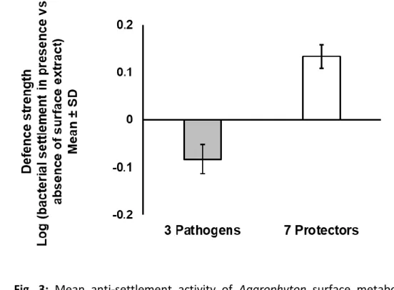
was it not possible to test all nineteen ‘protective’ strains. Thus, only five ‘significant 277
protectors’ i.e. Ralstonia sp., Shewanella aquimarina, Tenacibaculum skagerrakense, 278
Alteromonas stellipolaris, Tenacibaculum aestuarii and two ‘potential protectors’ i.e.
279
Cobetia marina and Nonlabens dokdonensis were tested. 106 µL suspensions of these 280
bacterial strains (O.D. 0.6-0.8) precultured in MB liquid medium (as described above) were 281
pipetted into the wells. The bacteria were allowed to settle for 3 h, and the settled cells that 282
13
could not be removed by rinsing two times with 110µL sterile seawater were stained with 283
the fluorescent DNA-binding dye Syto 9 (Invitrogen, GmbH). Fluorescence was subsequently 284
measured (excitation, 477–491 nm; emission, 540 nm) with a plate reader as a proxy for 285
bacterial settlement in terms of the attached cell density. All tested strains were allowed to 286
settle on all extracts.
287
The defence strength of Agarophyton surface metabolites is expressed as the ‘log effect 288
ratio,’ i.e., the logarithm of the fluorescence attributable to the settled bacteria of strain Y in 289
the presence of surface metabolites, divided by the fluorescence attributable to the settled 290
bacteria of strain Y in the absence of surface metabolites. A log effect ratio value of 0 (i.e., 291
an equal number of settled bacteria in wells with and without surface metabolites) 292
indicated that the tested surface metabolites had no effect on settlement, whereas a 293
negative log effect ratio value indicated a deterrent effect, and a positive log effect ratio 294
value indicated an attractant effect. Thus, a log effect ratio of -1 represents a 10-fold 295
reduction, whereas a value of +1 represents a 10-fold enhancement of bacterial settlement 296
caused by surface metabolites.
297 298
Defence strength = log (bacterial settlement in presence of Agarophyton surface metabolites) 299
(bacterial settlement in absence of Agarophyton surface metabolites) 300
301
Results 302
Agarophyton tip bleaching assay with epibacterial strains 303
(A) Bleaching assay with single isolates 304
14
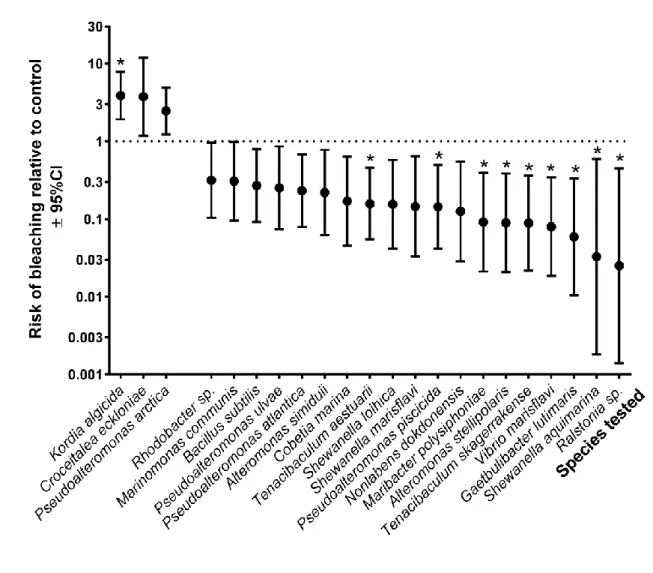
Out of 58 tested bacterial isolates Kordia algicida was found to significantly increase the risk 305
of tip bleaching (Table 1; Fig. 1, p<0.00086), compared to control treatments without 306
inoculation of bacteria and was a ‘significant pathogen’ after Bonferroni correction. Two 307
additional isolates (Pseudoalteromonas arctica and Croceitalea eckloniae) had the same 308
effect but were not significantly pathogenic after Bonferroni correction (Table 1; Fig. 1, 309
p<0.05) and were thus ‘potential pathogens’. Out of the remaining 55 isolates, 9 were found 310
to significantly reduce the risk of tip bleaching (Table 1; Fig. 1, p<0.00086) and were grouped 311
under ‘significant protectors’. 10 others had the same effect, although they were not 312
significantly protective after Bonferroni correction (Table 1; Fig. 1, p<0.05) and were called 313
‘potential protectors’. The remaining 36 isolates were found to be neutral, neither inducing 314
nor preventing bleaching (see Table S2; Fig. S2 in Supporting Information).
315
Similar numbers of microbiota that originated from native and non-native populations (30 316
and 28, respectively, see Table 1 and Table S2) of Agarophyton were tested in our bleaching 317
assay and double numbers of protective microbiota were detected from the non-native 318
range (3 from the native range and 6 from the non-native range, respectively, Table 1).
319
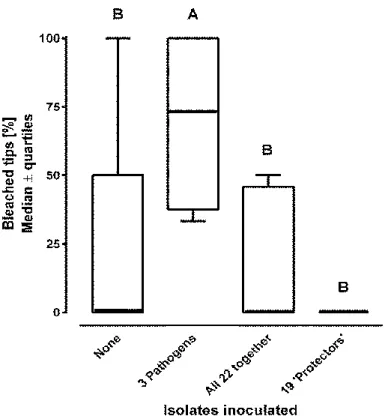
(B) Bleaching assay with combined ‘protectors’ and ‘pathogens’
320
When all three isolates (one ‘significant pathogen’ and two ‘potential pathogen’) that 321
induced bleaching individually at least with p < 0.05 were combined, a significant increase in 322
bleaching relative to the control was again observed (Fig. 2, p<0.05). However, combined 323
application of these three ‘pathogens’ and the nineteen ‘protective’ isolates that prevented 324
bleaching individually at least with p < 0.05 resulted in no such increase (Fig. 2, p<0.05). No 325
bleaching was observed when all 19 ‘protectors’ and no ‘pathogens’ were inoculated.
326 327
Defence capacity test of Agarophyton against pathogens and protectors 328
15
The effect of Agarophyton surface associated metabolites on bacterial settlement differed 329
significantly between the two groups of bacteria, i.e. ‘protectors’ and ‘inducers’ (Fig. 3, 330
Welch-corrected t-test, p < 0.0001). While the surface associated metabolites significantly 331
increased the settlement of ‘protectors’, the settlement of the bleaching ‘inducers’ was 332
significantly reduced by the surface associated metabolites.
333
334
Discussion 335
The data presented here demonstrate for the first time that aquatic macrophytes can use 336
surface associated chemicals not only to directly reduce settlement of pathogenic bacteria, 337
but also to recruit bacterial strains that provide protection from such pathogens. The 338
epimicrobiome of Agarophyton contains a component that protects the alga from 339
pathogens in a similar way as earlier demonstrated for two other seaweeds (G. conferta 340
(Weinberger et al., 1997) and D. pulchra (Longford et al., 2019)), and the settlement of such 341
protective bacteria on the surface of Agarophyton is not random. Surface associated 342
metabolites from the Agarophyton holobiont significantly deterred three strains that were 343
significantly and potentially pathogenic, while the metabolites had a probiotic effect 344
towards seven significantly and potentially protective strains that were tested. This confirms 345
the surface chemistry of Agarophyton has a similarly strong selective effect on bacterial 346
colonization as in Fucus vesiculosus (Lachnit et al., 2010) or Delisea pulchra (Longford et al., 347
2019). Moreover, it demonstrates for the first time that this selection is not only targeted to 348
exclude pathogens, but also targeted to attract protectors. Together with Lachnit et al., 349
(2010) and Kessler et al., (2018) our data strongly support the concept of chemically 350
mediated recruitment of microbes and not the ‘neutral’ hypothesis, according to which the 351
microbial community structure is determined randomly. Our data clearly support the 352
16
targeted deterrence hypothesis, as settlement of detrimental bacteria was chemically 353
suppressed. On the other hand, we cannot reject the ‘niche’ model, as multiple microbiota 354
were attracted by Agarophyton and possibly able to make use of resources provided by it.
355
Also, the ‘lottery’ hypothesis cannot be currently rejected, since the capacity of attracted 356
microbiota to coexist and share host resources is unknown.
357
Only 5% of the marine bacterial strains are cultivable (Haglund et al., 2002) and to date no 358
alternative technique has been developed to separate selected microbial components from 359
natural microbial communities and to test them in infection assays. Thus, only a small 360
fraction of all bacteria that are associated with the surface of Agarophyton could be isolated 361
and tested in our study. One representative out of 58 tested bacterial species, Kordia 362
algicida, was significantly capable to induce the tip bleaching symptom in Agarophyton. K.
363
algicida is already known to be detrimental to other organisms. It can kill diatom blooms in 364
a protease mediated molecular interaction (Paul & Pohnert, 2011) and a similar mechanism 365
cannot be excluded in the present case. Bleaching is often correlated with microbial cell wall 366
matrix degrading activity (Weinberger et al., 1994, 1997), but this was not the case in the 367
present study, as Kordia is incapable of agar degradation.
368
Two other isolates – which were also not agar degraders - also exhibited the potential to 369
induce bleaching symptoms in Agarophton, which strongly suggests that this capacity is not 370
unique. Interestingly, all three detrimental isolates originated from virtually healthy host 371
specimens. Thus, a relevant fraction of Agarophyton’s surface microbiome is obviously 372
composed of opportunistic pathogens that can induce bleaching symptoms under certain 373
conditions, similar as in several red seaweeds belonging to other species (Case et al., 2011;
374
Weinberger et al., 1994; Weinberger et al., 1997). Given that three out of 58 culturable 375
17
strains were significant or potential pathogens this fraction can be estimated to include 376
approximately 5 % of the microbiome. However, this percentage calculation is based on the 377
culturable proportion which is just a representative sample of the whole microbiome.
378
Of the remaining strains, 19 (9 ‘significant protectors’ and 10 ‘potential protectors’) could 379
reduce the risk of ‘spontaneous’ bleaching in thalli that were not intentionally inoculated 380
with pathogenic bacteria (Fig. 1). All the specimens of Agarophyton tested in our bleaching 381
induction assays were subjected to a pretreatment with two antibiotics that inhibited 382
bacterial cell wall synthesis, with the dual goal to remove opportunistic pathogens and to 383
disturb and weaken any protective components of the algal microbiome. The circumstance 384
that bleaching occurred ‘spontaneously’ at a low rate but could be prevented by an 385
important percentage of all tested isolates suggests that some opportunistic pathogens 386
survived the treatment with antibiotics but could then not become virulent when protective 387
bacteria were inoculated - similar as previously reported for bacteria that had been isolated 388
from Gracilaria ‘conferta’ and prevented thallus tip bleaching in this alga (Weinberger et al., 389
1997). The protective effect of various isolates on Agarophyton was further confirmed when 390
all the 19 protective strains (nine ‘significantly protective’ and ten ‘potentially protective’) 391
were tested together in combination with the 3 pathogenic strains (one ‘significant 392
pathogen’ and two ‘potential pathogens’) and a bleaching reduction was still documented.
393
Alltogether, our observations strongly hint at the presence of protective epibacteria on the 394
surface of Agarophyton. They could (again estimated from the number of identified isolates 395
in our tested strain collection) comprise at least 15% of all taxa present in this microbiome.
396
The presence of such beneficial bacteria has been previously demonstrated not only for 397
other Gracilarioids (Weinberger et al., 1997), but also for Delisea pulchra (Longford et al., 398
2019), corals (Rosenberg et al., 2007) and other seaweeds like the brown alga Fucus 399
18
vesiculosus, in which surface associated bacteria were found to inhibit the settlement of 400
macrofoulers (Nasrolahi et al., 2012).
401
The biofilms on seaweed surfaces represent highly competitive environments, with 402
microbes competing for refuge, nutrients and substratum, and interspecific antagonistic 403
effects of bacterial strains are not rare. For example, such effects have been previously 404
demonstrated for the brown alga Saccharina latissima (Wiese et al., 2009), the red alga 405
Delisea pulchra and the green alga Ulva australis (Penesyan et al., 2009). Release of 406
inhibitory components like antibiotics (Wiese et al., 2009) and/or quorum sensing inhibitors 407
(Romero et al., 2010) has been observed and could also explain the ‘protective’ effect 408
observed by us. Interestingly, one of the significant protective strains, Pseudoalteromonas 409
piscicida, belongs to a genus which comprises several species that are known to produce 410
antibacterial products to outcompete other bacteria for space and nutrients (Holmström &
411
Kjelleberg, 1999). Also Pseudoalteromonas piscicida has been recently demonstrated to 412
inhibit and/or kill competing bacteria - including several marine pathogens, such as Vibrio 413
vulnificus, Vibrio parahaemolyticus, Vibrio cholerae, Photobacterium damselae, and 414
Shewanella algae - through secretion of antimicrobial substances and the direct transfer of 415
digestive vesicles to competing bacteria (Richards et al., 2017). On the other hand, 416
Shewanella aquimarina exhibited a strong protective effect against Agarophyton tip 417
bleaching in the current study and the same was observed with two other potentially 418
protective species of the genus Shewanella, i.e. S. marisflavi and S. loihica. These 419
observations contrast with the findings that S. marisflavi is a pathogen of sea cucumbers (Li 420
et al., 2010) and other bacteria of the genus are pathogenic towards humans. The 421
mechanisms behind the protective effects on Agarophyton deserve further investigation.
422
The exact (additive or synergistic) contributions of the active epibacterial players in the 423
19
cross-infection experiment with all 19 ‘protectors’ combined with 3 ‘pathogens’ are not 424
known yet.
425
Beneficial roles of certain components in the bacterial communities around the rhizosphere 426
of terrestrial plants are well known. They can not only facilitate nutrient acquisition, but also 427
support plant growth under biotic and abiotic plant stress (Lareen et al., 2016; Mendes et 428
al., 2013). Seaweed-associated bacteria may also facilitate nutrient acquisition and provide 429
essential vitamins and growth factors (Wahl et al., 2012), and – as confirmed in the present 430
study – mediate biotic stress. However, while we have started to understand these benefits 431
and to gather evidence of a selective recruitment of bacteria both in terrestrial (Lebeis et al., 432
2015) and aquatic environments, empirical links between this selective recruitment of 433
communities and a health benefit for the host are still very rare in the aquatic realm. Kessler 434
et al., 2018 recently demonstrated for the marine macroalga Ulva mutabilis (Chlorophyta) a 435
mediating role of the algal osmolyte DMSP (dimethylsulfoniopropionate) in the attraction of 436
the beneficial bacterium Roseovarius sp. MS2, responsible for release of morphogenetic 437
compounds that ensure proper algal morphogenesis. In absence of these morphogenetic 438
compounds under axenic conditions, Ulva mutabilis develops into callus-like colonies 439
consisting of undifferentiated cells and abnormal cell walls. While microbial ‘gardening’ via 440
use of chemicals has been documented in terms of growth and development of seaweeds 441
(Kessler et al., 2018), our study demonstrates for the first time such a link between the 442
disease resistance capacity of a seaweed and beneficial selective gardening of ‘protective’
443
bacteria based upon surface chemistry.
444
Metabolites present on the surface of seaweeds or in the rhizosphere are a cocktail of 445
metabolites originating both from the algal or plant host and from associated surface 446
20
microbiota. Such surface associated metabolites from the algal holobiont are also known to 447
function as a defence against fouling by microfoulers (e.g. bacteria, diatoms) and 448
macrofoulers (e.g. barnacle larvae, mussels) (reviewed by Da Gama, 2014; Saha et al., 2017).
449
Also epibacteria from seaweeds are well known to have inhibitory activities against other 450
fouling organisms (Singh & Reddy, 2014). Thus, it is possible that beneficial bacteria 451
recruited by Agarophyton will not only act as a defence against pathogens but also against 452
other foulers, like filamentous algae. Using a transcriptomic approach, de Oliviera et al., 453
2012 demonstrated that the red seaweed host Laurencia dendriodea (rather the surface 454
associated bacteria) is involved in the biosynthesis of terpenoids (chemical defence 455
compounds against bacterial colonization and infection) through the mevalonate 456
independent pathway. For the Agarophyton holobiont, we do not know yet the identity of 457
surface associated bioactive compounds. Thus, it was not possible for us to distinguish the 458
relative contributions of surface metabolites originating from the algal host Agarophyton 459
and from surface associated microbiota. Mutants of Arabidopsis thaliana with suppressed 460
salicylic acid signalling pathways formed abnormal root microbiomes when compared to the 461
wild plants (Lebeis et al., 2015), which could suggest that the role of the host was more 462
important in this specific case. The contribution of seaweed microbiome metabolites 463
depends on the community composition, abundance and metabolic activity (Wahl et al., 464
2010) and may be expected to be more variable than that of the host. Selective effects 465
observed with surface associated metabolites coming from different algal individuals varied 466
relatively little in our study, which could suggest that metabolites generated by the host 467
have more importance. However, our knowledge of the species-species interactions of 468
cultivatable and non-cultivatible taxa associated with Agarophyton or other plants is 469
rudimentary at best. The involvement of multiple protective microorganisms in our and 470
21
several other cases (see above) strongly supports the view that the traditional conceptual 471
model emphasizing direct interactions of hosts and single microbes needs to be expanded to 472
a holobiont concept if seaweed- or plant-microbe interactions are to be understood (Egan et 473
al., 2013).
474
In conclusion, our study demonstrates for the first-time selective chemical ‘gardening’ of 475
protective epibacterial strains by a seaweed holobiont with regard to disease resistance 476
capacity. The combined effect of metabolites generated by the host alga and the protection 477
offered by associated microbial partners determines the virulence of harmful opportunistic 478
bacterial pathogens. A major component of the epibacterial community appears capable of 479
contributing to this protection against co-occurring pathogens, which suggests that 480
microbiota of very different taxonomic groups may provide the holobiont with the same 481
ecological function, which could be pivotal for the establishment of Agarophyton in new 482
environments. Thus, absence of protective microbiota in new environments might not be a 483
factor limiting the invasion success of Agarophyton.
484
As known for other seaweeds like the brown alga Fucus vesiculosus, bioactive surface 485
metabolites often act in synergism or additively and/or antagonistically, producing an 486
overall defensive or prebiotic effect on bacterial recruitment (Saha et al., 2011; Saha et al., 487
2012). The identification of metabolites responsible for such chemical ‘gardening’ effects via 488
classical bioassay guided fractionation techniques in the near future may allow us to 489
manipulate algal thallus microbiomes to enhance seaweed health, prevent bleaching 490
diseases and ensure production and sustainability in Agarophyton aquaculture.
491
Acknowledgements 492
22
This research was supported by a grant (CP1215) from the DFG Cluster of Excellence “Future 493
Ocean” to M. Saha. We thank the Institute of Clinical Molecular Biology of the Christian- 494
Albrechts-University Kiel (Germany) for performing Sanger sequencing, supported in part by 495
the DFG Clusters of Excellence “Inflammation at Interfaces” and “Future Ocean”.
496
Author contributions 497
M.S. isolated and identified the bacterial isolates, designed and performed the anti- 498
settlement experiments. F.W. designed and performed the tip bleaching assays. M.S. and 499
F.W. analysed the data. M.S. wrote the paper and F.W. contributed to the editing.
500
Author Declaration 501
The authors declare no conflict of interest. Data underlying this publication are freely 502
accessible and can be downloaded from the DRYAD data repository (Provisional DOI:
503
doi:10.5061/dryad.52j8p1r).
504
References 505
506
Bacilio-Jiménez, M., Aguilar-flores, S., Ventura-zapata, E., & Eduardo, P. (2003). Chemical 507
characterization of root exudates from rice (Oryza sativa) and their effects on the 508
chemotactic response of endophytic bacteria, Plant and Soil 249: 271–277.
509
Bell, W., & Mitchell, R. (1972). Chemotactic and growth responses of marine bacteria to algal 510
extracellular products. The Biological Bulletin, 143(2), 265–277.
511
Bengtsson, M. M., & Ovreas, L. (2010). Planctomycetes dominate biofilms on surfaces of the 512
kelp Laminaria hyperborea. BMC Microbiology, 10(1), 261.
513
Bordenstein, S. R., & Theis, K. R. (2015). Host biology in light of the microbiome: ten 514
23
principles of holobionts and hologenomes. PLoS Biology, 13(8), e1002226.
515
Burke, C., Steinberg, P., Rusch, D. B., Kjelleberg, S., & Thomas, T. (2011). Bacterial community 516
assembly based on functional genes rather than species. Proceedings of the National 517
Academy of Sciences of the USA, 108(34), 14288–14293.
518
Campbell, A. H., Harder, T., Nielsen, S., Kjelleberg, S., & Steinberg, P. D. (2011). Climate 519
change and disease: bleaching of a chemically defended seaweed. Global Change 520
Biology, 17(9), 2958–2970.
521
Case, R. J., Longford, S. R., Campbell, A. H., Low, A., Tujula, N., Steinberg, P. D., & Kjelleberg, 522
S. (2011). Temperature induced bacterial virulence and bleaching disease in a 523
chemically defended marine macroalga. Environmental Microbiology, 13(2), 529–537.
524
Cundell, A. M., Sleeter, T. D., & Mitchell, R. (1977). Microbial populations associated with the 525
surface of the brown alga Ascophyllum nodosum. Microbial Ecology, 4(1), 81–91.
526
Currier, W. W., & Strobel, G. A. (1976). Chemotaxis of Rhizobium spp. to plant root exudates.
527
Plant Physiology, 57(5), 820–823.
528
Egan, S., Harder, T., Burke, C., Steinberg, P., Kjelleberg, S., & Thomas, T. (2013). The seaweed 529
holobiont: Understanding seaweed-bacteria interactions. FEMS Microbiology Reviews, 530
37(3), 462–476.
531
Flint, H. J., Scott, K. P., Louis, P., & Duncan, S. H. (2012). The role of the gut microbiota in 532
nutrition and health. Nature Reviews Gastroenterology and Hepatology, 9(10), 577.
533
Gachon, C. M. M., Sime-Ngando, T., Strittmatter, M., Chambouvet, A., & Kim, G. H. (2010).
534
Algal diseases: spotlight on a black box. Trends in Plant Science, 15(11), 633–40.
535
24
Gurgel, C. F. D., Norris, J. N., Schmidt, W. E., Le, H. A. U. N. H. U., & Fredericq, S. (2018).
536
Systematics of the Gracilariales (Rhodophyta) including new subfamilies, tribes, 537
subgenera, and two new genera, Agarophyton gen. nov. and Crassa gen. nov., 374(1), 538
1–23.
539
Haglund, A.-L., Törnblom, E., Boström, B., & Tranvik, L. (2002). Large differences in the 540
fraction of active bacteria in plankton, sediments, and biofilm. Microbial Ecology, 43(2), 541
232–241.
542
Hartmann, A., Schmid, M., Van Tuinen, D., & Berg, G. (2009). Plant-driven selection of 543
microbes. Plant and Soil, 321(1–2), 235–257.
544
Hollants, J., Leliaert, F., De Clerck, O., & Willems, A. (2013). What we can learn from sushi: A 545
review on seaweed-bacterial associations. FEMS Microbiology Ecology, 83(1), 1–16.
546
Holmström, C., & Kjelleberg, S. (1999). Marine Speudoalteromonas species are associated 547
with higter organisms and produce biologically active extracellular agents. FEMS 548
Microbiology Ecology, 30 , 285–293.
549
Hooper, L. V, Midtvedt, T., & Gordon, J. I. (2002). How host-microbial interactions shape the 550
nutrient environment of the mammalian intestine. Annual Review of Nutrition, 22(1), 551
283–307.
552
Hurd, C. L. (2000). Review water motion , marine macroalgal physiology , and production.
553
Journal of Phycology, 36 (3), 453–472.
554
Kessler, R. W., Weiss, A., Kuegler, S., Hermes, C., & Wichard, T. (2018). Macroalgal–bacterial 555
interactions: Role of dimethylsulfoniopropionate in microbial gardening by Ulva 556
(Chlorophyta). Molecular Ecology, 27(8), 1808–1819.
557
25
Lachnit, T., Blümel, M., Imhoff, J., & Wahl, M. (2009). Specific epibacterial communities on 558
macroalgae: phylogeny matters more than habitat. Aquatic Biology, 5, 181–186.
559
Lachnit, T., Wahl, M., & Harder, T. (2010). Isolated thallus-associated compounds from the 560
macroalga Fucus vesiculosus mediate bacterial surface colonization in the field similar to 561
that on the natural alga. Biofouling, 26(3),247-55.
562
563
Lachnit, T., Meske, D., Wahl, M., Harder, T., & Schmitz, R. (2011). Epibacterial community 564
patterns on marine macroalgae are host-specific but temporally variable. Environmental 565
Microbiology, 13(3), 655–65.
566
Lane, A. L., Nyadong, L., Galhena, A. S., Shearer, T. L., Stout, E. P., Parry, R. M., … Kubanek, J.
567
(2009). Desorption electrospray ionization mass spectrometry reveals surface-mediated 568
antifungal chemical defense of a tropical seaweed. Proceedings of the National 569
Academy of Sciences of the USA, 106 (18), 7314-7319.
570
Lebeis, S. I., Paredes, S. H., Lundberg, D. S., Breakfield, N., Gehring, J., McDonald, M., … 571
Dangl, J. L. (2015). R es e a rc h | r e po r ts 27. Science, 349(6250), 860–864.
572
Li, H., Qiao, G., Li, Q., Zhou, W., Won, K. M., Xu, D., & Park, S. (2010). Biological 573
characteristics and pathogenicity of a highly pathogenic Shewanella marisflavi infecting 574
sea cucumber, Apostichopus japonicus. Journal of Fish Diseases, 33(11), 865–877.
575
Longford, S. R., Campbell, A. H., Nielsen, S., & Case, R. J. (2019). Interactions within the 576
microbiome alter microbial interactions with host chemical defences and affect disease 577
in a marine holobiont, 9, 1–13.
578
Mendes, R., Garbeva, P., & Raaijmakers, J. M. (2013). The rhizosphere microbiome:
579
26
significance of plant beneficial, plant pathogenic, and human pathogenic 580
microorganisms. FEMS Microbiology Reviews, 37(5), 634–663.
581
Moran, M. D., College, H., & Ae, W. (2003). Arguments for rejecting the sequential 582
Bonferroni in ecological studies, Oikos, 2, 1–3.
583
Nasrolahi, A., Stratil, S. B., Jacob, K. J., & Wahl, M. (2012). A protective coat of 584
microorganisms on macroalgae: inhibitory effects of bacterial biofilms and epibiotic 585
microbial assemblages on barnacle attachment. FEMS Microbiology Ecology, 81(3), 586
583–95.
587
Paul, C., & Pohnert, G. (2011). Interactions of the algicidal bacterium Kordia algicida with 588
diatoms: Regulated protease excretion for specific algal lysis. PLoS ONE, 6(6), e21032.
589
Penesyan, A., Marshall‐Jones, Z., Holmstrom, C., Kjelleberg, S., & Egan, S. (2009).
590
Antimicrobial activity observed among cultured marine epiphytic bacteria reflects their 591
potential as a source of new drugs. FEMS Microbiology Ecology, 69(1), 113–124.
592
Raaijmakers, J. M., Paulitz, T. C., Steinberg, C., Alabouvette, C., & Moënne-Loccoz, Y. (2009).
593
The rhizosphere: a playground and battlefield for soilborne pathogens and beneficial 594
microorganisms. Plant and Soil, 321(1–2), 341–361.
595
Richards, G. P., Watson, M. A., Needleman, D. S., Uknalis, J., Boyd, E. F., & Fay, P. (2017).
596
Mechanisms for Pseudoalteromonas piscicida-Induced Killing of Vibrios and Other 597
Bacterial Pathogens, Applied Environmental Ecology, 83(11), 1–17.
598
Romero, M., Martin-Cuadrado, A.-B., Roca-Rivada, A., Cabello, A. M., & Otero, A. (2010).
599
Quorum quenching in cultivable bacteria from dense marine coastal microbial 600
communities. FEMS Microbiology Ecology, 75(2), 205–217.
601
27
Rosenberg, E., Koren, O., Reshef, L., Efrony, R., & Zilber-Rosenberg, I. (2007). The role of 602
microorganisms in coral health, disease and evolution. Nature Reviews Microbiology, 603
5(5), 355.
604
Saha, M., Rempt, M., Gebser, B., Grueneberg, J., Pohnert, G., & Weinberger, F. (2012).
605
Dimethylsulphopropionate (DMSP) and proline from the surface of the brown alga 606
Fucus vesiculosus inhibit bacterial attachment. Biofouling, 28(6), 593–604.
607
Saha, M., Rempt, M., Grosser, K., Pohnert, G., & Weinberger, F. (2011). Surface-associated 608
fucoxanthin mediates settlement of bacterial epiphytes on the rockweed Fucus 609
vesiculosus. Biofouling, 27(4), 423–433.
610
Schiel, D. R., & Foster, M. S. (2006). The population biology of large brown seaweeds:
611
ecological consequences of multiphase life histories in dynamic coastal environments.
612
Annual Review of Ecology, Evolution, and Systematics, 37, 343–372.
613
Singh, R. P., & Reddy, C. R. K. (2014). Seaweed-microbial interactions: Key functions of 614
seaweed-associated bacteria. FEMS Microbiology Ecology, 88(2), 213–230.
615
Van Alstyne, K., & Houser, L. (2003). Dimethylsulfide release during macroinvertebrate 616
grazing and its role as an activated chemical defense. Marine Ecology Progress Series, 617
250, 175–181.
618
Wahl, M. (1989). Marine epibiosis. I. Fouling and antifoulinng: some basic aspects. Marine 619
Ecology Progress Series, 58, 175–189.
620
Wahl, M. (2008). Ecological lever and interface ecology: epibiosis modulates the interactions 621
between host and environment. Biofouling, 24(6), 427–38.
622
Wahl, M., Goecke, F., Labes, A., Dobretsov, S., & Weinberger, F. (2012). The second skin:
623
28
Ecological role of epibiotic biofilms on marine organisms. Frontiers in Microbiology, 3, 624
1–21.
625
Wahl, M., ShahnazL., Dobretsov, S., Saha, M., Symanowski, F., DavidK., … Weinberger, F.
626
(2010). Ecology of antifouling resistance in the bladder wrack Fucus vesiculosus:
627
Patterns of microfouling and antimicrobial protection. Marine Ecology Progress Series, 628
411, 33-48.
629
Weinberger, F. (2007). Pathogen induced defense and innate immunity in macroalgae.
630
Biological Bulletin, 35(1), 29–54.
631
Weinberger, F., Friedlander, M., & Gunkel, W. (1994). A bacterial facultative parasite of 632
Gracilaria conferta. Diseases of Aquatic Organisms, 18(2), 135–141.
633
Weinberger, F., Hoppe, H. G., & Friedlander, M. (1997). Bacterial induction and inhibition of 634
a fast necrotic response in Gracilaria conferta (Rhodophyta). Journal of Applied 635
Phycology, 9(3), 277–285.
636
Wiese, J., Thiel, V., Nagel, K., Staufenberger, T., & Imhoff, J. F. (2009). Diversity of antibiotic- 637
active bacteria associated with the brown alga Laminaria saccharina from the Baltic 638
Sea. Marine Biotechnology, 11(2), 287–300.
639
Yuan, J., Chaparro, J. M., Manter, D. K., Zhang, R., Vivanco, J. M., & Shen, Q. (2015). Roots 640
from distinct plant developmental stages are capable of rapidly selecting their own 641
microbiome without the influence of environmental and soil edaphic factors. Soil 642
Biology and Biochemistry, 89, 206–209.
643
644
29 645
![Table 1. Epibacterial strains [isolated from Agarophyton vermiculophyllum (GV) populations]](https://thumb-eu.123doks.com/thumbv2/1library_info/5317166.1679435/30.892.100.818.400.1145/table-epibacterial-strains-isolated-from-agarophyton-vermiculophyllum-populations.webp)