AUS DER ABTEILUNG FÜR PLASTISCHE-, HAND-, UND WIEDERHERSTELLUNGSCHIRURGIE
(PROF. DR. DR. MED. LUKAS PRANTL) DER FAKTULTÄT FÜR MEDIZIN DER UNIVERSITÄT REGENSBURG
Semi-automated extraction of stromal vascular fraction for
autologous cell therapy
Inaugural - Dissertation zur Erlangung des Doktorgrades
der Medizin
der
Fakultät für Medizin der Universität Regensburg
vorgelegt von Alexander Hanke
2016
AUS DER ABTEILUNG FÜR PLASTISCHE-, HAND-, UND WIEDERHERSTELLUNGSCHIRURGIE
(PROF. DR. DR. MED. LUKAS PRANTL) DER FAKULTÄT FÜR MEDIZIN DER UNIVERSITÄT REGENSBURG
Semi-automated extraction of stromal vascular fraction for
autologous cell therapy
Inaugural - Dissertation zur Erlangung des Doktorgrades
der Medizin
der
Fakultät für Medizin der Universität Regensburg
vorgelegt von Alexander Hanke
2016
Dekan: Prof. Dr. Dr. Torsten E. Reichert 1. Berichterstatter: Prof. Dr. Dr. Lukas Prantl
2. Berichterstatter: Prof. Dr. Dr. Torsten E. Reichert Tag der mündlichen Prüfung: 26. April 2017
3
1. Directory
1. Directory ... 3
2. Summary (German) ... 6
3. Summary ... 8
4. Abbreviations ... 10
5. Introduction ... 11
5.1. Stem Cells ... 11
5.2. Mesenchymal Stem Cells ... 11
5.3. Sources of Mesenchymal Stem Cells ... 12
5.4. MSCs from Bone Marrow and Adipose Tissue ... 13
5.5. Advantages of Stem Cells from Adipose Tissue ... 14
5.6. SVF and ASCs for autologous therapy ... 15
5.7. Rationale for the use of a Medical Device ... 17
5.8. Medical devices ... 17
5.9. Molecular markers ... 18
5.9.1. CD34 ... 18
5.9.2. CD45 ... 19
5.9.3. CD271 ... 19
6. Aim of the study ... 20
7. Material and methods... 21
7.1. Materials ... 21
7.1.1. Cells and tissues ... 21
7.1.2. Culture media and supplements ... 21
7.1.3. Fluids and other reagents ... 21
7.1.4. Cell Culture equipment ... 21
7.1.5. Enzymes ... 22
4
7.1.6. Antibodies and Kits ... 22
7.1.7. Equipment for preparation with a medical device... 22
7.1.8. Software ... 23
7.1.9. General Equipment ... 23
7.2. Extraction of the Stromal Vascular Fraction ... 24
7.2.1. Hand preparation ... 27
7.2.2. Preparation with a medical device ... 28
7.3. Flow Cytometry of the SVF ... 34
7.3.1. Technique of Flow Cytometry ... 34
7.3.2. Experimental setting ... 34
7.3.3. Preparation of the samples ... 36
7.3.4. Labeling ... 37
7.3.5. Measurement ... 38
7.3.6. Gating strategy ... 39
7.4. Cell culture ... 45
7.4.1. Medium and Culture Conditions ... 45
7.4.2. Trypsination and Splitting ... 45
7.4.3. Counting ... 45
7.4.4. Freezing ... 45
7.4.5. Thawing ... 46
7.5. Colony Forming Unit (CFU) Assay ... 47
7.6. Statistical analysis ... 49
8. Results ... 50
8.1. Flow Cytometry of the SVF ... 50
8.1.1. SVF ... 50
8.1.2. Cell concentration ... 50
8.1.3. Single positive Cells ... 52
5
8.1.4. Double positive Cells ... 54
8.1.5. Stain index ... 56
8.2. Colony Forming Unit (CFU) Assay ... 57
9. Discussion ... 61
9.1. Cell concentration varies between both methods ... 61
9.1.1. Explanation of the Preparation Failures ... 66
9.1.2. Suggestion for solution ... 67
9.2. Percentage of single and double positive cells ... 68
9.2.1. CD34 ... 68
9.2.2. CD45 ... 68
9.2.3. CD271 ... 69
9.2.4. CD34/CD45 double positive cells ... 69
9.2.5. CD34/CD271 double positive cells ... 70
9.2.6. CD45/CD271 double positive cells ... 70
9.3. Stain index ... 71
9.4. Colony Forming Unit (CFU) Assay ... 71
9.5. Application of the novel device for cell assisted lipotransfer (CAL) ... 71
9.6. Limitations of this work and outlook ... 72
9.7. Conclusion ... 73
10. References ... 74
11. Declaration (German)... 81
12. Acknowledgements (German) ... 82
13. Addendum ... 83
13.1. Figures ... 83
13.2. Tables ... 86
13.3. Curriculum Vitae (German) ... 87
6
2. Summary (German)
Einleitung: Die stammzellreiche Stromal Vascular Fraction (SVF) kann aus Lipoaspirat oder Fettgewebe durch enzymatische Verdau und anschließender Zentrifugation gewonnen werden. Bisher hat sich jedoch weder ein einheitliches Extraktionsverfahren, noch eine gängige Methode zur Anwendung am Patienten durchgesetzt. Ein neues kommerziell erhältliches halbautomatisches System zur Herstellung von SVF verspricht Sterilität, konstante Ergebnisse und Anwendbarkeit im klinischen Alltag. Ziel dieser Arbeit war es die Menge und Qualität der SVF, welche mit diesem System gewonnen werden kann, mit einer etablierten manuellen Labormethode zu vergleichen.
Material und Methodik: Die SVF wurde aus Lipoaspirat sowohl mit einem Prototyp der halbautomatischen UNiStation (NeoGenesis, Seoul, Korea) als auch mittels einer etablierten manuellen Labormethode extrahiert. Nach Lyse der verbliebenen Erythrozyten in der SVF erfolgte eine Messung mittels multiparametrischer Durchflusszytometrie (FACSCanto-II, BD Biosciences). Von Interesse war vorrangig die (Quantität) Gesamtzellzahl des gewonnenen Materials. Zusätzlich wurde die Qualität der SVF anhand des Stammzellmarkers CD34, dem Leukozytenmarker CD45 und dem Marker CD271 für hochproliferative Stammzellen untersucht. Des Weiteren wurden die prozentuale Verteilung dieser Marker in der SVF, doppelt positive Zellen und der stain index ermittelt.
Ergebnisse: Aus Lipoaspirat von sechs Patienten wurde sowohl mit der Maschine (d für „device“) als auch der (manuellen) Labormethode (h für „hand preparation“) eine makroskopisch sichtbare SVF erzeugt. Mit der maschinellen Extraktion war die Zellausbeute pro ursprünglichem Gramm Lipoaspirat jedoch tendenziell geringer (d:
1.1*105±1.1*105 vs. h: 2.0*105±1.7*105; p=0.06). Bei der Zusammensetzung der SVF zeigte sich der Anteil an CD34+ Zellen nach maschineller Extraktion signifikant reduziert (d: 57.3±23.8% vs. h: 74.1±13.4%; p=0.02). Im Gegensatz dazu lag der Anteil an CD45+ Leukozyten tendenziell höher (d: 20.7±15.8% vs. h: 9.8±7.1%;
p=0.07). Die Fraktion hochproliferativer CD271+ Zellen zeigte unabhängig von der Extraktionsmethode vergleichbarer Ergebnisse ohne signifikanten Unterschied (M:
13.4±11.6% vs. L: 12.9±9.6%; p=0.74). Es konnte kein Unterschied bzgl. des Anteils doppelt positiver Zellen für CD34+/CD45+ (d: 0.5±0.6% vs. h: 0.3±0.2%; p=0.21)
7
sowie CD34+/CD271+ (d: 1.9±2.3% vs. h: 2.4±2.0%; p=0.42) festgestellt werden.
CD45+/CD271+ doppelt positive Zellen konnten in keiner Probe nachgewiesen werden. Außerdem gab es keinen signifikanten Unterschied bezüglich des Stain Index (p>0.12).
Diskussion und Schlussfolgerung: Das halbautomatisierte System ermöglicht auf kleinstem Raum nennenswerte Mengen an SVF steril zu gewinnen. Diese SVF unterscheidet sich nur geringfügig in der Zusammensetzung gegenüber der manuellen SVF Extraktion. Insgesamt decken sich die Ergebnisse beider Methoden sehr gut mit den aus der Literatur bekannten Werten. Das halbautomatisierte System bietet die Möglichkeit, Forschung und Anwendung der SVF einen Schritt näher in die Klinik zu bringen.
8
3. Summary
Introduction: The stem cell rich Stromal Vascular Fraction (SVF) can be obtained by enzymatic digestion with a collagenase followed by centrifugation from patients’
lipoaspirate or fat tissue. To date neither a standardized extraction method nor a generally accepted application procedure exists for common use on patient. A novel commercially available semi-automated device for the extraction of SVF promises sterility, consistent results and usability in the clinical routine. The aim of this work was to investigate the quantity and quality of the SVF obtained by a semi-automated process in comparison to an established manual laboratory method.
Material and Methods: SVF was extracted from lipoaspirate by a prototype of the semi-automated UNiStation (NeoGenesis, Seoul, Korea) as well as by hand preparation with common laboratory equipment. The SVF was measured by multi- parametric flow-cytometry (FACSCanto-II, BD Biosciences) following the lysis of the remaining erythrocytes. The primary interest was the total cell number (quantity) of the extracted cells. In addition, the quality of the SVFs was investigated using the stem cell marker CD34, the leucocyte marker CD45 and the marker CD271 for highly proliferative stem cells. Furthermore, the distribution of these markers, double positive cells and the stain index were investigated.
Results: Lipoaspirate obtained from six patients was processed with both the novel device (d) as the hand preparation using laboratory equipment (h), always resulting in a macroscopically visible SVF. However, there was a tendency of a fewer cell yield per gram of used lipoaspirate with the device (d: 1.1*105±1.1*105 vs. h:
2.0*105±1.7*105; p=0.06). Regarding the composition of the SVF, the percentage of CD34+ cells was significantly reduced with the device (d: 57.3±23.8% vs. h:
74.1±13.4%; p=0.02). On the contrary there was a tendency to a higher percentage of CD45+ leukocytes (d: 20.7±15.8% vs. h: 9.8±7.1%; p=0.07). The percentage of highly proliferative CD271+ cells was comparable for both methods (d: 13.4±11.6%
vs. h: 12.9±9.6%; p=0.74). No significant difference was identified regarding the double positive cell fraction for CD34+/CD45+ (d: 0.5±0.6% vs. h: 0.3±0.2%; p=0.21) and CD34+/CD271+ (d: 1.9±2.3% vs. h: 2.4±2.0%; p=0.42). Double positive cells for CD45+/CD271+ were not detected in any sample. The stain index did not show a significant difference between the two extraction methods (p>0.12).
9
Discussion: The semi-automated system was able to provide considerable amounts of sterile SVF without requiring much space. The SVF extracted by the semi- automated process showed only little difference in its composition compared with the SVF obtained by the hand preparation. Taken together both methods showed comparable extraction results which are in accordance with the data from literature.
This semi-automated system offers an opportunity to take research and application of the SVF one step further to the clinic.
10
4. Abbreviations
AB Antibody
APC Allophycocyanin
ASCs Human adipose tissue-derived stem cells ATMP Advanced therapy medicinal products
CV Crystal violet
CAL Cell assisted lipotransfer DAPI 4′,6-Diamidin-2-phenylindol
FBS Fetal bovine serum
FSC Forwardscatter (Flow Cytometry)
HGF Hepatocyte growth factor
IDO indoleamine 2,3-dioxygenase
IGF1 Insulin-like growth factor 1
IL10 Interleukin 10
ISCT International Society for Cellular Therapy
MEM Minimum eagle medium
mg Milligram
ml Milliliter
µg Microgram
µl Microliter
P Passage
PBS Phosphate buffered saline
PE Phycoerythrin
PE-Cy7 Phycoerythrin-Cy7
PFA Paraformaldehyde
PGE2 Prostaglandin E2
rpm Rounds per minute
SVF Stromal vascular fraction
SSC Sidewardscatter (Flow Cytometry)
TGFβ Tissue growth factor β
U Unit
V Volume
VEGF Vascular endothelial growth factor
11
5. Introduction
5.1. Stem Cells
A defining feature of stem cells is their self-renewal while maintaining the potential to differentiate into various cell types and lineages. However, the potential of stem cells depends on their origin and possible lineage specification during early stages of differentiation: Therefore, zygotes are referred as totipotent, capable of giving rise to any cell of an embryo including extra-embryonic tissue like the placenta. The potential of zygotes to differentiate during embryonic development becomes limited to one of the three germ layers (i.e. ectoderm, mesoderm, endoderm (1)) which is defined as pluri-potency. The multi-potent character of differentiation defines cells that are only able to differentiate into cell types of one germ layer. The following cells are only capable of creating a specific tissue which makes them progenitor cells. This leads to tri-, bi- and finally uni-potential cells which are the basis for mature tissue cells. By reaching the stage of a progenitor cell, they lose their telomerase activity and thus their potential for self-renewal. This restricts them to the Hayflick limit of 50- 70 population doublings before cell senescence and programmed cell death (2).
However, a limited number of cells keep their properties of self-renewal and differentiation. They are required for continual maintenance and repair of tissues and organs throughout the organism's lifespan and are specified as adult stem cells (3).
5.2. Mesenchymal Stem Cells
There is a hierarchy between adult stem cells, equal to the hierarchy during embryogenesis. Most of them are tissue specific precursor cells with low differentiation potential. They can differentiate into a few cell types only. Some other stem cells have a higher differentiation potential and can differentiate into a variety of cells types. Between the 1960s and 1970s one type of adult stem cells was identified as a subpopulation of bone marrow cells, however different from hematopoietic stem cells. They demonstrated a fibroblast like morphology, culture plastic adherence and osteogenic differentiation potential (4). In 1990, these cells were termed
12
"mesenchymal stem cells" (MSCs) for the first time by Caplan (5). In 2006, a concise definition was established by the International Society for Cellular Therapy (ISCT).
Mesenchymal stem cells were defined as plastic adherent cells with a special phenotype (positive for CD105+, CD73+, CD90+ and negative for CD45-, CD34-, CD14- or CD11b-, CD79a- or CD19- and HLA-DR-) and with the ability to differentiate into osteoblasts, adipocytes and chondroblasts in vitro (6). Nowadays, it is known that MSCs exist in bone marrow and many other tissues, e.g. in adipose tissue, skin, muscle, kidney, dental pulp and the heart (7). Moreover, recent in vitro experiments consistently provided evidence that MSCs can not only differentiate into mesenchymal cell lineages (as the previously named osteoblasts, adipocytes, and chondrocytes) but also into non-mesenchymal cell lineages as skeletal muscle cells, hepatocytes, endothelial or neuronal cells under appropriate culture conditions (8).
Growing evidence suggests that MSCs might have a close relation to pericytes (9,10). Their exact origin still remains unclear. However, the interest in MSCs for novel cellular therapies grew over the last decade, accompanied by an increasing number of scientific publications.
5.3. Sources of Mesenchymal Stem Cells
MSCs can be found in various tissues (11) and have been investigated for their capacity of self-renewal and differentiation. For research purpose, and especially clinical application, a minimal invasive, easily accessible and abundant source of MSCs is required. Over the past decades MSCs were primarily isolated from bone marrow aspirates and are referred to as bone marrow mesenchymal stem cells (BMSCs). In 2002 Zuk et. al. (12) published the successful isolation of MSCs from adipose tissue for the first time, and provoked great interest in these cells and especially in their potential for clinical applications. The isolated heterogeneous cell mixture is commonly referred to as stromal vascular fraction (SVF) and the plastic adherent subpopulation as adipose tissue-derived stem cells (ASCs).
There exist various methods and variants for harvesting fat tissue. Direct fat resection from the abdomen is reported to have the highest cell yield compared to axilla and flank adipose tissue (13). However, liposuction appears to be the more suitable extraction technique given the fact that this technique is less invasive for the
13
patient, along with higher and maintained viability of cells (14). Thus, adipose tissue represents a valuable source for mesenchymal stem cells irrespective of the extraction method.
5.4. MSCs from Bone Marrow and Adipose Tissue
MSCs from bone marrow and adipose tissue share similar cell properties, but are not exactly identical. Therefore, direct comparison of published experimental outcomes is limited. On the one hand, BMSCs and ASCs demonstrate plastic adherence and spindle-like morphology (15), and are both able to differentiate into non- mesenchymal cell lineages as skeletal muscle cells, heart muscle cells and neuronal cells in vitro (16). On the other hand, they show a similar, however not identical expression of surface proteins. For example CD36 and CD49d is expressed on ASCs only, while CD49f, CD104 and CD106 are only present on BMSCs (17). Moreover, another difference is the expression of CD34. While cells of fresh isolated SVF are highly positive for CD34, the resulting ASCs stop expressing this marker and tend to be negative for this surface protein (18), as reported for BMSCs. A new and distinct definition of the SVF and ASCs was established by the International Society for Cellular Therapy in 2013 based on these new insights (19): General markers for stem cells were defined in the SVF (positive for CD34+ and negative for CD31-, CD45-, CD235a-) and ASCs (positive for CD73+, CD90+ and negative for CD31-, CD45-).
However, similar beneficial potential have been reported for BMSCs and ASCs despite the differences in surface antigen expression. The beneficial effect of these cells has been explained not only by their differentiation potential but even more by the secretion of soluble factors. On the one hand, there are growth factors like insulin-like growth factor 1 (IGF1) and vascular endothelial growth factor (VEGF), which are produced by BMSCs as well as by ASCs (20). On the other hand, there are immune-modulative and immune-regulative factors like indoleamine 2,3- dioxygenase (IDO), tissue growth factor β (TGFβ), prostaglandin E2 (PGE2), hepatocyte growth factor (HGF) and interleukin 10 (IL10). These factors are capable to modulate the adaptive immune and innate immune system in various ways (21).
Thus, BMSCs and ASCs alter the micro-environment by secreting bioactive proteins
14
which induce an anti-inflammatory response, and is currently investigated for regenerative therapies (3,22).
A tremendous interest on a regenerative therapeutic strategy exists for example for ischemic diseases, like myocardial infarction. A myocardial infarction causes a cascade of inflammation, scarring and heart failure. MSCs have been reported to limit the extend of cardiac remodeling and improve the ejection fraction in vitro and in vivo, independent of MSCs origin (23). It has been shown, that only 1% of the injected cells are present at the injection site after 24h (24). Therefore, it is suggested that the positive effects are not mainly caused by differentiation of MSCs or contribution of contractile elements. The beneficial effect might be related to the MSCs' paracrine mechanisms that regulate the immune response and also to the secretion of IGF1 and VEGF preventing apoptosis of cardiomyocytes under hypoxia (25).
5.5. Advantages of Stem Cells from Adipose Tissue
Bone marrow is commonly obtained by punctuation of the iliac crest or vertebral bodies. Aspirations of high amounts up to more than one liter are possible (26).
However, bone marrow is connected to the blood circulation and higher aspiration volumes cause dilution of the bone marrow with peripheral blood (27). This results in a lower concentration of nucleated cells while harvesting huge volumes of bone marrow. Therefore, it has been suggested, that bone marrow should be aspirated carefully in multiple small fractions of each 1ml to 4ml at every spot and level in the bone marrow (28). In addition, high volume aspirations require general anesthesia since the aspiration volume in local anesthesia is limited to about 40ml (16). Further aspiration is associated with high anxiety and pain of the patient (29). Bone marrow aspirates show a range of 1*107 to 5*107 nucleated cells/ml varying due to dilution (30,31).
The liposuction procedure can be performed to obtain tissue amounts starting from 50ml up to 3000ml whereas the amount of harvested tissue determines anesthesia (i.e. local or general anesthesia) (32). The lipoaspirate (or excised fat tissue) is digested with a collagenase, resulting in a heterogeneous cell mixture which is
15
referred as stromal vascular fraction (SVF). The concentration of nucleated cells per gram fat or milliliter lipoaspirate has been reported as 1*105 to 5*105 which is lower than reported yields in bone marrow (33–35). However, only 0,001%-0,002% of the nucleated cells in bone marrow represent MSCs, while the SVF contains 1-2% of MSCs (36). Thus, a total number of 100 to 1000 MSCs can be obtained from 1ml bone marrow aspirate but 1g fat tissue or 1ml lipoaspirate can yield 5000 MSCs (16).
Taken together, the easy and safe access to fat tissue, the possibility of large-volume harvests and the higher concentration of MSCs underline the favorable usage of adipose tissue for regenerative therapies and tissue engineering.
5.6. SVF and ASCs for autologous therapy
Mesenchymal stem cells based therapies originated from adipose tissue are currently under investigation in pre-clinical and clinical trials. Direct application of heterogeneous SVF after extraction is currently investigated but also ASCs which are selected as a subpopulation from the SVF by cell culture. The application of a selected subpopulation requires a second therapeutically intervention but enables preservation for later application and also in vitro tissue engineering purposes.
However, cell culturing bears the risk of contamination and induces significant changes in the expression of surface proteins (18) and the cell phenotype (37).
Furthermore, these changes are depending on cell culture supplements or cell culture plate, leading to a high variability between different culture protocols (38). The use of the SVF has become an even more attractive alternative for autologous cell- based therapies since immediate application does not require cell culture procedures.
The application of freshly isolated SVF in a rat model of a chronic myocardial infarction provided evidence that the left ventricular ejection fraction significantly improved from 26.46% (95% confidence interval: 17.48% - 32.02%) to 38.25% (95%
confidence interval: 28.77% - 47,73%) three months after treatment. No significant results were documented for the control group in a three month follow up (39).
An increased interest amongst orthopedic and trauma surgeons for ASCs was caused by the discovery of osteogenic differentiation potential of these cells.
16
Currently, bone autografts from the iliac crest are used for augmentation of damaged or lost bone. The iliac crest represents a limited source and therefore, alternative resources are under investigation. Different types of three dimensional ceramic and biodegradable scaffolds were combined with ASCs and tested in different animal models. An increased vascularization and a better distribution of applied cells within the scaffold has been reported. This effect was also apparent when the scaffolds were combined with freshly isolated SVF highlighting the advantages of SVF for future bone tissue engineering (40).
A positive effect on hypertrophic scars has been shown for both SVF and cultured ASCs in a humanized skin graft model of nude mice. A significant reduction of the scar thickness and the amount of collagen was evident two weeks after the application of either SVF or ASCs compared to a control group. However, the beneficial effect was higher for ASCs due to the fact that the heterogeneous SVF contains cells that have an impact on the scar remodeling capacity of stem cells itself (41).
In addition, freshly isolated SVF has been used for cell assisted lipotransfer (CAL) for soft tissue augmentation. The reason for this is mainly that solely re-injection of lipoaspirate is associated with an unpredictable loss of graft volume between 20% to 90% due to a lack of vascularization accompanied with central necrosis and fibrosis (42). The SVF supported lipo-autograft has been reported with better graft survival, enhanced vascularization and low complication rate (43). Therapeutic options are currently tested for breast reconstruction, breast augmentation, facial lipoarthrophy and other aesthetic or reconstructive indications. Recently, Yoshimura et al. reported promising results concerning engraftment for the treatment of breast implant complications (44). However, further research has to be performed in order to determine reasonable clinical application with standardized protocols and safety guidelines, especially regarding CAL in patients with a tumor history (45).
17
5.7. Rationale for the use of a Medical Device
SVF-based therapies for clinical use require standardized and predictable results in cell quality and quantity. In addition, extraction of the SVF has to be performed in an operating room with a sterile system preventing contamination (46).
The aim of a medical device for SVF extraction is to provide well defined and even automated extraction steps. This assures that all procedures will be conducted in accordance with the laws for autologous tissue transplantation since National regulations and a European regulation (1394/2007/EC) on advanced therapy medicinal products (ATMPs) exist (45,47). But in every case it is necessary that the application has to be performed in the shortest possible time to reduce the time of the lipo-aspirate and the SVF cells outside the body. Finally, there is a financial aspect regarding the costs of the device and also the consumables that are needed for every single treatment.
In summary, the following demands are required from a medical device:
- Standardized protocol
- Sterility and usage in the operating room
- Fast application regarding patient's safety and law regulations - Reasonable price for the device and consumables
5.8. Medical devices
Various medical devices were launched during the past years to satisfy the requirements for a cell therapy, whereas every device is based on a different extraction strategy. Aronowitz et Ellenhorn (48) compared four commercially available systems for cell assisted lipotransfer (CAL): In particular, an open manual system including a biosafety hood (Multi Station, PNC), a closed semi-automated processing system (Cha-Station, CHA Biotech), a closed fully automated processing system (Celution, Cytori) and a closed manual processing system (Lipokit, Medi Khan) were compared with each other. While all systems were able to provide a SVF, the overall cell yield and the reproducibility of the results varied significantly between the systems. The best and most constant results (2,41*105 cells per gram
18
lipoaspirate) were achieved with the fully automated system. The variability of the extracted cell product can be minimized but requires the highest price for the device and the consumables.
The medical device UniStation (NeoGenesis Co. Ltd, Seoul, Korea) which is a semi- automated system for SVF extraction and CAL was characterized and evaluated in the present study. The Extraction results were compared to a common laboratory hand preparation method using identical samples of lipo-aspirate for direct reference purpose. Advantages of this new device are the compact design and the few and easy to handle consumables, which enable the application in every operating room or laboratory. Furthermore, a high amount of 800ml lipoaspirate can be processed at the same time which might be supportive for further research and a more standardized development of cell-based therapies.
5.9. Molecular markers
The main aim of this work was to characterize and compare the extracted SVF population between the novel medical device and a hand preparation method. The identification of stem cells was performed by multi-parametric flow cytometry using different fluorescent dye conjugated antibodies. As proposed by the ISCT (19), a combination of positive and negative molecular markers was chosen in order to obtain as much information as possible with a limited number of antibodies.
Antibodies against CD34 and CD271 were chosen since these molecular markers are known to be expressed on mesenchymal stem cells in adipose tissue (49).
Furthermore, CD45 was used as a negative control for mesenchymal stem cells since this leucocyte marker is not expressed on stem cells but on leucocytes (6). All applied molecular markers for characterization of extracted cells are described in detail in the following sections.
5.9.1. CD34
The surface protein CD34 is mainly expressed on hematopoietic stem cells and on several other cell lineages and precursor cells (50). 60-80% of the SVF cells are positive for CD34 and are reported to play an important role in vasculogenesis (51).
19
Interestingly, a CD34 down-regulated expression becomes apparent during in vitro cultivation of fresh isolated SVF (18).
5.9.2. CD45
The transmembrane phosphatase CD45 can be found on all mature blood cells, except erythrocytes and platelets (52) and therefore is often referred to as the leukocyte common antigen. This is why the CD45 antibody was used to identify leucocytes in the SVF but also to confirm no expression on CD34 positive MSCs as reported in various studies (6,53). Thus possible influences of the preparation method on the number of leukocytes could be investigated.
5.9.3. CD271
CD271, also known as LNGFR (low affinity nerve growth factor) or p75NTR (neutrophin receptor), is a surface protein expressed on MSCs with a high proliferative potential (54). This marker was proposed as suitable for the identification of MSCs originating from adipose tissue since CD271+ positive subpopulations were identified in the SVF (55). However, the number of CD271+ positive cells decreases in adipose tissue and consequently in the SVF with patients’ age (56).
20
6. Aim of the study
The aim of the study was to investigate the influence of a new semi-automated process on the cell quality and quantity of stromal vascular fraction (SVF) and adipose tissue-derived stem cells (ASCs) from lipoaspirates. SVF was either isolated by a semi-automated process using a new medical device or by conventional hand preparation using laboratory equipment. The total number of cells was determined in the SVF extracted using both methods. In addition, cell populations of the extracted cells were investigated for three cell surface markers (i.e. CD34, CD45, CD271) by flow cytometry. Moreover, ASCs were cultured from both extraction techniques and characterized for their CFU capacity.
21
7. Material and methods
7.1. Materials
7.1.1. Cells and tissues
Lipoaspirate Gained with informed consent from
elective tumescent liposuction Stromal vascular fraction (SVF) Extracted from lipoaspirate
(described in 7.2 Extraction of the stromal vascular fraction)
Human adipose-tissue derived stem cells (ASCs)
Defined as all cells of the SVF that adhere to cell culture plastic
7.1.2. Culture media and supplements
DMEM, Low Glucose, Pyruvate (Gibco®) Life technologies, Darmstadt, Germany Minimum Essential Medium Eagle Alpha
Modification (α-MEM)
Sigma-Aldrich, Taufkirchen, Germany Fetal bovine serum (FBS):
South America Premium
PAN Biotech, Aidenbach, Germany Penicillin (10000U/ml) Streptomycin
(10mg/ml)
Sigma-Aldrich, Taufkirchen, Germany GlutaMAX™ Supplement Life technologies, Darmstadt, Germany
7.1.3. Fluids and other reagents Dulbecco's Phosphate Buffered Saline (PBS)
Sigma-Aldrich, Taufkirchen, Germany Paraformaldehyde (PFA) Powder Sigma-Aldrich, Taufkirchen, Germany Crystal Violet Powder Carl Roth, Karlsruhe, Germany
Dimethylsulfoxide (DMSO) Sigma-Aldrich, Taufkirchen, Germany Ammonium chloride (NH2Cl) Sigma-Aldrich, Taufkirchen, Germany Potassium hydrogencarbonate (KHCO3) Sigma-Aldrich, Taufkirchen, Germany Ethylenediaminetetraacetic (EDTA) Sigma-Aldrich, Taufkirchen, Germany
7.1.4. Cell Culture equipment
Filtertop cell culture flask, 175cm², sterile Greiner Bio One, Frickenhausen, Germany
Polystyrene Culture Dish 60X15mm BD Falcon, Heidelberg, Germany
22
Polystyrene Culture Dish 100X20mm BD Falcon, Heidelberg, Germany Cryogenic Vial self standig 2ml Simport Scientific, Beloeil, Canada CoolCell® Alcohol-Free Cell Freezing
Containers
PELOBiotech, Planegg, Germany Tubes, 15 or 50ml, PP, graduated,
conical bottom, blue screw cap, sterile
Greiner Bio One, Frickenhausen, Germany
Serological pipette 5ml, 10ml or 20ml individually packed
Greiner Bio One, Frickenhausen, Germany
Eppendorf Easypet Eppendorf, Wesseling-Berzdorf,
Germany
Steriflip Filter Unit 50ml 100µm MERCK Millipore, Schwalbach, Deutschland
7.1.5. Enzymes
Trypsin (EC 3.4.21.4) PAN Biotech, Aidenbach, Germany Liberase MNP-S (EC 3.4.24.-) Roche, Mannheim, Germany
7.1.6. Antibodies and Kits CD34 R-PE-conjugated mouse-anti- human monoclonal Antibody (clone 563)
BD Pharmingen, Heidelberg, Germany PE Mouse IgG1, κ Isotype Control BD Pharmingen, Heidelberg, Germany PE-Cy™7conjugated Mouse Anti-Human
CD45 (clone HI30)
BD Pharmingen, Heidelberg, Germany PE-Cy™7 conjugated Mouse IgG1 κ
Isotype Control
BD Pharmingen, Heidelberg, Germany CD271 (LNGFR) APC antibodies Human
(clone ME20.4-1.H4)
Miltenyi Biotec, Bergisch Gladbach, Germany
IgG1 APC isotype control antibodies Miltenyi Biotec, Bergisch Gladbach, Germany
AccuCheck Counting Beads Life technologies, Darmstadt, Germany 5ml Polystyrene Round-Bottom Tube
with Cell-Strainer Cap
BD Bioscience, Heidelberg, Germany
7.1.7. Equipment for preparation with a medical device
UNiStation Neogenesis, Seoul, Korea
UNiSyrige Neogenesis, Seoul, Korea
Steel Cap for syringe Neogenesis, Seoul, Korea Steel transfer piece Neogenesis, Seoul, Korea Original-Perfusor Syringe 50ml (Luer
Lock)
B Braun, Melsungen, Germany
23 7.1.8. Software
ScopePhoto (version: 3.0.12.792) ScopeTek
BD FACSDiva (version: 7.0) BD Biosciences, Heidelberg, Germany
Office 2007 Microsoft
SPSS Statistics (version 20) IBM, Chicago, USA Graph Pad Prism (version 5.01) Graph Pad Software
7.1.9. General Equipment
Laminar Flow Fume Hood M18 Schulz, Sprockhövel, Germany
Heracell™ 240i CO2 Incubator Thermo Scientific, Dreieich, Germany Inverse mikroscope “Wilovert S” with 8M
Pixels camera
Hund, Wetzlar, Germany
BD FACSCanto II Flow Cytometer BD Biosciences, Heidelberg, Germany
Water bath WB10 Memmert, Schwabach, Germany
Tubes, 50ml, PP, graduated, conical bottom, blue screw cap, sterile
Greiner Bio One, Frickenhausen, Germany
Serological pipette 5ml, 10ml or 20ml individually packed
Greiner Bio One, Frickenhausen, Germany
Eppendorf Easypet Eppendorf, Wesseling-Berzdorf,
Germany
Steriflip Filter Unit 50ml 100µm MERCK Millipore, Schwalbach, Deutschland
Shaker ("Schüttler") SM30 with Incubator ("Inkubationshaube") TH 30
Edmund Bühler, Hechingen, Germany Precicion scale EW 6200-2NM Kern, Balingen, Germany
Multifuge 3s, table centrifuge Thermo Scientific, Dreieich, Germany Biofuge fresco, for 24x2ml Thermo Scientific, Dreieich, Germany Vortex Genie 2 Scientific Industries, Bohemia, New York,
USA
Neubauer improved, depth 0,1mm Marienfeld Superior, Lauda-Königshofen, Germany
24
7.2. Extraction of the Stromal Vascular Fraction
Patients undergoing elective liposuction gave written consent for harvesting subcutaneous fat tissue which was in accordance with the guidelines of the Declaration of Helsinki for biomedical research and approved by the institutional ethics committee of the University Medical Center of Regensburg (Nr. 08/117).
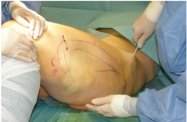
Lipoaspirate was obtained by tumescent infiltration followed by liposuction (Figure 1).
Figure 1 Liposuction procedure after tumescent infiltration at the left outer thigh. The area of liposuction is marked prior to the operation in accordance with patients request for aesthetic outcome. The liposuction cannula is maintained parallel to the surface during the suction procedure while the left hand is providing direction guidance.
The lipoaspirate contains rests of tumescent, debris, blood, oil and various cells integrated in fat tissue. These cells are released after tissue digestion with the MNP- S Liberase (Roche). Washing and centrifugation of digested tissue provide an inhomogeneous cell product consisting of lymphocytes, progenitor cells, mesenchymal stem cells and erythrocytes which is referred to as stromal vascular fraction (SVF).
The extraction was performed by "Hand preparation" with common laboratory equipment and by a "Preparation with a medical device" using the UniStation
25
(NeoGenesis Co. Ltd, Seoul, Korea), a prototype of a heatable centrifuge with a shake setting. Both will be described precisely in the following sections and are based on two crucial steps:
1. Disintegration of the cells by digestion with a collagenase (MNP-S Liberase, Roche)
2. Isolation of the cells by centrifugation
SVF sample pairs were obtained since 80 ml lipoaspirate from each patient was extracted by both methods using 40 ml by each method. Flow cytometry was applied for all samples and pairs. The extraction methods were compared regarding overall cell number, the percentage of single positive cells, double positive cells and the stain index of the investigated markers (i.e. CD34, CD45, CD271 as described previously in 5.9 Molecular markers used in this work). In addition, SVF samples from both extraction procedures were cultured in order to obtain ASCs which were characterized by their CFU Capacity.
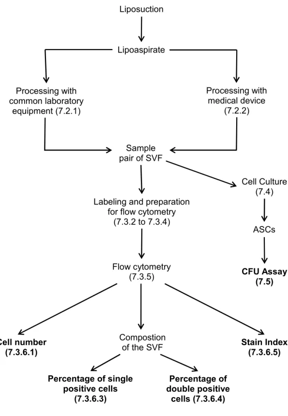
The order of the methods and the outcome parameters used in this work are shown below in a flow diagram (Figure 2).
26
Figure 2 Flow diagram of the used methods and outcome parameters (marked bold).
Liposuction
Lipoaspirate
Processing with common laboratory
equipment (7.2.1)
Processing with medical device
(7.2.2)
Sample pair of SVF
Cell Culture (7.4)
ASCs
CFU Assay (7.5) Labeling and preparation
for flow cytometry (7.3.2 to 7.3.4)
Flow cytometry (7.3.5)
Cell number (7.3.6.1)
Compostion of the SVF
Percentage of single positive cells
(7.3.6.3)
Percentage of double positive
cells (7.3.6.4)
Stain Index (7.3.6.5)
27 7.2.1. Hand preparation
7.2.1.1. Description
Common laboratory equipment was used, in particular a sterile fume hood, a shaking incubator, a sterile filtering system, sterile serological pipettes and sterile centrifugation tubes.
7.2.1.2. Protocol
A total of 40ml lipoaspirate was processed and for better handling separated into 20ml portions. Each of the 20 ml portions was placed into sterile centrifugation tubes of 50ml volume and equal volume of DMEM mixed with 2.5 mg to 5 mg of the MNP-S Liberase were added (i.e. 0.125mg-0.25mg MNP-S Liberase per 1 ml of lipoaspirate).
Immediately after incubation, the sample was incubated on a shaker with 100rpm at 37°C for 45min. The digested tissue was vigorously pipetted up and down ten times with a 25ml serological pipette to release remaining tissue bound cells. The suspension was filtered through a 100µm sterile filter system and centrifuged at 500g for 5min. The supernatant containing tumescent, debris and blood with an oil layer on top was carefully removed with a serological pipette without disturbing the cell pellet.
The remaining pellet containing the SVF was re-suspended in 10ml of PBS. The steps of centrifugation, discarding of the supernatant and re-suspension in PBS were performed two more times until the SVF pellet was clean and neither oil nor tissue residue remained.
28
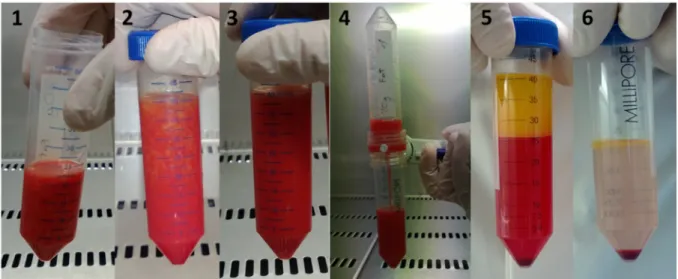
Figure 3 Hand preparation. 20ml of lipoaspirate (1) are mixed with equal volume of 20ml DMEM and 2.5-5mg of MNP-S Liberase (2). The tissue is digested after 45min of incubation at 37°C and shaking at 100rpm (3) and can be filtered through a sterile 100µm filter system (4). After the following centrifugation, the oily and fluid phase on the top can be clearly distinguished (5). After discarding of the supernatant and washing with PBS, the pellet representing the SVF is clearly visible on the bottom of the centrifuge tube (6).
Afterwards, 5ml of culture medium (αMEM containing 20% FBS) were added to inhibit the activity of the MNP-S Liberase. The tube was centrifuged at 300g for 5 minutes, and the supernatant was removed with a serological pipette. The resulting SVF pellet was immediately re-suspended in PBS for further experiments or in culture medium for in vitro experiments. After re-suspending in culture medium, the cell suspension was plated in a cell culture flask and handled as described in 7.4 Cell culture. Cells from the SVF which were able to adhere to cell culture plastic under cell culture conditions (7.4.1 Medium and culture conditions) were defined as ASCs.
7.2.2. Preparation with a medical device
The medical prototype device UniStation (NeoGenesis Co. Ltd, Seoul, Korea) for semi-automated extraction of the SVF was used in order to investigate consistent extraction results (i.e. cell count, cell composition, cell viability).
7.2.2.1. Description of the used tools
The medical device contains a heatable centrifuge (Figure 4) with a shaking plate that can be placed on top of the centrifuge (Figure 5). The sterile syringes used for processing provide a standard Luer Lock (ISO 594-1:1986) for connection with sterile
29
needles, sterile caps, and a sterile transfer piece to connect two syringes (Figure 6).
Therefore, a closed sterile compartment can be established between the syringes and the transfer piece. A detachable handpiece can be connected to the plunge by a thread and is removed prior to centrifugation to fit into the centrifuge.
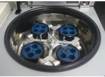
A four sequence program was used for the extraction of the SVF and is specifid in detail in Table 1. The medical device is preheated to 37°C prior to extraction process and temperature is maintained at this level for all steps.
Figure 4 The interior of the medical device shows the centrifuge with 16 slots for syringes.
The heater is located behind the silver surroundings.
Figure 5 Shaking plate placed on top of the centrifuge for the incubation step (program A2) offering space for eight syringes. No centrifugation steps can be performed when the shaker is installed.
30
Figure 6 Syringe and transfer system to establish a sterile compartment for the extraction of the SVF. The syringes are separately packed (a) and can be filled with up to 50ml (b). The plunge (e) can be unscrewed to save space in the centrifuge. The Caps (d) and the transfer piece (c) all have the standardized Luer Lock system.
Table 1 Different programs / steps that are performed for the extraction of the SVF with short description of the action.
Program / Step Action
A1 - Fat washing Centrifugation at 700g for 5min
A2 - Shaking incubation Shaking incubation: The shaking plate is added to the top of the centrifuge and is alternately turning at 2g for 30min
A3 - Separation of the SVF Centrifugation at 800g for 5min A4 - Washing the SVF Centrifugation at 800g for 3min
7.2.2.2. Protocol
40ml of lipoaspirate were transferred into a sterile syringe and closed with a cap.
After the centrifugation (Program A1) the lower part containing blood and tumescent was discarded (Figure 7) whereas solid tissue and oil remained in the syringe. 5- 10mg of the MNP-S Liberase was dissolved in an extra syringe with as much DMEM was required to reach the syringe volume of 40ml after discarding the lower part.
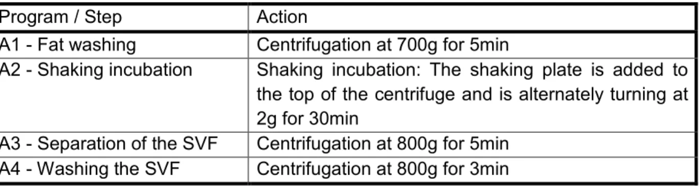
Thereafter, both syringes were connected with a transfer piece and the solution containing the MNP-S Liberase was added to the remaining tissue (Figure 8).
31
Figure 7 Discarding of tumescent solution and blood accumulated in the lower part of the syringe after the first centrifugation step of the lipoaspirate. Solid tissue and oil remained in the syringe for further processing.
Figure 8 The collagenase was added to the centrifuged lipoaspirate: The upper syringe contains DMEM with 10mg of MNP-S Liberase with a volume that is required to obtain 40ml in the lower syinge containing the lipoaspirate after the first centrifugation step and removal of the lower part. Both are connected via the silver transfer piece and the MNP-S Liberase solution is added by applying slight pressure on the plunge of the upper syringe.
32
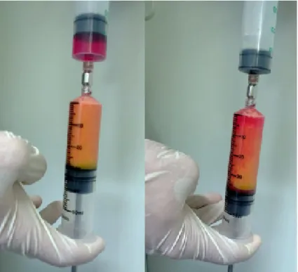
The shaking plate was installed to the centrifuge in order to start the digestion of the tissue with Program A2. Afterwards, the shaking plate was removed and the syringe was inserted into the centrifuge again (Program A3). After this centrifugation, the oil was separated from the layer of tumescent, blood and the SVF at the bottom (Figure 9).
Figure 9 Digested tissue before (a) and after centrifugation (b). The content is separated into an upper oil layer, a layer of tumescent, blood and a small SVF pellet at the bottom.
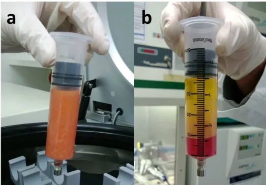
Thereafter, the syringe was uncapped without disturbing the separated layers. The plunge was pulled up 1mm to prevent cell loss in the syringe cap while uncapping as described in the manual and demonstrated in an instructional video by the manufacturer (57). The syringe was carefully connected to the transfer piece with a second empty syringe. The lower 5ml containing the SVF were transferred to the new syringe and the primary syringe was removed with the transfer piece from the secondary syringe. The remaining tissue in the primary syringe was discarded. 35ml of PBS were added to the secondary syringe, containing the SVF. The final volume reached 40ml and the syringe was centrifuged for the last time (Program A4).
After centrifugation, the SVF was apparent in the lower 5ml of the syringe and was transferred to a sterile 50ml centrifugation tube (Figure 10). The MNP-S Liberase activity was inhibited by adding 5ml of culture medium (αMEM containing 20% FBS).
The tube was centrifuged at 300g for 5minutes, and the supernatant was removed
33
with a serological pipette. The resulting SVF pellet was immediately re-suspended in PBS for further experiments or in culture medium for cultivation purpose as described in 7.4 Cell culture. Cells from the SVF that were able to adhere to cell culture plastic under cell culture conditions (7.4.1 Medium and culture conditions) were defined as ASCs.
Figure 10 Transferring the SVF into a 50ml centrifugation tube. The SVF is evident as a small red pellet at the bottom of the syringe and is released with the lower 5ml of PBS.
34
7.3. Flow Cytometry of the SVF
7.3.1. Technique of Flow Cytometry
The cell pellet is resuspended and the cell suspension is pumped through a steadily reducing cannula to a diameter where all cells are tightly connected. A laser beam of defined wavelength is adjusted to the cannula and deflected by passing cells.
Different detectors are able to measure the resulting light or fluorescence. One detector, directed along with the laser beam, called forward scatter (FSC), and another detector, directed perpendicular to the laser beam, called sideward scatter (SSC). Moreover, fluorescence detectors are included into the system to detect different wavelengths (multi-parameter flow cytometry). While the FSC signal correlates mainly with the volume, the SSC signal correlates with the granularity of the passing cells. The fluorescence detectors can provide further information about the cell, when fluorescent substances or antibody markers are used. All detector information, gained from a single passing cell, is combined and described as an event. The result is an accumulation of events corresponding to the cells in the suspension.
7.3.2. Experimental setting
Three antibody markers were used to determine the different surface proteins expressed by cells of the SVF. In particular, CD34, CD45 and CD271 were examined and are summarized in Table 2. Each marker was conjugated to a different fluorescent dye. R-Phycoerythrin (PE) Phycoerythrin-Cy7 (PE-Cy7) and Allophycocyanin (APC) were used because of their different emission spectrum. The combinations of antibodies with the corresponding fluorescence and the excitation and emission wavelengths are listed in Table 2.
35
Table 2 Short description of the examined surface proteins and specifications of the fluorescents conjugated to the used antibodies.
Surface protein
Short description of the surface protein
Fluorescent
conjugated to the surface protein antibody
Excitation wavelength (nm)
Emission wavelength (nm)
CD 34 Expressed mainly by hematopoietic progenitor cells but also different kinds of mesenchymal stem cells.
R-Phycoerythrin (PE)
480, 565, 743
767
CD 45 Called the "leucocyte common antigen" (LCA) and is expressed an all human leukocytes.
Phycoerythrin-Cy7 (PE-Cy7)
480, 565 578
CD 271 Also known as LNGFR (low-affinity nerve growth factor receptor) it can be found on mesenchymal stem cells with high proliferative potential
Allophycocyanin (APC)
650 660
Counting beads were added since the concentration of cells in the SVF was of interest and the flow cytometer only counts events. Counting beads have a special FSC and SSC signal that made bead-events clearly distinguishable from cell-events.
Two different types of beads with different FSC and SSC signals were used in a 1:1 ratio. To calculate the cell concentration (Ccells), the number of cell-events (Ncells), the number of bead-events (NBeads) and the known concentration of beads in the suspension (CBeads) are required according to the following formula:
(1) Ccells = Ncells*CBeads/NBead
Cell concentration per gram original lipoaspirate (Ccells/gram lipo) was of interest for better comparability given the fact that the cell concentration varies by the processed amount of lipoaspirate. Thus, the volume of PBS in which the SVF was re-suspended (VPBS) and the initial mass of the lipoaspirate (mlipo) was recorded besides the previously identified cell concentration (Ccells). In case only a part of the SVF was used, the partition of the SVF used for the experiment (p) was also taken into
36
account. In case the whole SVF was used the variable p equals 1. The cells per gram lipoaspirate were calculated using the following equation:
(2) Ccells/gram lipo = Ccells*VPBS/p*mlipo
By applying formula (1) into formula (2) the final equation is obtained and further used in 7.3.6.1 Cell concentration of single positive cells:
Ccells/gram lipo = Ncells*CBeads*VPBS/NBead*p*mlipo
7.3.3. Preparation of the samples
The SVF was extracted for each sample of lipoaspirate with two methods using a hand preparation or medical device. In this work, six pairs of corresponding SVFs were obtained and treated equally as described in the following:
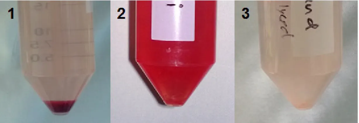
The freshly isolated SVF was re-suspended in PBS. The cell suspension was centrifuged for 5min at 500g and the supernatant was discarded to remove residual red blood cells. The remaining cell pellet was re-suspended in 15ml of erythrocyte lysis buffer, containing 168 mM Ammoniumchloride (NH2Cl), 10 mM Potassiumhydrogencarbonate (KHCO3), 1 mM Ethylenediaminetetraacetic acid (EDTA lysed at pH 8.0) and 0.5 μg/mL 4’,6-diamidino-2-phenylindole (DAPI). The suspension was kept at room temperature for 15min and afterwards centrifuged for 10min at 300g. The color of the SVF pellet changed from red to white when erythrocytes were appropriate lysed (Figure 11). The supernatant containing the lysed erythrocytes was discarded.
The erythrocyte free pellet was re-suspended in PBS and divided to the required sample size in 2ml Eppendorf cups. The cells were stored in PBS at 4°C up to a maximum of 24h when not immediately processed for flow cytometry.
37
Figure 11 Deep red color of the SVF pellet in PBS after isolation (1). Re-suspended pellet in erythrocyte lysis buffer (2). The white SVF pellet is barely visible after erythrocyte lysis in PBS (3).
7.3.4. Labeling
For every flow cytometric analysis, different samples from one SVF were required.
Hence the SVF was split into four 2ml Eppendorf cups. The following procedure was applied for both SVF preparation methods.
Three isotype control samples with antibodies of no specific binding were prepared to detect background-events. In addition, one test sample containing all three antibodies and counting beads was used to gather the desired information about the SVF. Samples with single antibody controls and unstained controls were measured first to calibrate and adjust gate settings of the flow cytometer.
Every 2ml Eppendorf cup contained a defined part of the SVF re-suspended in PBS after erythrocyte lysis. All cups were centrifuged at 300g for 5 minutes, the supernatant was discarded and the pellets were re-suspended in 100µl PBS. The antibodies were added in the dark as shown in Table 3 and incubated for 30min at room temperature.
38
Table 3 Description of the samples and volume of antibody solution added.
Name of the sample (pellet re-suspended in 100µl PBS)
Antibodies added Volume
Control Isotype PE PE isotype control (BD Pharmingen)
5µl Control Isotype PE-Cy7 PE-Cy7 isotype control
(BD Pharmingen)
5µl Control Isotype APC APC isotype control
(Miltenyi Biotec)
10µl Sample with all Antibodies Anti-human CD34 R-PE
conjugated
(BD Pharmingen)
20µl
Anti-human CD45 PE-Cy7 conjugated
(BD Pharmingen)
5µl
Anti-human CD 271 APC conjugated
(Miltenyi Biotec)
10µl
All steps after the incubation were performed in dark environment. 1.5ml PBS were added to each cup, and mixed gently by pipetting up and down. Thereafter, each cup was centrifuged for 5min at 300g. The supernatant with non-bound antibodies was discarded and the cell pellets of the isotype control samples were re-suspended in 800µl PBS. The test sample with the three antibodies was re-suspended in 700µl of PBS and 100µl of the counting bead solution was added. Each sample was loaded with a total volume of 800µl.
All samples were transferred into polystyrene round-bottom tubes through a cell- strainer cap retaining all particles which were too large to pass through the flow cytometer. All samples were stored on ice in the dark until the measurement with the flow cytometer.
7.3.5. Measurement
Each sample was vortexed for 10 seconds prior to flow cytometry, to ensure the homogeneity of its suspension. The measurement was finished when at least 30.000 events of the three isotype control samples and at least 250.000 events of the test
39
sample were recorded. The pressure was adjusted, so that the flow rate never exceeded 3000 events per second to ensure precise measurement.
7.3.6. Gating strategy
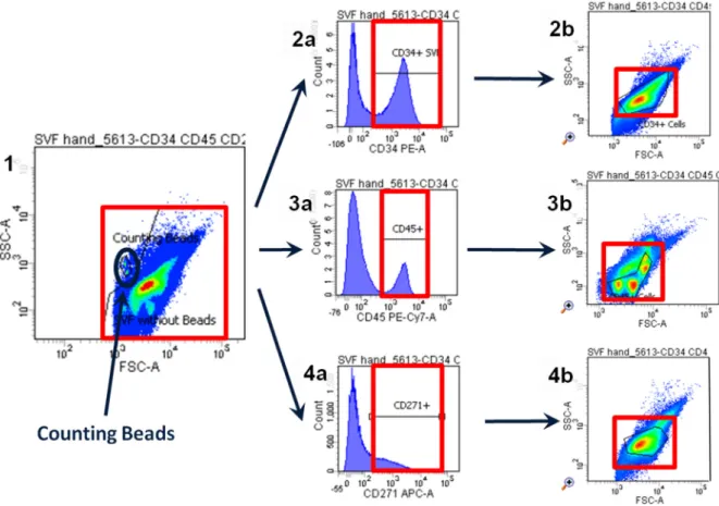
All recorded events were depicted in a dot plot diagram whereas the forwardscatter (FSC) signal represents the X-axis and the sidewardscatter (SSC) signal displays the Y-axis. The counting beads for determination of the cell concentration can easily be distinguished on the plot as they have a defined FSC-SSC pattern. This pattern can be clearly separated from the remaining events. These remaining events showed an inhomogeneous distribution and the amount of populations varied between the samples. While starting the evaluation by investigating the cell concentration for single positive cells (7.3.6.1 Cell concentration of single positive cells) the distribution of the events became more apparent. A main population was detected with positive signals for both stem cell markers (i.e. CD34 and CD271) (7.3.6.2 Definition of the main population) which was used for the investigation of single and double positive cells.
7.3.6.1. Cell concentration of single positive cells
The gating started with analyzing the number of cells per gram lipoaspirate which were positive for the investigated markers. First the counting beads were gated (Figure 12, number 1) resulting in the number of beads (NBead). Secondly the remaining population was gated and thereafter shown in a histogram for the three markers. Positive events were gated due to the equal isotype controls of every one of the three markers (Figure 12, number 2a, 3a and 4a). This procedure ensured that only events with sufficient fluorescence intensity were taken into consideration and counted as positive. The positive events for CD34+, CD45+ and CD271+ were plotted in three further FSC-SSC dot plots. Thereby the subpopulations were identified and finally gated (Figure 12, number 2b, 3b and 4b) resulting in the number of cells for each marker (Ncells). Finally, the concentration of cells per gram lipoaspirate was calculated by the formula previously explained in 7.3.2 Experimental setting using obtained results of Nbeads and Ncells.
Ccells/gram lipo = Ncells*CBeads*VPBS/NBead*p*mlipo
40
The sum of all cells positive for the three markers was calculated to estimate the overall cell number as well the overall cell concentration.
Figure 12 Gating strategy to obtain the cell concentration of the cells positive for one marker in the lipoaspirate. The complete SVF is shown in a FSC-SSC dot plot (1). All events are gated and shown in histograms for CD34 (2a), CD45 (3a) and CD271 (4a). All positive cells are drawn back to a FSC-SSC dot plot (2b, 3b, 4b) and gated for the cell population. The concentration could be calculated with the help of counting beads (marked blue in 1).
7.3.6.2. Definition of the main population
The main population was gated based on two principles: Firstly, cells’ physical appearance in the FSC-SSC dot plots indicating viability. Secondly, the population was defined when the criteria for stem cells were fulfilled (19). Thus, the population of interest included cells which were positive for CD34+ and negative for CD45-. Furthermore high proliferative cells positive for CD271+ (54) were included. The different markers were gated separately and shown in a FSC-SSC dot plot in the process of finding the cell concentration. Thereby the CD34+ and CD271+ positive events representing the stem cells arranged in one population in the same area of
41
the dot plots. On the other hand the CD45+ positive events representing the lymphocytes were located in different subpopulations. Based on these finding, a new gate was created in the initial dot plot containing viable mesenchymal stem cells and were termed main population and used for the analyses described in the following.
7.3.6.3. Percentage of single positive cells
The composition of the stem cells in the SVF was determined by investigating the percentage of single positive cells for the markers within the previously defined main population. Histograms for each marker were created for this specific cell population.
A gate was set in every histogram which symbolize positive cells for each marker (Figure 13) and were adjusted in reference to the isotype control samples. Finally, the percentage of events shown as positive in the matching isotype control was subtracted from the percentage of positive cells in the sample in order to reduce background noise. In case of a negative difference, no positive cells were assumed and the percentage was set to zero.
42
Figure 13 Gating strategy for the percentage of single positive cells within the defined main cell population. The FSC-SSC dot plot shows a main cell population circled red and two smaller populations on the left upper side that are the two different counting beads (1). The histograms on the right show the fluorescence of PE associated with CD34 (2), PE-Cy7 associated with CD45 (3) and APC associated with CD271 (4). The gates for positive cells were set in reference to the isotype controls, so that almost no events of the isotype control could be found in this gate.
7.3.6.4. Percentage of double positive cells
Following the gating of the percentage of single positive cells in the defined main population cells were investigated for the remaining other two markers. Thus all cells gated for a particular marker were depicted in a dot plot whereas each axis was labelled with the two other markers (e.g. all CD34+ cells were shown in a CD45 and CD271 dot plot). The plot was split up into four different gates: One gate representing only single positive (Figure 14, 3c), two containing double positive (Figure 14, 3a and 3d), and the last triple positive cells (Figure 14, 3b) for the investigated markers. The gates were set according to the isotype controls. The percentage of double positive cells in the single positive cell population was analyzed.
43
While the percentage of double positive cells in the main population was investigated two values resulted for every marker combination, sample and preparation method due to the gating procedure. For example, one marker was gated and further investigated for the other two markers. The same procedure was performed two times starting with a different marker (e.g. the percentage of CD34+/CD271+ double positive cell can be obtained by first gating for CD34 and then CD271 or the other way around).
Figure 14 Example for gating double positive cells. The events gated as cells in the FSC- SSC dot plot (1) are further gated in a histogram to obtain the CD34 positive cells (2). Those are shown in a dot plot with the two axes defined by the two other markers: CD45 and CD271 (3). The cross divides the plot defining four new gates. The cells of interest lay in gate 3a and 3d. They show CD34+ and CD271+ cells (3a) and CD34+ and CD271+ cells (3d). Just CD34 single positive cells can be found in gate 3c, while cells positive for all three markers lay in gate 3b.
7.3.6.5. Stain index
Autofluorescence, background noise and the dyes used for the multiparameter flow cytometry can influence the results. A different approach to the comparison of the main population of the SVF samples was the stain index (58). The stain index is a normalization of the fluorescence signals and gives information about the fluorescence intensity.
44
The stain index uses three values per sample (Figure 15) all resulting from the gate set around the samples main population: The mean fluorescence intensity of the sample (MFIsample). And two values origin from the matching isotype control: The mean fluorescence intensity of the isotype control (MFIisotype) and its width represented by its standard deviation (SDisotype). With these three values the stain index is calculated for every single marker as shown below:
Stain Index = (MFIsample -MFIisotype) / 2* SDisotype
Figure 15 Obtaining the stain index: The mean fluorescence intensity (MFIIsotype) and its standard deviation (SDisotype) are taken from the istoype control of one marker (1). From the same marker the mean fluorescence intensity of the sample (MFISample) is obtained (2). The stain index is calculated: Stain Index = (MFIsample - MFIisotype) / 2* SDisotype (58).