Slaats, Asberg, van Keimpema and Kruijswijk: Estimation of adenosine deaminase in pleural efTusions 677 J. Clin. Chem. Clin. Biochem.
Vol. 23, 1985, pp. 677-682
A Continuous Method for the Estimation
of Adenosine Deaminase Catalytic Concentration
in Pleural Effusions with a Hitachi 705 Discrete Analyser
By £. H. Slaats, E. G. M. Th. Asberg Department of Clinical Chemistry A. R. J. van Keimpema
Department of Pulmonary Diseases and H. Kruijswijk
Department of Clinical Chemistry,
Hospital "Onze Lieve Vrouwe Gasthuis", Amsterdam, The Netherlands
«
(Received April 24/June 14, 1985)
Summary: An assay of adenosine deaminase activity in pleural effusions is described. For the continuous determination of adenosine deaminase, the liberated ammonia is estimated by coupling the liberated NH3 with 2-oxoglutarate. The reaction is followed by the decrease of NADH absorbance at 340 nm. The assay was optimized for a Hitachi 705 analyser, with respect to pH, adenosine concentration and glutamate dehydrogenase activity. The assay is linear to an adenosine deaminase catalytic concentration of 110 U/l.
Elevated adenosine deaminase activities are föund in pleural effusions of patients with tuberculosis, empyema and mesothelioma. Although elevated adenosine deaminase activity in pleural effusion is not pathognomonic for tuberculpsis, it may be valuable äs a first screening parameter.
Eine Methode zur kontinuierlichen Messung von Adenosindesaminase in Pleuraexsudaten mit dem diskreten Analysator Hitachi 705
Zusammenfassung: Eine Bestimmungsmethode für Adenosindesaminase in Pleuraexsudaten wird beschrieben.
Zur kontinuierlichen Bestimmung von Adenosindesaminase wird das freigesetzte Ammoniak mit 2-Oxogluta- rat umgesetzt. Die Reaktion wird durch Messung des NADH-Verbrauchs bei 340 nm kontinuierlich verfolgt.
Die Methode wurde hinsichtlich pH, Adenosinkonzentration und katalytischer Konzentration von Glutamat- dehydrogenase für den Hitachi 705 optimiert. Die Methode ist linear bis 110 U/l Adenosindesaminase. Erhöhte katalytische Konzentrationen werden in Pleuraexsudaten bei Tuberkulose, Empyem und Mesotheliom gefun- den. Obwohl die erhöhte Adenosindesaminase nicht pathognomonisch für Tuberkulose ist, kann sie als ein erstes Merkmal für das Screening wertvoll sein.
Introduction for the differentiation of lymphoid cells, particular T Adenosine deaminase (EC 3.5.4.4) is a widely dis- cells (1) and maturation of monocytes to macro- iributed enzyme, which catalyses the hydrolysis of phages (2). Its deficiency in the lymphocytes results adenosine to inosine and ammonia in the purine in severe combined immunodeficiency disease (3, 4).
catabolic pathway. Adenosine deaminase is essential Elevated adenosine deaminase activity is found in
J. Clin. Chem. Clin. Biochem. / Vol. 23, 1985 / No. 10
678 Sluats, Asberg, van Keirnpema and Kruijswijk: Estimation of adenosine deaminase in pleural effusions serum of patients with infectious mononucleosis (5),
acute leukaemia (8), typhoid fever (6), liver diseases (7, 10) and sarcoidosis (18). In pleural effusions of patients with tuberculosis, empyema and rheumatoid pleural effusion, higher adenosine deaminase ac- tivities are reported than in malignant or para- pneumonic pleural effusions (9 — 13). The adenosine deaminase activity may be measured by de- termination of the rate of either consumption of adenosine or of formation of inosine or ammonia.
The determination of produced ammoiiia is mostly used in routine measurements. The ammonia can be determined by the indophenol reaction (Berthelot) (14, 15) or by coupling the liberated NHa to 2-oxo- glutaric acid with glutamate dehydrogenase (EC 1.4.1.3). The last method has the advantage that the overall reaction can be followed by the rate of consumption of NADH.
We have adapted this method for measurement with a Hitachi 705 discrete analyser, in order to st dy the usefulness of adenosine deaminase determination in the evaluation of the origin of pleural effusions.
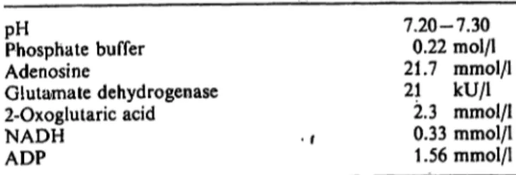
Tab. 1. Final concentfations of reagents.
Materials and Methods Reagents
1. Phosphate buffer 0.25 mol/1 pH 7.5:
39.0 g Na2HP04 · 2H2O, 4.2 g KH2PO4 in l litre distilled water.
2. Adenosine (Boehringer Mannheim, FRG) 300 mmol/1):
suspend 2.0 g in 20 ml distilled water and add dropwise 360 g/kg HC1 until the adenosine is dissolved. Adjust to 25 ml with distilled water. Stable for l month at —20 °C.
3. Glutamate dehydrogenase in glycerol, 1200 kU/1 (Boehringer Mannheim, FRG, 127710).
4. 2-Oxoglutaric acid (Boehringer Mannheim, FRG) 0.2 mol/1:
dissolve 2.9 g in 100 ml distilled water and neutralize. Stable for l month at -20°C.
5. NADH, disodium salt (Boehringer M nnheim, FRG, Grade I), ± 21 mmol/1:
dissolve 15 mg/ml distilled water. Prepare fresh daily.
6. ADP, disodium salt (Boehringer Mannheim, FRG) 90 mmol/1:
dissolve 425 mg ADP in 10 ml distilled water.
In the final reagent 15 ml phosphate buffer is mixed with 300 μΐ glutamate dehydrogenase Suspension, 200 μΐ 2-oxoglutaric acid solution, 250 μΐ NADH solution, 300 μΐ ADP solution and 1250 μΐ adenosine solution. The pH is adjusted to 7.25 + 0.05. The final concentrations of the reagents are given in t ble 1.
The unspecific activity is measured under the same conditions with buffer instead of adenosine in the final reagent.
pHPhosphate buffer Adenosine
Glutamate dehydrogenase 2-Oxoglutaric acid NADH
ADP
7.20-7.30 0.22 mol/1 21.7 mmol/l 21 kU/1
2.3 mmol/1 0.33 mmol/1 1.56 mmol/1
Samples
The pleural effusions were centrifugated at 1000g 15 min and stored at —20 °C until assayed. The adenosine deaminase ac- tivity was unaltered for at least 4 months at —20 °C.
No difference was found between serum, heparin plasma and EDTA plasma. Haemolytic samples with a concentration up to 0.24 mmol/1 haemoglobin have no effect on the adenosine deaminase activity (tab. 2).
Tab. 2. Influence of haemolysis on the adenosine deaminase activity in serum at 37 °C
Adenosine deaminase (U/0
Serum 23.5 H- 0.08 mmol/1 haemoglobin 22.9 H- 0.12 mmol/1 haemoglobin 23.9 + 0.24 mmol/1 haemoglobin 23.3
Procedure f.
The Hitachi 705 discrete analyser (Boehringer Mannheim, FRG) was operated according to the usual protocpl, the settings being shown in table 3. The optimazation of the cpntinuous method was performed on an Acta II spectrophotometer (Beck- man Instr., NY, USA).
The molar lineic absorbance, ε = 622 m2/mol at 340 nm, was used for NADH. With a reference wavelength of 660 nm on the Hitachi 705 discrete analyser, the molar iineic absprbatice, ε 340/660 of 589 m2/moi was used.
The activity in U/l is defmed s the quantity of NH3 produced in μπιοί per minute per litre.
Tab. 3. Parameter listing for detefmination of adenosine deaminase activity at 37 °C on a Hitachi 705 discrete analyser.
Assay code Sample volume
Wavelength l Wavelength 2 Factor Abs. limit rate
2 (rate)-20-28 20 μ!
350 μΐ Ο660 nm 340 nm 31612000 (0.2 A)
Slaals, Asbcrg, van Kcimpema and Kruijswijk: Estimation of adenosinc deaminase in pleural effusions 679
Rcsults
p H - o p t i m u m
To determine the optimal pH for the assay, phosphate buffers ranging in pH (Vorn 6.9 to 7.5 are used. An optimal pH for the enzyme activity in the final re- agent was found between 7Λ and 7.3 (fig. 1). In the final procedure a final pH of 7.25 ± 0.05 was chosen.
150
αc 100
i
Wo
•o£
50
'"N
6.8 7.0 7.2
PH
7.4 7.6
Fig. 1. Influence of pH on the adenosine deaminase activity in pleural effusion (o) and serum (o).
Optimal concentration of reagents
The reaction time in the assay is limited by the use of the Hitachi 705 analyser, the measurement has to be completed within 10 minutes. As the endogenous ammonia and other Substrates causing non-specific NADH consumption are eliminated within ± 6 min- utes, there is a period in which the decrease of the absorbance each 20 seconds can be measured and the linearity of the assay can be controlled. In figure 2 the rate of ammonia consumption is plotted s a function of the glutamate dehydrogenase activity. A glutamate dehydrogenase activity of 20 kU/1 in the presence of 1.5 mmol/1 ADP and 4 mmol/1 2-oxo- giutarate is sufficient to eliminate 700 μιηοΐ/ΐ am- monia within 6 minutes. ADP in a concentration of 1.5 mmol/1 is added to activate the glutamate de- hydrogenase, according to the literature (16).
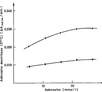
The adenosine concentration is varied between 5 and 27 mmol/1. The influence of the adenosine con- centration on the adenosine deaminase activity is given in figure 3. From a Lineweaver-Burk plot a
0.400 -
0.300
o 0.200 -
0.100 -
t Lmin J
Fig. 2. Consumption rate of NH3 (700 μπιοΐ/ΐ) s function of the glutamate dehydrogenase activity at 37 °C: 11 U/l (o), 22 U/l (D), 44 U/l (A).
0.030
0.020
0.010
Michaelis constant is calculated: Km = 4.8
10 20 Adenosine C mmol / l ]
Fig. 3. Influence of the adenosine concentration on the adeno- sine deaminase activity in pleural effusion (o) and serum (n).
mmol/1 in pleural effusion from tuberculosis and Km = 3.4 mmol/1 in a serum, thereby demonstrating the presence of various isoenzymes (4, 14). At a final adenosine concentration of 21.4 mmol/1, the adenosine deaminase activities are 86% and 89% of Kmax for the pleural effusion and serum, respectively. Higher adenosine concentration in the final reagent are not used because the adenosine tends to crystallize out.
J. Clin. Chem. Clin. Biochem. / Vol. 23, 1985 / No. 10
680 Slaats, Asbcrg, van Kcimpcma and Kruijswijk: Estimation of adenosine deaminase in pleural effusions
200
Precision
The intra-run precision in the lower r nge is 2.4%
(x = 26.0, n = 25) and in the elevated r nge 2.1%
(x = 88.4, n = 25).
Unspecific activity
The unspecific activity in pleural effusions was meas- ured in 119 patient samples. The mean activity was 4.6 U/l and the r nge (x ± 1.96 σ) is 0.2 - 9.5 U/l.
A maximal unspecific activity of 12.3 U/l was found.
Adenosine deaminase in pleural effusions The adenosine deaminase activities among the various groups of lung diseases are compared in figure 5. The patients are subdivided into 5 groups:
I tuberculosis (4 x), II empyema (7 x), III mesothelioma (lOx),
IV other malignant neoplasms (32 x),
V patients with pneumothorax (9x), para- pneumonic effusions (14x), haematothorax (2 x), congestive heart failure (2 x), and unspe- cific pleural effusions (10 x).
One patient in group V, with a Budd-Chiari syndrome with ascites, had an unspecific pleural effusion in which the adenosine deaminase activity was 81 U/l.
However, the adenosine deaminase activity in serum (117 U/l) was also elevated. No significant difference is found between group IV and V (Student t-test).
From these two groups a distribution-free 98th
percentile of 44 U/l is found.
150
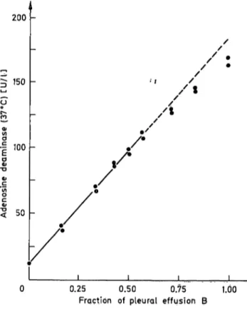
Linearity
The upper limit of enzyme activity at which a linear response can be obtained is determined by using mixtures of pleural effusions with a low adenosine deaminase level (sample A: 11 U/l) and a high adeno- sine deaminase level (sample B: 1.84 U/l). The fraction of pleural effusion B in the mixture of pleural effusion A and pleural effusion B is given in figure 4. The calculated regression line is: ^ ADA (obs) U/l = 173 · fraction of sample B + 11.1. | 100 Using an upper limit of 110 U/l a correlation co-
efficient r = 0.9990 is obtained.
o
ΌS
50
0 0.25 0.50 0.75 1.00 Frqction pf pleural effusion B
Fig. 4. Linearity of the adenosine deaminase assay. Pleural effusion B (184 U/l) is mixed with pleural effusion A (11 U/l).
1 . - . . _ .
:
• · «| · · · ·
• • • • • j · · ·
•4|HH· *
•M» ![>·
I Tuberculosis I Empyema
ΠΙ Mesothelioma .y Other malignant - neoplasm
Υ Other 0 50 100
Adenosine deaminase (37°C) CU/U
Fig. 5. Adenosine deaminase activity in pleural effusion in various disOrders.
The line (— ) shows the 98th percentile of group IV and V.
Discussion
Pleural effusions are frequently a diagnostic problem in clinieal practice. A careful diagnostic evaluation of the aetiology of the effusion cannot be est blished in about 20% of patients (11). The estimation of adenosine deaminase in pleural effusions appeared to be a valuable aid in the diagnosis of tuberculous pleural effusions. Although the incidence of tu- berculosis has decreased, this disease still has to be taken into account in the differential diagnosis of pleural effusions.
Slaats, Asberg, van Keimpema and Kruijswijk: Estimation of adenosine deaminase in pleural effusions 681
Our intention was to look for a fast and simple test to make or exclude the diagnosis of tuberculosis, in order to avoid the necessity to prepare cultures for the identification of Mycobacterium tuberculosis, or to prepare biopsies of parietal pleura to look for epithelioid granulomas. The method for de- termination of adenosine deaminase activity by me- ans of the Berthelot reaction (11, 15) is rather time- consuming, therefore we adapted and optimized the enzymatic method for the estimation of the adenosine deaminase activity on a Hitachi 705 discrete analyser.
The resulting enzymatic method gave a high quality performance.
Ratech & Hirschhorn (4) have investigated the kinetic properties and molecular mass of the adenosine de- aminase (ADA) isoenzymes. They reported a mon- omeric 35000 dalton isoenzyme ADAj, an 280000 dalton isoenzyme, containing 35 000 dalton catalytic units with a nonenzymatic 200000 dalton combining glycoprotein ADAi,Gp and a 100000 dalton iso- enzyme ADA2. The reported Km values for AD AI and ADA1$GP are 51 μπιοΐ/ΐ and 75 μιηοΐ/ΐ, respectively.
For ADA2, Km = 2800 μπιοΐ/ΐ. Also the Kmax values for AD A! and ΑΟΑί%Ορ are more than 10 times less than that for ADA2.
Notably, a child with severe combined immuno- deficiency (SCID), showed a complete absence of ADA] and ADA^op in lymphocytes, erythrocytes and other tissues. The serum ADA2 activity was nor- mal. These different kinetic properties of the adeno- sine deaminase isoenzymes are a confirmation of the work of Ellis & Goldberg (16).
In tuberculous pleural effusion, we found a Km of 4.8 mmol/1 for the adenosine deaminase enzyme. We were therefore pr bably dealing with the ADA2 type iso- enzyme. For this reason, it becomes clear that the correlation with the number of lymphocytes in the pleural effusion, s sought by Pettersori (17) and Ocana (l 2 a) will not be found; the lymphocytes con- tain only ADAj type isoenzyme and in our and their assay the adenosine concentration is too high. At the adenosine concentration used, the conversion rate V
is about 88% of Fmax for adenosine. With the large difference in Kmax for the ADA] type and ADA2 type isoenzyme (4), the ADA, is obscured in a medium where ADA2 type enzyme is present in at least the same order of magnitude. Accordingly, and in con- tradiction with the literature (10, 15), our assay is not influenced by haemolysis. However, further study is necessary to clarify the origin and characterization of adenosine deaminase in tuberculous pleural ef- fusions. Possibly the adenosine deaminase in tu- berculous pleural effusions originates from the epi- thelioid granulomas.
Our clinical results are shown in figure 5. The di- agnosis of tuberculosis was confirmed by culture.
There was no evidence of culture negative tu- berculous pleural effusions in our population. From this small data group it appears that tuberculous pleural effusions have a significantly higher adenosine deaminase activity than pleural effusions of group IV and V (Student t-test, p < 0.005). With the exception of the results of Blake et al. (11), our results are in concordance with the literature (tab. 4). There is a discrepancy between the results of Blake et al. (11) and the other reported data (tab. 4). An explanation cannot be given, because Blake et al. give no speci- fication of the used pH, buffer concentration and glutamate dehydrogenase activity.
As can be seen from figure 5, the adenosine de- aminase activity in pleural effusions does not dis- criminate between tuberculosis, empyema and me- sothelioma, when there is a normal blood level of adenosine deaminase activity. In the literature high values of adenosine deaminase activity are also de- scribed in pleural effusions from patients with rheu- matoid arthritis (17). Consequently, elevated adeno- sine deaminase activity in pleural effusions is not pathognomonic for tuberculosis.
Acknowledgement
We gratefully acknowledge Dr. J. P. M. Wagenaar, head of the department of pulmonary diseases, and colleagues for providing several pleural effusions from patients.
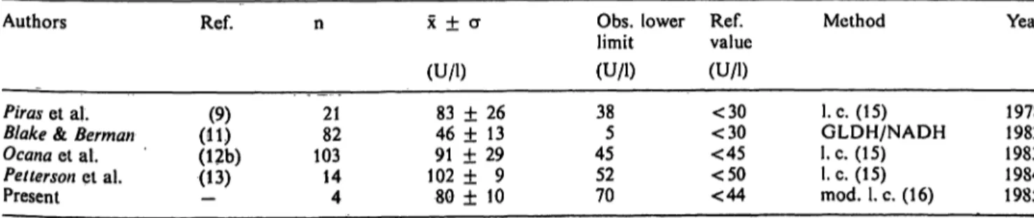
Tab. 4. Reported adenosine deaminase activity in tuberculous pleural effusions at 37 °C Authors
Piras et al.
Blake & Berman Ocana et al.
Pelterson et al.
Present
Ref.
(11)(9) (12b) (13)
n
2182 10314 4
χ ± σ (U/l)
83 ± 26 46 ± 13 91 ± 29 102 ± 9 80 ± 10
Obs. lower limit (U/l) 385 4552 70
Ref.value (U/l)
<30
<30
<45
<50
<44
Method
I.e. (15) GLDH/NADH I.e. (15) I.e. (15) mod. I.e. (16)
Year
19781982 19831984 1985
J. Clin. Chem. Clin. Biochem. / Vol. 23, 1985 / No. 10
682 Slaats, Asberg, van Keimpema and Kruijswijk: Estimation of adenosirie deaminase in pleural effusions References
1. Hovi, T., Smyth, J. F., Allison, A.C. & Williams, S. C.
(1976) Clin. Exp. Immunol. 23, 395-403.
2. Fisher, D. N., Van der Weyden, M. B., Snyderman, R. &
Kelley, W. N. (1976) J. Clin. Invest. 58, 399-407.
3. Giblett, E. R., Anderson, J. E., Cohen, F., Pollara, B. &
Meuwissen, H. J. (1972) Lancet //, 1967-1969.
4. Ratech, H. & Hirschhorn, R. (1981) Clin. Chim. Acta 115, 341-347.
5. Koehler, L. H. & Benz, E.J. (1962) Clin. Chem. 8, 133-140.
6. Giusti, G. (1970) Pol. Aren. Mew. Wewn. 44, 525.
7. Goldberg, D. M. (1965) Br. Med. J. /, 353-355.
8. Coleman, M. S., Greenwood, M. F., Hutton, J. J., Holland, R, Lampkin, B., Krill, C. & Kastelic, J. E. (1978) Blood 52, 1125-1131.
9. Piras, M. A., Gakis, C., Badroni, M. & Andreoni, G. (1978) Br. Med. J. 2, 1751-1752.
10. Hankiewicz, J. & Koterwa, A. (1978) Mat. Med. Pol. 3, 180-183.
11. Blake, J. & Berman, P. (1982) SA Med. J. 62, 19-21.
12. Ocana, L, Martinez-Vasquez, J, M., Segura, R. M., Fernandez-de Sevilla, T. & Capdevila, J. A.
a) (1983) Chest 84, 51-53.
b) (1984) Chest 86, 273-274. 'r
13. Petterson, t., Ojala, K. & Weber, T. H. (1984) Acta Med.
Scand. 215, 299-304.
14. Giusti, G. & Gakis, C. (1971) Enzyme 12, 417-425.
15. Giusti, G. (1974) In: Methoden der enzymatischen Analyse, 3rd ed., Bergmeyer, (H. U., ed.) Verlag Chemie, pp.
1134-1150.
16. Ellis, G. & Goldberg, D. M. (1970) J. Lab. Clin, Med. 75, 507-517.
17. Petterson, T., Klockars, M. & Weber, T. (1984) Chest 86, 18. Taylor, A. (1984) Clin. Chem. 30, 499-500.273.
Dr. E. H. Slaats
Department of Clinical Chemistry Onze Lieve Vrouwe Gasthuis le Oosterparkstraat 179 NL-1091 HA Amsterdam