Lipid raft integrity affects GABA A receptor, but not NMDA receptor modulation by psychopharmacological compounds
Caroline Nothdurfter1,2, Sascha Tanasic1,2, Barbara Di Benedetto1,2, Manfred Uhr2, Eva-Maria Wagner2, Kate E. Gilling3, Chris G. Parsons3, Theo Rein2, Florian Holsboer2, Rainer Rupprecht1,2and Gerhard Rammes2,4
1Department of Psychiatry and Psychotherapy, University Regensburg, Regensburg, Germany
2Max-Planck-Institute of Psychiatry, Munich, Germany
3In Vitro Pharmacology, Preclinical Research and Development, Merz Pharmaceuticals GmbH, Frankfurt am Main, Germany
4Department of Anesthesiology, Technische Universita¨t Mu¨nchen, Munich, Germany
Abstract
Lipid rafts have been shown to play an important role for G-protein mediated signal transduction and the function of ligand-gated ion channels including their modulation by psychopharmacological compounds. In this study, we investigated the functional significance of the membrane distribution of NMDA and GABAAreceptor subunits in relation to the accumulation of the tricyclic antidepressant desipramine (DMI) and the benzo- diazepine diazepam (Diaz). In the presence of Triton X-100, which allowed proper separation of the lipid raft marker proteins caveolin-1 and flotillin-1 from the transferrin receptor, all receptor subunits were shifted to the non-raft fractions. In contrast, under detergent-free conditions, NMDA and GABAAreceptor subunits were detected both in raft and non-raft fractions. Diaz was enriched in non-raft fractions without Triton X-100 in contrast to DMI, which preferentially accumulated in lipid rafts. Impairment of lipid raft integrity by methyl-b- cyclodextrine (MbCD)-induced cholesterol depletion did not change the inhibitory effect of DMI at the NMDA receptor, whereas it enhanced the potentiating effect of Diaz at the GABAAreceptor at non-saturating con- centrations of GABA. These results support the hypothesis that the interaction of benzodiazepines with the GABAAreceptor likely occurs outside of lipid rafts while the antidepressant DMI acts on ionotropic receptors both within and outside these membrane microdomains.
Received 3 September 2012 ; Reviewed 24 September 2012 ; Revised 11 October 2012 ; Accepted 18 October 2012 ; First published online 10 December 2012
Key words: Antidepressants, benzodiazepines, GABAAreceptor, lipid rafts, NMDA receptor.
Introduction
Lipid rafts are distinct microdomains within the cell membrane which are enriched in cholesterol and glyco- sphingolipids (Pike, 2004). These heterogeneous and dynamic structures float within the membrane bilayer al- lowing lipids and proteins to move in and out of these membrane microdomains (Kusumi and Suzuki, 2005).
The concept of lipid rafts has attracted considerable in- terest because these structures may affect membrane traf- ficking and signal transduction processes (Tsiu-Pierchala et al., 2002 ; Patel et al., 2008).
There is increasing evidence that lipid raft association can also influence neurotransmitter receptor function by affecting transmitter binding, receptor trafficking and clustering (Allen et al., 2007). In post-mortem brain tissue
of patients with major depression, G-protein subunit (Gs)awas more likely associated with lipid rafts, where it turned out to be less likely coupled to adenylyl cyclase (Donati et al., 2008). Moreover, Gsa-mediated signalling was enhanced by chronic treatment with antidepressants, which prevented the accumulation of Gsa in lipid rafts (Donati and Rasenick, 2005). With regard to ligand-gated ion channels, the serotonin (5-HT)3A receptor has been detected in raft-like domains (Eisensamer et al., 2005).
Likewise the GABAA receptor subunit a1 has been reported to be lipid raft associated (Dalskov et al., 2005), whereas a differential distribution of synaptic and extra- synaptic GABAA receptor subunits has recently been demonstrated (Li et al., 2007). Also the localization of NMDA receptors within the cell membrane appears to depend on the respective subunit composition (Hering et al., 2003 ; Guirland et al., 2004).
A variety of antidepressants and antipsychotics accumulate in raft-like domains, which exert their functional antagonistic effects at the 5-HT3 receptor (Eisensamer et al., 2003, 2005 ; Rammes et al., 2004). More
Author for correspondence : Dr C. Nothdurfter, Department of Psychiatry and Psychotherapy, University Regensburg, Universita¨tsstrasse 84, 93053 Regensburg, Germany.
Tel.:+49 941 941 2059 Fax:+49 941 941 2065 Email: Caroline.Nothdurfter@medbo.de
by guest on September 13, 2016http://ijnp.oxfordjournals.org/Downloaded from
recently, we showed that 5-HT3 receptor function depends on lipid raft integrity, whereas the ability of antidepressants to modulate this receptor is retained after lipid raft impairment (Nothdurfter et al., 2010).
So far, no data are available on the significance of lipid rafts for the modulation of NMDA and GABAA
receptors – crucial members of the most abundant excit- atory and inhibitory neurotransmitter systems in the central nervous system – by antidepressants and benzo- diazepines. We therefore investigated the effect of lipid raft integrity on the allosteric modulation of GABAA
receptor function by diazepam (Diaz) and on the modu- lation of NMDA receptor function by the tricyclic anti- depressant desipramine (DMI). Moreover, the membrane domain distribution of the receptor subunits and com- pounds was investigated using different conditions of li- pid raft isolating procedures.
Method and materials Chemicals and drugs
For stock solutions DMI (10 mM; Sigma-Aldrich, Germany) was dissolved in H2O and Diaz (5 mM; Sigma- Aldrich) in 80 % ethanol.
Cell cultures
HEK 293 cells (European Collection of Cell Cultures, UK), WSS-1 cells (ATCC LGC Promochem, Germany) and HEK 293 cells stably expressing the human NMDA receptor NR1A/2A, (NMDA-HEK ; Merck, Germany), were cultured in Dulbecco’s modified eagle medium (Invitrogen, Germany) supplemented with 10 % foetal calf serum (FCS), 1 % sodium pyruvate and antibiotics.
Cells were reseeded onto fresh Petri dishes twice weekly after treatment with 1 % trypsin-EDTA (Invitrogen). Cells were cultured at 37xC, 5 % CO2and 95 % humidity.
Primary hippocampal neurons were obtained from rat embryos (E20–E21) and were transferred to Ca2+- and Mg2+-free Hank’s-buffered salt solution (Invitrogen).
Cells were dissociated in 0.66 % trypsin/0.1 % DNAse following 0.05 % DNAse/0.3 % ovomucoid incubation.
After centrifugation, cells were resuspended in MEM (Invitrogen) and plated on poly-DL ornithine/laminin coated Petri dishes. Cells were cultured in NaHCO3/ Hepes-buffered MEM supplemented with 5 % FCS and 5 % horse serum. Glial mitosis was inhibited by cytosine- b-D-arabinofuranoside.
Electrophysiological recordings
Voltage clamp recordings were made in the whole-cell configuration at a holding potential ofx70 mV. Com- pounds were applied using a fast superfusion device with a stepper motor-driven (Warner Instruments LLC, USA) application pipette, made from modified double- barrelled theta glass. This arrangement allows complete
exchange of solutions superfusing all receptors expressed on the cell surface within milliseconds. Currents were recorded using an EPC-9 amplifier (HEKA, Germany).
Patch clamp pipettes were pulled from borosilicate glass using a horizontal puller (P-97 Puller ; Sutter Instruments, USA), with resistances of 3–6 MV when filled with in- tracellular solution. Intracellular solution for NMDA- HEK : 120 mMCsCl ; 10 mMEGTA ; 1 mMMgCl2; 0.2 mM CaCl2; 10 mMglucose ; 22 mMTEACl ; 2 mMATP ; 0.2 mM cAMP ; pH 7.35. Corresponding extracellular bath sol- ution : 140 mMNaCl ; 3 mMKCl ; 10 mMglucose ; 10 mM Hepes, 0.2 mMCaCl2; 4.5 mMsucrose ; pH 7.35.D-Serine (5mM) was present in all solutions sufficient to cause 80–85 % activation of glycine B receptors. Intracellular solution for GABAA receptor mediated currents from WSS-1 cells : 140 mM KCl ; 11 mMEGTA ; 10 mMHepes- KOH ; 2 mMMgCl2; 0.1 mMATP ; pH 7.35. Corresponding extracellular bath solution : 162 mM NaCl ; 5.3 mM KCl ; 0.67 mM NaH2PO4; 0.22 mM KH2PO4; 5.6 mM glucose ; 15 mM Hepes ; 2 mM CaCl2; pH 7.4. Antagonism of NMDA receptor-mediated currents was measured in terms of the magnitude of the steady-state blocked cur- rent as percentage of the control current. Five cumula- tively increasing concentrations of the respective compound were applied in the continuous presence of agonists and the control current for each substance concentration was calculated by a linear projection from the steady-state current before and after substance ap- plication, i.e. for the first concentration control current=
0.85rcurrent before antagonist plus 0.15rrecovery cur- rent. For the second substance concentration, these same currents were multiplied by 0.7 and 0.3 respectively and so on. The potency of the compound was assessed by plotting the mean percentage current magnitude against the antagonist concentration and then a curve was fit using the logistic equation for which the variable para- meters IC50and Hill coefficient were free and the range and background values were normally set to 100 and 0, respectively. For analysis of GABA-induced currents in WSS-1 cells only results of stable cells showing at least 50 % recovery of responses to GABA following the re- moval of drugs entered the final analysis. To compensate for this effect the % antagonism at each concentration was based on both the control and the recovery current by assuming a linear time-course for the rundown.
Sucrose density gradient centrifugation
Two confluent Petri dishes of cultured cells were washed in ice-cold PBS before resuspension in 800ml high-salt Hepes buffer (20 mM Hepes, 5 mM EDTA, 1M NaCl, pH 7.4) supplemented with protease-inhibitor cocktail.
For solubilization 1 % Triton X-100 was added directly to the saline buffer as described previously (Nothdurfter et al., 2007). Following homogenization and sonification, the suspension was diluted 1 : 1 with 80 % sucrose solution prepared in high-salt Hepes buffer. The solution was then
by guest on September 13, 2016http://ijnp.oxfordjournals.org/Downloaded from
placed at the bottom of a centrifuge tube (Beckman Coulter, USA) and overlaid with 1400ml 35 % sucrose and finally with 1000ml 5 % sucrose (both prepared in low-salt Hepes buffer : 20 mM Hepes ; 5 mM EDTA ; 0.5M NaCl ; pH 7.4). Samples were ultracentrifuged at 40 000 rpm (2 10 000g) in a Beckman SW60 rotor for 4 h at 4xC.
Subsequently, 10 fractions (400ml each) were taken from the top to the bottom of the gradient.
The experimental protocols for mouse brain tissue fractionation were approved by the Ethical Committee on Animal Care and Use of the Government of Bavaria, Germany. Brains of male C57Bl6 mice aged 28–42 d were put into ice-cold artificial cerebrospinal fluid ; sagittal slices of the medial prefrontal cortex (350mM) were prepared using a microtome (HM 650 V ; Microm International, Germany). Tissue of one animal was homogenized/sonified in 1 ml high-salt Hepes buffer supplemented with protease-inhibitor cocktail. The suspension was centrifuged at 1000g, 10 min, 4xC. The supernatant was again centrifuged at 8000g, 10 min, 4xC. For solubilization the supernatant of the second centrifugation step was supplemented with 0.4 % Triton X-100 and incubated for 30 min on ice. The sol- ution (1850ml) was then diluted 1 : 1 with 80 % sucrose, overlaid with 3200ml 35 % sucrose and 2300ml 5 % sucrose. After ultracentrifugation in an SW41 rotor at 40 000 rpm (2 50 000g) for 4 h at 4xC, 13 fractions (710ml each) were collected.
Cholesterol depletion
Cholesterol depletion using low doses of methyl-b- cyclodextrine (MbCD) to maintain cell viability for elec- trophysiological recordings was performed as described previously (Nothdurfter et al., 2010). Briefly, cells were incubated with 0.25 or 0.5 mM MbCD for 12 h at 37xC in serum-free DMEM prior to the electrophysiological recordings.
Western blotting
Equal volumes of sucrose gradient fractions were ad- justed 1 : 5 with Laemmli sample buffer. For the detection of GABAAreceptor subunit c2, samples were heated at 70xC, 10 min ; all other samples were denaturated at 95xC, 5 min. SDS-PAGE was performed on 12 % gels.
After transfer to a nitrocellulose membrane and blocking (TBS-T/5 % milk powder), blots were probed with spe- cific antibodies : transferrin receptor (mouse monoclonal, Zymed, USA, dilution 1 : 1000) ; caveolin-1 (mouse monoclonal, BD Biosciences, USA, dilution 1 : 3000) ; flotillin-1 (mouse monoclonal, BD Biosciences, dilution 1 : 1000) ; GABAA receptor subunit a1 (goat polyclonal, Santa Cruz Biotechnologies, USA, dilution 1 : 500) ; GABAA receptor subunit b2 (rabbit polyclonal, Phosphosolutions, Germany, dilution 1 : 200) ; GABAA
receptor subunit c2 (Centre of Brain Research of the
University of Vienna, Austria, dilution 1 : 1000) ; NMDA receptor subunit NR1A (goat polyclonal, Santa Cruz Biotechnologies, dilution 1 : 300). After incubation with horseradish peroxidase-coupled secondary antibodies, specific antibody binding was visualized by ECL chemi- luminescence (Amersham Biosciences, Germany).
Quantification of DMI and Diaz
For the quantification of DMI, 250ml sucrose gradient fractions, 750ml H2O, 0.1 ml internal standard solution and 1 ml 2Msodium hydrogen carbonate buffer (adjusted to pH 10.5) were mixed and supplemented with 5 ml n-hexane/1.5 % isoamyl-alcohol. After centrifugation (4000g, 15 min), the organic layer was transferred to a tube containing 0.25 ml 0.18M phosphoric acid, mixed and centrifuged at 4000gfor 10 min. The organic layer was discarded and an aliquot of the aqueous phase was injected for chromatographic separation. For HPLC analysis, a Beckman Coulter (USA) 166 variable-wave- length UV detector and a Beckman Coulter gradient pump 126 Solvent Module equipped with a Beckman Coulter autoinjector 508 autosampler were used.
Separations were made on four coupled Chromolith per- formance RP-18e 100r4.6 mm columns (Merck KGa A).
The mobile phases (A, 0.22 % orthophosphoric acid ad- justed to pH 3.5 ; B, acetonitrile) were degassed in an ultrasonic bath immediately before use. The column temperature was 30xC, the flow of the mobile phase was 1.5 ml/min. A mobile phase gradient was used for the chromatographic analysis. DMI was determined by UV absorption 214 nm wavelength. Samples were calibrated using spiked samples at different concentrations. The quantification was performed by calculating the internal standard peak :area ratio of the analyte and a regression model was fitted to the peak :area ratio of the compound to internal standardvs.concentration.
Diaz concentrations were measured using liquid chromatography/mass spectrometry/mass spec- trometry, which consisted of an Agilent 1100 HPLC sys- tem (Agilent technologies, Germany) and an API 4000 triple quadrupole mass spectrometer (Applied Bio- systems, Germany) equipped with a turbo ion spray ionization interface in positive MRM mode. Chromato- graphic separation was performed on a GEMINI C 18, 50 mmr2.0 mm (Phenomenex, USA) with particle size 5mM. The mobile phase consisted of methanol-H2O (85 : 15, v/v) containing 0.1 % formic acid and 10 mMam- monium formate. Isocratic separation was performed with a flow rate of 0.2 ml/min and a run time of 4 min. Column oven temperature was adjusted to 40xC.
Protriptyline was selected as the internal standard.
The column effluent was monitored at the following transitions : Diaz m/z 285.1p154.0 and the internal standard protriptyline 264.3p155.1. Diaz was extracted with a modified liquid/liquid extraction procedure with acetonitrile.
by guest on September 13, 2016http://ijnp.oxfordjournals.org/Downloaded from
Lowry protein assay
Protein concentration was determined by a modified method of Lowry et al. (1951).
Cholesterol assay
Cholesterol concentrations were determined color- imetrically with a commercial assay kit (Boehringer, Germany).
Statistical analysis
Differences in electrophysiological parameters were compared usingttests for paired samples. As a nominal level of significance,a=0.05 was accepted.
Results
Isolation of lipid rafts
In view of the controversial discussion about lipid raft association of proteins in relation to the use of detergents, we analysed the membrane distribution of the lipid raft marker proteins flotillin-1/caveolin-1 and the transferrin receptor, a bona fide non-raft protein, in the absence and presence of detergent. HEK 293 cells (Fig. 1a) and mouse neocortex (Fig. 1b) were fractionated by sucrose gradient density centrifugation. In the absence of detergent flotillin-1 and caveolin-1 as well as the transferrin recep- tor were detected both in low buoyant density (LBD) and in high buoyant density (HBD) fractions of sucrose gra- dients of HEK 293 cells and mouse neocortex. In contrast, the addition of Triton X-100 shifted the transferrin receptor to the HBD fractions, whereas flotillin-1 and caveolin-1 were retained in the LBD fractions, but lost in the HBD fractions. With regard to the distribution of cholesterol within the cell membrane, cholesterol con- centrations were enriched predominantly in the LBD fractions of mouse neocortex (Fig. 1c, upper panel).
Interestingly, the use of detergent did not affect the cholesterol distribution within membrane factions (Fig. 1c, lower panel). Thus, methodological issues such as the use of detergent are crucial for the designation of proteins to be raft-associated or not, while cholesterol distribution in the respective membrane fractions re- mains unaffected. Consequently, we studied the mem- brane distribution of ionotropic receptor subunits and psychopharmacological compounds both under deter- gent-free and detergent-containing conditions in the fol- lowing experiments.
Effect of lipid raft integrity for NMDA receptor modulation by DMI
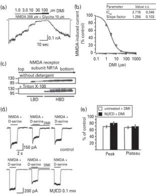
The antidepressant DMI reduced NMDA-induced cation currents through human NMDA receptor subunits NR1A/2A in a dose-dependent fashion with an IC50
value of 2.7¡0.3mM (Fig. 2a,b). The control NMDA
currents and NMDA currents in the presence of different concentrations of DMI reached steady-state after ap- proximately 5 s, thereby assuring correct current analysis after receptor desensitization has been accomplished.
To investigate the membrane localization of the NMDA receptor, we fractionated NMDA-HEK cells by sucrose gradient density centrifugation and probed the respective fractions with an antibody against the NMDA receptor subunit NR1A (Fig. 2c). When no detergent was used, immunoreactivity was detected both in the low buoyant density fractions (LBD, at the top of the gradient) and in the high buoyant density fractions (HBD, at the bottom of the gradient). The use of Triton X-100 completely shifted the NMDA receptor subunit NR1A to the HBD fractions. To affect lipid raft integrity, we partially depleted cholesterol from cell membranes by means of low-dose MbCD treatment thereby retaining cell viability for subsequent electrophysiological record- ings as described previously (Nothdurfter et al., 2010).
Pre-treatment of primary hippocampal neurons with 0.1 mMMbCD for 12 h did not alter the inhibitory effect of DMI on NMDA/D-serine-evoked cation currents (Fig. 2d,e).
55 55
55
without detergent
without detergent + Triton X-100
+ Triton X-100
+ Triton X-100 bottom
bottom bottom
flotillin-1
flotillin-1
Fraction number caveolin-1
caveolin-1 transferrin R
transferrin R
flotillin-1 caveolin-1 transferrin R flotillin-1 caveolin-1 transferrin R top
Cholesterol (µg/ml)
without detergent bottom top
top top
22 22
22
95 95
95
300 250 200 150 100 50 0
1 2 3 4 5 6 7 8 9 10 11 12 13
Fraction number Cholesterol (µg/ml)
250 200 150 100 50
0 1 2 3 4 5 6 7 8 9 10111213 55 22 95
(a)
(c)
(b)
Fig. 1.Effect of detergent on the separation of flotillin-1/
caveolin-1 from the transferrin receptor and cholesterol distribution. HEK 293 cells (a) and mouse brain neocortex (b) were homogenized, sonicated in high-salt buffer and fractionated by sucrose gradient density centrifugation.
Fractions were collected and analysed by SDS-PAGE. Replicate gels were immunoblotted with antibodies against flotillin-1, caveolin-1 and the transferrin receptor (transferrin R). One part of HEK 293 cells or mouse neocortical tissue was prepared without detergent, the other part was solubilized with Triton X-100. (c) Sucrose gradient fractions of mouse neocortical tissue were assayed for cholesterol concentrations. One part of the tissue was prepared without detergent, the other part was solubilized with Triton X-100. Results represent the means¡S.E.M. of three independent experiments.
by guest on September 13, 2016http://ijnp.oxfordjournals.org/Downloaded from
Membrane distribution of desipramine and diazepam and colocalization with the NMDA and GABAAreceptor
To determine whether the addition of detergent also affects the differential accumulation of psychopharma- cological compounds, we quantified the concentrations of DMI in sucrose gradient fractions of HEK 293 cells after incubation with 10mMDMI. As shown previously (Eisensamer et al., 2005), DMI was enriched in the LBD fractions using a detergent-free preparation procedure.
However, the addition of Triton X-100 caused a shift of DMI to the HBD fractions (Fig. 3). Thus, the membrane distribution of both proteins and antidepressive com- pounds appears to be sensitive to the use of detergent.
Considering the antagonistic effects of DMI at the NMDA receptor we investigated whether the GABAA
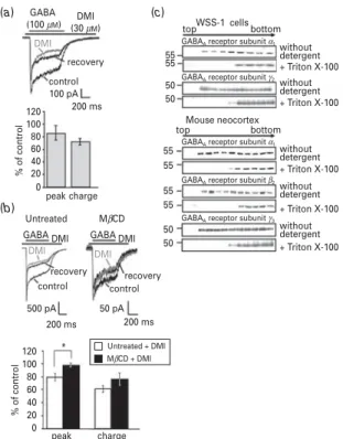
receptor is also sensitive to modulation by DMI. Com- pared to NMDA-induced currents, GABAA receptor- mediated currents of WSS-1 cells were only slightly inhibited by DMI even at high concentrations, which are far above those achieved under clinical conditions (Glotzbach and Preskorn, 1982 ; Fig. 4a). Following ap- plication of 30mMDMI, chloride currents were reduced only to 73.0¡5.2 % of control (charge of currents ex- pressed as the area under the curve in relation to the charge obtained in the absence of DMI, which is set as 100 %). With a reduction to 86.6¡9.8 %, the peak of re- ceptor currents was even less affected. To investigate whether this rather moderate modulatory potency of DMI at the GABAA receptor is related to a divergent distribution of the compound and receptor subunits within the cell membrane, we studied the membrane localization of GABAAreceptor subunits in WSS-1 cells
1.0
NMDA 200 µM + Glycine 10 µM 3.0 10
0.1 nA 10 sec
top 130
95 130 95
bottom without detergent + Triton X-100
LBD
NMDA + D-serine
NMDA + D-serine
NMDA + D-serine
NMDA + D-serine NMDA + D-serine
150 pA
200 pA
control
MβCD 0.1 mM
Peak Plateau MβCD + DMI
2 s
2 s
NMDA + D-serine
untreated + DMI 100
% of control
80 60 40 20 HBD
DMI
DMI NMDA receptor
subunit NR1A
Parameter Slope factor IC50 100
80 60 40 20 0
0.1 1 10 DMI (µM)
100 1000 MMDA-induced current (% control)
2.716 1.256
0.346 0.103 Value S.E. 30 100µMDMI
(a) (b)
(c)
(d) (e)
Fig. 2.Inhibitory effect of desipramine (DMI) on NMDA-evoked cation currents and membrane distribution of the NMDA receptor subunit NR1A. (a) Cation currents of HEK 293 cells stably expressing NR1/NR2A composed NMDA receptors (NMDA-HEK) were recorded in a whole-cell voltage-clamp configuration. Currents were recorded under a continuous stimulation with 200mMNMDA in the presence of 10mM
glycine. Cells were exposed to increasing concentrations of DMI (10 to 100mM). The panel shows representative NR1/2A receptor currents. The upper bar indicates increasing concentrations of DMI ; the lower bar indicates the presence of NMDA/glycine. (b) Dose–response relationship of DMI against the current amplitude. Results are expressed in % of the control NMDA-induced currents obtained without DMI and represent the means¡S.E.M. of five independent
experiments. (c) Membrane fractions of NMDA-HEK cells were prepared by means of sucrose gradient density centrifugation and were analysed by SDS-PAGE. Replicate gels were immunoblotted with a polyclonal antibody against the NMDA receptor subunit NR1. One part of the cells was prepared without detergent, the other part was solubilized with Triton X-100. LBD, low buoyant density fractions ; HBD, high buoyant density fractions. (d) Cation currents of primary hippocampal neurons were recorded in a whole-cell voltage-clamp configuration. 200mMNMDA/5mMD-serine was applied for 2 s in the absence or presence of 3mMDMI. One portion of cells was pre-treated with 0.1 mMmethyl-b-cyclodextrine (MbCD) for 12 h prior to the recordings (lower panel), the other portion of cells remained untreated (control ; upper panel). The upper bar indicates the presence of NMDA/
D-serine, the lower bar indicates the presence of DMI. One representative recording is shown, respectively. (e) Peak amplitude and plateau of untreated cells and after pre- treatment with 0.1 mMMbCD for 12 h of seven independent experiments are shown in the presence of 3mMDMI in relation to values in the absence of DMI, which are set as 100 % (control). Data are shown as means¡S.E.M. % of control.tTests
45 40 35 30 25 20 15 10 5 0
+ Triton X-100
Fraction number
1 2 3 4 5 6 7 8 9 10
Specific DMI concentration
Fig. 3Accumulation of desipramine (DMI) in membrane fractions of HEK 293 cells in the presence of detergent (Triton X-100). HEK 293 cells were incubated with 10mMDMI for 30 min at 37xC. After homogenization and sonification cells were fractionated by sucrose gradient density centrifugation.
Fractions were collected and the concentrations of DMI were determined by HPLC. Results are expressed as specific concentration of DMI (ng DMI/mg protein). Data are
means¡S.E.M. of three independent experiments of homogenates solubilized with Triton X-100. Under detergent-free conditions, DMI was preferentially enriched in the low buoyant density fractions (data not shown ; Eisensamer et al., 2005).
for paired samples revealed no statistically significant differences between peaks and plateaus of untreated and MbCD pretreated cells.
by guest on September 13, 2016http://ijnp.oxfordjournals.org/Downloaded from
(a1andc2) and mouse neocortex (a1,b2andc2; Fig. 4c).
Similarly to the NMDA receptor, all GABAA receptor subunits were detected both in LBD and HBD fractions of cells and tissue using a detergent-free protocol, but only in the HBD fractions when using Triton X-100. Thus, the membrane distribution of GABAAreceptor subunits does not explain the considerably weaker effect of DMI at this ligand-gated ion channel. Interestingly, the inhibitory potency of DMI on GABAAreceptor-mediated currents appeared to be partially reversed under cholesterol- depleting conditions (charge : 61.5¡5.0 % untreated vs.
75.5¡10.7 % MbCD pre-treated ; peak : 79.2¡5.6 % un- treatedvs.97.5¡3.4 % MbCD pre-treated ; Fig. 4b).
Benzodiazepines are potent positive allosteric mod- ulators of GABAA receptors. We therefore investigated the modulatory properties of Diaz on GABAAreceptor function in relation to the membrane distribution of GABAA receptor subunits and that of Diaz. As shown previously (Davies et al., 2000), 1mMDiaz markedly po- tentiated GABA-evoked chloride currents of WSS-1 cells (235.7¡15.2 % ; charge of the currents expressed as the area under the curve in relation to the charge obtained in the absence of Diaz, which is set as 100 % ; data represent
GABA (100 µM) DMI
(30 µM) top
top
bottom
bottom without detergent + Triton X-100 without detergent
without detergent + Triton X-100
+ Triton X-100 without detergent + Triton X-100 without detergent + Triton X-100 GABAA receptor subunit α1
GABAA receptor subunit α1
GABAA receptor subunit γ2
GABAA receptor subunit β2
GABAA receptor subunit γ3
WSS-1 cells
Mouse neocortex DMI
recovery control
% of control
100 pA 200 ms 120
100 80 60 40 20 0
peak charge
% of control
120 100 80 60 40 20
0 peak charge Untreated MβCD
MβCD + DMI GABA DMI GABA DMI
DMI DMI
recovery recovery control
500 pA 50 pA
200 ms
* Untreated + DMI 200 ms control
55
55 55 55 55 50 50 55 50 50
(a)
(b)
(c)
Fig. 4.Effect of desipramine (DMI) on GABA-evoked chloride currents and membrane distribution of GABAAreceptor subunits. (a) 30mMDMI was applied to WSS-1 cells containing recombinanta1c2and low levels of endogenousb3GABAA
receptor subunits. Chloride currents were recorded in a whole- cell voltage-clamp configuration under conditions of a 500 ms stimulation with 100mMGABA. The upper panel shows a representative original recording. The upper bar indicates the presence of GABA ; the lower bar indicates the presence of DMI. The lower panel shows peak amplitude and charge expressed as the area under the curve (AUC) of the GABA- evoked currents in relation to values obtained in the absence of DMI, which is set as 100 % ( % of control). Data are presented as the means¡S.E.M. of ten independent experiments. For calculation of the final DMI effect, values from control and recovery traces were included into the analysis. (b) 30mMDMI was applied to WSS-1 cells in serum-free medium (untreated) and after cholesterol depletion induced by 0.5 mMmethyl-b- cyclodextrine (MbCD). Chloride currents were recorded in a whole-cell voltage-clamp configuration under conditions of a 500 ms stimulation with 100mMGABA. The upper panel shows representative original recordings. The upper bar indicates the presence of GABA ; the lower bar indicates the presence of DMI. The lower panel shows peak amplitude and charge expressed as the AUC of the GABA-evoked currents in relation to values obtained in the absence of DMI, which is set as 100 % ( % of control). Data are presented as the means¡S.E.M. of five independent experiments, respectively. For calculation of the final DMI effect, values from control and recovery traces were included into the analysis.Ttests for paired samples revealed a statistically significant difference between peaks of untreated and MbCD pre-treated cells ; *p<0.05. (c) WSS-1 cells and mouse neocortical tissue were homogenized, sonicated and fractionated by sucrose gradient density centrifugation either in the presence or absence of detergent. Fractions of the gradient were collected and analysed by SDS-PAGE. Replicate gels were immunoblotted with antibodies against the GABAA
GABA (10 µM) Diaz
(1 µM) recovery
800 HEK 293 cells
Mouse neocortex
1 2 3 4
Fraction number 5 6 7 8 9 10
1 2 3 4
Fraction number 5 6 7 8 91011 12 13 600
400 Specific Diaz Specific Diaz concentrationconcentration 200
0
0 50 100 150 200 250 300 control
Diaz
500 pA 1 s
(a) (b)
Fig. 5.Potentiation of GABA-evoked chloride currents by diazepam (Diaz) and membrane distribution of Diaz. (a) Diaz (1mM) was applied to WSS-1 cells containinga1b3c2GABAA
receptor subunits. Chloride currents were recorded in a whole-cell voltage-clamp configuration under conditions of 5 s stimulation with 10mMGABA. A representative original recording is shown. The upper bar indicates the presence of GABA ; the lower bar indicates the presence of Diaz. (b) HEK 293 cells (upper panel) or mouse brain neocortex (lower panel) were incubated with 1mMDiaz for 30 min at 37xC. After
homogenization and sonification cells and tissue were fractionated by sucrose gradient density centrifugation in the absence of detergent. Fractions were collected and analysed for the concentrations of Diaz by mass spectrometry. Results are expressed as specific concentration of Diaz (ng Diaz/mg protein). Data are expressed as means¡S.E.M. of three independent experiments.
receptor subunitsa1,b2(for mouse neocortex) andc2. The expression of theb3subunit in WSS-1 cells was below the limit of detection by Western blotting.
by guest on September 13, 2016http://ijnp.oxfordjournals.org/Downloaded from
the means¡S.E.M. of five independent experiments ; Fig.
5a). This potentiating effect was antagonised by 10mM
flumazenil (data not shown). The distribution of Diaz within the cell membrane was determined in sucrose gradient fractions of HEK 293 cells and mouse neocortex after incubation with 1mMDiaz (Fig. 5b). In contrast to DMI, which was enriched in the LBD fractions, Diaz preferentially accumulated in the HBD non-raft fractions already under detergent-free conditions. This distri- bution pattern was not altered when Triton X-100 was used for preparation of lipid rafts (data not shown). Thus, both ligand-gated ion channel subunits and psycho- pharmacological compounds show different distribution patterns within the cell membrane with regard to lipid raft association.
Effect of lipid raft integrity for GABAAreceptor modulation by Diaz
Impairment of lipid raft integrity by pre-treatment of WSS-1 cells with MbCD for 12 h dose-dependently diminished GABA-evoked chloride currents (Fig. 6a–c).
The shift of the GABA dose–response curve to the right after MbCD treatment (Fig. 6d) indicates reduced agonist effectiveness when receptors are not integrated in lipid rafts. When receptors were activated at submaximal GABA concentrations (Fig. 6e) the GABA enhancing effect of Diaz i.e. increase in GABA potency was even more pronounced under MbCD pre-treatment condi- tions. However, the maximal Diaz-induced potentiation of receptor currents did not change (Fig. 6f), i.e. maximal currents observed with saturating concentrations of GABA were the same both in the presence and absence of Diaz.
Because cholesterol depletion decreased membrane stability and deteriorated patch-clamp recording con- ditions the generation of a concentration–response curve in the presence of Diaz was not achievable with 0.5 mM MbCD. However, leak current and membrane resistance of HEK cells after treatment with 0.5 mM MbCD were comparable to those cells that were untreated. Pre- treatment with 0.25 mMMbCD reduced membrane chol- esterol content to 82.10 % of control (data not shown).
Consequently, this concentration did not result in a suf- ficient shift to the right of the GABA concentration–
response curve as observed at 0.5 mM MbCD. These results indicate that substantial amounts of GABAAre- ceptors were still incorporated into lipid rafts.
Discussion
In the present study we investigated the membrane dis- tribution of NMDA and GABAA receptor subunits in
30 µM GABA
200 pA
200 pA
100 pA
120
% of control
100 90 60 40 20 0
10–1 100 101 GABA (mM) Untreated Untreated : IC50 = 10.647 MβCD 0.5 mM
Untreated MβCD 0.5 mM
MβCD:
nH = 1.057 nH = 1.087 IC50 = 24.747
Untreated Untreated
Untreated + Diaz
Untreated : IC50 = 10.647 MβCD 0.25 mM MβCD:
Diaz:
Diaz MβCD:
MβCD + Diaz
nH = 1.0261 nH = 1.303 nH = 1.739 nH = 2.074 IC50 = 12.323 IC50 = 3.015 IC50 = 4.295 102 103 104
10–1 100 101 GABA (mM)
102 103 104 200 ms
200 ms
200 ms
control
charge
* *
charge
0.25 mM MβCD
0.5 mM MβCD 30 µM
GABA 30 µM GABA
% of control 30 mM GABA% of control 1 µM
Diaz
30 µM GABA
30 µM GABA
30 µM 1 µM GABA Diaz
30 µM GABA
30 µM GABA
30 µM 1 µM GABA Diaz
250 200 150 100
120 100 80 60 40 20 0 50
0 % of control 100 mM GABA
250 200 150 100 50 0
(a) (e)
(f) (b)
(c)
(d)
Fig. 6Effect of methyl-b-cyclodextrine (MbCD) induced cholesterol depletion of WSS-1 cells on GABA-evoked chloride currents and their modulation by diazepam (Diaz). (a)
Potentiating effect of Diaz at the GABAAreceptor of WSS-1 cells.
Chloride currents were recorded in a whole-cell voltage-clamp configuration. 30mMGABA were applied for 500 ms in the absence or presence of 1mMDiaz. The upper bars indicate the presence of GABA, the lower bar indicates the presence of Diaz. Representative recordings before and after application of Diaz and subsequent washout are shown. Potentiating effect of Diaz at the GABAAreceptor of WSS-1 cells after cholesterol depletion induced by 0.25 mMMbCD (b) or 0.5 mMMbCD (c).
Chloride currents were recorded in a whole-cell voltage-clamp configuration. GABA (30mM) were applied for 500 ms in the absence or presence of 1mMDiaz. The upper bars indicate the presence of GABA, the lower bar indicates the presence of Diaz.
Representative recordings before and after application of Diaz and subsequent washout are shown. Cells were treated with 0.25/0.5 mMMbCD for 12 h prior to the recordings. (d) Dose–response relationship of the GABA-evoked current charge through the GABAAreceptor of untreated WSS-1 cells and of WSS-1 cells pre-treated with 0.5 mMMbCD. Results are normalized to the maximum receptor current induced by 3 mM
GABA and presented as means¡S.E.M. of seven independent experiments. (e) Charges of untreated cells and after pre-treatment with 0.5 mMMbCD for 12 h evoked by 30mM
GABA (left panel) or 100mMGABA (right panel) in the presence of 1mMDiaz of five independent experiments in relation to values in the absence of Diaz, which are set as 100 % (control).
Data are shown as means¡S.E.M. % of control (currents evoked with 30 or 100mMGABA). Statistically significant differences between charges of untreated and MbCD pre-treated cells (ttest for paired samples, *p<0.05). (f) Dose–response relationship of the GABA-evoked current charge through the GABAAreceptor of untreated WSS-1 cells and of WSS-1 cells pre-treated with 0.25 mMMbCD. Results are normalized to the maximum receptor current induced by 3 mMGABA and presented as means¡S.E.M. of five independent experiments. Also presented are means¡S.E.M. of normalized charges of untreated WSS-1 cells treated with 1mMDiaz of four independent experiments and
means¡S.E.M. of normalized charges of WSS-1 cells pre-treated with 0.25 mMMbCD treated with 1mMDiaz of four independent experiments.nH, Hill coefficient.
by guest on September 13, 2016http://ijnp.oxfordjournals.org/Downloaded from
relation to their allosteric modulation by DMI and Diaz.
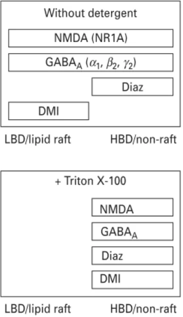
The assignment of receptor subunits and drugs to specific membrane fractions turned out to be strongly dependent on the preparation procedure. A partial colocalization of receptor subunits and the compounds within the cell membrane was found under detergent-free conditions and a complete overlap in non-raft fractions when using detergent (Fig. 7). Impairment of lipid raft integrity by cholesterol depletion did not substantially alter the modulatory potency of DMI at the NMDA and GABAA
receptor. However, impairment of lipid raft integrity af- fected the positive allosteric modulation of the GABAA
receptor by benzodiazepines.
Preparation of lipid rafts heavily relies on the purifi- cation method employed. Commonly, lipid rafts are considered as membrane fractions that are resistant to non-ionic detergents such as Triton X-100 (Brown and Rose, 1992). In fact, we found that the use of detergent allowed proper separation of the lipid raft marker pro- teins caveolin-1 and flotillin-1 from the transferrin re- ceptor, which is widely considered as a non-raft protein (Nothdurfter et al., 2007). This is in line with previous findings reporting a dose-dependent effect of Triton X-100 on the separation of raft from non-raft proteins (Brady et al., 2004). On the other hand, the use of detergent-free protocols has been suggested to more closely reflect the physiological situation (Schnitzer et al., 1995a, b ; Smart et al., 1995 ; Song et al., 1996 ; Luria et al., 2002 ; Eisensamer et al., 2005 ; Persaud-Sawin et al., 2009).
Accordingly, different compositions and properties of lipid rafts have been reported depending on the extrac- tion method used (Foster et al., 2003 ; Kim et al., 2004 ;
Sprenger and Horrevoets, 2007 ; Garner et al., 2008 ; Pike, 2009). Consequently, we studied the membrane distribution of ligand-gated ion channels and psycho- pharmacological compounds under both detergent-free and detergent-containing conditions in the present study.
The detergent-free protocol revealed a much more differential distribution pattern of ionotropic receptor subunits and compounds investigated, although the separation of prototypical markers of raft and non-raft proteins appeared more challenging.
Concerning the NMDA receptor, the NR1A subunit has been located in non-raft membrane fractions of rat forebrain when using an Optiprep gradient without detergent solubilization (Wu et al., 1997). In a study by Hering et al. (2003), NR1 was found both in raft and in non-raft fractions under detergent-containing conditions.
A non raft-association has been reported for the NR1, NR2A and NR2B subunits using Triton X-100 as a deter- gent (Suzuki et al., 2001). Besshoh et al. (2007) found lipid raft association of the NR2A subunit under detergent- containing conditions, whereas the NR2B subunit in rat brain tissue shifted from post-synaptic densities to lipid rafts with increasing age. A study by Ponce et al. (2008) suggested an important role of lipid raft integrity for NMDA receptor function, showing that the reduction of cholesterol levels by simvastatin might protect from NMDA-induced neuronal damage by reducing the as- sociation of the NR1 subunit with lipid rafts. In our study, impairment of lipid raft integrity by cholesterol depletion did not affect the modulatory potency of DMI at the NMDA receptor, which suggests that the membrane lo- calization of this receptor and compound plays a minor role for its modulation by this antidepressant. In some contrast to the NMDA receptor, lipid raft impairment partially reversed the antagonistic effect of DMI at the GABAA receptor, although this effect appeared only marginally. Detergent-free lipid raft preparation yielded a clear distribution of both DMI and partially GABAA
receptor subunits in the raft fractions. Thus, cholesterol depletion might interfere with the pharmacological effects of DMI on GABAA receptors by e.g. impairing ligandrreceptor interaction, thereby weakening the antagonistic potency.
With regard to lipid raft association of GABAA re- ceptors, an almost exclusive lipid raft association has been claimed for thea1subunit of rat cerebellar granule cells after purification with the detergent Brij 98 (Dalskov et al., 2005). Following multiple solubilization steps with Triton X-100 of rat forebrain tissue, distinct GABAA
receptor subunit combinations have been attributed to different cell membrane compartments (Li et al., 2007).
Theaandbsubunits in the absence ofcsubunits, which might represent extra-synaptic receptors, have been detected in detergent-resistant membranes/lipid rafts. In contrast, synaptic assemblies containingaandbsubunits together with thec2subunit showed only negligible lipid raft association.
Without detergent NMDA (NR1A) GABAA (α1, β2, γ2)
Diaz DMI
HBD/non-raft
LBD/lipid raft LBD/lipid raft
+ Triton X-100 NMDA GABAA Diaz DMI
HBD/non-raft
Fig. 7.Schematic diagram showing the distribution of receptors and compounds in lipid raft and non-raft membrane fractions of sucrose gradients. Diaz, diazepam ; DMI, desipramine ; LBD, low buoyant density fractions ; HBD, high buoyant density fractions.
by guest on September 13, 2016http://ijnp.oxfordjournals.org/Downloaded from
In our study, the use of the detergent Triton X-100 shifted NMDA and GABAAreceptor subunits to the HBD fractions of sucrose gradients. However, when using a detergent-free protocol, a much more differential distri- bution pattern of ionotropic receptors within the cell membrane could be observed. NMDA receptor (NR1A subunit) and GABAAreceptor subunits (a1,b2,c2) were distributed both over the LBD and the HBD fractions of the sucrose gradient. In contrast, the 5-HT3receptor was located predominantly in the lipid raft-associated LBD fractions (Eisensamer et al., 2005 ; Nothdurfter et al., 2010). Presumably, physiological and pharmacological processes are more reliably reflected using detergent-free protocols.
Currently, only few data are available concerning the distribution of psychopharmacological drugs within membrane fractions. The tricyclic antidepressant DMI, which modulates the 5-HT3receptor in a non-competitive manner (Eisensamer et al., 2003), was predominantly ac- cumulated in raft-like domains and colocalized with the 5-HT3 receptor protein under detergent-free conditions as shown previously for various other antidepressants and antipsychotics (Eisensamer et al., 2005). However, the addition of Triton-X-100 during preparation com- pletely shifted DMI to the HBD fractions, but had no ef- fect on the cholesterol distribution within the cell membrane. These results are in contrast to the benzodia- zepine Diaz, which was enriched in the HBD fractions already under detergent-free conditions. Thus, in spite of their common lipophilic properties, various psycho- pharmacological compounds display a differential as- sociation with lipid rafts, which might contribute to their distinct psychopharmacological profile.
It has been shown previously that the tricyclic anti- depressant DMI acts as a functional antagonist at the NMDA receptor (Sernagor et al., 1989 ; Watanabe et al., 1993 ; Szasz et al., 2007). Our experiments performed with NMDA receptors expressed in HEK cells confirm these results. Furthermore, currents showed a nearly complete recovery to the control steady-state level after removal of the antagonist. This observation and the fact that the original cytosolic content is mainly dominated by the in- tracellular pipette solution with no protein left, renders a DMI-mediated mechanism via receptor internalisation unlikely and rather speaks in favour of a direct antagon- ism at the receptor. Although the concentrations required for these modulatory effects are in the low micromolar range, they are nevertheless of clinical relevance in view of a high brain : plasma ratio of tricyclic antidepressants (Glotzbach and Preskorn, 1982). On the other hand, DMI delivered at rather high concentrations only slightly af- fected GABA-evoked chloride currents, thereby indicat- ing that the effect of DMI on GABAAreceptors appears to be less potent as compared to NMDA receptors and is not of clinical relevance. Thus, the colocalization of DMI with subunits of ionotropic receptors seems not to be sufficient for a potent allosteric receptor modulation in general.
The benzodiazepine Diaz markedly potentiated GABA-evoked chloride currents, which is in line with previous studies (Davies et al., 2000). Diaz was coloca- lized with GABAAreceptor subunits in the HBD fractions already under detergent-free conditions, which supports the hypothesis that synaptic GABAAreceptors, which are sensitive to benzodiazepines, are located predominantly within non-raft fractions (Li et al., 2007). To investigate the role of the membrane localization of the GABAAreceptor for its modulation by benzodiazepines we impaired lipid raft integrity by MbCD-induced cholesterol depletion.
MbCD treatment diminished GABAAreceptors currents per se, similarly to the results previously found for the 5-HT3receptor (Nothdurfter et al., 2010). The potentiating effect of Diaz appeared to be enhanced at lower GABA concentrations under cholesterol-depleting conditions.
However, due to technical limitations related to choles- terol depletion, we could not perform Schild plot analysis which would have provided definite clarity about the effects of benzodiazepines at the GABAAreceptor under conditions of cholesterol depletion. Nevertheless, our re- sults suggest that, at submaximal GABA concentrations, benzodiazepines show an increased effectiveness at GABAA receptors after cholesterol depletion. Together with the preferential accumulation of Diaz in non-raft fractions, these results argue for the hypothesis that the interaction of benzodiazepines with the GABAAreceptor likely occurs outside lipid rafts.
Acknowledgements
HEK 293 cells stably expressing the NMDA receptor NR1A/2A were a generous gift from Merck, Darmstadt, Germany. The GABAAreceptor subunitc2antibody was a generous gift from Werner Sieghart from the Centre of Brain Research of the University of Vienna, Austria.
The authors thank Christian Namendorf and Annette Schubert for performing the quantification of psycho- pharmacological compounds, Peter Altrichter (Centre of Brain Research of the University of Vienna) for assistance with the Western blots of the GABAAreceptor subunitc2 and Thomas Kirmeier and Julia S. Kessler for valuable discussion.
Statement of Interest None.
References
Allen JA, Halverson-Tamboli RA, Rasenick MM (2007) Lipid raft microdomains and neurotransmitter signalling. Nat Rev Neurosci 8 :128–140.
Besshoh S, Chen S, Brown IR, Gurd JW (2007) Developmental changes in the association of NMDA receptors with lipid rafts.
J Neurosci Res 85 :1876–1883.
Brady JD, Rich TC, Le X, Stafford K, Fowler CJ, Lynch L, Karpen JW, Brown RL, Martens JR (2004) Functional role of
by guest on September 13, 2016http://ijnp.oxfordjournals.org/Downloaded from