A comprehensive approach in high-grade glioma management: position statement from the
Neuro-Oncology Scientific Club (NOSC), Shiraz, Iran
Ein umfassender Ansatz zur Behandlung hochgradiger Gliome:
Positionspapier des Neuro-Oncology Scientific Club (NOSC), Shiraz, Iran
Abstract
Establishing a robust teamwork model in the practice of neuro-oncology requires continued interdisciplinary efforts. The Neuro-Oncology Scientific
Mansour Ansari
1Ahmad Mosalaei
1Club (NOSC) initiative is an interdisciplinary clinical forum promoting
Niloufar Ahmadloo
1the comprehensive approach across involved disciplines in the manage-
Alireza Rasekhi
2ment of central nervous system (CNS) malignancies. With its provincial founding panels and national steering board, NOSC has been operational
Bita Geramizadeh
3in Iran since 2011. This initiative has pursued its mission through inter-
Ali Razmkon
4val strategic meetings, tumor boards, case discussions as well as pub-
Kazem Anvari
5lishing neuro-oncology updates, case study periodicals, and newsletters.
A provincial meeting of NOSC in Shiraz put together insights from inter-
Mohammad Afarid
6national practice guidelines, emerging evidence, and expert opinions
Ali Dadras
6,7to draw a position statement on high-grade glioma management in
Leila Nafarieh
6adults. The present report summarizes key highlights from the above clinical forum.
Mohammad
Mohammadianpanah
1Keywords:neuro-oncology, interdisciplinary, high-grade glioma, position statement, Shiraz
Hamid Nasrolahi
1Seyed Hasan Hamedi
1Zusammenfassung
Die Schaffung eines robusten Teamwork-Modells in der Praxis der Neuro-Onkologie erfordert fortgesetzte interdisziplinäre Anstrengungen.
Shapour Omidvari
1Mohammad Nami
6,8,9,10Der Neuro-Oncology Scientific Club (NOSC) ist ein interdisziplinäres kli-
nisches Forum, das den umfassenden Ansatz bei der Behandlung von 1 Department of Radiation Oncology, School of Malignomen des zentralen Nervensystems (ZNS) über alle Fachrichtun-
Medicine, Shiraz University gen hinweg fördert. Seit 2011 ist NOSC mit seinen regionalen Grün-
of Medical Sciences, Shiraz, Iran
dungspanels und dem nationalen Steuerungsgremium im Iran tätig.
Diese Initiative hat ihre Mission durch strategische Intervalltreffen, Tu-
mor Boards, Falldiskussionen sowie die Veröffentlichung von Neuroon- 2 Department of Radiology, School of Medicine, Shiraz kologie-Updates, Fallstudienzeitschriften und Rundschreiben verfolgt.
University of Medical Sciences, Shiraz, Iran Eine regionale Tagung des NOSC in Shiraz brachte Erkenntnisse aus
internationalen Praxis-Leitlinien, neue Belege und Expertenmeinungen
zusammen, um ein Positionspapier zur Behandlung hochgradiger Gliome 3 Department of Pathology, School of Medicine, Shiraz bei Erwachsenen zu erstellen. Der vorliegende Bericht fasst die wich-
tigsten Highlights aus dem oben genannten klinischen Forum zusam- men.
University of Medical Sciences, Shiraz, Iran Schlüsselwörter:Neuroonkologie, interdisziplinär, hochgradiges Gliom,
Positionspapier, Shiraz
4 Department of
Neurosurgery, School of Medicine, Shiraz University of Medical Sciences, Shiraz, Iran
5 Cancer Research Center, Mashhad University of Medical Sciences, Mashhad, Iran
6 Behestan Medical Scientific Committee, Behestan Group, Tehran, Iran 7 Institute of Biochemistry
and Biophysics (IBB), University of Tehran, Tehran, Iran 8 Department of
Neuroscience, School of Advanced Medical Sciences and Technologies, Shiraz University of Medical Sciences, Shiraz, Iran 9 Neuroscience Laboratory
(Brain, Cognition and Behavior), Department of Neuroscience, School of Advanced Medical Sciences and Technologies, Shiraz University of Medical Sciences, Shiraz, Iran 10 Shiraz Neuroscience
Research Center, Shiraz University of Medical Sciences, Shiraz, Iran
1 Interdisciplinary care in
neuro-oncology: five years with NOSC
The Neuro-Oncology Scientific Club (NOSC) has been an ongoing interdisciplinary initiative fostering teamwork in optimal management of brain tumors across Iran since 2011. Over the past six years and under the NOSC um- brella, experts and practitioners from the disciplines in- volved in brain tumor care have come together to bridge science, knowledge and practice gaps through research endeavors, educational forums and concurrence on clin- ical pathways in brain tumor management, respectively [1], [2], [3], [4], [5], [6], [7], [8], [9], [10].
The NOSC comprises provincial and nation-wide steering boards through which its educational and clinical activities are scheduled, planned and implemented. The provincial NOSC founding panel in Shiraz, Southern Iran, hosted an interactive clinical forum entitled: “setting standards for optimal care to high-grade glioma patients through inter- disciplinary efforts” where over 50 experts and profession- als from related disciplines including neurosurgery, radi- ation oncology, pathology, neuroradiology, neurology, and clinical neuroscience participated to discuss the most updated evidence.
Discussions during this NOSC forum revolved around:
1. the significance of a comprehensive approach to diagnosis, treatment and follow-up in CNS malignan- cies,
2. updates on the molecular pathogenesis of glio- blastoma multiforme (GBM),
3. advanced imaging technologies in high-grade gliomas, 4. novel surgical approaches in glioma,
5. non-surgical management of high-grade gliomas (HGGs).
The present report highlights the communicated insights during the above NOSC meet-up and summarizes the agreed position by Shiraz-NOSC’s expert panel on “optim- izing interdisciplinary care in HGGs”.
2 Malignant gliomas and the unsettled clinical burden
The diagnosis, morphological classification, and efficient treatment of HGGs are regarded as challenging clinical encounters. The clinicopathological data are often insuf- ficiently satisfactory to realize the biology of tumors and to timely identify the therapeutic implications as well as the prognosis [11], [12].
The primary glioblastoma multiforme (GBM) predomin- antly feature astrocytic differentiation while its secondary types may result from astrocytic, oligodendroglial or mixed tumoral transformation of glial cells.
According to the World Health Organization (WHO) classi- fication, GBM is regarded as the most common primary brain tumor with preponderating astrocytic differentiation.
GBM is basically identified through atypical, mitotic, and pleomorphic glial cells together with necrosis, vascular thrombosis, micro-vascular proliferation [13], [14]. The more recent WHO update in 2016 has incorporated mo- lecular markers in glioma classification. This classification includes new genetically identified entities and variants, allowing more diagnostic precision, and a new layered approach to diagnosis. It includes newly recognized en- tities, variants, and patterns, such as IDH (isocitrate de- hydrogenase) mutant and wild-type entities. The update also includes designation of new, genetically defined en- tities such as loss of heterozygosity (LOH) of 1p 19q [15].
The new WHO classification however focuses exclusively on diagnosis. While it includes more molecular assay findings criteria, the specific methods to obtain them are less specified [15].
Promulgating the new classification to our practice re- quires a balancing act not only to address the needs of both clinicians and patients, but also clinical trial experts, population health researchers, policymakers, and health insurers. A key question to address is “whether in our practice a CNS malignancy is to be defined based on histology and genetics or remains based on histology alone”.
The widespread availability of the new diagnostic techno- logies needs to be ensured when recommending these tests for diagnosis and classification purposes.
Apart from our country’s major provinces which currently have access to such tests (i.e. IDH-1 mutation test via immunohistochemistry and LOH of 1p 19q test via fluor- escence in situ hybridization), institutions or regions lacking access to such diagnostic tools need to be accom- modated before the routine use of such test for diagnosis purpose is recommended by our working group. Where such diagnostic tests are not readily accessible, the “not otherwise specified” (NOS) category would be applied when the pathology report is based on the recent WHO classification.
HGGs comprise a large number of primary brain neo- plasms. Though we have started epidemiological surveys using the NOSC brain tumor collaborative registry (BTCR), the prevalence record for HGGs in Iran is yet to be estab- lished [3]. In general, GBM is known to account for up to 15% of the intracranial and 60–70% astrocytic tumors, respectively [16], [17], [18].
Similar to a population-based study conducted in Europe which showed that the incidence of GBM peaks at almost the age of 60, and over 80% of the cases were older than 50 years, interim results from our BTCR suggest a com- parable trend [6], [18]. In agreement with the above study, our so far records confirm that GBM is more com- mon in males than females [6].
Given the infiltrative nature of HGGs and their interference with critically functional and eloquent brain regions, they are typically not amenable to total resection. With a high capacity for micro-infiltration, spreading and rapid pro-
gression, survival with GBM tends to be below one year in almost half of the patients [19], [20].
Upon confirmation of the primary or secondary GBM dia- gnosis, the overall survival (OS) is roughly similar. However in secondary GBM, OS depends on the grade of the ori- ginal pathology which transformed to GBM [21].
When the genetic aspects of the tumors are studied, a large number of mutations may be observed. In many instances, while proto-oncogenes are invigorated, onco- gene-protecting factors are suppressed. Studies have suggested a long list for gene mutations both for the primary and secondary GBM [22], [23], [24].
In terms of the GBM location, they may appear in any subcortical region of either hemispheres. While almost in two third of the cases GBMs are found in temporal and parietal lobes, they are observed in frontal and occipital lobes in almost 25% and 16% of the cases, respectively.
The fronto-temporal presentation of HGGs and GBM in particular is also frequent [25], [26].
Tumors may infiltrate via the white matter tracts and through the corpus callosum to conquer the contralateral hemisphere. In children, the presence of HGGs in striatum and the thalamic region is not uncommon. Some infre- quent or exceptional locations of the tumor include the ventricles, brainstem, cerebellum, and spinal cord [27].
The clinical manifestations of GBM cases may largely vary. In fact, many of the symptoms suggestive for the pathology (including headache, nausea and vomiting, and clouding of consciousness) may root in the increased intracranial pressure or mass effect through invasion, compression, and edema. Symptoms arising from the latter may include seizures, focal neurological deficit, and altered cognitive functions [2], [6].
GBM is generally regarded as among the most common primary CNS tumors. Though this neoplasm is currently categorized more often histologically, genetic studies have recently been positioned as an integral part of the diagnosis, disease profiling, and prognostic assessments [2], [10], [15].
In practice, the treatment approaches are pursued based on the patient’s age, performance status, and baseline neurocognitive status. In many instances, the standard care comprises surgical resection followed by chemoradi- ation and adjuvant chemotherapy in cognitively competent patients younger than 70 years old. In elderly subjects however, radiotherapy and palliative care is perhaps all that is sought [28].
Despite the best possible care today, GBM prognosis has remained poor given the aggressive nature of the tumor.
Therefore it is crucial to ensure that not only an extended progression-free survival but also an acceptable quality of life is provided through treatments. In terminal cases where treatments are withheld, best supportive or pallia- tive care has to be provided to ameliorate symptoms as much as possible [28].
3 Clinical assessment and follow-up of patients with GBM
The general, neurological and cognitive examination constitute the key imperatives while evaluating a case with GBM. When baseline measures are established, an improvement in the patient’s clinical status can be wit- nessed, in terms of intracranial hypertension, focal neurological signs or cognitive profile. The assessment needs to be dynamic based on the treatment response [28], [29].
As the clinical status improves, evaluation of the patient may advance to more sophisticated tests including neuropsychological evaluations, language assessments, neurocognitive profiling, and rehabilitation or studies where the patient’s cooperation is definitional, including the functional magnetic resonance imaging (fMRI) of functional near infra-red spectroscopy (fNIRS) [30].
The functional status of patients is usually reported by the Karnofsky performance status (KPS) score or Eastern Cooperative Oncology Group (ECOG) scale, where patients with KPS of 70 and above or ECOG score of 0 to 1 have maintained their autonomy and can actively engage in activities of daily living [31], [32].
Other than comprehensive neurological examination, patients would need to be assessed for neurocognitive performance and their quality of life. As such pre- and post-treatment quality of life and neurocognitive manifest- ations would be comparatively measured as a part of clinical evaluations and follow-up [12], [28]. Some vali- dated tools which are used for neurocognitive assessment include testing batteries such as Addendrook’s Neurocog- nitive Examination (ACE), Repetitive Battery for the Assess- ment of Neuropsychological Status (RBANS), the Trail Making Test and the Multilingual Aphasia Examination [30].
Given the availability of the neurocognitive assessment and rehabilitation platform in our setting [30], the NOSC panel agreed that such evaluations (baseline, post-oper- atively, and following treatments) using neuropsychologic- al, cognitive tests and functional neuroimaging modalities should be incorporated into our glioma treatment work- flow [33].
4 Treatment protocols and approaches
The choice of treatment is essentially determined by the patient’s age, general medical condition, KPS, character- istics of the lesion, extent of surgical removal, survival benefits against risks and patient’s willingness [28].
The open surgery, navigation-guided or stereotactic biopsy taking can be done to provide histopathological sample to confirm diagnosis. The selection of the biopsy site is crucial since taking samples from necrotic, edematous areas and areas near the subarachnoid space with the potential of bleeding may be misleading or life-threaten-
ing. A multidisciplinary neuro-oncology group may best decide on the most appropriate site to be biopsied [5].
Moreover, the surgical resection of the lesion is done to help decompressing the cerebral tissue, especially when the compressive effects threats patient’s life or function.
Compared to biopsy only, surgery is known to improve the patient’s condition and to contribute to extended survival [5], [8]. The improved condition of patients follow- ing surgery may enable their access to further treatment measures and the opportunity to respond [8].
To minimize the post-surgical sequellae, functional sur- gery and awake craniotomy settings have been employed in some centers across our country. To maximize the functional outcome, proximity to eloquent areas (motor, language, sensory, visual), baseline focal neurologic signs and deep location of the tumor (basal ganglia, ventricles, brainstem) need to be well considered upon surgery [2], [6].
Baseline neuropsychological assessments and brain mapping measures for surgical planning would provide useful baseline information. Furthermore, stereotactic radiosurgery or safe maximal resection will be done, the latter including intraoperative recording through electro- corticography or somatosensory evoked potentials where applicable. The setup has already been established in well-equipped neuroscience laboratories in Shiraz [30].
4.1 Primary and complementary treatments in GBM
The choice of treatment in patients younger than 70 years with a KPS of 70 and above is based on the latest EANO guideline [28]. Maximal surgical excision is in fact the practice at first place. Although some years ago, the pro- gnostic impact of surgery was questioned, different studies confirm that in high grade gliomas, extensive surgery is linked to a more favorable prognosis [34], [35], [36], [37], [38].
The standard treatment in GBM is based on the results from an international, multi-center open-labeled, random- ized controlled phase III study, where patients who were randomized to undergo radiotherapy (RT) alone or concur- rent chemoradiation with temozolomide (TMZ) at 75 mg/m2followed by 6 cycles of TMZ at 150–200 mg/m2 where compared in terms of overall survival (OS) [21].
According to Stupp’s study, a larger two-year OS rate was observed in cases receiving RT+TMZ than RT alone (26%
vs. 10%). In a follow-up investigation on the same study population, 9% of GBM patients who followed this treat- ment survived up to 5 years, while this was only 1% in the RT alone arm. Additionally, the impact on OS was superior in patients with positive methylation status in the O6-methylguanine-DNA-methyltransferase (MGMT) promoter [21].
More recently, further possible treatments targeting to arrest angiogenesis have also been experienced in clinical setting. For instance, in a multi-center, international, randomized phase III trial known as AVAglio, an anti-an- giogenic therapy provided survival benefits in newly dia-
gnosed GBM patients [39], [40]. This study randomly assigned patients into two arms whereby the first under- went the standard protocol by Stupp et al. plus placebo, while the second followed the similar standard treatment plus a concurrent dose of bevacizumab at 10 mg/kg i.v., the treatment continuing for 3 additional weeks. Results of the above investigation indicated an improved progres- sion-free survival (PFS) and quality of life in the concurrent treatment arm. This could have largely been due to a re- duced use of steroids secondary to the add-on therapy using bevacizumab. This study however failed to demonstrate improvement in OS [39].
In the current practice of neuro-oncology, bevacizumab is particularly indicated in recurrent GBM settings. The regimen is normally administered at 10 mg/kg i.v. bi- weekly, or 15 mg/kg i.v. every 3 weeks [12], [28]. This approach is being followed in our practice given the availability of bevacizumab.
Other available alternative chemotherapeutic regimen to consider include the procarbazine, lomustine and vin- cristine (PCV) combination protocol [28].
In the recurrent GBM setting, some challenging cases who fail to respond to the above, may be enrolled in in- vestigational setups including immunotherapy (PD-1, PD- L1 or CTLA-4 receptor antibodies) [41], [42].
4.2 Treatment follow-up
In controversial cases, an interdisciplinary neuro-oncolo- gical assessment would help determining the most advis- able therapeutic and follow-up approaches.
In fact, a crucial component in the patients’ treatment is proper follow-up through interval clinical, imaging and laboratory assessments. The follow-up is normally pur- sued with the post-operative control MRI to define the extent of residual lesion following surgery. In addition, patients undergo multimodal imaging using MRI to assess their tumor behavior as well as the response to treatment [4], [6].
Moreover, on-treatment laboratory assessments also become applicable to evaluate patients clinically for possible chemotherapy-induced side effects prior to each treatment cycle (typically, every 4 weeks) [28].
In case of uncertainties with regard to tumor behavior in regular MRI, multimodal MRI scans comprising diffusion and perfusion sequences with apparent diffusion capacity (ADC) maps would provide additional information on the extent of the lesion and regional cerebral blood volume (rCBV). To elucidate whether true or pseudo-progression has occurred, uncertain areas might be evaluated using the magnetic resonance spectroscopy (MRS). Such ima- ging assessments would clarify contrast enhancement or hyper-intensity signals in T1 and T2 sequences, respect- ively. Other than the above and when accessible, [18F]- fluoro-ethyl-L-tyrosine PET would be the best evaluated option to explore the biological activity of the lesion [12], [28], [43].
4.3.Control MRI schedule
Post-operatively, the first MR scan is recommended at 48 hours. Further to regular follow-up imaging upon treatment, a diffusion-weighted MRI might be required to define the presence of any subacute contrast enhance- ment which might be mistaken for progression while in fact resulting from radiation necrosis in typical instances [12].
While the need for control MRI upon completion of radio- therapy treatment is a matter of debate, the first control MRI is strongly advised following the 2nd or 3rd cycle of adjuvant chemotherapy. Control MRIs would then be taken every 8–12 weeks to evaluate the patient’s re- sponses to therapy [12], [28].
4.4 Neuroimaging technical
characteristics and considerations
The control and follow-up MRI need to be practiced using the same equipment, acquisition protocol, contrast and topographical references, to technically allow comparable data [44].
The sequences applied to follow-up MR scans include the T1 3D and gadolinium-contrasted T1 3D spoiled gradient recalled (SPGR) images providing three-plane reconstruction and size calculation through segmentation as well as T2 or T2 fluid-attenuated inversion recovery (FLAIR) with thin cuts [44].
In practice, CT scans have no place in follow-up assess- ment of patients with CNS tumors unless complications including hydrocephalus or hemorrhage are doubted [13].
4.5 Response assessment in neuro-oncology (RANO) criteria
The RANO criteria, based on the MRI scans, are currently referenced for response assessment in neuro-oncology.
The radiological response to a given therapy is evaluated after measuring baseline tumoral dimensions before and after the treatment. It should always be noted that radi- ological response assessment particularly upon permea- bility changes after treatment with anti-angiogenic ther- apies is challenging. Thus, confirmatory imaging needs to be considered 4 weeks following a radiological re- sponse [45], [46].
With respect to the radiological response, key terms in- cluding complete response (CR), partial response (PR), stable disease (SD), and progressive disease are applied.
Disease progression is usually considered when T2/FLAIR non-enhancing lesions are increased in size on stable or increased corticosteroid doses compared to baseline or earlier assessments [46].
When following up the disease, the possibility of pseudo- progression needs to be taken into account. In other words, early tissue reactions within the first few months should be differentiated from similar effects which in fact
result from true progression. Some factors to consider when labeling true progression include:
1. contrast enhancement beyond 12 weeks after the completion of radiotherapy,
2. contrast enhancement in a non-irradiated zone, 3. an increase in non-enhancing and hyper-intense sig-
nals on T2/FLAIR,
4. excluded underlying causes (ischemia, post-radiother- apy changes, seizure activity, infection or demyelina- tion) explaining the changing T2/FLAIR signals, and 5. altered T2/FLAIR signals indicating tumor infiltration
outside the radiation field [46].
5 The NOSC’s agreed clinical pathway in glioma care
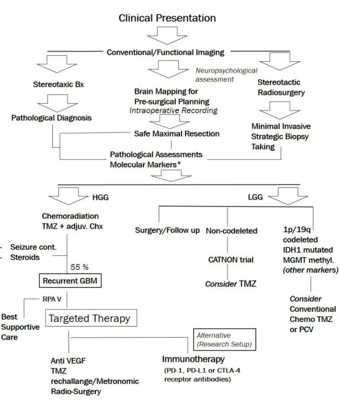
Based on the international consensus statements and the review of current guidelines [15], [28], our working group’s agreed approach to patients with malignant gliomas, including diagnostic aspects (i.e., early diagnosis, history, clinical examination, neuroimaging, preoperative management, biopsy and resection, histological classifi- cation and grading, molecular diagnostics) as well as general therapeutic approach (i.e., surgical therapy, ra- diotherapy, pharmacotherapy, and other therapeutic ap- proaches) is depicted in Figure 1.
The routine use of molecular markers (PD-1, PD-L1 or CTLA-4 receptor antibodies) especially for the classifica- tion purpose, as recommended by 2016 WHO guideline, is currently not feasible and yet to be established in our setting.
6 Summary and recommendations
In line with the mission statement of the NOSC in Iran, and in order to draw an updated clinical decision guide for the interdisciplinary practice of neuro-oncology, and the treatment of high-grade gliomas in particular, the panel of experts from Shiraz NOSC agreed on a clinical pathway (Figure 1). Based on this clinical pathway al- gorithm following the clinical suspicion, conventional/func- tional neuroimaging are taken, after which stereotactic biopsy would be taken to ensure pathological diagnosis.
The specimen is then assessed for molecular markers.
Patients will initially undergo baseline neuropsychological assessments and further brain mapping measures for surgical planning. Stereotactic radiosurgery or safe max- imal resection will be done, latter including intraoperative recording through electrocorticography or somatosensory evoked potentials where applicable.
Post-operatively, the newly diagnosed HGG patients would undergo the protocol of chemo-radiation and adjuvant chemotherapy with TMZ established by Stupp et al. [12].
Seizure control and optimized steroid use are to be con- sidered.
In recurrent HGG patients, those with poor performance status receive best supportive care, whereas patients with fair to favorable performance either receive tar- geted/re-challenge therapy (anti-angiogenic agent, TMZ re-challenge/metronomic dosing, radiosurgery, re-irradi- ation or TMZ+anti-angiogenic agent) or enter the investi- gational setups including immunotherapy.
Providing an optimal care to patients with CNS malignan- cies requires a multidisciplinary team approach. The key benefits include well-organized coordination of multiple providers, direction for complicated cases, open commu- nication amongst care teams, education, and clinical trial access. Over the past 6 years, the NOSC panel has strived to coordinate multidisciplinary care and influence care decisions in most of the major provinces across Iran.
The following parts of the international consensus state- ments [15], [28] have been established by the NOSC panel for implementation in our local practice:
1. The standard of care for anaplastic astrocytoma in- cludes resection as feasible or biopsy, followed by involved field radiotherapy.
2. The standard of care for glioblastoma (age <65–70 years) includes resection as feasible or biopsy, fol- lowed by involved-field radiotherapy and concomitant and adjuvant (six cycles) TMZ chemotherapy.
3. Chemotherapy with TMZ or PCV is as effective as ra- diotherapy in the treatment of anaplastic gliomas, including anaplastic astrocytomas.
4. Elderly patients who are not candidates for TMZ con- current with radiotherapy followed by adjuvant TMZ should be treated with radiotherapy (e.g., 15 doses of 2·66 Gy) alone or temozolomide (5/28) based on MGMT promoter methylation status
5. Upon recurrence, standards of care for GBM are nitro- sourea regimens, TMZ rechallenge, and bevacizumab as options for pharmacotherapy.
6. Patients with 1p/19q co-deleted anaplastic oligo- dendroglial tumors (assessed in our country through FISH method in major provinces) should not be treated with radiotherapy alone, but should receive chemo- therapy with alkylating agents with or without radio- therapy.
Meanwhile, some remaining parts of the international consensus, especially the recent WHO molecular classi- fication for gliomas, are planned to be established in the coming years when almost all our CNS malignancy care accounts are equipped with such molecular testing set- ups (1p/19q co-deletion, MGMT promoter methylation, IDH1/2 mutation).
While there is strong agreement on the role of clinical decision making and education, the countrywide imple- mentation of recommendations need to be continuously monitored and ensured by the NOSC provincial founding panels across the country.
Figure 1: The NOSC’s agreed clinical pathway for the diagnosis and treatment of patients with glioma. Early diagnosis, history, clinical examination, neuroimaging, preoperative management, biopsy and resection, histological classification and grading,
molecular diagnostics as well as surgical therapy, radiotherapy, pharmacotherapy, and other therapeutic approaches are considered in the pathway. The pathways would be expected to receive updates in coming years when molecular markers
testing set-up and novel therapies become widely accessible in our setting.
Notes
Acknowledgment
The authors would like to thank Dr. Dindoust P, Salarian A, Hejazi-Farahmand, SAR for supporting this clinical forum. The ‘NOSC, Shiraz’ received scientific and admin- istrative support from the Department of Radiation Onco- logy, Shiraz University of Medical Sciences, Shiraz, Iran, as well as the MSD Medical team at Behstan Darou PJS, Tehran, Iran.
Competing interests
The present report outlined the communications and ex- perts’ opinions during the NOSC meet-up 2015, Shiraz, Iran. The authors declare no competing interest upon data review, talk delivery during the meeting, interactive discussions and preparation of the present report. MN provided medical consultancy to Behestan Medical Sci- entific Committee, Behestan Group, Tehran, Iran.
Preprint publication
A first version of this manuscript was published as a not peer-reviewed draft in the PeerJ Preprints [47].
References
1. Anvari K, Bahadorkhan G, Nekooi S, Taghizadeh A, Kheradmand H, Nowferesti G, et al. Towards the Real Interdisciplinary Approach in Treating Brain Tumors: Report from the Neuro- Oncology Scientific Club opening meeting - NOSC 2011-13 October- Mashhad, IR Iran. Webmedcentral.
2011;2(10):WMC002381. DOI:
10.9754/journal.wmc.2011.002381
2. Anvari K, Bahadorkhan G, Silanian-Toussi M, Rahighi S, Ghavamnasiri M, Saeedi M, et al. From fundamental brain tumor science to interdisciplinary bedside care; the outcome report from the Neuro-Oncology Scientific Club second meet-up (NOSC- 2), 19th April 2012, Mashhad, Iran. Int J Med Clin Res.
2012;3(5):168-75. DOI: 10.9735/0976-5530.3.5.168-175 3. Torabi-Nami M, Hejazi Farahmand S, Mohammadzadeh F. Neuro-
oncology scientific club and the national Iranian brain tumor registry. Neuro Oncol. 2012 Sep;14 Suppl 3:P.282. DOI:
10.1093/neuonc/nos183
4. Amouheidari A, Hemati S, Sabouri M, Emami J, Mehrzad V, Hekmatnia H, et al. The Nexus between Interdisciplinary Approach and Extended Survival in CNS Tumors, Neuro-Oncology Scientific Club (NOSC) Meeting Report, 27 December 2012, Isfahan, Iran.
Res Cancer Tumor. 2013;2(1):1-9. DOI:
10.5923/j.rct.20130201.01
5. Haddad P, Zali A, Tabatabaeefar M, Nikoofar A, Hadizadeh Kharazi H, Ghadyani M, et al. Turning Interdisciplinary Brain Tumor Science into Survival; Report from the NeuroOncology Scientific Club Opening Session, NOSC 2012 -19 January- Tehran, IR Iran. Rep Opinion. 2012;4(2):42-53. DOI:
10.7537/marsroj040212.07
6. Haddad P, Shazadi S, Samiei F, Kharrazi HH, Tabatabaeefar M, Rakhsha A, Faranoosh M, Torabi-Nami M, Dadras A, Liaghi A, Nafarieh L. An overview of neuro-oncology research and practice in Iran, three years with the NOSC initiative. Int J Clin Exp Med.
2015;8(3):3946-55.
7. Faranoush M, Torabi-Nami M, Mehrvar A, Hedayati-Asl A, Tashvighi M, Parsa R, et al. Classifying Pediatric Central Nervous System Tumors through near Optimal Feature Selection and Mutual Information: A Single Center Cohort. Middle East J Cancer.
2013;4(4):153-62.
8. Anvari K, Bahadorkhan G, Etemad-Rezaie H, Silanian-Toussi M, Nekooei S, Samini F, et al. Prognostic Factors and Treatment Outcome in Glial Brain Tumors; Data from the Third Neuro- oncology Scientific Club's Input Forum, 2013, Mashhad. Iran. Br J Med Med Res. 2014;4(18):3538-53. DOI:
10.9734/BJMMR/2014/9142
9. Anvari K, Hosseini S, Rahighi S, Toussi MS, Roshani N, Torabi- Nami M. Intracranial meningiomas: Prognostic factors and treatment outcome in patients undergoing postoperative radiation therapy. Adv Biomed Res. 2016;5:83. DOI:
10.4103/2277-9175.182214
10. Ansari M, Nasrolahi H, Kani AA, Mohammadianpanah M, Ahmadloo N, Omidvari S, Mosalaei A. Pediatric glioblastoma multiforme: A single-institution experience. Indian J Med Paediatr Oncol. 2012 Jul;33(3):155-60. DOI: 10.4103/0971-
5851.103142
11. Kim ES, Satter M, Reed M, Fadell R, Kardan A. A novel, integrated PET-guided MRS technique resulting in more accurate initial diagnosis of high-grade glioma. Neuroradiol J. 2016 Jun;29(3):193-7. DOI: 10.1177/1971400916639962 12. Stupp R, Brada M, van den Bent MJ, Tonn JC, Pentheroudakis
G; ESMO Guidelines Working Group. High-grade glioma: ESMO Clinical Practice Guidelines for diagnosis, treatment and follow- up. Ann Oncol. 2014 Sep;25 Suppl 3:iii93-101. DOI:
10.1093/annonc/mdu050
13. Evans MR, Evans SB. Glioblastoma multiforme: a devastating diagnosis. Pract Neurol. 2016 Oct;16(5):416-8. DOI:
10.1136/practneurol-2016-001424
14. Alcedo-Guardia R, Labat E, Blas-Boria D, Vivas-Mejia PE.
Diagnosis and New Treatment Modalities for Glioblastoma: Do They Improve Patient Survival? Curr Mol Med. 2016 Apr 29. DOI:
10.2174/1566524016666160429120150
15. Louis DN, Perry A, Reifenberger G, von Deimling A, Figarella- Branger D, Cavenee WK, Ohgaki H, Wiestler OD, Kleihues P, Ellison DW. The 2016 World Health Organization Classification of Tumors of the Central Nervous System: a summary. Acta Neuropathol. 2016 Jun;131(6):803-20. DOI: 10.1007/s00401- 016-1545-1
16. Burton E, Ugiliweneza B, Woo S, Skirboll S, Boaky M. A Surveillance, Epidemiology and End Results-Medicare data analysis of elderly patients with glioblastoma multiforme:
Treatment patterns, outcomes and cost. Mol Clin Oncol. 2015 Sep;3(5):971-978. DOI: 10.3892/mco.2015.590
17. Darefsky AS, King JT Jr, Dubrow R. Adult glioblastoma multiforme survival in the temozolomide era: a population-based analysis of Surveillance, Epidemiology, and End Results registries. Cancer.
2012 Apr;118(8):2163-72. DOI: 10.1002/cncr.26494 18. Baldi I, Huchet A, Bauchet L, Loiseau H. Épidémiologie des
glioblastomes [Epidemiology of glioblastoma]. Neurochirurgie.
2010 Dec;56(6):433-40. DOI: 10.1016/j.neuchi.2010.07.011 19. Kong BH, Moon JH, Huh YM, Shim JK, Lee JH, Kim EH, Chang
JH, Kim DS, Hong YK, Kim SH, Lee SJ, Kang SG. Prognostic value of glioma cancer stem cell isolation in survival of primary glioblastoma patients. Stem Cells Int. 2014;2014:838950. DOI:
10.1155/2014/838950
20. Carlberg M, Hardell L. Decreased survival of glioma patients with astrocytoma grade IV (glioblastoma multiforme) associated with long-term use of mobile and cordless phones. Int J Environ Res Public Health. 2014 Oct;11(10):10790-805. DOI:
10.3390/ijerph111010790
21. Stupp R, Hegi ME, Mason WP, van den Bent MJ, Taphoorn MJ, Janzer RC, Ludwin SK, Allgeier A, Fisher B, Belanger K, Hau P, Brandes AA, Gijtenbeek J, Marosi C, Vecht CJ, Mokhtari K, Wesseling P, Villa S, Eisenhauer E, Gorlia T, Weller M, Lacombe D, Cairncross JG, Mirimanoff RO; European Organisation for Research and Treatment of Cancer Brain Tumour and Radiation Oncology Groups; National Cancer Institute of Canada Clinical Trials Group. Effects of radiotherapy with concomitant and adjuvant temozolomide versus radiotherapy alone on survival in glioblastoma in a randomised phase III study: 5-year analysis of the EORTC-NCIC trial. Lancet Oncol. 2009 May;10(5):459-66.
DOI: 10.1016/S1470-2045(09)70025-7
22. Lohkamp LN, Schinz M, Gehlhaar C, Guse K, Thomale UW, Vajkoczy P, Heppner FL, Koch A. MGMT Promoter Methylation and BRAF V600E Mutations Are Helpful Markers to Discriminate Pleomorphic Xanthoastrocytoma from Giant Cell Glioblastoma.
PLoS ONE. 2016;11(6):e0156422. DOI:
10.1371/journal.pone.0156422
23. Sarmiento JM, Mukherjee D, Black KL, Fan X, Hu JL, Nuno MA, Patil CG. Do Long-Term Survivor Primary Glioblastoma Patients Harbor IDH1 Mutations? J Neurol Surg A Cent Eur Neurosurg.
2016 May;77(3):195-200. DOI: 10.1055/s-0035-1566121 24. Franceschi S, Lessi F, Aretini P, Mazzanti CM, Menicagli M, La
Ferla M, De Gregorio V, Caramella D, Naccarato AG, Bevilacqua G, Bonadio AG, Pasqualetti F. Molecular portrait of a rare case of metastatic glioblastoma: somatic and germline mutations using whole-exome sequencing. Neuro Oncol. 2016 Feb;18(2):298-300. DOI: 10.1093/neuonc/nov314 25. Tuovinen N, de Pasquale F, Caulo M, Caravasso CF, Giudice E,
Miceli R, Ingrosso G, Laprie A, Santoni R, Sabatini U. Transient effects of tumor location on the functional architecture at rest in glioblastoma patients: three longitudinal case studies. Radiat Oncol. 2016 Aug;11(1):107. DOI: 10.1186/s13014-016-0683- x
26. Paldor I, Drummond KJ, Kaye AH. IDH1 mutation may not be prognostically favorable in glioblastoma when controlled for tumor location: A case-control study. J Clin Neurosci. 2016 Dec;34:117-120. DOI: 10.1016/j.jocn.2016.05.016
27. Akbari H, Macyszyn L, Da X, Bilello M, Wolf RL, Martinez-Lage M, Biros G, Alonso-Basanta M, O Rourke DM, Davatzikos C. Imaging Surrogates of Infiltration Obtained Via Multiparametric Imaging Pattern Analysis Predict Subsequent Location of Recurrence of Glioblastoma. Neurosurgery. 2016 Apr;78(4):572-80. DOI:
10.1227/NEU.0000000000001202
28. Weller M, van den Bent M, Hopkins K, Tonn JC, Stupp R, Falini A, Cohen-Jonathan-Moyal E, Frappaz D, Henriksson R, Balana C, Chinot O, Ram Z, Reifenberger G, Soffietti R, Wick W; European Association for Neuro-Oncology (EANO) Task Force on Malignant Glioma. EANO guideline for the diagnosis and treatment of anaplastic gliomas and glioblastoma. Lancet Oncol. 2014 Aug;15(9):e395-403. DOI: 10.1016/S1470-2045(14)70011-7 29. Stupp R, Tonn JC, Brada M, Pentheroudakis G; ESMO Guidelines
Working Group. High-grade malignant glioma: ESMO Clinical Practice Guidelines for diagnosis, treatment and follow-up. Ann Oncol. 2010 May;21 Suppl 5:v190-3. DOI:
10.1093/annonc/mdq187
30. Ashjazadeh N, Boostani R, Ekhtiari H, Emamghoreishi M, Farrokhi M, Ghanizadeh A, Hatam G, Hadianfard H, Lotfi M, Mortazavi SM, Mousavi M, Montakhab A, Nili M, Razmkon A, Salehi S, Sodagar AM, Setoodeh P, Taghipour M, Torabi-Nami M, Vesal A.
Operationalizing Cognitive Science and Technologies' Research and Development; the "Brain and Cognition Study Group (BCSG)"
Initiative from Shiraz, Iran. Basic Clin Neurosci. 2014;5(2):104- 16.
31. Jones LW, Cohen RR, Mabe SK, West MJ, Desjardins A, Vredenburgh JJ, Friedman AH, Reardon DA, Waner E, Friedman HS. Assessment of physical functioning in recurrent glioma:
preliminary comparison of performance status to functional capacity testing. J Neurooncol. 2009 Aug;94(1):79-85. DOI:
10.1007/s11060-009-9803-x
32. Janinis J, Efstathiou E, Panopoulos C, Samantas E, Aravantinos G, Christodoulou C, Skarlos D. Phase II study of temozolomide in patients with relapsing high grade glioma and poor performance status. Med Oncol. 2000 May;17(2):106-10. DOI:
10.1007/BF02796204
33. Anvari K, Ersi MF, Etemad-Rezaie H, Nekooie S, Arbabi F, Ganjeifar B, et al. P21.04 Clinical pathway in glioma management; from NOSC (Neuro-Oncology Scientific Club) meetings to bedside practice. Neuro Oncol. 2016;18(suppl 4):iv81-iv2. DOI: 10.1093/neuonc/now188.292
34. Badhiwala JH, Nassiri F, Almenawer SA. GBM surgery in the elderly-time to be more aggressive? Clin Neurol Neurosurg. 2016 Feb;141:131-2. DOI: 10.1016/j.clineuro.2015.10.009 35. Sun GC, Wang F, Chen XL, Yu XG, Ma XD, Zhou DB, Zhu RY, Xu
BN. Impact of Virtual and Augmented Reality Based on Intraoperative Magnetic Resonance Imaging and Functional Neuronavigation in Glioma Surgery Involving Eloquent Areas.
World Neurosurg. 2016 Dec;96:375-382. DOI:
10.1016/j.wneu.2016.07.107
36. Krivosheya D, Prabhu SS, Weinberg JS, Sawaya R. Technical principles in glioma surgery and preoperative considerations. J Neurooncol. 2016 Nov;130(2):243-252. DOI: 10.1007/s11060- 016-2171-4
37. Pessina F, Navarria P, Cozzi L, Ascolese AM, Simonelli M, Santoro A, Tomatis S, Riva M, Fava E, Scorsetti M, Bello L. Value of Surgical Resection in Patients with Newly Diagnosed Grade III Glioma Treated in a Multimodal Approach: Surgery,
Chemotherapy and Radiotherapy. Ann Surg Oncol. 2016 Sep;23(9):3040-6. DOI: 10.1245/s10434-016-5222-3 38. Satoer D, Visch-Brink E, Dirven C, Vincent A. Glioma surgery in
eloquent areas: can we preserve cognition? Acta Neurochir (Wien). 2016 Jan;158(1):35-50. DOI: 10.1007/s00701-015- 2601-7
39. Chinot OL, Nishikawa R, Mason W, Henriksson R, Saran F, Cloughesy T, Garcia J, Revil C, Abrey L, Wick W. Upfront bevacizumab may extend survival for glioblastoma patients who do not receive second-line therapy: an exploratory analysis of AVAglio. Neuro Oncol. 2016 Sep;18(9):1313-8. DOI:
10.1093/neuonc/now046
40. Sandmann T, Bourgon R, Garcia J, Li C, Cloughesy T, Chinot OL, Wick W, Nishikawa R, Mason W, Henriksson R, Saran F, Lai A, Moore N, Kharbanda S, Peale F, Hegde P, Abrey LE, Phillips HS, Bais C. Patients With Proneural Glioblastoma May Derive Overall Survival Benefit From the Addition of Bevacizumab to First-Line Radiotherapy and Temozolomide: Retrospective Analysis of the AVAglio Trial. J Clin Oncol. 2015 Sep;33(25):2735-44. DOI:
10.1200/JCO.2015.61.5005
41. Platten M, Bunse L, Wick W, Bunse T. Concepts in glioma immunotherapy. Cancer Immunol Immunother. 2016 Oct;65(10):1269-75. DOI: 10.1007/s00262-016-1874-x
42. Lamano JB, Ampie L, Choy W, Kesavabhotla K, DiDomenico JD, Oyon DE, Parsa AT, Bloch O. Immunomonitoring in glioma immunotherapy: current status and future perspectives. J Neurooncol. 2016 Mar;127(1):1-13. DOI: 10.1007/s11060-015- 2018-4
43. Hutterer M, Nowosielski M, Putzer D, Jansen NL, Seiz M, Schocke M, McCoy M, Göbel G, la Fougère C, Virgolini IJ, Trinka E, Jacobs AH, Stockhammer G. [18F]-fluoro-ethyl-L-tyrosine PET: a valuable diagnostic tool in neuro-oncology, but not all that glitters is glioma. Neuro Oncol. 2013 Mar;15(3):341-51. DOI:
10.1093/neuonc/nos300
44. Bagheri MH, Ahmadloo N, Rezaian S. Artifacts in magnetic resonance imaging after surgical resection of brain tumors. Magn Reson Imaging. 2013 Jun;31(5):700-2. DOI:
10.1016/j.mri.2012.11.001
45. Jaspan T, Morgan PS, Warmuth-Metz M, Sanchez Aliaga E, Warren D, Calmon R, Grill J, Hargrave D, Garcia J, Zahlmann G. Response Assessment in Pediatric Neuro-Oncology: Implementation and Expansion of the RANO Criteria in a Randomized Phase II Trial of Pediatric Patients with Newly Diagnosed High-Grade Gliomas.
AJNR Am J Neuroradiol. 2016 Sep;37(9):1581-7. DOI:
10.3174/ajnr.A4782
46. Huang RY, Rahman R, Ballman KV, Felten SJ, Anderson SK, Ellingson BM, Nayak L, Lee EQ, Abrey LE, Galanis E, Reardon DA, Pope WB, Cloughesy TF, Wen PY. The Impact of T2/FLAIR Evaluation per RANO Criteria on Response Assessment of Recurrent Glioblastoma Patients Treated with Bevacizumab. Clin Cancer Res. 2016 Feb;22(3):575-81. DOI: 10.1158/1078- 0432.CCR-14-3040
47. Ansari M, Mosalaei A, Ahmadloo N, Rasekhi A, Geramizadeh B, Razmkon A, Afarid M, Dadras A, Nafarieh L, Mohammadianpanah M, Nasrolahi H, Hamedi SH, Omidvari S, Nami M. A connect- approach in high-grade glioma management; Position statement from the Neuro-Oncology Scientific Club (NOSC), Shiraz, Iran.
PeerJ Preprints. 2016;4:e2575v1. DOI:
10.7287/peerj.preprints.2575v1
Corresponding author:
Mohammad Nami, MD, PhD
Department of Neuroscience, School of Advanced Medical Sciences and Technologies, Shiraz University of Medical Sciences, Shiraz, Iran
torabinami@sums.ac.ir
Please cite as
Ansari M, Mosalaei A, Ahmadloo N, Rasekhi A, Geramizadeh B, Razmkon A, Anvari K, Afarid M, Dadras A, Nafarieh L,
Mohammadianpanah M, Nasrolahi H, Hamedi SH, Omidvari S, Nami M.
A comprehensive approach in high-grade glioma management: position statement from the Neuro-Oncology Scientific Club (NOSC), Shiraz, Iran. GMS Ger Med Sci. 2017;15:Doc05.
DOI: 10.3205/000246, URN: urn:nbn:de:0183-0002463
This article is freely available from
http://www.egms.de/en/journals/gms/2017-15/000246.shtml
Received:2017-01-21 Revised:2017-02-09 Published:2017-02-28
Copyright
©2017 Ansari et al. This is an Open Access article distributed under the terms of the Creative Commons Attribution 4.0 License. See license information at http://creativecommons.org/licenses/by/4.0/.