PMMA vertebroplasty in patients with malignant vertebral destruction of the thoracic and lumbar spine
PMMA Vertebroplastie bei Patienten mit malignen Destruktionen der thorakalen und lumbalen Wirbelsäule
Abstract
Object: Patients with osteolytic metastases frequently suffer from serious local and radicular pain. Pathophysiologically, local pain arises from
Michael Winking
1Jens-Peter Stahl
2skeletal instability, whereas radicular pain originates from compression
Matthias Oertel
1of nerve roots by local tumor growth. Causal treatment of osteolytic
Reinhard Schnettler
2metastases in disseminated malignant disease is very difficult. Resection of vertebrae, in combination with ventro-dorsal stabilization, is a complex
Dieter-Karsten Böker
1treatment for patients with a limited life expectancy. Percutaneous polymethylmethacrylate (PMMA) vertebroplasty is a new and easy
method of relieving patients' pain. In addition, it is both cost effective 1 Neurosurgical Clinic, Universitätsklinikum, Justus- and safe. Pain is reduced immediately after treatment. Due to the re-
gained vertebral stability, early mobilization of the patients is possible. Liebig-Universität, Giessen, Germany
Methods: A total of 22 patients with osteolytic malignancies of the
thoracic and lumbar spine were treated with PMMA vertebroplasty. 2 Trauma Surgery,
Universitätsklinikum, Justus- Prior to and after surgery, then six weeks and six months after discharge
Liebig-Universität, Giessen, Germany
from hospital, patients answered the Oswestry Low Back Pain Disability (OLBPD) Questionnaire for assessment of treatment-related change in disability. Percutaneous vertebroplasty was performed in a total of 19 patients. In three patients with tumor related compression of nerve roots an open neurolysis was performed followed by vertebroplasty.
Results: A total of 86% of patients reported a significant pain reduction.
Vertebroplasty was highly beneficial for patients with pain related to local instability of the spine, but less so in patients with additional nerve root compression. Extravasation of PMMA beyond the vertebral margins was observed in 23% of the cases. No treatment-related clinical or neurological complications were seen.
Conclusions: PMMA vertebroplasty is a useful and safe method of pain relief for patients with malignant osteolytic diseases of the thoracic and lumbar spine.
Zusammenfassung
Einleitung: Patienten mit osteolytischen Metastasen leiden häufig an Schmerzen zweier Qualitäten: den lokalen und den radikulären Schmerz.
Pathophysiologisch kann der lokale Schmerz auf die knöcherne Insta- bilität zurückgeführt werden, wohingegen der radikuläre Schmerz aus der Kompression der Nervenwurzeln durch lokales Tumorwachstum resultiert. Eine Kausaltherapie osteolytischer Metastasen, die Ausdruck der disseminierten Aussaat einer malignen Erkrankung sind, ist schwierig. Die Resektion von Wirbeln in Kombination mit einer ventro- dorsalen Stabilisation ist für diese Patienten mit sehr begrenzter Über- lebenszeit ein komplexes Behandlungsverfahren. Die perkutane Poly- methylmetacrylat (PMMA) Vertebroplastie ist eine neue und einfache Methode die Schmerzen der Patienten zu vermindern. Zusätzlich ist sie kostengünstig und komplikationsarm. Der Schmerz wird unmittelbar nach Anwendung gelindert. Wegen der wiedergewonnenen knöchernen Stabilität ist eine frühzeitige Mobilisation der Patienten möglich.
Methoden: Insgesamt wurden 22 Patienten mit osteolytischen Maligno- men der thorakalen und lumbalen Wirbelsäule mit der PMMA Vertebro- plastie behandelt. Nach Aufnahme, vor sowie sechs Wochen und sechs Monate nach Entlassung beantworteten die Patienten zur Beurteilung von behandlungsbedingten Änderungen ihrer Beschwerden den Os- westry Low Back Pain Disability (OLBPD) Fragebogen. Die perkutane Vertebroplastie wurde bei 19 Patienten eingesetzt. Bei drei Patienten mit tumorbedingter Kompression von Nervenwurzeln wurde die perku- tane Vertebroplastie nach offener Neurolyse vorgenommen.
Ergebnisse: Insgesamt 86% der Patienten berichteten über eine signifi- kante Schmerzreduktion. Die Vertebroplastie hatte einen hohen Nutzen bei Patienten mit Schmerzen, die durch eine lokale Instabilität hervor- gerufen waren. Eine geringere Wirkung zeigte sich in Fällen mit zusätz- licher Nervenwurzelkompression. Der Austritt von PMMA über die Wir- belkörpergrenzen wurde bei 23% der Fälle beobachtet. Es traten keine behandlungsbedingten neurologischen Komplikationen auf.
Schlussfolgerung: Die PMMA Vertebroplastie ist eine nützliche und si- chere Methode zur Schmerzlinderung bei Patienten mit osteolytischen Metastasen der thorakalen und lumbalen Wirbelsäule.
Introduction
Severe pain after compression fractures of the spine is a common medical problem. Vertebral compression fractures occur either due to mineral loss of the bone in osteoporosis, or due to vertebral destruction in benign or malignant tumors. Percutaneous vertebroplasty is a method to augment bone and to relieve the pain by the injection of polymethylmethacrylate into a collapsed ver- tebral body [1]. In patients with malignancies, the infil- trating tumor destroys the integrity of the vertebrae, which is followed by vertebral collapse causing severe pain.
Regardless of the etiology, vertebral compression frac- tures yield to disabling pain in all patients. Prior to the introduction of percutaneous vertebroplasty, this pain has been treated conservatively with analgesics, bed rest and external bracing. However, these treatments provided only variable efficacy [2]. The minority of patients began to get pain relief within a few days or weeks after their fracture. The majority had severe and persistent pain for weeks or months.
Surgery is rarely considered a therapy for patients with metastatic bone infiltration. Vertebroplasty is useful for the treatment of selected patients with spinal malignan- cies. In general, the technique has been applied to pa- tients with a limited life span, and those who are con- sidered to be poor surgical candidates. Vertebroplasty may be performed to provide pain reduction, spinal sta- bilization, or both. Extensive osteolysis particularly in- volving the posterior vertebral cortex may lead to a leak- age of the material used for vertebroplasty into the spinal canal. As a result spinal cord or nerve root compression may occur. In contrast to benign compression fractures, the treatment of neoplastic lesions often requires modi- fication of the techniques used for vertebroplasty. Com- bination of vertebroplasty with post treatment irradiation is possible and useful.
The advantage of vertebroplasty for treatment of neo- plastic disease was supported in a previous series. Weil
et al. [3] reported a clear improvement of pain in 24 (73%) of 33 procedures in a series of patients treated for metastatic lesions by vertebroplasty. In five patients (14%) they observed minor complications, all related to cement extravasation. Three patients developed radicular pain.
In a follow up study on 37 patients "marked pain relief"
was noted in 22 (59%) cases. Extra-vertebral cement leakage was detected by CT in 29 of the 40 treated ver- tebrae. In almost all cases, the leaks were small and had no clinical relevance [4]. Comparing the treatment of benign osteoporotic fractures with vertebral malignancies, vertebroplasty has similar results with regards to pain relief. However, complications were more frequent in patients with malignancies. This study was conducted to test the hypothesis that the reduction of pain after percu- taneous vertebroplasty is an immediate and long lasting phenomenon that is highly beneficial for patients with malignant osteolytic processes in the spine.
Methods
Between January and July 2002 patients with osteolytic malignancies of the thoracic and lumbar spine without neurological deficits were included into the study. The main indication for treatment was pain.
Preoperatively, an intensive diagnostic workup was per- formed in all patients including spinal MRI for detection of tumor extension, localization, infiltration of the spinal canal and paravertebral tissue; spinal CT of the affected vertebrae for evaluation of paravertebral infiltration and bony destruction and a conventional x-ray of the affected spinal region.
The degree of metastatic infiltration of the vertebral bodies was quantified by the Tokuhashi scoring system [5].
Table 1: Five step quantification of the results of Oswestry Low Back Pain Disability Questionnaire
Life quality questionnaire
For assessment of changes in quality of life after vertebro- plasty all patients received the Oswestry Low Back Pain Disability Questionnaire before, two days, six weeks and six months after vertebroplasty [6]. This questionnaire is divided into ten sections assessing back pain related limitations of various activities of the daily life. Each sec- tion contains six statements. The patient marks the one statement in each section which describes his limitation most accurately. Each section is scored on a 0-5 scale, 5 representing the greatest disability. The scores for all sections added together, give a possible score of 50. If a section is not completed because it is inapplicable, the final score is adjusted to obtain a percentage. The validity and reliability of the questionnaire was proofed by several studies [7], [8], [9]. A 5-step disability score was used to group the percentage results (Table 1). Results from the pre- and post-treatment questionnaire were compared.
Pain assessment
Two additional pain scales were used. The visual analogue scale was used to estimate the patients' pain perception prior to, two days, six weeks and half a year after vertebro- plasty. This scale with a range of 0-10 is a standard method in pain analysis. 0 stands for no pain, 10 for in- tolerable pain.
All patients receiving local anesthesia for vertebroplasty were additionally asked about their comfort levels during the operation procedure. A five step scale was used im- mediately after treatment. The scale was defined as fol- lows:
1. no problems
2. short lived and well tolerable pain 3. moderate pain
4. distinct pain during the procedure 5. intolerable pain.
Patients, who received general anesthesia for surgical neurolysis followed by vertebroplasty were excluded from this part of analysis.
Analgesic medication
Preoperatively most patients received an analgesic combination regime for pain treatment. Changes in anal-
gesia after vertebroplasty were assessed using a 5 step classification (Table 2).
Treatment procedure
Bilateral vertebroplasty was performed in the operating room using fluoroscopy. All patients, except those with segmental pain related to tumor compression of nerve roots, were treated under local anesthesia with Xandicain 1%. Midazolam was given as mild sedation, if needed.
Patients with additional radicular pain were treated in general anesthesia.
During positioning on the operating table, attention was paid to upholster the kyphotic or lordotic spinal malalign- ment.
Needle placement for percutaneous vertebroplasty was performed using standard fluoroscopy (Siremobil 2000, Siemens Erlangen, Germany).
A commercially available kit was used for vertebroplasty (Stryker Coorporation, Jersey City, NJ, U.S.A.). The kit contains needles, 1 and 3 cc syringes for injection, and a vacuum-assisted cement mixing device, as well as a package of Howmedica Surgical Simplex P™ cement.
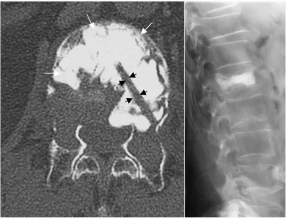
The pedicles were identified under anterior-posterior (AP), bilateral oblique and lateral view. An approach directly lateral of the pedicles was used to minimize the risk of spinal cord or nerve root injury. Bilaterally, the vertebral body was punctured with a 13 gauge needle. After fluoro- scopic control with contrast medium (Solutrast, 2cc, Byk Gulden, Germany) PMMA was injected into the damaged vertebrae. In a post treatment CT-scan the access for vertebral body puncture is shown (Figure 1).
Three hours after the vertebroplasty procedure, patients were allowed to get up and walk without additional exter- nal orthesis.
Data analysis
Mean +/-SEM values were calculated and statistical analysis was performed using students t-test and Wil- coxon test for paired values. Statistical significance was accepted for p<0.05.
Table 2: Grading of analgesic drug usage
Figure 1: Computed tomography of vertebral body L1 after vertebroplasty.
Arrows mark the puncture canal for PMMA injection. The white staining marks the PMMA inside the vertebral body. Small picture on the right: post-treatment X-ray
Results
Twenty-two patients with secondary malignancies of the thoracic and lumbar vertebral column were examined.
The mean age of the patients was 59.5 ± 1.9 years. The male to female ratio was 2.1:1. The diagnosis of vertebral metastasis was made after back pain prompted a radiolo- gical diagnostic in all patients.
Back pain was the sole indication for treatment in this study. No patient showed a tumor related neurological deficit. Mostly, terrible pain related to body movement limited the mobility of the patients. Three patients were immobilized continuously due to pain at every movement of their body. Further five patients rested mainly in bed because of unforeseeable pain attacks during walking.
Twelve patients were categorized into the group of severe disability, whereas two patients showed only moderate disability.
In two cases of pulmonary cancer and in one of prostate cancer additional radicular pain was observed besides back pain. These three patients were grouped in the bed rest group (Figure 4, Table 1). MRI in all three patients showed local nerve root compression by the tumor in combination with osteolytic destruction of two vertebral bodies and pedicles. These three patients with infiltration of the neighboring vertebrae were first treated by hemil- aminectomy and local tumor removal for surgical neuro- lysis. Vertebroplasty followed subsequently as a second step during the same general anesthesia.
Histology
The primary tumor was histologically confirmed in all pa- tients. Primary tumor sites were lungs, prostate and breasts in 50%, 27% and 18% of patients, respectively.
One patient (5%) had a spinal compression due to a
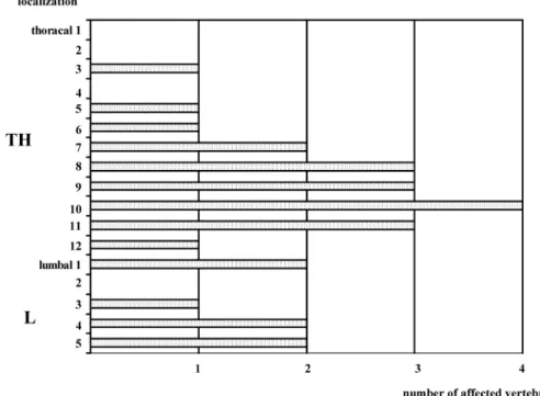
Figure 2: Distribution of affected vertebrae in the cohort of 22 patients.
Note that 15 of 26 (58 %) of affected vertebrae are located in the lower thoracic and upper lumbar spine (Th 8-L1). None of the vertebrae were located in the cervical spine.
plasmocytoma in a vertebrae. Most of the patients re- ceived standard treatment for the primary tumor including surgery and combined radio-chemotherapy prior to ver- tebroplasty.
Localization
Four patients showed an infiltration of more than one vertebral body. In two of them the adjacent to L 4 and 5 and in one case the vertebral bodies of TH 10 and 11 were affected. One patient showed an infiltration of mul- tiple vertebral bodies. Clinical symptoms were apparent for a metastasis in vertebral body of L 3. He was treated only in the symptomatic localization.
Middle and lower thoracic vertebral column was the pre- dominant localization of metastases. A lower number of cases were observed in lumbar spine (Figure 2).
Pain assessment
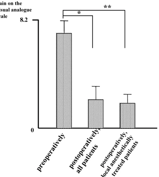
According to the visual analog scale, the median pain value preoperatively was 6.4 (2.3 to 9.1). Two days after treatment, the visual analogue scale showed a general reduction of pain. Median pain value decreased signifi- cantly to 2.1 (0.8 to 4.6; p<0.01). Excluding the three patients from the analysis that were treated by combina- tion of hemilaminectomy and vertebroplasty, pain im- proved to 1.5 (0.8 to 2.9; p<0.01) (Figure 3).
Immediately after vertebroplasty the patients treated under local anesthesia were asked for their discomfort during the procedure. The subjective judgment of the patients showed no major problems. Using the five step division for their "comfort" during vertebroplasty, the median strain on the whole treatment including position-
ing of the patient, puncture procedure and duration was 2 (range 1 to 3).
Preoperative analgesic drug usage was reduced during 24 hours after vertebroplasty. Three patients expressing minor local pain preoperatively did not receive any medi- cation at 24 hours after vertebroplasty. One of these pa- tients remained free of analgesic drugs at six months after vertebroplasty. Especially in patients with opioide and morphine medication drug reduction was performed carefully. At six weeks after operation the number of pa- tients with class 3 and 4 medication was significantly re- duced. No patient receiving class 4 drugs preoperatively became drugfree. But they were grouped into lower drug classes or were treated with reduced amounts of their prior medication. These circumstances explain the num- ber of NSAID and class 2 drugs treated patients already after six months.
Additional treatment with benzodiazepine for muscle re- laxation or neuroleptics was also reduced during six weeks after operation and remained on a lower postop- erative level.
OLBPD-Questionnaire
The most important point of the study and main objective of the treatment was the reduction of osteolysis related vertebral pain and the improvement of the patients' quality of life. Quality of life was assessed pre- and post- operatively by the OLBPD-questionnaire. Before complet- ing the questionnaire two days after vertebroplasty the patients had to do physical exercises like walking, climb- ing stairs, knee-bend and bending over. As shown in Fig- ure 4, all patients reported a pain relief. The average im- provement was 1.7 ± 0.1 steps of the disability score
Figure 3: Pain assessment by visual analogue scale pre- and postoperatively.
(* for P < 0.001, ** for P < 0.001, n = 22)
Figure 4: The Oswestry Low Back Pain Disability Questionnaire.
Disability before and after vertebroplasty. All patients improved significantly after the procedure.
(p<0.01). Treatment in the subgroup of patients with local and radicular pain was less successful. Local pain improved well. However, residual radicular pain resulted in a minor improvement. Overall, Wilcoxon statistical analysis of the relief of disability showed a highly signifi- cant improvement after vertebroplasty (p<0.01). Although a slight worsening occured during the six month postop- erative time, quality of life persisted on a significantly higher level than preoperatively.
Complications
Extravasation of PMMA outside the vertebral margins was defined as minor treatment complication. This complica- tion was observed in five patients (23%). No patient de- veloped any clinical or neurological symptoms. Further treatment related complication did not occur. Periopera- tive mortality was 0%.
Discussion
Summary of findings
In this study a total of 22 patients with back pain due to osteolytic metastasis of various primary tumors were treated with vertebroplasty. Overall a significant reduction of pain could be achieved. The patients tolerated the procedure well. Although the total amount of analgesic drugs was not reduced significantly, several patients were treated satisfactorily with reduced amounts of analgesics or with drugs of lower classes. The OLBPD-assessment revealed a significant reduction of pain leading to im- proved daily life activity and quality of life within two days after vertebroplasty. During the following six months quality of life diminished slowly but remained significantly improved.
Patients were ambulatory within three hours after the procedure. No new neurological deficits were observed and the mortality of the procedure was 0%. Therefore, the procedure is considered to be safe. The results on pain reduction and improved quality of life in combination with reduced pain medication indicate the benefit of vertebroplasty in patients with spinal metastases.
Treatment options in patients with osteolytic metastasis
Osteolytic metastases and myeloma are the most fre- quent malignant osteolytic lesions of the spine. Severe back pain and disability represent the major symptoms in affected patients. Mechanical pain is the predominant type of pain in patients with osteolytic metastasis.
Therapeutic management depends on the number of af- fected vertebrae, the spinal level and the osteolytic loca- tion of the tumor within the vertebra. Epidural extension of tumor tissue in the spinal canal together with the neurological signs determines the therapeutic regimen.
In general, the baseline condition of the patient sets the
limitations of the therapeutic extend. The timing of the treatment is primarily determined by the severity of pain and disability.
Radiation therapy gives partial or complete relief of the pain in more than 90% of patients [10]. However, this analgesic effect is not obtained until 10-20 days after the procedure. Strengthening of osteolytic vertebra after irradiation is minimal with a high remaining risk of painful vertebral collapse [10].
Vertebrectomy and surgical reinforcement of osteolytic vertebral bodies is not practical in most patients because of the multifocal nature of the disease. Therefore, minimal invasive methods with the aim of pain release combined with a stabilizing compound will be the method of choice.
The immediate analgesic effect of vertebroplasty can be explained by a so called "in situ immobilization" of verte- bral body fracture. Pathophysiologically, the cement is inserted into the bone and stabilizes the bone immedi- ately. In addition, the polymerisation heat may destroy pain fibers in the bone itself.
Indications for vertebroplasty
Clinical appearance
The best results are seen in patients who complain of a severe, focal, and mechanical back pain, requiring bed rest and major analgesic drugs, related to a neoplastic vertebral collapse without epidural involvement [11]. In a study on 37 patients with osteolytic metastases pain decreased in 97.3% of patients [12]. In a two years follow- up narcotic and analgesic drug usage decreased by 63%
[13]. In our study focal pain during movement represented the best indication for vertebroplasty.
The indications for vertebroplasty and open surgery are different. Decompression of spinal canal and nerve roots are main goals of surgical therapy. A combination of the two methods may be indicated if vertebroplasty facilitates surgery. Vertebroplasty provides an anterior stabilization of the vertebral column that may avoid an anterior surgi- cal approach. If necessary posterior surgical stabilization can be performed with smaller orthesis following dorsal decompression.
Radiological appearance
The vertebral collapse must not be complete. In case of an osteolysis with a high risk of vertebral collapse, it is indicated to perform vertebroplasty already in asympto- matic metastatic vertebral body lesions in order to prevent the collapse [11]. The amount of cement inserted does not correlate with the reduction of pain [4]. There is no reason for a complete filling of the lesion. It is better to avoid a leakage of cement with the risk of cord compres- sion, especially in extensive cortical osteolytic lesions.
The amount of PMMA used in this study ranged between 3 and 6 ml.
Vertebroplasty and adjacent therapies
Radiotherapy should be performed after vertebroplasty.
Radiation therapy does not interfere with the mechanical
properties of PMMA [14]. Additionally, irradiation comple- ments the analgesic effect of vertebroplasty.
Vertebroplasty seems to have an antitumoral effect. This can explain the low number of cases of local recurrence even if vertebroplasty is not completed by irradiation. The antitumoral effect may be explained by the local toxicity of PMMA and the heat of polymerisation and ischemia induced by the injection of cement in a limited tumoral tissue volume [15], [16], [3].
Complications of vertebroplasty
In general, more complications are reported after vertebro- plasty of malignant lesions than of osteoporotic fractures [3]. Most complications are due to leakage of cement, in cases of cortical osteolysis. The use of a relatively viscous cement mixture may prevent leakage in this situation [17], [4], [18]. Needle placement should be guided by the particular anatomy of each specific lesion. In general, anterior needle placement is desirable so that the cement is injected as far from the spinal canal as possible. Mul- tiple needle placements and separate cement injections may be necessary to fill the vertebral body adequately without compromising of the spinal canal or the neural foramen. The goal of cement injection is to provide ad- equate support for the anterior column. The complication rate for vertebroplasty in malignant spinal lesions is 10%
[11]. Three types of acute symptoms can be observed:
increased pain not associated with cement leak, radicu- lopathy, and spinal cord compression. Radiculopathy is the major complication of vertebroplasty with a risk of 4% [11]. The thoracic and lumbar transpedicular ap- proach of the vertebral body avoids leaks into the external part of the neural foramina along the needle track and the risk of radiculopathy [11]. In osteolytic vertebral col- lapse and narrow pedicles of thoracic vertebral column, an approach immediately lateral to the pedicle will provide additional safety in order to avoid opening of the spinal canal or damaging of a nerve root. Vertebroplasty under local anesthesia should be considered for the treatment of patients with osteolytic vertebral collapse [19], [18].
Awake patients allow the clinical detection of neurological symptoms during the injection. In our study under local anesthesia, patients tolerated the procedure well and reported only little pain. None of the patients canceled the procedures due to intolerable pain.
Conclusions
Percutaneous vertebroplasty developed by Galibert et al.
[1] as treatment option for patients with malignant de- struction of the thoracic and lumbar vertebral column provides fast pain relief by in situ immobilization of des- troyed vertebra. Painless mobilization immediately after treatment restores the patients' quality of life and allows early discharge from the hospital.
Acknowledgements
We are thankful to Dioni Rovello-Freking, RN, NP Univer- sity of California Los Angeles, UCLA Medical Center for a critical review and language editing of the paper.
References
1. Galibert P, Deramond H, Rosat P, Le Gars D. Note préliminaire sur le traitement des angiomes vertébraux par vertebroplastie acrylique percutanée. Neurochirurgie 1987;33:166-8.
2. Cortet B, Cotton A, Boutry N, Flipo RM, Duquesnoy B, Chastanet P, Delcambre B. Percutaneous vertebroplasty in the treatment of osteoporotic vertebral compression fractures: an open prospective study. J Rheumatol 1999;26:2222-8.
3. Weil A, Chiras J, Simon JM, Rose M, Sola-Martinez T, Enkaoua E. Spinal metastases: indications for and results of percutaneous injection of acrylic surgical cement. Radiology 1996;199:241-7.
4. Cotton A, Dewatre F, Cortet B, Assaker R, Leblond D, Duquesnoy B, Clarisse J. Percutaneous vertebroplasty for osteolytic metastases and myeloma: effects of the percentage of lesion filling and the leakage of methyl methacrylate at clinical follow- up. Radiology 1996;200:525-30.
5. Tokuhashi Y, Matsuzaki H, Toriyama S, Kawano H, Ohsaka S.
Scoring system for the preoperative evaluation of metastatic spine tumor prognosis. Spine 1990;15:1110-3.
6. Fairbank JCT, Couper J, Davis JB, O´Brien JP. The Oswestry low back pain disability questionnaire. Physiotherapy 1980;66:271- 3.
7. Airaksinen O, Herno A, Turunen V, Saari T, Suomlainen O. Surgical outcome of 438 patients treated surgically for lumbar spinal stenosis. Spine 1997;22:2278-82.
8. Frost H, Lamb SE, Klaber-Moffett JA, Fairbank JC, Moser JS. A fitness programme for patients with chronic low back pain: 2- year follow-up of a randomised controlled trial. Pain 1998;75:273-9.
9. Little DG, MacDonald D. The use of the percentage change in Oswestry Disability Index score as an outcome measure in lumbar spinal surgery. Spine 1994;19:2139-43.
10. Shepherd S. Radiotherapy and the management of metastatic bone pain. Clin Radiol 1988;39:547-50.
11. Deramond H, Deprister C, Gailbert P, Le Gars D. Percutaneous vertebroplasty with polymethylmethacrylate. Technique, indications, and results. Radiol Clin North Am 1998;36:533-46.
12. Cortet B, Cotton A, Boutry N, Dewatre F, Flipo RM, Duquesnoy B, Chastanet P, Delcambre B. Percutaneous vertebroplasty in patients with osteolytic metastases or multiple myeloma. Rev Rheum Engl Ed 1997;64:177-83.
13. Amar AP, Larsen DW, Esnaashari N, Albuquerque FC, Lavine SD, Teitelbaum GP. Percutaneous transpedicular polymethylacrylate vertebroplasty for the treatment of spinal compression fractures.
Neurosurgery 2001;49:1105-15.
14. Murray JA, Bruels MC, Lindberg RD. Irradiation of
polymethylmethacrylate. In vitro gamma radiation effect. J Bone Joint Surg Am 1974;56:311-2.
15. Brado M, Hansmann HJ, Richter GM, Kauffmann GW.
Interventionelle Therapie von primären und sekundären Tumoren der Wirbelsäule. Orthopäde 1998;27:269-73.
16. Chiras J, Depriester C, Weill A, Sola-Martinez MT, Deramond H.
Vertebroplasties percutanées. Technique et indications. J Neuroradiol 1997;24:45-59.
17. Barr JD, Barr MS, Lemley TJ, McCann RM. Percutaneous vertebroplasty for pain relief and spinal stabilization. Spine 2000;25:923-8.
18. Martin JB, Jean B, Sugiu K, San Millan Ruiz D, Plotin M, Murphy K, Rufenacht B, Muster M, Rufenacht DA. Vertebroplasty: clinical experience and follow-up results. Bone 1999;25 (2 Suppl):11S- 15S.
19. Bostrom MP, Lane JM. Future directions. Augmentation of osteoporotic vertebral bodies. Spine 1997;22 (24 Suppl):38S- 42S. Erratum in: Spine 1998;23:1922.
Corresponding author:
Dr. Michael Winking
Neurochirurgische Klinik, Justus-Liebig-Universität, Klinikstr. 29, 35392 Giessen, Germany, Tel.:
0641/9945501, Fax: 0641/9945509 michael.winking@neuro.med.uni-giessen.de
Please cite as
Winking M, Stahl JP, Oertel M, Schnettler R, Böker DK. PMMA vertebroplasty in patients with malignant vertebral destruction of the thoracic and lumbar spine.Ger Med Sci. 2003;1:Doc08.
This article is freely available from
http://www.egms.de/en/gms/2003-1/000008.shtml
Received:2003-07-28 Published:2003-11-20
Copyright
©2003 Winking et al. This is an Open Access article distributed under the terms of the Creative Commons Attribution License
(http://creativecommons.org/licenses/by-nc-nd/3.0/deed.en). You are free: to Share — to copy, distribute and transmit the work, provided the original author and source are credited.