Disc height and anteroposterior translation in fused and adjacent segments after lumbar spine fusion
Degenerationen und Instabilitäten in angrenzenden Segmenten nach Spondylodesen: Eine Pilotstudie basierend auf Distorsionskorrigierter Röntgenanalyse (DCRA)
Abstract
In a series of 46 patients the effects of spinal fusion upon intervertebral height and sagittal alignment in operated and non-operated segments
Michael Pfeiffer
1Oliver Haas
2were retrospectively evaluated on digitized radiographs. Data was
Martin Huber-Stentrup
1compared with age- and gender-normalized standard values. The object-
Christian Georg
3ive was to evaluate the influence of different types of spine fusions primarily upon adjacent segments, particularly in terms of degeneration
and sagittal profile of the lumbar spine.
Wolfgang Frobin
4Incidence of adjacent segment degeneration (ASD) is still highly contro-
versial. However, not every degeneration adjacent to spinal fusion must 1 HELIOS Rosmann Klinik, Hauptabteilung Orthopädie, Breisach, Germany be caused by the fusion and responsibility of the fusion for ASD may
vary with its range and type.
Distortion Corrected Roentgen Analysis (DCRA) was utilized. DCRA is a proven valid, reliable, observer-independent, and accurate tool for as-
2 Klinik für Orthopädie und Rheumatologie, Philipps- Universität, Marburg, Germany
sessment of these parameters over time and in comparison with "nor- mal" cohorts. With this method the exact posture of the patients needs
not to be known. 3 Medizinisches Zentrum für
Radiologie, Klinik für There was little evidence for serious fusion-related ASD within an aver-
age of 40 months follow-up. No difference could be detected for rigid Strahlendiagnostik, Marburg, Germany
vs. non-rigid fusion and instrumented vs. non-instrumented techniques.
Temporary postoperative distraction effects could be detected in oper- 4 Institut für Experimentelle Biomechanik, Westfälische ated and non-operated segments. Absolute preoperative values for in-
tervertebral height and vertebral slip were age-related. Retrospectively, Wilhelms-Universität, Münster, Germany the choice of segments for fusion was clearly based upon radiological
criteria. Thus we conclude that radiological parameters have an obvious clinical relevance for decision-making and need to be quantified. Within the limitations of this pilot study, true fusion related ASD seems to be infrequent.
Zusammenfassung
In einer Serie von 46 Patienten wurde der Effekt hinterer Wirbelsäulen- Versteifungseingriffe (Spondylodesen) auf die Höhe des Zwischenwir- belraumes und das sagittale Profil der Lendenwirbelsäule auf digitali- sierten Röntgenbildern untersucht. Dabei wurden operierte und angren- zende Bewegungssegmente retrospektiv im Zeitverlauf evaluiert. Die Daten wurden mit alters- und geschlechtsnormierten Standardwerten verglichen. Ziel war es, den Einfluss verschiedener Fusionsverfahren auf den Verschleiß angrenzender Segmente zu quantifizieren.
Die Inzidenz sogenannter Anschlussdegenerationen nach Spondylodesen ist stark umstritten. Nicht jede solche Degeneration muß aber auch durch die Spondylodese verursacht sein, Alterungsprozesse sind schwer abgrenzbar.
Distorsionskorrigierte Röntgenanalyse (DCRA) kam für diesen Zweck zum Einsatz. DCRA ist ein nachweislich valides, reliables, untersucher- unabhängiges und genaues Verfahren zur Messung der genannten
wenig rigiden Implantaten sowie zwischen Versteifungen mit und ohne Implantaten. Temporäre postoperative Distraktionseffekte bestanden in operierten und angrenzenden Segmenten. Die Absolutwerte für die Höhe des Zwischenwirbelraumes und Wirbelgleiten waren altersabhän- gig. Retrospektiv zeigte sich, dass für die Wahl des Spondylodesever- fahrens radiologische Kriterien für den Operateur den Ausschlag gaben.
Daraus ergibt sich die Notwendigkeit, diese radiologischen Parameter auch zu quantifizieren. DCRA ist hierfür gut geeignet. Im Rahmen der Aussagekraft dieser Pilotstudie erscheint jedoch die fusionsinduzierte Anschlussdegeneration weniger häufig als erwartet.
Introduction
To date a major concern of lumbar spinal fusion in pa- tients with low back pain (LBP) remains the unsolved problem of consecutive adjacent segment degeneration (ASD), often leading to "instability" and further operations.
The new Distortion Compensated Roentgen Analysis (DCRA) method opens the field for non-invasive analysis of adjacent segment behavior after spinal fusion, which must be seen on the background of results from other methods:
In the past there have been made several attempts to measure ASD in-vivo via intervertebral distance ("disc height", dh) and sagittal translation vector ("slip, olisthesis, translation",tr) on plain x-rays. The results were inconsistent: Even 16-20 years after fusion no significant ASD was found in a majority of cases on plain x-rays [1]
if compared to a non-operated control group. Other au- thors described ASD in 100% of adjacent segments more than five years after fusion [2], [3] and significantly more often than in patients with lumbar decompression only [4].
This study attempts to answer the following basic ques- tions:
1.: Do disc height and sagittal plane displacement in a cohort of patients with spinal fusions change with time and are such changes (if present) different in the oper- ated and the non-operated (adjacent) segments?
2.: Do changes in disc height and displacement (if present) differ for
a) rigid vs. non-rigid fusion techniques,
b) instrumented vs. non-instrumented techniques?
Methods
Patients
Complete time series of technically sound lumbar spine radiographs from 50 patients having undergone spinal fusion were randomly chosen from our radiological archive list. Each series consisted of a.p. and lateral views taken
preoperatively, postoperatively prior to discharge and at follow-up. All radiographs had been taken in standing posture between 9 and 12 o'clock in the morning. Four cases with overt material failure or resorption of bone mass were excluded. So a total of 46 series of fusion patients could be evaluated.
The mean age of the patients was 46.3 years (SD 10.6);
mean follow-up time was 40 months (24-68). There were 29 male and 17 female patients, 25 of whom had a single-level fusion (two vertebrae) and 21 a two-level fu- sion (three vertebrae). Thirty had a primarily degenerative cause, 16 exhibited spondylolysis on lateral projections as underlying pathology. Twenty-five obtained a "rigid"
procedure with posterior bilateral pedicle screw fixation with an average in vitro bending stiffness of 3.07 Nm/mm, axial stiffness of 929 N/mm, and torsional stiffness of 2.58 Nm/° without crosslinks. Instrumenta- tion was eventually reinforced with "O'Brien-type" anterior (retroperitoneal) interbody bicortical homologous iliac crest grafting (ALIF) [5]. Twenty-one obtained non-rigid procedures, in 18 cases single-level posterolateral spondylodesis at L5-S1, according to Wiltse [6] or single level ALIF without any metal implants. 3 patients had a non-rigid titanium cable augmented fusion. A total of 28 patients thus had spinal metal instrumentation, 18 were non-instrumented. Posterior non-instrumented fusion al- ways was confined to the pre-sacral segment. No PLIF and no "floating fusion" was involved in the study.
Measurement of disc height and displacement
To monitor disc height and sagittal plane displacement in the operated and non-operated segments of patients and controls, DCRA [7] was employed. This computer- assisted method, based on measurements from plain lateral radiographic views, has previously been proven to be accurate and reliable. In addition, a database of nor- mal, gender- and age-appropriate disc height and sagittal plane displacement has been compiled using DCRA. There is one other computer-assisted method available [8], but without large series providing normal values. Methods
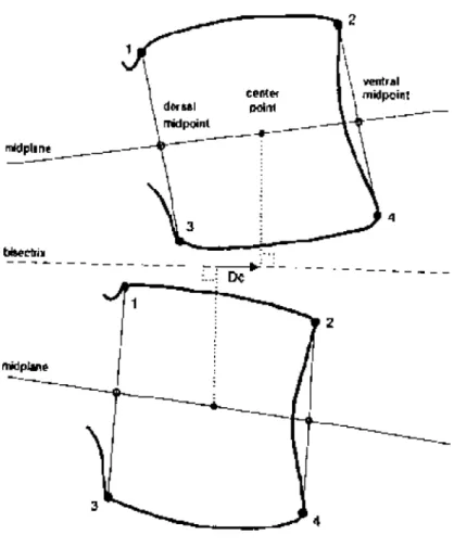
Fig. 1: Definition of auxiliary landmarks and geometric measures:
Midplanes angle: Angle between neighboring midplanes, counted positive if the wedge opens ventrally.
Mean depth: Mean of cranial depth between corner points 1 and 2 and caudal depth between corner points 3 and 4. Two other dorsal corner points, which are not included in the protocol but necessary for re-identification, are not depicted here.
Sagittal translationtras given by the distance Dc between the projections of the center points measured in direction of the bisectrix, and divided by the mean depth of the cranial vertebra. Positive values are counted for anterior displacement of the
cranial vertebra.
The sum of the perpendicular distance of corner points 4 and 2 from the bisectrix between the two midplanes divided by the mean depth of the cranial vertebra gives the "disc height",dh.
without computer assistance, too numerous to mention here in detail, share common limitations and are not ac- curate enough for a precise measurement of disc height [9].
In the following, the DCRA method is only briefly reiter- ated; for details, the reader is referred to Frobin et al. [7].
Calculation of disc height and sagittal plane displacement is based on corners 1-4 located on the outer contour of vertebral bodies (Figure 1) following a new protocol compensating for distortion in central projection, off- center position, axial rotation and lateral tilt of the spine.
Positive values oftrdesignate a ventral displacement of the cranial vertebra with respect to the caudal vertebra.
To account for radiographic magnification and variation in stature, both measures are divided by the mean depth of the caudal vertebral body. Thus, disc height and sagittal plane displacement as obtained by DCRA are di- mensionless numbers. If it were desired to convert disc height and displacement into millimeters, the parameters would have to be multiplied by the vertebral depth. To
obtain approximate numbers, one may simply assume a general depth value of 35 mm. We do not recommend this conversion, however, since in reality all the values are related to their individual caudal vertebra.
Values of disc height and displacement as defined above depend on the angle of lordosis. A correction is applied transforming the raw data to data at standard angles of lordosis. This permits comparison of data among individu- als and with a normal database. The DCRA measurement procedure comprises of mapping the vertebral contours on a transparent foil and digitization of the contour lines (point density 5/mm, precision 0.125 mm). Series of programs check geometric properties of the contours, locate corners, calculate derived geometric measures and compute angle-corrected disc height and sagittal plane displacement.
For the description of the results of this work, the follow- ing nomenclature is used: The subscripts "pre", "post"
and "follow up" designate the preoperative, postoperative (at discharge), and follow up values of disc heightdhand
ive=dhfu-dhpost
Δdhfu= disc height at follow up - disc height preoperative
=dhfu-dhpre
Δtrpre= displacement postoperative - displacement preop- erative=trpost-trpre
Δtrpost= displacement at follow up - displacement postoper- ative=trfu-trpost
Δtrfu= displacement at follow up - displacement preoper- ative=trfu-trpre
Error study and statistical tools
DCRA measurement precision (SD) has previously been determined to 0.014 (or 4.2%) for disc height and 0.014 for displacement [7] and been validated against Roentgen Stereophotogrammetric Analysis [10]. In this study, only one author (O.H.), having been uninvolved in the surgical procedures, performed all measurements from radio- graphs. Potential inter-observer errors thus only come into play when comparing pre-operative disc height and displacement of the segments to be fused with the normal age- and gender-appropriate value of the existing data- base. Inter-observer variance was quantified by re-evalu- ating angle-standardized disc height and dorso-ventral displacement of 23 segments in the region L3-4 to L5/S1 from radiographs, which originally contributed to the normal database of Frobin et al. [7]. This actual mean difference between the two evaluations was 0.016 for dhand 0.011 fortr.
Parametric statistical methods were
for 1.: Paired T-Test and T-Test for unpaired samples and calculation of linear correlation coefficients;
for 2.: ANOVA with post-hoc significance tests, and T-Test for unpaired samples, respectively.
All criteria for test applicability were checked prior to calculation (tests for homogeneity of variances, normal distribution, etc.). Significance level for the (two-tailed) tests was generally set to p < 0.05. All these calculations were made in SPSS for Windows, v. 10.0.5 (SPSS Inc., Chicago, USA) or NCSS™ 5.x Series (J.L. Hintze, Kaysville, USA) after importing spreadsheets from Excel 2000™
(Microsoft Corp., Redmond, USA).
Results
1. All patients with spine operations:
Meandhpreof all segments between the cranial adjacent and the caudal adjacent segment ranged from 0.338 to 0.385. Mean dhpost was 0.388 to 0.410. Mean dhfu amounted between 0.346, and 0.3730. Table 1 gives
in all fused segments and in the cranial adjacent seg- ment. The changes in the caudal adjacent segment were insignificant.
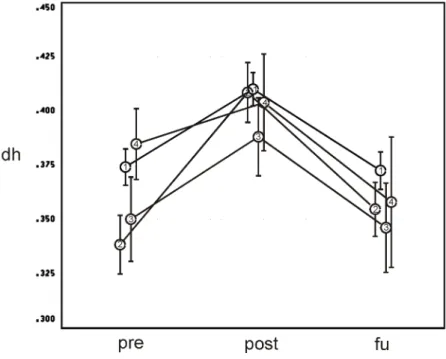
Meandhfudecreased significantly, compared todhpost, in all fused segments and in the cranial adjacent segment, thus receding back to values indistinguishable fromdhpre. The corresponding dh changes in the caudal adjacent segment remained insignificant. Meandhfuvs.dhpre, ac- cordingly, showed insignificant changes. Compare Figure 3 for graphic depiction of these data.
Mean trpre vertebrae ranged from -0.107 (the negative value indicating a slight retroposition of the superior ver- tebrae) to 0.014. Meantrpostwas -0.104 to 0.006. Mean trfuyielded -0.103 to 0.042. Compare again Table 1.
Meantrpostcompared totrpreremained virtually unchanged.
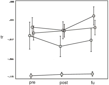
The same was true fortrfuvs.trpost, except for the second fusion vertebrae (in case of two-level fusion): Here trfu was significantly increased overtrpost.
Only in the second fusion vertebraetrfuwas also signifi- cantly greater thantrpre. For all other vertebrae the corres- ponding values did not exhibit significant changes. Com- pare Figure 4 for the above mentioned values.
None of all above mentioned differences comparing fol- low-up and preoperative values were significantly correl- ated with follow-up time.
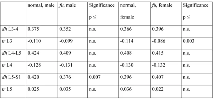
For the majority of segments, L4-5 and L5-S1 (11 patients obtained L4-5 fusion, 12 L5-S1, and 17 L4-S1), mean dhpreandtrprewere compared to a database of age- and gender-appropriate normal values [7]. The results are depicted in Table 2.
While operated segments deviated from normal, indicat- ing lower disc height and increased anterior translation, the correspondingdhprevalues of the adjacent segments and the trpre values of the cranial and caudal adjacent vertebrae were mostly well within the range of the normal values: In Figure 3 and Figure 4 the error bars of the cranial and caudal adjacent segments in case of dhpre, and of the cranial adjacent vertebrae in case oftrpre, are clearly separated from the pathological values of the other segments. The observation that the error bar of the upper vertebrae of the caudal adjacent segment is not equally separated on Figure 4 must be considered non- pathologic and can be explained by Table 3. Also in nor- mal persons, anterior slip is the more pronounced, the more caudally the vertebra is situated. Values from the - in this study - most frequent adjacent segments at follow- up mostly showed no significant differences in comparis- on to normal values.
There was a significant negative correlation ofdhprewith age at the cranial adjacent segment (r = -0.391) and the topmost (or, in case of one-level fusion, only) fused seg- ment (r = -0.442). At the cranial adjacent vertebral body trpre was positively correlated with age (r = 0.336). The
Tab. 1 (Fig. 2): Differences Δ of disk height (dh) and displacement (translation, tr) over time in dimensionless numbers:
For "dh" columns - 1: unfused cranial adjacent segment, 2: topmost (only fused segment in case of one-level fusion) fusion segment, 3: second fusion segment (in case of two-level fusion), 4: unfused caudal adjacent segment.
For "tr" columns - 1: cranial adjacent vertebra (upper vertebra of the cranial adjacent segment), 2: topmost fusion vertebra, 3:
second fusion vertebra, 4: caudal adjacent vertebra (upper vertebra of the caudal adjacent segment).
Δdhpre= disc height postoperative - disc height preoperative =dhpost-dhpre Δdhpost= disc height at follow-up - disc height postoperative =dhfu-dhpost
Δdhfu= disc height at follow up - disc height preoperative =dhfu-dhpre Δtrpre = displacement postoperative - displacement preoperative =trpost-trpre
Δtrpost= displacement at follow up - displacement postoperative =trfu-trpost Δtrfu= displacement at follow up - displacement preoperative =trfu-trpre
Fig. 3: Development of "disc height" dh
(Mean and Standard Errors, dimensionless numbers) preoperatively (pre), postoperatively (post), and at follow-up (fu).
1: unfused cranial adjacent segment,
2: topmost fusion segment (only fused segment in case of one-level fusion), 3: second fusion segment (in case of two-level fusion),
4: unfused caudal adjacent segment.
Note temporary distraction and consecutive loss off correction in all segments. Adjacent segments start at a higher "baseline"
than fusion segments.
changes ofdhandtrover the time of analysis neverthe- less did not exhibit such correlation.
Fig. 4: Development of "sagittal translation" tr (data depicted accordingly to Fig. 3).
1: cranial adjacent vertebra (upper vertebra of the cranial adjacent segment);
2: topmost fusion vertebra;
3: second fusion vertebra;
4: caudal adjacent vertebra (upper vertebra of the caudal adjacent segment).
Note the lack of anterior displacement of 1 and the nearly constant sagittal displacement of 1 and 4. Slight reduction of anterior displacement in the fusion segments is temporary.
Tab. 2 (Fig. 5): dhpreand trpreof in the present study most frequently operated segments/vertebrae compared to normal values in dimensionless numbers
(L4-5, n = 11; L5-S1, n = 12; L4-S1, n = 17).
Tab. 3 (Fig. 6): dhpreof in the present study most frequently operated superior adjacent (L3-4, n = 28; L4-5, n = 12) and inferior adjacent (L5-S1, n = 14) segments and trprefor their corresponding vertebrae compared to normal values (dimensionless
numbers)
2a. Changes of disc height and displacement related to the initial stiffness of the fusion construct:
Mean Δdhpre, Δdhpost, and Δdhfuwere at none of the seg- ments significantly different for the two different stiffness groups.
Mean Δtrprewas significantly different for the topmost fu- sion and the second fusion vertebrae: Rigid trpost de- creased, non-rigidtrpostincreased overtrpre.
Meantrpostwas significantly different for the second fusion vertebrae: Towards follow-up, the sagittal translation of initially rigidly fixed vertebrae over their inferior counter- part increased more than if operated non-rigidly (reverse to Δtrpre). For none of the vertebrae, mean Δtrfuwas signi- ficant.
2b. Changes of disc height and
displacement for instrumented vs. non- instrumented fusion technique:
Mean differences Δdhpre and Δdhpost of all segments showed virtually the same results for both techniques.
Only Δdhfushowed a significant difference between both groups at the topmost fusion segment: Other than in non- instrumented segments, the instrumented intervertebral height decreased (compression). Here it must be borne in mind that non-instrumented techniques in our study often involved anterior cortical grafts, obviously causing some segment distraction.
Mean Δtrprewas significant for the topmost fusion verteb- rae and the second fusion vertebrae: Instrumentedtrpost decreased ("reposition"), non-instrumentedtrpostincreased overtrpre.
Mean Δtrpostwas significant for the cranial adjacent ver- tebrae and the second fusion vertebrae: Towards follow- up, the sagittal translation of the cranial adjacent verteb- rae over their inferior instrumented counterpart increased, over non-instrumented vertebral bodies it decreased. For the instrumented second fusion vertebrae the sagittal displacement increased more than in case of non-instru- mented technique. Yet, again mean Δtrfuwas for none of the vertebrae significantly different.
Discussion
To date, despite an enormous increase of lumbar fusion procedures, we still do not know exactly what is going on in case of ASD. There are some impediments of all ana- lytical methods:
- Despite the fact that there is little evidence for significant correlation between radiological changes and clinical outcome [3], not even in case of pseudarthrosis [11], x- ray results are often key points for surgical decision making in suspected ASD.
- Ageing may overshadow genuine radiological effects of the fusion - thus long-term analyses with more than 10 years of follow-up and/or without Matched-Pair design are not very helpful in this respect.
It is obvious that a preoperative reduction of disc height and increase of anterior sagittal translation is influential for choosing a segment for spinal fusion. This "pathology"
is usually recognized by the surgeon without any caliper measurement, probably by comparing the disc height and sagittal slip of an incriminated segment to the other segments and/or some "normal" appearance derived from his personal experience. The measurement tech- nique applied here overall supports the assessments made with bare eyes: Segments chosen for fusion are narrower than adjacent segments and narrower than
segments need not be symptomatic.
Since considerable postoperative distraction and/or re- position also occur in adjacent segments and in segments with non-instrumented posterior fusion, some factor un- related to the fusion technique itself must be involved.
Interaction of prone and/or flexed position on the opera- tion table and bed rest with diminished activity after the operation may lead to a (temporary) restoration of disc height and alignment, as known from diurnal changes [12], [13].
Roentgen Stereophotogrammetric Analysis (RSA) studies were inconclusive and showed partly increase or decrease of the translatory movement of the juxtafused segment [14]. Unfortunately, most of RSA studies focus on the fused segments only, maybe also due to the ethical problem of exposing adjacent segments for mere insertion of tantalum markers. RSA pointed out the fact that even in cases with assumed consolidation some residual ("micro")motion in the "fused" segment may persist [15], [16]. The influence of such residual motion upon adjacent segments yet is completely unclear.
The higher the postoperative distraction and reposition within the operated as well as in the adjacent segments, the higher is, not surprisingly, the loss of correction to- wards follow-up. The more pronounced effect of intraop- erative reposition (decrease oftr) with the rigid instrumen- tation is counteracted by marked recurrence of slip to- wards follow-up. With a non-rigid method less postoper- ative reposition within the fusion is predictably obtained but in the end the choice of methods does not matter for the alignment of the vertebrae involved.
The differences between the preoperativedhandtrval- ues and the follow-up values for the adjacent segments compared between rigid vs. non-rigid technique as for instrumented vs. non-instrumented technique are small, indicating an, if at all, minor influence of the operation strategy on the result. As the design of the analysis was that of a pilot study no "a priori" Power Analysis could be carried out. A post-hoc Power Analysis, however, makes obvious that in the "worst case" total sample sizes of n = 190 would be necessary to further increase the test power (1-β) from 0.50 to 0.95, thus reducing the β-error for sufficient exclusion of an effect of the operative technique on these differences. For most comparisons, however, the test power ranges between 0.80 and 0.90, thus cor- roborating the aforementioned conclusion.
Here, adjacent segments prior to the operation and again at follow-up were normal in comparison to values from large cohort studies. This leads to the conclusion that ASD measured in radiological terms usually does not oc- cur up to 68 months after spinal fusion.
Non-instrumented and instrumented as well as rigid and non-rigid fusion fail to show different effects upon adja-
as long as they are confined to the presacral segment (large area for bone grafting and small distance between transverse processes and alae sacri). Independently from this study, the clinical results for the surgical methods involved are currently being analyzed prospectively with a three-year follow up. Preliminary results, as compared to the literature, have been good enough to warrant fur- ther use of our repertoire.
We cannot exclude that other than the evaluated fusion techniques indeed promote ASD. Beneficial effects of fusion for the patients are reflected but not necessarily explained by the results of this study. The study could also lend its methodology to a comparative analysis of the effects of disc endoprostheses upon adjacent seg- ments, which are often promoted as being superior in this respect - yet reliable data is still lacking.
Since except for computer assisted techniques none of the traditional methods based upon conventional radio- graphs is valid and accurate enough the latter can no longer be considered state of the art in studies interindi- vidually comparing (adjacent) segment pathology. They should thus be abandoned even though their application is alluringly less circumstantial and cumbersome. Further research seems necessary to increase the acceptance of DCRA on a wider scale.
Acknowledgements
With acknowledgements to Prof. Dr. rer. nat. Paul Brinckmann, Institut für Experimentelle Biomechanik, Westfälische Wilhelms-Universität, Domagkstrasse 3, D- 48129 Münster, Germany
References
1. Van Horn JR, Bohnen LM. The development of discopathy in lumbar discs adjacent to a lumbar anterior interbody spondylodesis. A retrospective matched-pair study with a postoperative follow-up of 16 years. Acta Orthop Belg 1992;58:280-6.
2. Dennis S, Watkins R, Landaker S, Dillin W, Springer D.
Comparison of disc space heights after anterior lumbar interbody fusion. Spine 1989;14:876-8.
3. Miyakoshi N, Abe E, Shimada Y, Okuyama K, Suzuki T, Sato K.
Outcome of one-level posterior lumbar interbody fusion for spondylolisthesis and postoperative intervertebral disc degeneration adjacent to the fusion. Spine 2000;25:1837-42.
4. Guigui P, Wodecki P, Bizot P, Lambert P, Chaumeil G, Deburge A. Influence à long terme de l'arthrodèse associée sur les niveaux adjacents dans le traitement des sténoses lombaires. A propos d'une série comparative de 127 cas à 9 ans de recul moyen.
Rev Chir Orthop Reparatrice Appar Mot 2000;86:546-57.
5. O'Brien JP, Dawson MH, Heard CW, Momberger G, Speck G, Weatherly CR. Simultaneous combined anterior and posterior fusion. A surgical solution for failed spinal surgery with a brief review of the first 150 patients. Clin Orthop 1986;(203):191-5.
6. Wiltse LL. Spondylolisthesis in children. Clin Orthop 1961;21:156- 63.
7. Frobin W, Brinckmann P, Biggemann M, Tillotson M, Burton K.
Precision measurement of disc height, vertebral height and sagittal plane displacement from lateral radiographic views of the lumbar spine. Clin Biomech 1997;12 Suppl 1:S1-S63.
8. Quint DJ, Tuite GF, Stern JD, Doran SE, Papadopoulos SM, McGillicuddy JE, Lundquist CA. Computer-assisted measurement of lumbar spine radiographs. Acad Radiol 1997;4:742-52.
9. Saraste H, Brostrom LA, Aparisi T, Axdorph G. Radiographic measurement of the lumbar spine. A clinical and experimental study in man. Spine 1985;10:236-41.
10. Leivseth G, Brinckmann P, Frobin W, Johnsson R, Stromqvist B.
Assessment of sagittal plane segmental motion in the lumbar spine. A comparison between distortion-compensated and stereophotogrammetric roentgen analysis. Spine 1998;23:2648- 55.
11. Suzuki T, Pearcy MJ, Tibrewal SB, Wilson D, Duthie RB. Posterior intertransverse fusion assessed clinically and with biplanar radiography. Int Orthop 1985;9:11-7.
12. Krag MH, Cohen MC, Haugh LD, Pope MH. Body height change during upright and recumbent posture. Spine 1990;15:202-7.
13. Ledsome JR, Lessoway V, Susak LE, Gagnon FA, Gagnon R, Wing PC. Diurnal changes in lumbar intervertebral distance, measured using ultrasound. Spine 1996;21:1671-5.
14. Axelsson P, Johnsson R, Stromqvist B. The spondylolytic vertebra and its adjacent segment. Mobility measured before and after posterolateral fusion. Spine 1997; 22:414-7.
15. Johnsson R, Stromqvist B, Axelsson P, Selvik G. Influence of spinal immobilization on consolidation of posterolateral lumbosacral fusion. A roentgen stereophotogrammetric and radiographic analysis. Spine 1992;17:16-21.
16. Pape D, Fritsch E, Kelm J, Müller K, Georg T, Kohn D, Adam F.
Lumbosacral stability of consolidated anteroposterior fusion after instrumentation removal determined by roentgen stereophotogrammetric analysis and direct surgical exploration.
Spine 2002;27:269-74.
Corresponding author:
Dr. Michael Pfeiffer
HELIOS Rosmann Klinik, Hauptabteilung Orthopädie, Zeppelinstr. 37, 79206 Breisach, Germany, Tel.: +49 (0)7667 84360, Fax: +49 (0)7667 84264
mpfeiffer@breisach.helios-kliniken.de
Please cite as
Pfeiffer M, Haas O, Huber-Stentrup M, Georg C, Frobin W. Disc height and anteroposterior translation in fused and adjacent segments after lumbar spine fusion.Ger Med Sci. 2003;1:Doc05.
This article is freely available from
http://www.egms.de/en/gms/2003-1/000005.shtml
Received:2003-04-28 Published:2003-09-04
Copyright
©2003 Pfeiffer et al. This is an Open Access article distributed under the terms of the Creative Commons Attribution License
(http://creativecommons.org/licenses/by-nc-nd/3.0/deed.en). You are free: to Share — to copy, distribute and transmit the work, provided the original author and source are credited.