• Keine Ergebnisse gefunden

Osmoregulation of the proU operon at a post-transcriptional level in Escherichia coli

N/A
N/A
Protected

Academic year: 2021

Aktie "Osmoregulation of the proU operon at a post-transcriptional level in Escherichia coli"

Copied!
77
0
0

Wird geladen.... (Jetzt Volltext ansehen)

Volltext

(1)

Osmoregulation of the proU operon

at a post-transcriptional level in Escherichia coli

Inaugural-Dissertation zur

Erlangung des Doktorgrades

der Mathematisch-Naturwissenschaftlichen Fakultät der Universität zu Köln

vorgelegt von

Kanstantsin Kavalchuk

Köln

2011

(2)

Berichterstatter/in:

Prof. Dr. Karin Schnetz PD Dr. Niels Gehring

Tag der mündlichen Prüfung: 28. Juni 2011

(3)

Contents

Abbreviations ... 2

Zusammenfassung... 3

Summary ... 4

1. Introduction ... 5

1.1 Adaption of E. coli to osmotic stress and the role of the proU operon in osmoadaptation... 5

1.2 Osmoregulation of proU transcription ... 8

1.3 Post-transcriptional regulation of gene expression and potential mechanisms of post-transcriptional regulation of proU... 11

1.4 Objectives of the PhD project ... 14

2. Results ... 15

2.1 Osmoregulation of proU at post-transcriptional level ... 15

2.1.1 Transcriptional and post-transcriptional osmoregulation of proU... 16

2.1.2 Osmoregulation of ProV protein expression ... 18

2.1.3 ProV protein stability under different osmolarity conditions ... 19

2.2 Osmoregulation of RNase III processing of proU mRNA... 20

2.2.1 RNase III processing of proU mRNA in the conserved region ... 20

2.2.2 Effect of rnc mutation on proU osmoregulation ... 24

2.2.3 Effect of RNase III processing site mutation on proU osmoregulation... 27

2.2.4 RNase III initiates rapid degradation of proU mRNA after hypoosmotic stress ... 32

2.2.5 RNase III processing of minimal proU substrates... 36

2.3 RNA chaperones Hfq, StpA, ProQ and proU osmoregulation ... 40

2.3.1 Osmoregulation of proU in hfq mutant cells ... 40

2.3.2 Osmoregulation of proU in stpA mutant cells ... 42

2.3.3 Osmoregulation of proU in proQ mutant cells... 43

3. Discussion ... 44

3.1 Post-transcriptional osmoregulation of proU is based on osmoregulation of RNase III processing of the proU mRNA ... 44

3.2 The role of RNase III processing in proU osmoregulation... 46

3.3 Possible mechanisms of osmoregulation of RNase III processing of proU mRNA ... 47

3.4 The integrative model of proU osmoregulation... 49

4. Materials and Methods ... 51

4.1 Media and antibiotics... 51

4.2 Standard molecular biology techniques ... 51

4.3 Plasmids ... 51

4.4 Bacterial strains... 53

4.5 Transformation by CaCl

2

method ... 56

4.6 Electrocompetent cells and electroporation ... 56

4.7 Integration of plasmids into the attB site of the chromosome... 57

4.8 Transduction with phage T4GT7 ... 57

4.9 Deletion of chromosomal genes... 58

4.10 β-galactosidase assay ... 58

4.11 SDS-PAGE, Western blotting and the determination of protein stability... 59

4.12 RNA analysis by Denaturating Urea-PAGE ... 59

4.13 5' RACE protocol for mapping of processed 5' ends of mRNA ... 60

4.14 Site-specific mutagenesis of the plasmid-borne proV gene ... 61

4.15 Site-specific mutagenesis of the chromosome-encoded proU ... 62

4.16 Real-time RT-PCR and calculation of RNA half-life ... 64

5. Bibliography... 66

Acknowledgements ... 74

Erklärung... 75

(4)

Abbreviations

5´ RACE rapid amplification of cDNA 5´ ends Amp ampicillin

Cm chloramphenicol FRT (FRT site) Flp recombinase target sites

Km kanamycin Rif rifampicin

σ

70

RpoD, primary sigma factor, encoded by gene rpoD

σ

S

RpoS, stationary phase sigma factor, encoded by gene rpoS

SNP single-nucleotide polymorphism

Spec spectinomycin

sRNA small regulatory RNA

TAP tobacco acid pyrophosphatase

Tet tetracycline

(5)

Zusammenfassung

Das E. coli proU-Operon besteht aus den drei Genen proV, proW und proX. Diese kodieren einen ABC-Transporter für die Aufnahme der kompatiblen Solute Prolin und Glyzinbetain bei hyperosmotischen Stressbedingungen. Die Expression des proU-Operons ist osmoreguliert: Das Operon ist bei niedriger Osmolarität reprimiert und wird durch hyper- osmotischen Stress induziert. Frühere Publikationen zeigten, dass die Osmoregulation des proU-Operons auf Ebene der Transkriptionsregulation erfolgt. Der proU-Promotor wird bei hoher Osmolarität stärker. Zusätzlich wird die Transkription durch das Nukleoid-assozierte Protein H-NS bei niedriger Osmolarität reprimiert. Nicht publizierte Ergebnisse der Arbeitsgruppe deuteten auf eine zusätzliche Post-transkriptionelle Ebene der Osmoregulation hin.

In dieser Arbeit wurde die post-transkriptionelle Osmoregulation von proU charakterisiert. Ergebnisse dieser Arbeit zeigen, dass die proU-mRNA durch RNase III an den Positionen +217/+218 und +281/+282 (relativ zum Transkriptionsstart) prozessiert wird. Die Prozessierung erfolgt innerhalb eines doppelsträngigen Bereichs einer in Enterobacteriaceae hoch konservierten RNA-Sekundärstruktur von proU. Diese Prozessierung durch RNase III ist osmoreguliert. RNase III prozessiert die proU-mRNA effizient bei Wachstum in Medium niedriger Osmolarität. Durch hohe Osmolarität wird die Prozessierung inhibiert. Die Prozessierung der proU-mRNA durch RNase III verursacht die post-transkriptionelle Osmoregulation von proU, da die Hemmung der RNase III-Prozessierung durch Mutation die post-transkriptionelle Osmoregulation aufhebt. Außerdem zeigen die Daten, dass die Stabilität der proU-mRNA durch die RNase III kontrolliert wird. Die proU-mRNA ist bei hoher Osmolarität relativ stabil. Die Halbwertszeit der mRNA an der Prozessierungsstelle beträgt 65 bis 75 Sekunden. Bei hypo-osmotischem Stress führt die Prozessierung durch RNase III zu einem schnellen Abbau der proU-mRNA mit einer Halbwertszeit von 4 bis 7 Sekunden. Diese Ergebnisse deuten darauf hin, dass die primäre Rolle der Prozessierung durch RNase III die schnelle Degradation der proU-mRNA bei hypo-osmotischen Stressbedingungen ist.

Die Ergebnisse dieser Arbeit werden in ein generelles Modell der Osmoregulation von proU integriert. Hyperosmotischer Stress bewirkt die Derepression der Transkription von proU und die Stabilisierung der proU-mRNA durch Hemmung der RNase III Prozessierung.

Hypo-osmotischer Stress führt zur Repression der proU Transkription und zum schnellen Abbau der proU-mRNA, der durch RNase III initiiert wird.

Die hohe Konservierung der proU-Sequenz im Bereich der mRNA Sekundärstruktur

um die RNase III-Prozessierungsstelle deutet darauf hin, dass die RNase III Prozessierung

auch in anderen Enterobacteriaceae stattfindet. Der molekulare Mechanismus, welcher der

Osmoregulation der RNase III Prozessierung unterliegt, ist noch offen.

(6)

Summary

The E. coli proU operon consists of three genes, proV, proW and proX. These encode an ABC transporter for uptake of osmoprotectants which help the cell to survive hyperosmotic stress. The expression of the proU operon is osmoregulated: it is repressed under low osmolarity conditions and induced by hyperosmotic stress. Previous studies demonstrated that the proU operon is osmoregulated at the transcriptional level. The proU promoter is osmoresponsive. In addition, transcription is repressed by the abundant nucleoid-associated protein H-NS under low osmolarity conditions. A recent study indicated that proU is also osmoregulated at the post-transcriptional level.

In this work post-transcriptional osmoregulation of proU was characterized. Results presented here demonstrate that the proU mRNA is processed by RNase III at positions +217/+218 and +281/+282 relative to the transcription start site. These processing sites are located within a highly conserved stem-loop secondary structure, which is cleaved by RNase III in a double-stranded part. RNase III-mediated processing of the proU mRNA is osmoregulated. RNase III processes proU mRNA efficiently under low osmolarity conditions, while under high osmolarity conditions processing is inhibited. Processing of proU RNA by RNase III is the cause for post-transcriptional osmoregulation of proU, as blocking of RNase III processing by mutations cancels post-transcriptional osmoregulation. Further, proU mRNA stability is largely dependent on the activity of RNase III processing. The proU mRNA is relatively stable at high osmolarity with an estimated half-life of the mRNA next to the processing site of 65 to 75 seconds. After hypoosmotic stress RNase III rapidly processes the proU mRNA which leads to quick mRNA degradation with an estimated half-life of 4 to 7 seconds. These results suggest that the primary function of RNase III in proU osmoregulation is to initiate rapid degradation of proU mRNA under hypoosmotic stress conditions.

The data obtained in the presented work are integrated in the general model of proU osmoregulation. Hyperosmotic stress leads to derepression of proU transcription and stabilisation of proU transcript due to inhibition of RNase III processing. Hypoosmotic stress leads to repression of proU transcription and rapid degradation of proU mRNA initiated by RNase III.

The high conservation of the proU sequence and proU mRNA secondary structure

around the RNase III processing site suggests that RNase III processing is involved in proU

osmoregulation in other enterobacterial species. The molecular mechanism underlying

osmoregulation of RNase III processing of the proU mRNA requires further investigation.

(7)

1. Introduction

Escherichia coli inhabits the small intestine of humans and other vertebrates as a commensal organism. Some E. coli strains are pathogenic causing intestinal diseases. In addition, E. coli can colonize other parts of the human body and cause various extra-intestinal diseases, including urinary tract infections. E. coli can also survive outside of their host in soil, water or on plants surface. It is therefore not surprising that E. coli, as other bacteria, has to cope with different environmental stresses, such as temperature variations (cold and heat shock), pH variation (acid and alkaline stress), starvation, oxidative stress, action of antibiotics, heavy metals etc. E. coli employs various stress response systems to withstand these stresses. Each stress leads to the activation of expression of a specific set of stress response genes. Upon the stress E. coli RNA polymerase dissociates from the house-keeping transcriptional sigma factor σ

70

and associates with one of the alternative transcriptional sigma factors. RNA polymerase associated with the alternative sigma factor recognizes the promoters and activates the transcription of stress response genes. In addition, transcription of stress response genes is often regulated by two-component signaling systems. The changes in extracellular environment are sensed by the membrane bound component of two-component systems. The signal is further transferred to the response regulator that activates the transcription of the target genes. Expression of stress response genes can also be regulated at a post-transriptional level, e.g. by stress responsive small regulatory RNAs. In addition, activity and degradation of stress response proteins can be regulated. In this study, the response of E. coli to osmotic stress was analyzed. Specifically, I analyzed osmoregulation of expression of the proU operon at a post-transcriptional level.

1.1 Adaption of E. coli to osmotic stress and the role of the proU operon in osmoadaptation

One of the most common stresses E. coli face is the osmotic stress. Osmotic stress is caused by a change in the concentration of solutes in the intracellular environment. The cellular membranes and the peptidoglycan cell wall are relatively permeable to water, but the membrane is normally not permeable to inorganic ions or organic molecules. Therefore change of the solute concentration outside of the cell leads to water efflux out of the cell during hyperosmotic stress or to water influx during hypoosmotic stress. Hyperosmotic stress leads to cell dehydration and fall in turgor pressure, cytoplasm shrinkage, overcrowdedness of the cytoplasm with macromolecules, inhibition of enzyme activities, disruption of transmembrane transport and protein synthesis, and cell growth arrest (Altendorf et al., 2009).

Hypoosomotic stress leads to massive influx of water and similarly to disruption of normal

physiological processes. Severe hypoosomotic stress leads such a great increase of cell turgor

that the cell wall can not hold the internal pressure and the cells burst (Altendorf et al., 2009).

(8)

To withstand osmotic stresses E. coli have to control the fluxes of water and solutes in and out of the cell. Both outer and cytoplasmic membranes are semipermeable and allow the passage of water and uncharged small molecules like glycerol or ethanol. The outer membrane in addition carries porins, such as OmpF and OmpC, that allow the passage of larger molecules like sugars and aminoacids (Nikaido, 2003). The peptidoglycan layer allows passage of even larger molecules including small globular proteins (Dijkstra and Keck, 1996).

Therefore, water, inorganic ions and small organic molecules can relatively easily move between the external environment and the periplasmic space. The principal regulation of solutes and water transport occurs with transporters localized in the cytoplasmic membrane.

Aquaporin, AqpZ, and aquaglyceroporin, GlpF, both facilitate the passage of water across the cytoplasmic membrane (Stroud et al., 2003). However, E. coli can not actively transport water across the membrane and instead controls water fluxes indirectly by transport of solutes.

Several systems are responsible for the adaptation of E. coli to osmotic stress (Fig. 1).

The main mechanism that helps E. coli to cope with hypoosmotic stress involves mechano- sensitive channels belonging to three classes MscL, MscS and MscM (Booth et al., 2007).

These channels have low selectivity and are permeable to different solutes including inorganic ions and small organic molecules. Hypoosmotic stress leads to increase of turgor and as a result to the deformation of the cytoplasmic membrane. Msc channels sense this deformation, open and release solutes to the periplasm (Booth et al., 2007). This lowers the internal pressure and saves cell from burst.

The response to hyperosmotic stress is more complex. The first response of E. coli to

hyperosmotic stress is the accumulation of potassium ions (Sleator and Hill, 2002; Altendorf

et al., 2009). The uptake of K

+

is principally achieved by two homologous transporters TrkG

and TrkH (Schlosser et al., 1995), and by the transporter KdpFABC (Greie and Altendorf,

2007). Expression of the latter is regulated by the two-component signalling system KdpDE

(Heermann and Jung, 2010). In addition, the Kup transporter (TrkD) plays a minor role in K

+

uptake during hyperosmotic stress (Trchounian and Kobayashi, 1999). Accumulation of K

+

is

counterbalanced by the simultaneous accumulation of glutamate anions that are synthesized

inside the cell (McLaggan et al., 1994). This increase in the potassium glutamate

concentration in the cytoplasm prevents water loss and in addition draws some water back

from the external environment. However the increase in the concentration of inorganic ions

has a detrimental effect on cell physiology, inhibiting protein biosynthesis and cell growth

(Altendorf et al., 2009). Therefore, as a second step of hyperosmotic stress response, these

inorganic ions are substituted by organic osmoprotectants called compatible solutes.

(9)

Figure 1. Osmoregulation systems of E. coli. Transporters ProP, ProU, BetU and BetT import compatible solutes; transporters TrkG/H and KdpFABC (expression controlled by two-component signaling system KdpDE) import potassium; AqpZ is a water channel; OtsA/B and BetA/B synthesize osmoprotectants trehalose and glycine betaine, respectively; glutamate is synthesized to counterbalance K

+

uptake; mechanosensitive channels MscL/S/M release osmolytes after hypoosmotic stress. Figure taken from Wood (2006).

Compatible solutes are organic substances that are relatively inert and do not interfere with the cell physiology. E. coli uses as compatible solutes amino acids and derivatives (like proline and glycine betaine) and some carbohydrates (mainly trehalose). The main mechanism of compatible solutes accumulation is the uptake from the external environment. E. coli possesses four principal transporters of compatible solutes: ProP, ProU, BetT and BetU (the latter is found only in one third of E. coli isolates) (Wood, 2006; Sleator and Hill, 2002;

Kempf and Bremer, 1998; Altendorf et al., 2009). BetT transports choline into the cell, which

is then converted to glycine betaine. BetU is responsible for uptake of betaines like glycine

betaine. ProU has a rather broad substrate specificity. It transports glycine betaine and proline

betaine with high affinity and also a number of other substrates with lower affinity including

proline, ectoine, taurine, choline, carnitine, dimethyl glycine, homobetaine, and

γ-butyrobetaine (Lucht and Bremer, 1994; Kempf and Bremer, 1998). ProP also has a broad

(10)

substrate specificity and transports a similar set of substrates as ProU (Kempf and Bremer, 1998). Compatible solutes are usually present in the natural external environment, where they are released after organisms death or excreted by plants or microorganisms. E. coli can use different transporters to take up any compatible solute available. If no compatible solutes are present in the medium, E. coli can synthesize trehalose from glucose and use it as an osmoprotectant (Kempf and Bremer, 1998). Accumulation of compatible solutes helps the cell to rehydrate the cytoplasm and leads to transport of potassium out of the cell.

Additionally, compatible solutes in contrast to ions are excluded from the surface of proteins which leads to the hydration of the protein surface and stabilization of protein conformation (Kempf and Bremer, 1998; Sleator and Hill, 2002).

1.2 Osmoregulation of proU transcription

The activity of osmoregulatory systems in E. coli is tightly regulated. The regulation of function of transporters occurs on two levels. Firstly, the activity of transporters is regulated in response to osmolarity change. For example ProP, the most studied example in E. coli, functions itself as an osmosensor and is activated by hyperosmotic stress (Wood et al., 2001;

Wood, 2006). Secondly, the expression of transporters is regulated. Several studies showed that hyperosmotic stress leads to repression of rRNA transcription and simultaneously to upregulation of expression of up to 400 genes (Weber et al., 2005). Many genes induced by hyperosmotic stress are thought to be transcribed by polymerase associated with the alternative stress sigma factor σ

S

(Weber et al., 2005). It was proposed that accumulation of potassium glutamate serves as a primary signal leading to blockage of transcription from the majority of σ

70

promoters (including rRNA transcription) and stimulation of transcription from a majority of σ

S

promoters and selected σ

70

promoters (Gralla and Huo, 2008; Gralla and Vargas, 2006; Lee and Gralla, 2004).

The regulation of expression of the proU operon has been intensively studied in Salmonella enterica serovar Typhimurium and in E. coli. The proU operon (Fig. 2) consists of three structural genes proV, proW, and proX (Dattananda and Gowrishankar, 1989;

Gowrishankar, 1989). Proteins encoded by these genes form a transport system that belongs

to the family of ATP-binding cassette transporters (ABC-transporters). ProW is an integral

cytoplasmic membrane transporter, consisting of seven transmembrane segments, with a

periplasmic N-tail and short cytoplasmic C-tail (Whitley et al., 1994; Haardt and Bremer,

1996). ProV is a hydrophylic cytoplasmic ATPase associated with ProW at the cytoplasmic

membrane (Mimmack et al., 1989; May et al., 1989). ProX is a periplasmic protein that has

high affinity to glycine betaine and proline betaine and lower affinities to some other

osmoprotectants (see above). The activity of the ProVWX transporter was shown to be

osmoregulated (May et al., 1986; Faatz et al., 1988).

(11)

Figure 2. E. coli proU operon. Structural genes proV, proW, proX transcribed from σ

S

and σ

70

promoters.

Coordinates indicate the position of σ

S

promoter (-192), proV translation start (+60) and 3´ end of proV gene (+1260) in comparison to transcription start (+1) from σ

70

promoter. H-NS was shown to bind proU at least between -230 to +270 region (Badaut et al., 2002; Lucht et al., 1994), H-NS binding nucleation sites +25 and +130 are shown. The -315 to +1260 proU region used in this work to study proU osmoregulation is indicated in the lower part of the figure.

The proU operon is expressed at an extremely low level under low osmolarity conditions. The expression is elevated up to several hundred times by hyperosmotic stress (Gowrishankar, 1985) and remains high in hyperosmotic medium (Lucht and Bremer, 1994).

Transcription of proU is lead by two promoters. The main promoter is located 60 bp upstream of the start codon of the first structural gene proV, and the activity of this promoter is osmoregulated (Gowrishankar, 1989; May et al., 1989; Stirling et al., 1989; Lucht and Bremer, 1991; Park et al., 1989; Mellies et al., 1994). The transcription from this promoter was experimentally shown to be σ

70

-dependent (Jovanovich et al., 1989; Yim et al., 1994).

The second promoter is σ

S

-dependent and maps 250 nt upstream of the proV translational start site (Gowrishankar, 1989; Dattananda et al., 1991; Manna and Gowrishankar, 1994;

Rajkumari et al., 1996). However, this σ

S

promoter seems to be not important for proU osmoregulation, and transcription from this promoter could only be activated under specific conditions (cold shock or after mutation of hns, hfq, rho genes) (Rajkumari and Gowrishankar, 2001).

Two different mechanisms were proposed to be responsible for osmoinduction of proU promoter activity. Firstly, the promoter was proposed to be induced by K

+

ions that influx into the cell during the initial phase of hyperosmotic stress response (see above). Indeed, induction of proU depends on the concentration of K

+

in growth medium during hyperosmotic stress (Sutherland et al., 1986). Also proU expression was shown to be induced by potassium glutamate in cell-free in vitro assays, although the degree of proU osmoinduction in these assays was lower than that observed in vivo (Ramirez et al., 1989; Jovanovich et al., 1989;

Prince and Villarejo, 1990; Ueguchi and Mizuno, 1993). Secondly, proU was found to be

repressed by the global regulator H-NS (heat-stable nucleoid structuring protein). No specific

regulators were found to be involved in osmoregulation of proU transcription. Initially, it was

proposed that H-NS induced changes in DNA supercoiling might be responsible for proU

promoter osmoregulation (Higgins et al., 1988). However, in another study supercoiling was

found to have no effect (Ramirez and Villarejo, 1991). In the meantime it was shown that

H-NS forms extended complexes on the DNA which cause repression of transcription as

detailed below.

(12)

H-NS is a nucleoid-associated protein, which presumably regulates the structure of the bacterial chromosome and which functions as a global transcriptional repressor (Dillon and Dorman, 2010; Dorman, 2004). The mutation of the hns gene was found to affect proU expression and osmoregulation (Higgins et al., 1988; May et al., 1990; Lucht and Bremer, 1991). In fact, the proU operon serves as a key model system for the analysis of H-NS-mediated repression and regulation. H-NS represses proU by binding both within the coding sequence of proV (Dattananda et al., 1991; Overdier and Csonka, 1992; Owen-Hughes et al., 1992) and within the promoter region and upstream of the promoter (Lucht et al., 1994). For repression of proU by H-NS its binding within the coding region is crucial (Overdier and Csonka, 1992; Owen-Hughes et al., 1992; Nagarajavel et al., 2007). At the proU locus H-NS covers AT-rich DNA stretches located next to the promoter (Lucht et al., 1994). In addition, two identical 10 bp sequences located around +25 and +130 relative to the transcription start of the proU locus were found to be high affinity H-NS binding sites (Bouffartigues et al., 2007). H-NS presumably first binds to these two high affinity (nucleation) sites and then laterally oligomerizes along the DNA by occupying lower affinity sites which results in an extended repression complex at the proU promoter (Bouffartigues et al., 2007). The H-NS repression complex spreads as far as at least 230 nt upstream of the transcription start site (Lucht et al., 1994) and up to 270 nt downstream of transcription start (Badaut et al., 2002; Lucht et al., 1994). H-NS binding leads to repression of proU transcription (Ueguchi and Mizuno, 1993) presumably by inhibiting binding of RNA polymerase (Jordi and Higgins, 2000; Nagarajavel et al., 2007). H-NS repression of proU is more effective under low osmolarity conditions and depends on the promoter activity (Nagarajavel et al., 2007).

Results of two studies also suggested that proU expression might be regulated by an

H-NS homologue, StpA (Free et al., 1998; Sonden and Uhlin, 1996). As H-NS, StpA is a

nucleoid-associated protein that functions a transcriptional repressor, and in addition as an

RNA chaperone (Dorman, 2004). In one study, overexpression of StpA was shown to repress

proU expression in both wild type and hns mutant cells (Sonden and Uhlin, 1996). In another

study, stpA deletion did not affect proU expression neither in wild type cells nor in an hns null

mutant, however upregulated proU expression in cells expressing only the N-terminal part of

H-NS (Free et al., 1998). StpA might potentially affect proU expression either at the

transcriptional level or at post-transcriptional level as a RNA chaperone. Two other nucleoid-

associated proteins HU (Manna and Gowrishankar, 1994) and IHF (Lucht and Bremer, 1991)

were also found to have some effect on proU expression, however the effect was minor and

probably indirect. In contrast Fis, another important nucleoid-associated protein, has no effect

on proU expression (Lucht and Bremer, 1991).

(13)

1.3 Post-transcriptional regulation of gene expression and potential mechanisms of post-transcriptional regulation of proU

The only two mechanisms that were clearly demonstrated to contribute to proU osmoregulation are the osmoresponsiveness of the promoter activity and repression by H-NS.

However, a recent study suggested that proU expression is still osmoregulated even when the native proU promoter is replaced by the constitutive lacUV5 promoter and when expression was analyzed in hns mutant cells (Madhusudan S., 2007). Also osmoregulation of translational proU-lacZ fusions was stronger than that of the respective transcriptional proU-lacZ fusions. These results suggested that in addition to osmoregulation of transcription, proU might also be osmoregulated at a post-transcriptional level. Further results by Madhusudan (2007) suggest that the proU mRNA is processed by RNase III and degraded by RNase E.

Post-transcriptional regulation of gene expression includes the regulation of mRNA maturation, mRNA stability and the translation of the mRNA. The expression can further be regulated post-translationally by regulation of protein stability and post-translational protein modifications. RNases are the enzymes responsible for mRNA maturation and degradation. In bacteria the maturation of mRNA is carried out by endonucleases and includes the processing of polycistronic mRNA in intergenic regions, and the processing in 5´ untranslated regions.

Degradation of mRNA is a multistep process including the action of different RNases. The degradation is usually initiated by endonucleases, and then the mRNA is degraded by exonucleases. Short oligonucleotide products are finally degraded by oligonucleases (Arraiano et al., 2010; Condon, 2007; Deutscher, 2006; Kushner, 2007; Li and Deutscher, 2004).

RNase E is a principal endonuclease in E. coli, that takes part in maturation of rRNAs and tRNAs and also in degradation of many mRNAs (Condon, 2007; Deutscher, 2006).

RNase E is the central component of a multienzyme complex, the degradasome. In addition to RNase E, the degradasome consists of exonuclease PNPase, helicase RhlB and enolase.

Degradosome plays a central role in mRNA degradation (Carpousis, 2007).

RNase III is an endoribonuclease which primary role is maturation of rRNAs and some tRNAs (Arraiano et al., 2010; Drider and Condon, 2004; Conrad and Rauhut, 2002).

RNase III also takes part in mRNA processing. RNase III facilitates degradation of some mRNAs by specific cleavage within untranslated regions or within coding sequences.

RNase III can also process polycistronic mRNA to generate separate mRNAs and process 5´ untranslated region to activate mRNA translation. RNase III cleaves double-stranded RNA:

either double-stranded regions formed by single RNA molecules or complexes of a small

regulatory RNA with mRNA target. RNase III cleavage is site-specific. No conserved

sequences recognized by RNase III were found. However, efficiency of processing and

enzyme binding depends on sequences located at the cleavage site and within 10 bp from the

cleavage site (Pertzev and Nicholson, 2006; Zhang and Nicholson, 1997). In E. coli,

(14)

RNase III is encoded by a single gene rnc and its expression is autoregulated (Bardwell et al., 1989)

RNase III is involved in stress response in E. coli. RNase III together with the sRNA DsrA regulates the expression of the stress-response sigma factor σ

S

(Resch et al., 2008).

Therefore, RNase III should indirectly affect the expression of σ

S

-dependent genes involved in stress response. RNase III together with the sRNA GadY regulates expression of the acid response genes gadX and gadW (Opdyke et al., 2010). RNase III also regulates expression of bolA, encoding a protein involved the regulation of cell morphology and the general adaptation to stress (Freire et al., 2006).

RNase III processing is affected by some stress conditions. Activity of RNase III is inhibited by the stress-responsive regulator YmdB under cold stress conditions (Kim et al., 2008). YmdB directly binds to RNase III and inhibits RNase III activity. RNase III processing is also downregulated during stationary-phase growth (Kim et al., 2008). Further, the rnc, pnp, and bdm transcripts are all processed by RNase III (Bardwell et al., 1989; Regnier and Hajnsdorf, 1991; Sim et al., 2010). Also, the expression of these three genes is upregulated by hyperosmotic stress (Sim et al., 2010). It was therefore proposed that RNase III activity is downregulated by hyperosmotic stress and this downregulation leads to the osmotical upregulation of rnc, pnp and bdm expression (Sim et al., 2010).

Regulatory RNAs play an active role in post-transcriptional regulation of gene expression in bacteria (Waters and Storz, 2009). Many regulatory RNAs belong to the class of small regulatory RNAs (sRNAs) and usually have a size of 50-400 nucleotides (Frohlich and Vogel, 2009; Liu and Camilli, 2010; Thomason and Storz, 2010). Most sRNAs have homology to their target mRNA and carry out their function by basepairing to the target.

Some of these sRNAs, termed cis-encoded sRNAs or antisense sRNA, are encoded in the

same locus as the target mRNA (Thomason and Storz, 2010). Cis-encoded sRNAs are

transcribed from the DNA strand opposite its target mRNA in the opposite direction. Other

sRNAs, termed trans-encoded sRNAs, are transcribed from their own locus separately from

the target gene. In contrast to cis-encoded sRNAs, trans-encoded sRNAs have only limited

homology to their targets. Basepairing of sRNAs to mRNA targets can lead to activation or

inhibition of mRNA translation, to initiation of mRNA processing and degradation by

RNases, or on the contrary to stabilization of mRNA (Frohlich and Vogel, 2009; Waters and

Storz, 2009; Liu and Camilli, 2010; Thomason and Storz, 2010). Binding of cis-encoded

sRNA to mRNA target can also trigger termination of transcription of this mRNA (Thomason

and Storz, 2010). Some trans-encoded sRNAs do not have homology to mRNA targets and

instead indirectly affect gene expression via binding to regulatory proteins (Waters and Storz,

2009; Liu and Camilli, 2010). These sRNAs can bind to and inactivate negative regulatory

proteins stabilizing the target mRNA. Alternatively they can bind to RNA polymerase or

transcription factors inhibiting the transcription of target genes (Waters and Storz, 2009; Liu

and Camilli, 2010).

(15)

Another class of regulatory RNAs in bacteria are riboswitches (Waters and Storz, 2009;

Smith et al., 2010). These are RNA elements located in 5´ untranslated region of some mRNAs. These elements form a secondary structure that can be changed in response to different environmental stimuli. Riboswitches can bind various ligands, including some uncharged tRNA species, different small organic molecules (amino acids, nucleotides and derivatives) and inorganic ions. Interaction with the ligand leads to a conformational change of riboswitch. This conformational change results in activation or inhibition of translation, or alternatively in termination or antitermination of transcription (Waters and Storz, 2009; Smith et al., 2010).

The regulation of mRNA stability and the translation efficiency generally involves RNA chaperones. RNA chaperones change the tertiary structure or secondary structure of the RNA, and thus disrupt or on the contrary promote basepairing within single RNA molecules or between two separate RNA molecules (Rajkowitsch et al., 2007). The RNA chaperone Hfq is a global regulator which regulates mRNA translation, polyadenylation and mRNA stability (Brennan and Link, 2007; Chao and Vogel, 2010). Hfq also facilitates the binding of sRNAs to their mRNA targets. This sRNA-mRNA binding can lead to the inhibition or activation of translation or to the degradation of mRNA. One well studied example is the activation of rpoS mRNA translation by Hfq together with sRNA DsrA and RNase III (Resch et al., 2008).

Mutation of the gene coding for the RNA chaperone Hfq was reported to affect proU expression (Rajkumari and Gowrishankar, 2001; Madhusudan S., 2007). StpA, the H-NS homologue, also has RNA chaperone activity and might potentially regulate proU expression at the transcriptional or post-transcriptional level.

Some recent data indicate that protein ProQ might affect proU expression. ProQ

consists of two domains that have homology to RNA chaperones Hfq and FinO (Chaulk et al.,

2011). FinO is a plasmid-encoded RNA chaperone that together with the sRNA FinP inhibits

translation of traJ mRNA and consequently blocks the transfer of F plasmid by conjugation

(Arthur et al., 2011). RNA chaperone activity of ProQ was demonstrated by in vitro assays

(Chaulk et al., 2011). In vivo, mutation of proQ gene leads to the downregulation of

expression of proP encoding the ProP transporter (Chaulk et al., 2011). However, the

mutation of proQ does not affect ProP expression when proU operon is also deleted,

suggesting the possible interaction between proQ and proU (J. Wood, personal

communication).

(16)

1.4 Objectives of the PhD project

The general aim of the PhD project was to study the post-transcriptional osmoregulation of the proU operon. Specifically, the objectives of the current study were:

1) to elucidate the molecular mechanisms of osmoregulation of proU expression at post-transcriptional level

2) to examine the possible role of RNase III, Hfq, ProQ and StpA in post-transcriptional osmoregulation of proU

3) to build a model of proU osmoregulation including both transcriptional and

post-transcriptional mechanisms

(17)

2. Results

2.1 Osmoregulation of proU at post-transcriptional level

Previous analysis in the lab suggested that the proU operon might be osmoregulated at a post-transcriptional level (Madhusudan, 2007). In these studies repression of the proU operon was analyzed using a fragment of proU from position -315 to +303 relative to transcription start site of the σ

70

promoter (Fig. 2). This proU fragment was assumed to include all DNA elements of the proU locus known to be important for repression by H-NS (Nagarajavel et al., 2007). In addition, Madhusudan (2007) compared the osmoregulation of a transcriptional fusion of proU(-315 to +315) with the lacZ reporter gene and a reporter in which lacZ was translationally fused to the proU(-315 to +303) fragment. Osmoregulation of the translational fusion was higher, indicative of a post-transcriptional level of regulation. However, with these reporter constructs the degree of osmoregulation was significantly lower than what was observed in the literature where lacZ was fused further downstream of the transcription site of proU (Dattananda et al., 1991)

In my thesis I analyzed osmoregulation of the proU operon using a longer proU sequence from position -315 to +1260 relative to transcription start site of the σ

70

promoter (Fig. 2). This fragment includes the full-length sequence of the proV gene, extending up to position proU(+1260), and the regulatory region 315 bp upstream of the transcription start site (Fig. 2). As shown below osmoregulation of this longer proU(-315 to +1260) constructs was significantly stronger than that of the short proU(-315 to +315) fragment and in addition allowed detection of the ProV protein with Western blotting using a fusion with HA-tag. In this work I use the term proV without coordinates to indicate the full-length proV gene including the untranslated 5´ mRNA region, and the term proV with coordinates [e.g. proV(+1 to +303)] to indicate the corresponding truncations of the proV gene. The term proU refers to the proU operon in general. Term proU mRNA is used to designate mRNA transcript even when it is transcribed not from full-length proU but from a short proU fragment (e. g. proV).

The term proU promoter refers to the proU(-315 to +1) sequence, which includes both σ

70

and σ

S

promoters. In some constructs the proU promoter was substituted by the constitutive lacUV5 promoter. In all experiments E. coli strains were used in which the native proU operon was deleted.

In this work the expression of the proU operon was determined under different

osmolarity conditions. Cells were grown in a standard LB medium and osmolarity was varied

by adjusting the concentration of NaCl in the medium. Standard LB medium used in the

laboratory contains 0.085M NaCl. LB supplemented with 0.3M of NaCl was used as the high

osmolarity medium, and LB supplemented with 0.01M of NaCl was used as the low

osmolarity medium.

(18)

2.1.1 Transcriptional and post-transcriptional osmoregulation of proU

In a first set of experiments the new constructs which include the full-length proV gene were used to determine the contribution of the promoter region and of H-NS-mediated repression to osmoregulation. In addition, they were used to find out whether any additional factors are involved in osmoregulation. To determine the expression level of proV, translational and transcriptional fusions of the lacZ reporter gene to the 3' end of proV were constructed and integrated in the E. coli chromosome (Fig. 3). The strains were grown under different osmolarity conditions and expression of the proV-lacZ fusions was determined with β-galactosidase assay. Then the osmoinduction of expression of each construct was calculated as a ratio of the expression value in high osmolarity medium and the expression value under low osmolarity conditions.

Figure 3. Contribution of proU promoter, H-NS repression and post-transcriptional mechanisms to proU

osmoregulation. Transcriptional (A, C) and translational (B, D) fusions of proV with the lacZ reporter gene are

integrated in the chromosome. Expression of these reporter constructs from the original proU promoter (A, B)

and lacUV5 promoter (C, D) was determined in wt and hns mutant cells using β-galactosidase assay. Cells were

grown in LB with indicated NaCl concentrations. Bars show the determined expression level (black bars for wt

and white bars for hns mutant). Values are the mean of at least three calculations. Error bars represent standard

error of the mean. Strains used: A, wt S4066, hns S4113; B, wt S4068, hns S4115; C, wt S4064, hns S4111; D,

wt S4070, hns S4117. Note that different scales are used to indicate β-galactosidase activity.

(19)

To estimate the contribution of H-NS repression to osmoregulation, expression was compared in hns mutants and wild type strains. To estimate the contribution of proU promoter and upstream H-NS binding sites to osmoregulation, constructs expressing proV from the native proU promoter and from a constitutive lacUV5 promoter variant were used. In contrast to the native proU promoter, the activity of lacUV5 promoter is not affected by osmolarity conditions (Csonka et al., 1994).

The translational fusion of lacZ reporter to proV gene under control of the proU promoter should represent the behavior of the native proU locus with all the regulatory mechanisms involved (Fig. 3B). This construct was expressed at an extremely low level in wild type cells grown under low osmolarity conditions (Fig. 3B). The expression was induced more than 2000-fold in cells grown under high osmolarity conditions (Fig. 3B). As expected, both the proU promoter and H-NS repression contributed to this high degree of osmoinduction. The proU promoter was induced by hyperosmotic stress only 3.4-fold in wild type cells and 2-fold in hns mutant cells (Nagarajavel et al., 2007). However, substitution of the proU promoter by the constitutive lacUV5 promoter lead to a drop in osmoinduction of the translational proV-lacZ fusion from more than 2000-fold to only 8-fold (compare black bars in Fig.3B versus 3D). This large drop in osmoinduction reflects the importance of the proU promoter and possibly also of upstream H-NS binding sites for proU osmoregulation.

Comparison of osmoregulation of translational proV-lacZ fusion from the proU promoter in hns mutants versus wild type cells confirmed the role of H-NS repression in osmoregulation (Fig. 3B). In the hns mutant the expression was more than 50-fold increased under low osmolarity conditions in comparison to wild type cells (Fig. 3B). In contrast, no difference between expression in wild type and in hns mutants (and therefore no H-NS repression) was detected under high osmolarity conditions. As the result, the osmoregulation dropped from more than 2000-fold in wild type cells to only 41-fold in the hns mutant.

Interestingly, expression was still osmoregulated even when the proU promoter was

substituted by the constitutive lacUV5 promoter and when simultaneously H-NS repression

was abrogated. Namely, the expression of the translational proV-lacZ fusion from the lacUV5

promoter was still 3.2-fold osmoregulated in hns mutant cells (Fig. 3D). These results support

the idea that in addition to osmoregulated transcription initiation at the proU promoter and to

H-NS repression some other mechanism contributes to proU osmoregulation. In contrast to

the translational fusion, the transcriptional PlacUV5-proV-lacZ fusion was not osmoregulated

anymore in the hns mutant (Fig. 3C). Thus, this putative additional mechanism of

osmoregulation of proU expression operates at a post-transcriptional level. In conformity with

this, osmoinduction of the transcriptional proV-lacZ fusions expressed from both the proU

and the lacUV5 promoters was uniformly lower than osmoinduction of the corresponding

translational fusions in both the wild type and the hns mutants (Fig. 3A vs. B, and Fig. 3C vs

D).

(20)

2.1.2 Osmoregulation of ProV protein expression

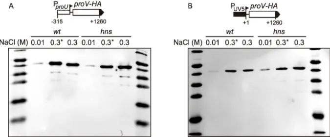
To ensure that the proV-lacZ translational fusion reflects regulation of native proU, osmoregulation was independently analyzed with Western blots. The proV gene was fused with a sequence coding for a C-terminal HA-tag. This proV-HA fusion was expressed from the proU promoter on low copy plasmids pKEKK11 (Fig. 4A) and from the lacUV5 promoter on plasmid pKEKK12 (Fig. 4B). The expression level of ProV-HA was determined in wild type and hns mutant cells that were either grown constantly under low and high osmolarity conditions, respectively, or first grown under low osmolarity conditions and then subjected to hyperosmotic stress by addition of NaCl to the medium. In conformity with the results of the β-galactosidase assays, only minimal levels of ProV-HA were detected under low osmolarity conditions. Expression was significantly higher in cells constantly grown in hyperosmotic medium. Strong osmoinduction and similar levels of ProV-HA were observed already ten minutes after hyperosmotic upshift. As expected, expression from the proU promoter was more strongly osmoregulated than from the lacUV5 promoter. Importantly, expression of ProV-HA was still osmoregulated when expressed from lacUV5 promoter in hns mutant cells (Fig. 4B). These results confirmed that in addition to H-NS repression and proU promoter activity some additional mechanism contributes to osmoregulation of the proU locus.

Figure 4. Western blot analysis of osmoregulation of ProV-HA expression. The proV gene fused with HA-

tag sequence was expressed from the proU promoter (plasmid pKEKK11) and the lacUV5 promoter (plasmid

pKEKK12) in wt cells (S3460) and hns mutant cells (S4162). Cells were grown constantly in LB supplemented

with 0.01M or 0.3M NaCl (labelled 0.01 and 0.3 respectively). Alternatively cells were first grown in LB with

0.01M NaCl to OD

600

of 0.5, and then NaCl concentration was increased to 0.3M, and samples were taken 10

minutes later (labelled 0.3*). Total cell lysates were analyzed with antibodies to HA-tag.

(21)

2.1.3 ProV protein stability under different osmolarity conditions

The results of above described experiments showed that proU is osmoregulated at a post-transcriptional level. Different mechanisms might be responsible for this post-transcriptional induction of proU by hyperosmotic stress:

1. proU mRNA might be more stable under high osmolarity conditions 2. proU translation might be more efficient under high osmolarity conditions 3. protein ProV might be more stable under high osmolarity conditions

The latter possibility was examined by comparing the stability of ProV-HA under low and high osmolarity conditions. Cells carrying the high copy plasmid pKEM78 expressing ProV-HA from lacUV5 promoter were grown under different osmolarity conditions. Protein synthesis was inhibited by addition of chloramphenicol (200 µg/ml) and samples were taken immediately before as well as five, ten, and twenty minutes after chloramphenicol addition.

The experiment showed that protein ProV is more stable under low osmolarity conditions (Fig. 5). Therefore the higher amount of ProV under high osmolarity conditions can not be explained by the increased ProV stability. The possibility of osmoregulation of proU mRNA stability and proU translation were examined in further experiments.

Figure 5. Stability of protein ProV-HA under different osmolarity conditions. Western blot analysis of

expression of ProV-HA under control of lacUV5 promoter from plasmid pKEM78. Cells (strain S3460) were

grown in LB medium supplemented with 0.01M or 0.3M NaCl. Protein synthesis was inhibited by the addition

of chloramphenicol to final concentration of 200 µg/ml. Samples were taken before, as well as 5, 10 and 20

minutes after chloramphenicol addition. ProV-HA protein was visualized with anti-HA antibodies. Four-times

more lysate of cells grown at low osmolarity was loaded on a gel. The Western blot analysis was done twice with

two independently obtained sets of samples. One representative Western blot image is shown. The corresponding

chart below shows the relative amount of ProV-HA protein remaining at each time point as determined by

Odyssey Infrared Imaging System software and plotted as a function of time. Each value is the average of four

measurement done for two independently obtained sets of samples.

(22)

2.2 Osmoregulation of RNase III processing of proU mRNA 2.2.1 RNase III processing of proU mRNA in the conserved region

RNase E and RNase III were shown to process proU mRNA, with one RNase III processing site reported between positions +216 and +217 (Madhusudan S., 2007). In this study, RNase processing of proU mRNA was further analyzed and the possibility of osmoregulation of RNase processing was tested.

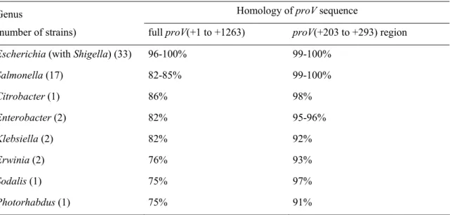
Processing of proU mRNA was examined with RACE (rapid amplification of cDNA ends) analysis of 5´ transcript ends (Wagner and Vogel, 2005). This method is a modification of the standard RACE protocol and includes an additional step of the treatment with tobacco acid pyrophosphatase (TAP) that removes diphosphate residue from the primary 5´ end of mRNA. The method enables mapping of both primary 5´ ends and 5´ ends formed after RNase processing. 5´ RACE was used to analyze the 5' region of proU mRNA extending from the known transcription site to position +496. The analyzed proU part covers a region between positions +203 and +293 that is highly conserved in Enterobacteriaceae species (Table 1) and therefore might be important for the regulation of proU.

Table 1. Conservation of the proV(+203 to +293) region in Enterobacteriaceae

Genus Homology of proV sequence

(number of strains) full proV(+1 to +1263) proV(+203 to +293) region

Escherichia (with Shigella) (33) 96-100% 99-100%

Salmonella (17) 82-85% 99-100%

Citrobacter (1) 86% 98%

Enterobacter (2) 82% 95-96%

Klebsiella (2) 82% 92%

Erwinia (2) 76% 93%

Sodalis (1) 75% 97%

Photorhabdus (1) 75% 91%

The sequence homology of full proV(+1 to +1263) sequence and proV(+203 to +293) sequence in Enterobacteriaceae species. Species are grouped in genera. Number of strains analyzed for each genus is provided in brackets. Note that Shigella strains belong to the species E. coli. The percent homology was determined by performing a BLAST search of full proV(+1 to +1263) sequence and proV(+203 to +293) sequence of E. coli K-12 substr. MG1655 against bacterial genome sequences.

To perform 5´ RACE, proV was expressed from the low copy plasmid pKEKK12 under

control of the lacUV5 promoter. Cells carrying the plasmid were grown under high and low

osmolarity conditions and total RNA was extracted. Part of each RNA sample (6 µg of RNA)

was treated with TAP to remove diphosphate from 5´ primary end of mRNA. Another part of

samples (also 6 µg of RNA) were not treated with TAP. RNA samples were then ligated to

RNA adapter by incubation with T4 RNA ligase. T4 RNA ligase ligates RNA adapter only to

(23)

the transcripts that carry 5´ monophosphate but not 5´ triphosphate. As a result, the RNA adapter is expected to be ligated to 5´ primary ends of mRNA only in samples treated with TAP. In contrast, the adapter is ligated to processing products in both TAP-treated samples and samples not treated with TAP. Therefore, comparison of TAP-treated and not treated samples should allow to distinguish 5´ primary ends from 5´ ends generated by processing.

RNA ligation products were purified and used as a template for cDNA synthesis with the gene-specific DNA oligonucleotide complementary to +467 to +486 region of proU. The resulting cDNA was amplified by PCR with gene-specific and 5´-adapter-specific primers, and the PCR products were analyzed with agarose gel electrophoresis (Fig. 6A). PCR products with the size corresponding to the expected size of primary transcript (530 bp), as well as two shorter products were detected in wild type cells (Fig. 6A). While both primary transcript and processing products were detected in cells grown under high osmolarity conditions, almost no primary transcript was detected under low osmolarity conditions. These results suggest that RNase processing of proU mRNA is more efficient under low osmolarity conditions. The same 5´ RACE analysis was also carried out with RNA isolated of a rnc mutant expressing a non-functional RNase III. Only the large PCR product corresponding to the primary transcript was detected in the rnc mutant. This suggests that the two shorter bands detected in the RNA isolated from wild type cells correspond to the products of RNase III processing.

To map the RNase III processing sites, individual DNA bands were purified from the agarose gel, cloned in vector pUC12 and sequenced (Fig. 6B, C). For cloning of each DNA band, sequences of five to six inserts in pUC12 were analyzed, since both primary and processed 5´ ends may vary by a few nucleotides. Sequencing confirmed that the larger DNA fragment (Fig. 6A, bands 1 and 2) corresponds to the primary transcript, although most of the inserts sequenced corresponded to transcripts which are truncated by a few bases at the 5' end (Fig. 6B, C). It is not clear whether these truncations is based on RNase processing in vivo or later during experimental procedures. The presence of truncated primary transcripts explains why, in contrast to our expectations, the primary transcript was detected in both TAP-treated samples and samples not treated with TAP (Fig. 6A). More importantly, sequencing of shorter DNA bands showed that the 5' ends of processing products corresponded to positions +218 and +282 relative to proU transcription start (Fig. 6B, C). Therefore RNase III processes the proU mRNA at positions +217/+218 and +281/+282.

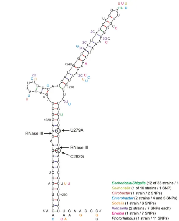
RNase III is known to process RNA in double-stranded regions. Therefore the secondary structure of proU mRNA was modeled using mfold software (Zuker, 2003) and RNase III processing sites were mapped on the predicted secondary structure. The proU mRNA was found to form an intensive stem-loop structure in conserved +203 to +293 region (Fig.7). The RNase III cleavage sites are located within the double-stranded region.

Processing in these sites would lead to the formation of two-nucleotide overhang at the 3´

ends of mRNA, a typical feature of RNase III processing.

(24)

Figure 6. 5´ RACE analysis of proU mRNA processing by RNases. (A) Gel electrophoresis of amplification products of cDNA. Each DNA band on a gel corresponds to the separate RNA species (primary proU transcript or processing product). Analysis of processing of proU mRNA in wild type cells and in rnc mutant cells is shown. The proU(+1 to +1260) mRNA was expressed from lacUV5 promoter on low copy plasmid pKEKK12.

Wild type (S3460) and rnc mutant cells (S4023) carrying pKEKK12 were grown in LB supplemented with either 0.01M or 0.3M of NaCl. RNA was extracted and further analyzed as described in text. (+) and (-) refers to TAP treatment prior to ligation with RNA adapter. PT - primary transcript. Numbers indicate the DNA bands that were subsequently purified from the gel, cloned in pUC12 and analyzed by sequencing. (B) and (C) Mapping of RNase III processing sites by sequencing. (B) The results of DNA bands sequencing. The band numbers in the table correspond to the band numbers in (A). Most DNA bands are a mixture of fragments with different 5´ ends.

Therefore, five to six clones were sequenced per cloning of each band. The number of clones sequenced, the positions of 5´ ends determined by sequencing and the number of clones with corresponding 5´ ends (in brackets) are shown. (C) Sequence of the plasmid pKEKK12 used for 5´ RACE analysis with RNase III processing sites. Sequence includes lacUV5 promoter and upstream part of proU analyzed by 5´ RACE. Primer T266 was used for PCR amplification of cDNA, and therefore corresponds to the 3´ end of the analyzed proU sequence. The location of 5´ ends of clones determined by sequencing are taken from the Fig. 6B and indicated by arrows. For each cloning, the most upstream position taken as the true 5´ end and depicted as a fat arrow with indication of position in comparison to transcription start (+1).

Not only the primary sequence but also the secondary structure of the stem-loop is highly conserved among Enterobacteriaceae species (Fig. 7). SNPs found in this region of proU in other species are predominantly located in single stranded parts of the mRNA (bulges and loops). SNPs found in double-stranded regions of mRNA in most cases also do not disrupt base pairing (e.g. G-C base pare changes to A-U or non-canonical G-U) (Fig. 7).

Conservation of both the primary sequence and the putative secondary structure of proU

mRNA around the RNase III cleavage site suggested that RNase III processing might be

important for proU osmoregulation. Indeed, the results of the 5´ RACE analysis suggested

that RNase III processes the proU mRNA more efficiently under low osmolarity conditions

and the processing is inhibited under high osmolarity conditions (Fig. 6B).

(25)

Figure 7. RNase III processes proU mRNA in a conserved region. Secondary structure of proU mRNA +202 to +300 region from E. coli K-12 substr. MG1655 was predicted using mfold software (Zuker, 2003). Numbers indicate the coordinates in the relation to the transcription start (+1). BLAST search of proV sequence of E. coli K-12 substr. MG1655 against the bacterial genomes was performed (see Table 1 for details) and SNPs found in 57 Enterobacteriaceae strains were mapped. SNPs are shown as single nucleotides, numbers in front of SNP indicate the number of strains this SNP was found. The list of Enterobacteriaceae genera with corresponding number of strains analyzed and number of SNPs is provided. The colors of font used in the list match the colors of the corresponding SNPs on the figure. RNase III cleavage sites mapped by 5´ RACE are shown with arrows.

Nucleotides mutated to disrupt RNase III processing are circled and shown with arrows.

(26)

2.2.2 Effect of rnc mutation on proU osmoregulation

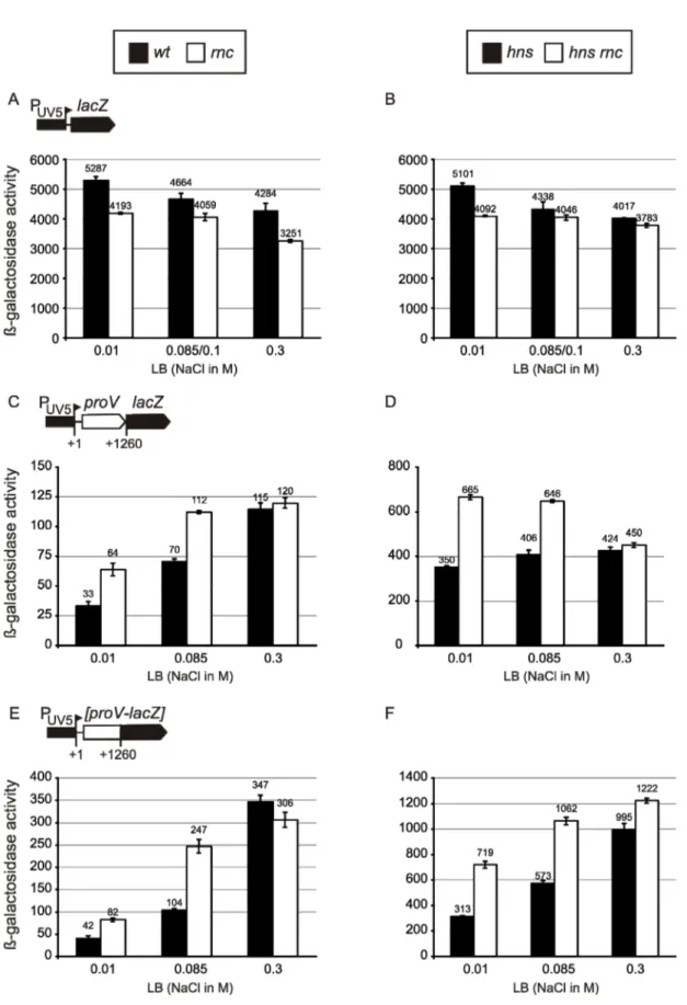
The possible involvement of RNase III in proU osmoregulation was further investigated by comparing the osmoregulation of proU in rnc mutants and the wild type. The rnc allele was transferred by transduction to strains which carry chromosomal copies of translational and transcriptional fusions, respectively, of proU with the reporter lacZ gene under control of lacUV5 promoter. Expression of proV-lacZ fusions in rnc and double rnc hns mutants was compared to the expression in wild type and the hns mutant under different osmolarity conditions (Fig. 8). Mutation of rnc lead to slight 1.1- to 1.3-fold downregulation of the expression of the control PlacUV5-lacZ construct (Fig. 8A, B) that might be due to the indirect effect of rnc mutation (see below). Importantly, the expression of proV-lacZ fusions under low osmolarity conditions was 1.9- to 2.3-fold higher in rnc mutants than in cells with the intact rnc gene, both in the wild type and in the hns mutant background (Fig. 8C to F). In contrast, expression of these constructs under high osmolarity conditions was not significantly affected by rnc mutation. These data further support the results of the 5´ RACE analysis that RNase III processing of the proU mRNA is inhibited under high osmolarity conditions.

The effect of the rnc mutation on proU expression was also analyzed using the proV-lacZ fusions expressed from the native proU promoter. Unexpectedly, mutation of rnc gene lead to the noticeable 1.4- to 1.8-fold downregulation of the expression of a control PproU-lacZ fusion (which lacks the proV coding sequence and RNase III processing sites) (Fig. 9A, B). The negative effect of rnc mutation on the expression of PlacUV5-lacZ and PproU-lacZ fusions may be partly non-specific. rnc mutant cells grow significantly slower than wild type cells and as a result the rnc mutation indirectly affects the expression of several hundreds of growth rate-dependent genes (Sim et al., 2010). However, the constructs expressed from the proU promoter might be further downregulated by mutation of rnc because RNase III is involved in activation of σ

S

expression (Resch et al., 2008). Therefore, mutation of rnc might lead to lower level of σ

S

and as the result to the lower expression from the σ

S

-dependent proU promoter.

Mutation of rnc had differential effects on the expression of the PproU-proV-lacZ fusions under different osmolarity conditions (Fig. 9C-F). Expression of all fusions was upregulated in the rnc mutant under low osmolarity conditions but downregulated under high osmolarity conditions. This probably results from the combination of two opposing effects:

upregulation of expression due to blocking of RNase III processing and downregulation of the

proU promoter.

(27)

Figure 8. Effect of rnc mutation on the osmoregulation of proV expressed from the constitutive lacUV5

promoter. Transcriptional (C, D) and translational (E, F) fusions of proV with the lacZ reporter gene, as well as

lacZ gene alone (A, B) are integrated in the chromosome. Expression of these reporter constructs from the

lacUV5 promoter was determined with β-galactosidase assay. Expression of constructs was compared in wt

versus rnc mutant cells (A, C, E) and in hns versus hns rnc double mutant cells (B, D, F). Cells were grown in

LB supplemented with the indicated NaCl concentrations. Bars show the determined expression level (black bars

for wt and hns mutants, and white bars for rnc and hns rnc mutants respectively). Values are the mean of at least

three calculations. Error bars represent standard error of the mean. Strains used: A, wt S1979, rnc T214; B, wt

S3122, hns rnc T216; C, wt S4064, rnc T97; D, hns S4111, hns rnc T99; E, wt S4070, rnc T100; F, hns S4117,

hns rnc T102. Note that different scales are used to indicate β-galactosidase activity. Expression values for

strains S1906 and S3122 are taken from (Madhusudan S., 2007).

(28)

Figure 9. Effect of rnc inactivation on the osmoregulation of proV expressed from the native proU

promoter. Transcriptional (C, D) and translational (E, F) fusions of proV with the lacZ reporter gene, as well as

lacZ gene alone (A, B) are integrated in the chromosome. Expression of these reporter constructs from the

original ProU promoter was determined with β-galactosidase assay. Expression of constructs was compared in

wt versus rnc mutant cells (A, C, E) and in hns versus hns rnc double mutant cells (B, D, F). Cells were grown in

LB supplemented with the indicated NaCl concentrations. Bars show the determined expression level (black bars

for wt and hns mutants, and white bars for rnc and hns rnc mutants respectively). Values are the mean of at least

three calculations. Error bars represent standard error of the mean. Strains used: A, wt S2048, rnc T380; B, wt

S3124, hns rnc T382; C, wt S4066, rnc T90; D, hns S4113, hns rnc T92; E, wt S4068, rnc T94; F, hns S4115,

hns rnc T95. Note that different scales are used to indicate β-galactosidase activity. Expression values for strains

S2048 and S3124 are taken from (Madhusudan S., 2007).

(29)

2.2.3 Effect of RNase III processing site mutation on proU osmoregulation

For 5´ RACE analysis of proU mRNA processing, RNase III activity was abolished by mutation of the rnc gene. However, the rnc mutation may have pleiotropic effects and may indirectly affect the expression of the lacZ reporter constructs. Therefore a different strategy of blocking RNase III processing was required to measure the effect of RNase III processing on proU expression. Instead of using the rnc mutant, the processing was blocked by mutation of the RNase III processing site of proU mRNA. Two single nucleotide mutations C282G and T279A that do not change the encoded amino acid sequence of the ProV protein were introduced at the RNase III processing site by site-specific mutagenesis (Fig. 7). The resulting proV mutants (proVC282G and proVT279A, respectively) were cloned into various reporter plasmids. Wild type proV sequence in plasmid pKEKK12, which carries proV-HA construct under control of the lacUV5 promoter, was substituted by the proV mutants resulting in plasmids pKEKK36 expressing proVT279A-HA and pKEKK38 expressing proVC282G-HA.

Using these plasmids 5´ RACE analysis was performed under the same conditions as described above for wild type proV expressed from pKEKK12. After gel electrophoresis only one large fragment corresponding to the primary transcript was detected for both of the proV processing site mutants (Fig. 10). Therefore both mutations completely abolished RNase III processing of proU mRNA.

Figure 10. 5´ RACE analysis of proU mRNA processing by RNases. Gel electrophoresis of amplification products of cDNA is shown. Each DNA band on a gel corresponds to the separate RNA species (primary proU transcript or processing product). Analysis of processing of wild type proU in both wild type cells (indicated wt) and in rnc mutant cells (indicated rnc), as well as processing of proU mutants in wild type cells (indicated proUC282G and proUT279A) is shown. Wild type and mutant forms of proU(+1 to +1260) transcripts were expressed from lacUV5 promoter from low copy plasmids: pKEKK12 for wild type proU, pKEKK38 for proUC282G, pEKK36 for proUT279A. Wild type (S3460) and rnc mutant cells (S4023) carrying these plasmids were grown in LB supplemented with either 0.01M or 0.3M of NaCl. RNA was extracted and further analyzed as described in text. (+) and (-) refers to TAP-treatment prior to ligation with RNA adapter. PT - primary transcript.

To estimate the effect of RNase III processing on proU osmoregulation, the expression

of wild type and mutant proV genes was measured with Western blot and with β-galactosidase

assays under different osmolarity conditions. The plasmids pKEKK12, pKEKK36 and

pKEKK38 expressing wild type and mutant proV-HA genes and used for 5´ RACE were now

also used for Western blot analysis. Expression was determined in wild type and hns mutant

cells carrying these plasmids under low osmolarity conditions and ten minutes after

hyperosmotic upshift. Both mutations of the RNase III processing site lead to significantly

(30)

higher expression levels of ProV-HA in cells grown under low osmolarity conditions (Fig. 11). This effect was evident in both wild type and hns mutant cells. In contrast, there was no significant effect of processing site mutations on ProV-HA expression under high osmolarity conditions. This supports the conclusion that RNase III processing of proU mRNA is effective under low osmolarity conditions and inhibited by hyperosmotic stress.

Figure 11. Western blot analysis of osmoregulation of proV with mutated RNase III processing site. Wild type proV and mutant proVC282G and proVT279A fused with HA-tag were expressed from lacUV5 promoter from the plasmids pKEKK11 (wild type proV, A and B), pKEKK38 (proVC282G, A), and pKEKK36 (proVT279A, B) respectively. Wild type (S3460) and hns mutant cells (S4162) carrying these plasmids were grown in LB supplemented with 0.01M NaCl to OD

600

of 0.5, NaCl was added to the final concentration of 0.3M and samples were taken immediately before and 10 minutes after this hyperosmotic stress. Total cell lysates were analyzed with antibodies to HA-tag.

Osmoregulation of the proV mutants was further studied using the proV-lacZ reporter

fusions which were integrated into the chromosome (Fig. 12). Expression of the translational

and transcriptional proU-lacZ fusions under the control of lacUV5 and proU promoter was

measured in wild type and hns mutant cells grown under different osmolarity conditions. Both

mutations of the RNase III processing site lead to similar higher expression level of all the

constructs tested (Fig. 12). The effect of abolishing RNase III processing was more

pronounced under low than under high osmolarity conditions for all constructs expressed

from lacUV5 and proU promoters in wild type as well as in hns mutant cells. Specifically,

mutations of processing site lead to only 1.3- to 1.9-fold increase of expression of constructs

under high osmolarity conditions. In contrast, under low osmolarity conditions mutations of

processing site lead to 2.4- to 4.4-fold increase in expression of transcriptional proV-lacZ

fusions (Fig. 12A, B, E, F) and 5.0- to 10.0-fold increase of expression of translational

proV-lacZ fusions (Fig. 12C, D, G, H). The latter difference between the translational and

transcriptional fusions is expected, because RNase III processing should affect the

translational proV-lacZ fusions stronger than the transcriptional fusions. To summarize, the

results of the β-galactosidase assays and Western blot analysis of expression of proV mutants

together with the results of 5´ RACE experiments clearly show that RNase III processing of

proU mRNA is much more efficient under low osmolarity than under high osmolarity

conditions.

Abbildung

Figure 1. Osmoregulation systems of E. coli. Transporters ProP, ProU, BetU and BetT import compatible  solutes; transporters TrkG/H and KdpFABC (expression controlled by two-component signaling system KdpDE)  import potassium; AqpZ is a water channel; OtsA
Figure 2. E. coli  proU operon. Structural genes proV,  proW,  proX transcribed from σ S  and σ 70  promoters
Figure 3. Contribution of proU promoter, H-NS repression and post-transcriptional mechanisms to proU  osmoregulation
Figure 4. Western blot analysis of  osmoregulation of ProV-HA expression. The proV gene fused with HA- HA-tag sequence was expressed from the proU promoter (plasmid pKEKK11) and the lacUV5 promoter (plasmid  pKEKK12) in wt cells (S3460) and hns mutant cell
+7

Referenzen

ÄHNLICHE DOKUMENTE

Based on the data obtained from the csrA::Tn5 strain, it is tempting to conjecture that under conditions where CsrA activity is inhibited, at the beginning of stationary phase for

is appropriate for studying specific parts of transcription units like different ribosome binding site (RBS) or promoters, but is limited for investigations of effects on

This structure, which represents a kinetically trapped reaction intermediate of DsbB, contains a charge-transfer complex between the Cys44 of DsbB and ubiquinone Q8 in its active

To identify important regulatory motifs in the promoter region, mutations were introduced into the conserved elements region by PCR (primers.. TATA boxes, CAAT and TAAG motifs

Fatty acid incubation and analysis Yeast cultures of the only effective transformation process with HO-poly-KanMX4-HO + GPD prom + fat-1t + term as demonstrated by repeated positive

With the means of HDACs and SMRT inhibitors the function of the recruited complex was verified and a novel mechanism of target gene dependent VDR- mediated

Photo- graphs were taken from time lapse microscopy during microfluidic per- fusion cultivation (A) and microscale batch cultivation (B) using LB medium supplemented with 100 mM (A)

The following facts support this hypothesis: (i) the Sim protein is synthesized as a precursor with a hydrophobic leader sequence of 20 amino acid resi- dues,