Idiopathic scrotal hematoma in a neonate
Idiopathisches Skrotalhämatom bei einem Neugeborenen
Abstract
Background: Neonatal scrotal hematoma is considered a surgical emergency in the neonatal period. Up to recently, immediate surgical
Sylvia
Gkantseva-Patsoura
1exploration was considered the gold standard for the diagnosis and
treatment in the underlying causes.
George Katsaras
2Petroula Georgiadou
1Objective:In this article, we present a case of idiopathic scrotal hema-
toma in a neonate.
Nektarios Lainakis
3Method:It was managed conservatively with clinical and ultrasonograph-
ic follow-up.
Eirini Liovarou
4Rita Theofanopoulos
1Result:The hematoma had gradually subsided, and any surgical inter-
vention was avoided to the neonate.
Martha Theodoraki
1Conclusion:With good clinical and imaging follow-up, some cases could be managed nonoperatively.
1 Neonatal Intensive Care Unit, General State Hospital of Keywords:neonate, scrotal hematoma, idiopathic
Nikaia “Agios Panteleimon”, Piraeus, Greece
Zusammenfassung
Hintergrund: Das neonatale Skrotalhämatom wird als chirurgischer Notfall in der Neugeborenenperiode angesehen. Bis vor Kurzem wurde
2 Paediatric Department, General Hospital of Pella – Hospital Unit of Edessa, Greece
die sofortige chirurgische Untersuchung als Goldstandard für die Dia- gnose und Behandlung der zugrunde liegenden Ursachen angesehen.
3 Paediatric Surgery Department, General State Ziel:In diesem Artikel stellen wir einen Fall eines idiopathischen Skro-
talhämatoms bei einem Neugeborenen vor.
Hospital of Nikaia “Agios Methode:Es wurde konservativ mit Beurteilung der Klinik und mittels
Ultraschallverlaufskontrollen beobachtet. Panteleimon”, Piraeus,
Greece Ergebnis:Das Hämatom war allmählich rückläufig und ein chirurgischer
Eingriff bei unserem Neugeborenen wurde vermieden. 4 Radiology Department, General State Hospital of Schlussfolgerung:Mit einer guten klinischen und bildgebenden Nach-
sorge könnten einige Fälle nicht operativ behandelt werden. Nikaia “Agios Panteleimon”, Piraeus, Greece
Schlüsselwörter:Neugeborene, Skrotalhämatom, idiopathisch
Introduction
Scrotal hematoma is the collection of blood inside the scrotum, which contains the testicle, the epididymis and the spermatic cord. Testicular hematomas can form within the testicular parenchyma or on the surface of the testicle. Scrotal hematoma can occur secondary to some intra-abdominal diseases, including intraperitoneal or retroperitoneal bleeding [1]. It is a rare genitourinary emergency in a neonate [2]. Some cases have clear eti- ology such as testicular torsion, inguinal hernia, adrenal hemorrhage, meconium peritonitis, hematocele, testicular tumor or birth trauma, whereas other cases have no definite cause. Because Doppler ultrasonography (DUS) in the small neonatal testis has previously been regarded as an inconsistently diagnostic method, scrotal explora-
tion was considered necessary for the diagnosis and treatment of these neonates [3], [4]. In this article, we present a case of idiopathic scrotal hematoma in a neonate which was managed conservatively with clinical and radiological follow-up, emphasizing the role of ultra- sonography in the evaluation of the several potential causes leading to an acute scrotum, and consequently sparing some neonates surgical exploration.
Case description
A late preterm male neonate (Ballard score: 36 weeks gestational age) was delivered by a 21-year-old woman at home with no professional care, no labors in pregnancy.
The neonate was transported by ambulance to the nearest hospital, where he was resuscitated and intu-
1/4 GMS German Medical Science 2021, Vol. 19, ISSN 1612-3174
Case Report
OPEN ACCESS
Neonatology
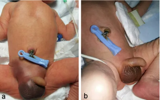
Figure 1: a) At the 3rdday of life, bruises are observed at both groins and the scrotum. b) The same with complete resolution after two weeks.
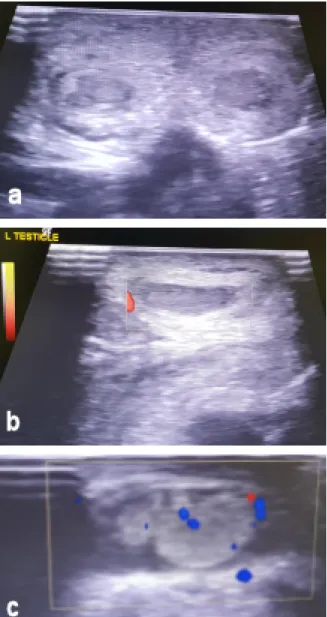
Figure 2: a) Ultrasonography of the right testicle. b) Ultrasonography of the left testicle. Both testicles are depicted inside the scrotum. The scrotal wall as well as the testicles are grossly thickened and edematous, while the echogenic structure and
vascularity of the testicles are normal.
bated. Afterwards, he was again transported by ambu- lance to our tertiary NICU at approximately the 6th hour of life. Since the neonate was hemodynamically stable at the time of admission, he was extubated. He was a small for gestational age neonate with 2000 gr body weight. The examination of the abdomen was normal.
Painless swelling of the scrotum and hydrocele were clinically observed. The neonate had a hematocrit of 48.6%, negative Coombs test and normal values of the coagulation parameters (activated partial thromboplastin time (aPTT), prothrombin time (PT), international normal- ized ratio (INR), platelet count and fibrinogen). He re- ceived vitamin K.
At the 3rdday of life, jaundice appeared, and phototherapy started. Furthermore, bruises were observed at both groins as well as at the whole scrotum (Figure 1a). The swelling of the scrotum was thickened. There was no clinical feature suggestive of trauma or bleeding diathesis.
The ultrasonography of the abdomen, the adrenals and the testicles were all normal. Clinical examination by a pediatric surgeon took place that excluded the acute scrotum.
At the 4thday of life, the phototherapy was stopped. Due to the progressing clinical condition at the groins and scrotum, we conducted a new ultrasonography at these areas. The scrotal wall was grossly thickened and edema- tous, while the echogenic structure and vascularity of both testicles were normal (Figure 2a, b). Based on the clinical image, the finding concerned a hematoma of the scrotum. In this phase, no hydrocele was observed.
The child was managed non-operatively. He was mon- itored clinically and radiologically. In the following days, the testicles maintained their normal echogenic structure and vascularity, however, thickening of the scrotum wall was still observed in the context of hematoma, while at the same time a hydrocele with echogenic elements on the left scrotum (hematoma) was observed to be more intensive (Figure 3a, b, c). Follow-up ultrasound scans revealed complete resolution of the earlier noted hema- toma at the scrotal wall.
2/4 GMS German Medical Science 2021, Vol. 19, ISSN 1612-3174
Gkantseva-Patsoura et al.: Idiopathic scrotal hematoma in a neonate
Figure 3: a) Ultrasonography of both testicles. b) Doppler ultrasonography of the right testicle. c) Doppler ultrasonography
of the left testicle. Both testicles maintain their normal echogenic structure and vascularity. Thickening of the scrotum wall is still observed in the context of hematoma. A hydrocele with echogenic elements on the left scrotum (hematoma) is
observed.
Discussion
Acute scrotal hematoma in the neonate might be a major surgical emergency [5]. Although the surgical exploration was considered the gold standard, clinical evaluation and ultrasonography have been shown to be of greatest value in the differential diagnosis of testicular diseases, as discussed in AWMF guideline 006/23 from 2015 [6], [7].
The most common causes for acute scrotal hematoma include testicular torsion, inguinal hernia, scrotal or testicular edema and scrotal trauma, whereas adrenal hemorrhage and adrenal neuroblastoma are rare but must also be considered. Idiopathic scrotal edema and hydrocele are among the minor causes. Spontaneous idiopathic hemorrhage of the scrotum, as found in our
patient, has also been described and is usually a diagno- sis of exclusion [1], [8], [9].
Our patient, at the 3rdday of life, presented with bruises at both groins as well as at the whole scrotum. There was no clinical feature suggestive of trauma or bleeding dia- thesis. The ultrasonography of the abdomen, the adrenals and the testicles were normal.
Ultrasonography, including power Doppler, is usually performed to differentiate conditions such us testicular torsion, scrotal or testicular edema, inguinal hernia, testicular trauma, or adrenal hemorrhage in an acute scrotum. In testicular torsion, there is hypovascularity of the affected testis, while testicular trauma may present with intratesticular hematoma or laceration of the tunica albuginea. Acute scrotal discoloration in neonates is usually caused by testicular torsion [10]. Also few cases of scrotal hematoma in neonates due to adrenal hemor- rhage have been reported in the literature [11]. Scrotal and abdominal ultrasonography can provide important information about the patient, and this would seem to be essential in order to avoid unnecessary invasive proced- ures. Spontaneous hemorrhage of the scrotum, as found in our patient, has also been described in the literature [12].
Except for spermatic cord torsion and inguinal hernia, hemoscrotum due to other causes may be managed with conservative treatment. We treated our patient nonoper- atively as well as with systematic clinical and sonographic follow-up examinations, and there was a gradual improve- ment of the symptoms (Figure 1b).
Conclusion
The diagnosis of neonatal scrotal hematoma should be considered in a newborn with scrotal swelling and bluish discoloration. Our case presentation constitutes further evidence and enhances the importance of Doppler in the indication for surgical exploration. Some risk factors can be identified, e.g. evidence of bleeding diathesis, maternal gestational diabetes and high birth weight, with evidence of delivery-related trauma or predisposing intra-abdominal lesions (adrenal hemorrhage, subcapsular liver hema- toma) on ultrasonography. A thorough clinical examination and ultrasonography are the cornerstones in establishing an early diagnosis.
Notes
Informed consent
Informed consent has been obtained from the patient’s mother for the publication of this case report.
Competing interests
The authors declare that they have no competing in- terests.
3/4 GMS German Medical Science 2021, Vol. 19, ISSN 1612-3174
Gkantseva-Patsoura et al.: Idiopathic scrotal hematoma in a neonate
Authors’ contributions
Sylvia Gkantseva-Patsoura and George Katsaras partici- pated in the conceptualization, investigation, methodol- ogy, writing and original drafting of the manuscript.
Petroula Georgiadou, Nektarios Lainakis, Eirini Liovarou and Rita Theofanopoulos were responsible for investiga- tion, methodology, validation, editing and reviewing the paper. Martha Theodoraki participated in supervision, editing, reviewing and validating the final version.
References
1. Lai LJ, Chen LM, Chu PY, Tseng MH, Chang CC, Lu CW. Neonatal adrenal hemorrhage associated with scrotal hematoma: an unusual case report and literature review. Pediatr Neonatol.
2012 Jun;53(3):210-2. DOI: 10.1016/j.pedneo.2012.04.010 2. Al-Salem AH. Acute Scrotum. In: Al-Salem AH, editor. An Illustrated
Guide to Pediatric Urology. 1st ed. Cham: Springer; 2017. p. 601- 17.
3. Santi M, Lava SAG, Simonetti GD, Bianchetti MG, Milani GP.
Acute Idiopathic Scrotal Edema: Systematic Literature Review.
Eur J Pediatr Surg. 2018 Jun;28(3):222-6. DOI: 10.1055/s-0037- 1603089
4. Morey AF, Brandes S, Dugi DD 3rd, Armstrong JH, Breyer BN, Broghammer JA, Erickson BA, Holzbeierlein J, Hudak SJ, Pruitt JH, Reston JT, Santucci RA, Smith TG 3rd, Wessells H; American Urological Assocation. Urotrauma: AUA guideline. J Urol. 2014 Aug;192(2):327-35. DOI: 10.1016/j.juro.2014.05.004 5. Gordhan CG, Sadeghi-Nejad H. Scrotal pain: evaluation and
management. Korean J Urol. 2015 Jan;56(1):3-11. DOI:
10.4111/kju.2015.56.1.3
6. Liang T, Metcalfe P, Sevcik W, Noga M. Retrospective review of diagnosis and treatment in children presenting to the pediatric department with acute scrotum. AJR Am J Roentgenol. 2013 May;200(5):W444-9. DOI: 10.2214/AJR.12.10036 7. Deutsche Gesellschaft für Kinderchirurgie e.V. (DGKCH);
Deutsche Gesellschaft für Urologie e.V. (DGU). S2k-Leitlinie Akutes Skrotum im Kindes- und Jugendalter. AWMF-Registernr.
006/023. Berlin: AWMF; 2015. Available from: https://
www.awmf.org/leitlinien/detail/ll/006-023.html
8. Wu SC, Chou CY. Neonatal idiopathic scrotal hemorrhage: report of one case. Clin Neonatol. 2005 Dec;12(2):64-6. DOI:
10.7098/CN.200512.0064
9. Karmazyn B, Steinberg R, Kornreich L, Freud E, Grozovski S, Schwarz M, Ziv N, Livne P. Clinical and sonographic criteria of acute scrotum in children: a retrospective study of 172 boys.
Pediatr Radiol. 2005 Mar;35(3):302-10. DOI: 10.1007/s00247- 004-1347-9
10. Lam WW, Yap TL, Jacobsen AS, Teo HJ. Colour Doppler ultrasonography replacing surgical exploration for acute scrotum:
myth or reality? Pediatr Radiol. 2005 Jun;35(6):597-600. DOI:
10.1007/s00247-005-1411-0
11. Gonçalves R, Abuabara A, Abuabara RFF, Feron CA. Scrotal hematoma as a sign of adrenal hemorrhage in newborns. Sao Paulo Med J. 2011 Mar;129(2):113-5. DOI: 10.1590/S1516- 31802011000200011
12. Jimoh BM, Chinwe EB, Adebisi AO, Ifeoma IC, Ogechi M, Oluwafemi AJ. Idiopathic Scrotal Hematoma in Neonate: A Case Report and Review of the Literature. Case Rep Urol.
2014;2014:1-3. DOI: 10.1155/2014/212914
Erratum
An author name was corrected.
Corresponding author:
George Katsaras
Paediatric Department, General Hospital of Pella – Hospital Unit of Edessa, End of Egnatia Str., 58200 Greece
gkatsaras84@gmail.com
Please cite as
Gkantseva-Patsoura S, Katsaras G, Georgiadou P, Lainakis N, Liovarou E, Theofanopoulos R, Theodoraki M. Idiopathic scrotal hematoma in a neonate. GMS Ger Med Sci. 2021;19:Doc01.
DOI: 10.3205/000288, URN: urn:nbn:de:0183-0002888
This article is freely available from
https://www.egms.de/en/journals/gms/2021-19/000288.shtml
Received:2020-09-17 Revised:2020-12-23 Published:2021-02-19
Published with erratum:2021-03-01
Copyright
©2021 Gkantseva-Patsoura et al. This is an Open Access article distributed under the terms of the Creative Commons Attribution 4.0 License. See license information at
http://creativecommons.org/licenses/by/4.0/.
4/4 GMS German Medical Science 2021, Vol. 19, ISSN 1612-3174
Gkantseva-Patsoura et al.: Idiopathic scrotal hematoma in a neonate