channel Kir5.1 in the distal convoluted tubule
DISSERTATION
ZUR ERLANGUNG DES DOKTORGRADES DER NATURWISSENSCHAFTEN (DR. RER. NAT.) DER FAKULTÄT FÜR BIOLOGIE UND VORKLINISCHE
MEDIZIN DER UNIVERSITÄT REGENSBURG
Vorgelegt von
Catarina Isabel Rina Quintanova aus Vila Franca de Xira, Portugal
Im Jahr 2017
Das Promotionsgesuch wurde eingereicht am: 05.12.2017
Die Arbeit wurde angeleitet von: Prof. Dr. med. Richard Warth
Unterschrift:
________________________
(Catarina Quintanova)
-1-
Table of Contents
Table of Contents ... 1
1. Introduction ... 4
1.1 Distal Convoluted Tubule ... 4
1.1.1 Reabsorption of Na+ in the DCT ... 7
1.1.2 Regulation of DCT K+ transport ... 7
1.1.3 Ca2+ reabsorption in the DCT ... 9
1.2 DCT disorders ...10
1.2.1 Gitelman’s syndrome ...10
1.2.2 Familial hyperkalemic hypertension (Gordon syndrome) ...10
1.2.3 EAST/SeSAME syndrome ...11
1.3 Classification and function of K+ channels ...11
1.3.1 Inwardly rectifying K+ channels ...12
1.3.1.1 KCNJ10 (Kir4.1) ...13
1.3.1.2 KCNJ16 (Kir5.1) ...14
2. Objectives ...17
3. Materials and Methods ...18
3.1 Material ...18
3.1.1 Instruments ...18
3.1.2 Laboratory Material ...19
3.1.3 Substances ...20
3.1.4 Enzyme, Kits ...22
3.1.5 Software ...23
3.1.6 Oligonucleotide ...23
3.1.7 Antibody ...24
3.1.8 Buffers and solutions ...24
3.2 Methods ...27
3.2.1 Mice ...27
3.2.1.1 KCNJ16 knockout model ...27
3.2.1.2 Isolation of genomic DNA ...27
3.2.1.3 Genotyping of KCNJ16 knockout mice ...28
3.2.1.4 Diet 4% NaCl ...28
3.2.2 Cell line ...30
3.2.3 Histological Methods ...30
-2-
3.2.3.1 Tissue fixation by retrograde arterial perfusion ...30
3.2.3.2 Cryo preparation and sectioning ...31
3.2.3.3 X-Gal stain: detection of β-galactosidase activity ...31
3.2.3.4 Immunofluorescence ...31
3.2.4 Molecular Biological Methods ...32
3.2.4.1 Site directed Mutagenesis ...32
3.2.4.2. Isolation of total RNA from Kidneys ...32
3.2.4.3 Reverse Transcription (cDNA-Synthase) ...33
3.2.4.4 Quantitative Polymerase chain reaction (Real-time PCR) ...34
3.2.5 Patch Clamp Experiments of transfected cells ...34
3.2.5.1 Patch clamp measurements in whole cell mode ...35
3.2.5.2 Patch clamp measurements in single channel mode ...35
3.2.6 Ca2+ Measurements with Fura-2 ...36
3.2.6.1 Ca2+ measurements in isolated DCTs ...36
3.2.7 Plethysmography ...37
3.2.8 Statistics ...38
4. Results ...39
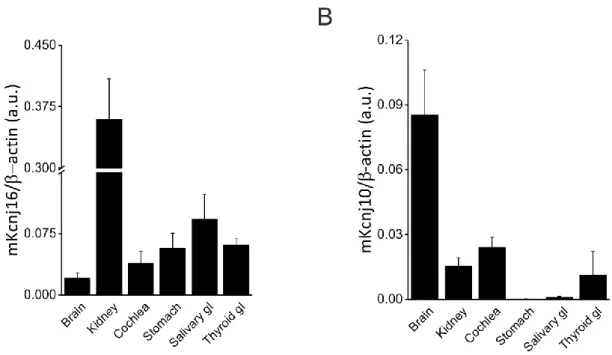
4.1 Expression and localization of KCNJ16 ...39
4.1.1 KCNJ16 expression in mouse tissues ...39
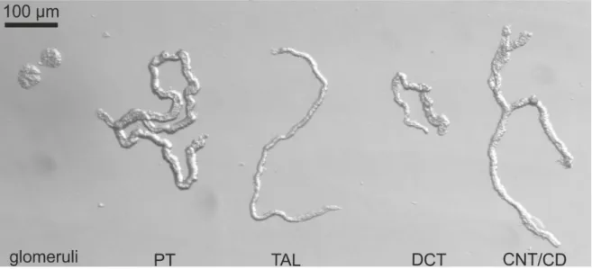
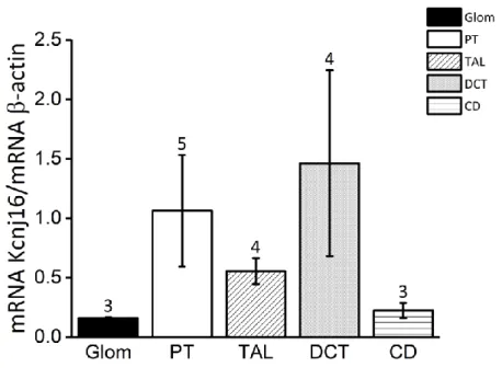
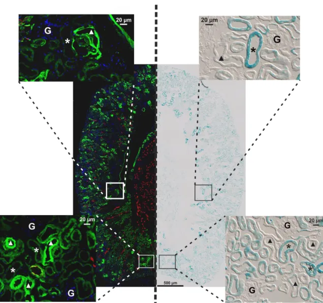
4.1.2 Kir5.1 (Kcnj16) localization in the kidney ...39
4.1.3 Electrophysiological experiments ...43
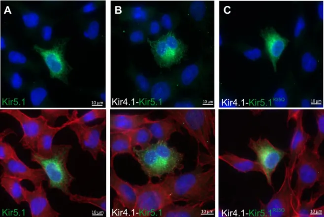
4.1.3.1 Immunofluorescence of transfected cells ...43
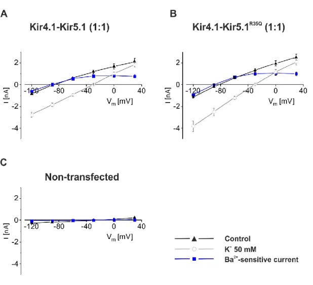
4.1.3.2 Whole cell measurements ...44
4.1.3.3 Effects of Kir5.1 mutation at single channel level ...48
4.2 Kir5.1-/- Mouse Model ...52
4.2.1 Basal electrolyte excretion of adult mice ...52
4.2.2 Effect of amiloride on electrolyte excretion levels ...54
4.2.3 Effect of high Na+ diet on electrolyte excretion levels ...55
4.2.4 Measurement of electrolytes in blood samples ...58
4.2.5 Respiratory response of Kir5.1-/- ...60
4.2.6 Ca2+ measurements on isolated tubules ...64
4.2.5.1 Superfused DCT tubules ...64
4.2.5.2 Perfused DCT tubules ...69
5. Discussion...72
5.1 Kcnj16 expression ...72
5.2 Role of KCNJ16 in the kidney ...72
5.2.1 Electrophysiological characterization of the Kir5.1R35Q mutation ...73
-3-
5.2.2 Kir5.1-mediated K+ sensing in DCT ...74
5.3 Phenotype of Kir5.1-/- mice ...76
5.3.1 Electrolyte balance in Kir5.1-/- mice ...76
5.3.2 Effect of high Na+ diet in Kir5.1-/- mice ...78
5.3.2 Role of KCNJ16 in respiration ...80
6. Summary ...82
7. Zusammenfassung ...84
8. References ...86
9. Supplements ...93
10. List of Figures ...95
11. List of Tables ...97
12. List of abbreviations ...98
13. Attachment ... 100
13.1 Congresses ... 100
14. Acknowledgments ... 101
-4-
1. Introduction
1.1 Distal Convoluted Tubule
The kidney plays an important role in maintaining body electrolytes and fluid balance, blood pressure and in the regulation and maintenance of systemic acid-base balance. The nephron is the structural and functional unity of the kidney (Figure 1). This functional unit filters the blood, reabsorbs the filtered electrolytes, solutes and fluid and excretes excessive electrolytes and water.1
Figure 1. Schematic representation of a single nephron.
The nephron is the microscopic structural and functional unit of the kidney. It is composed of a renal corpuscle and a renal tubule. In the glomerulus, ultra-filtrate is continuously generated by filtration of the blood. In the adherent renal tubule, certain substances and water are either reabsorbed or secreted. The ultra-filtrate first reaches the proximal tubule―the main side of reabsorption―followed by the U-shaped loop of Henle including the thick ascending limb, where the filtrate is concentrated.
Fine-tuning of the urine composition takes place in the distal convoluted tubule (red) and in the collecting ducts.
The distal convoluted tubule (DCT) is the shortest segment of the nephron and is the portion of the nephron that is immediately downstream from macula densa and ends in the connecting tubule/collecting duct system (CD). Despite its size, DCT plays an important role in regulating the extracellular fluid volume and electrolyte homeostasis in a variety of homeostatic processes that include sodium (Na+) reabsorption, potassium (K+) secretion, calcium (Ca2+) and magnesium (Mg2+) handling (Figure 2). The DCT can be further divided into two sub-segments, DCT1 and DCT2, which are distinguished by their response to the mineralocorticoid aldosterone. Aldosterone is a steroid hormone that is released from the adrenal glands in response to hyperkalemia and volume depletion. DCT2 is more sensitive to aldosterone due to the expression of an enzyme called 11-β hydroxysteroid dehydrogenase 2 (11-βHSD2) that metabolizes cortisol. Cortisol has a structure similar to
-5-
aldosterone, therefore by metabolizing cortisol to cortisone the enzyme 11-βHSD2 prevents the binding to the mineralocorticoid receptor expressed in the DCT2. The enzyme 11- βHSD2 is also expressed in connecting tubule (CNT) and CCD making these three segments known as the aldosterone sensitive distal nephron.2,3,4,5,6
DCT is morphologically unique since its extensive basolateral membrane has numerous deep infoldings and the cells are particularly rich in mitochondria. Because of very high rates of transcellular ion transport, the DCT requires a considerable amount of ATP high activity of Na+/K+-ATPase to maintain ionic gradients. It is therefore, not surprising that DCT cells possess the highest density of Na+/K+-ATPase along the nephron.7,8
-6-
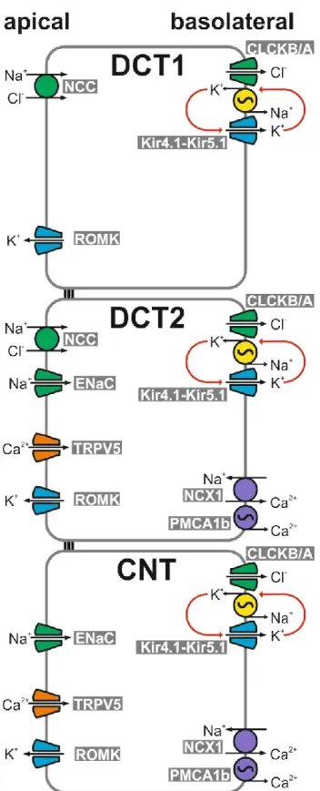
Figure 2. Schematic model of distal convoluted tubule and connecting tubule.
In the early DCT (DCT1), apical Na+ reabsorption is exclusively mediated by NCC, whereas in the late DCT (DCT2) and connecting tubule (CNT). Na+ reabsorption is also mediated by the epithelial Na+ channel ENaC. Na+ transport by ENaC is coupled to the activity of the renal outer medullary K+ channel (ROMK) mediating K+ secretion into the urine. The Na+/K+-ATPase mediates basolateral Na+ efflux and K+ uptake. The latter is functionally coupled to K+ recycling through Kir4.1-Kir5.1.
Apical Ca2+ transport is mediated by TRPV5 in the late DCT and connecting tubule. In the cytosol, Ca2+ is bound by calbindin and then transported out of the cell by NCX1 or plasma membrane Ca2+
ATPases (PMCA).
-7-
1.1.1 Reabsorption of Na+ in the DCT
Regulation of Na+ excretion is essential to control and maintenance of extracellular fluid balance and blood pressure control. Na+ is mainly reabsorbed by proximal tubules (PT) and thick ascending limbs (TAL) with fine-tuning occurring in the distal convoluted tubule (DCT), connecting tubule (CNT) and collecting ducts (CD). The DCT cells are responsible for reabsorbing 5-10% of the filtered Na+ and chloride (Cl-) from the filtrate. The uptake of Na+ in the DCT is mediated through the action of the NaCl cotransporter (NCC). NCC, also known as thiazide-sensitive Na+ and Cl- cotransporter, is localized in the apical membrane of DCT1. This process is electroneutral, since for one Na+ cation one Cl- anion enters the cell. NCC inhibition by thiazides, a class of diuretics, is used in the clinical setting for the treatment of arterial hypertension and edema. Thiazides inhibit Na+ reabsorption by blocking NCC thereby decreasing the workload for the Na+/K+-ATPase. In parallel, urinary Na+, K+ and Mg2+ excretion increases. However, Ca2+ is less excreted (hypocalciuria).
Hypocalciuria was postulated to be generated by two different mechanisms: either by increased active Ca2+ uptake via the vanilloid transient receptor potential channel 5 (TRPV5) in the DCT, or by thiazide-induced hypovolemia resulting in enhanced passive Ca2+ uptake in the proximal convoluted tubule.9 Since the TRPV5-/- mice still developed hypocalciuria when given thiazide diuretics, an increased uptake of Ca2+ in the DCT seems less likely.2,9–14
Besides Na+ uptake through NCC, Na+ can also be taken up in DCT2 by the amiloride- sensitive epithelium Na+ channel (ENaC). ENaC, mediates Na+ influx and causes a depolarization of the apical membrane. This depolarization enhances the driving force for K+ secretion across the apical membrane through the "renal outer medullary K+ channel"
(ROMK). The reabsorbed Na+ is extruded at the basolateral side by the Na+/K+-ATPase.
The activity of the Na+/K+-ATPase fuels further Na+ transport across the tubular cells by keeping the intracellular Na+ concentration low and the K+ concentration high (Figure 2). 6,15
1.1.2 Regulation of DCT K+ transport
As mentioned above, sustained reabsorption of Na+ by either NCC or ENaC requires basolateral Na+ extrusion via the Na+/K+-ATPase. For ongoing activity of Na+/K+-ATPase, that exchanges three Na+ for two K+, the basolateral availability of K+ can become a limiting factor, especially at high transport rates and when the basolateral space is narrow, i.e. in the basolateral infoldings of the plasma membrane. Therefore, basolateral K+ channels serve two important functions: i) they hyperpolarize the membrane (which is important for voltage-dependent processes such as Cl- exit through channels) and ii) the recycle of K+
-8-
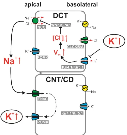
thereby allowing the ATPase to run at high transport rates. The functional coupling of the ATPase and the basolateral K+ conductance (K+ leak) is called "pump-leak coupling".2,6,8,16 Recently, a more prominent role of the DCT in the regulation of K+ homeostasis was suggested by Terker and co-workers.17 They hypothesized that DCT cells can actively sense changes in plasma [K+] and indirectly modify K+ secretion in CNT and CCD through regulation of the NCC. Reduced NCC activity leads to an enhanced Na+ flux to CNT and CCD where Na+ is then replaced by K+. They suggested, that the plasma K+ sensor Kir4.1 (and Kir4.1-Kir5.1) that senses increases in plasma [K+] by translating it into a depolarization of DCT cells. This depolarization, in turn results in increased intracellular [Cl-] by reducing the driving force for Cl- efflux via ClCKb channels. Increased intracellular [Cl-] then was proposed to lead a decrease in phosphorylation of NCC via the inhibition of the Cl--sensitive WNK-SPAK/OSR1 kinase pathway ultimately leading to decreased Na+ reabsorption in the early DCT (Figure 3).17,18 The resulting elevated luminal Na+ ions would be reabsorbed in more distal segments via ENaC leading to ROMK-mediated secretion of K+ and normalization of elevated plasma K+ levels.
Narrow regulation of plasma K+ between 3-6 mM concentrations is vital for life. Depending on plasma K+ levels, the kidney can vary the fraction excretion of K+ from 1% (net reabsorption) to 200% (net secretion) meaning that the kidney is a main contributor to K+ homeostasis. This flexibility is a property of the aldosterone-sensitive distal nephron including collecting ducts. In the early DCT, secretion of K+ is low and increases along the distal tubule parallel to the increase in lumen-negative transepithelial voltage observed in the late DCT. ROMK mediated K+ transport depends on the driving force for K+ secretion which is mainly determined by the aldosterone-regulated ENaC-mediated Na+ reabsorption.
The increased activity of ENaC depolarizes the apical membrane, which is a stimulus for K+ efflux through ROMK. Other channels may be involved in K+ secretion Maxi-K+ channels (BK channels). BK channels are triggered in states of high urinary flow (during diuretic use, for example) leading to an increase of K+ secretion under these conditions.2,5,19
-9-
Figure 3. Model of K+ sensing and regulation in the distal tubules.
In the distal convoluted tubule DCT, elevated plasma K+ levels are sensed by heteromeric Kir4.1- Kir5.1 channels leading to depolarization of the basolateral membrane potential (Vm), which in turn inhibits Cl- efflux via ClCKB channels. The resulting increased intracellular [Cl-] blocks the Cl-- dependent kinases in the WNK/SPAK pathway, which in turn inhibits the activity of the Na+/Cl-- cotransporter (NCC) resulting in elevated luminal Na+ levels. Apical Na+ is reabsorbed in the more distal connecting tubule (CNT) and collecting duct (CD) by the epithelial Na+ channel (ENaC) ultimately leading to K+ efflux via the renal outer medullary K+ channel (ROMK) effectively correcting the elevated plasma K+ levels.
1.1.3 Ca2+ reabsorption in the DCT
Extracellular Ca2+ is tightly regulated and serves as intracellular messenger in numerous essential cellular events such as immune response, muscle contraction, hormone secretion and regulation of enzymes. Ca2+ is regulated in the renal tubules by several calciotropic hormones including parathyroid hormone (PTH), 1,25-dihydroxyvitamin D3 (vitamin D3), calcitonin and Ca2+ itself. PTH and vitamin D3 are known to increase Ca2+ reabsorption in TAL, DCT and CNT. Plasma Ca2+ also controls renal Ca2+ absorption through altered PTH secretion and by binding to the Ca2+-sensing receptor (CaSR) in the TAL. To facilitate paracellular Ca2+ reabsorption along renal tubules, transepithelial voltage difference needs to be favorable paracellular Ca2+ passage, meaning a positive voltage in the apical side.
For transcellular reabsorption, Ca2+ can enter through apical channels and needs to be extruded in an energy consuming process on the basolateral side.9,20–22
-10-
In the kidney, transcellular Ca2+ reabsorption is restricted to the late DCT and CNT. Only 5- 10% of filtered Ca2+ is absorbed in DCT in a three-step process. In the DCT, the transport of Ca2+ is passively mediated by TRPV5 channels on the apical membrane. The reabsorbed Ca2+ is then bound to cytosolic calbindin-D28K, which is involved in transcellular Ca2+
diffusion and buffers cytosolic Ca2+ to maintain a low intracellular concentration. Ca2+ then is basolaterally extruded by secondary active transport through Na+ /Ca2+ exchanger (NCX1) and across Ca2+-ATPase (PMCA 1-4). NCX1 controls intracellular Ca2+ levels by extruding Ca2+ from the cell. In the kidney, NCX1 is expressed in the basolateral membrane of DCT2 and CNT and actively transport Ca2+ back to basolateral side due to electrochemical gradient created by Na+/K+-ATPase playing a key role in the process of renal transcellular Ca2+ reabsorption.10,12,20,23,24
1.2 DCT disorders
Mutations in renal ion transport systems can lead to distinct salt-losing nephropathies, which demonstrate the kidneys’ crucial role in fluid and electrolyte homeostasis.
1.2.1 Gitelman’s syndrome
Gitelman’s syndrome mimics the effect of long-term thiazide treatment and is an autosomal recessive salt-wasting kidney disease characterized by hypokalemia, hypomagnesemia, decrease of Ca2+ in urine and elevated blood pH (metabolic alkalosis). The disorder is caused by genetic mutations resulting in improper function of the thiazide-sensitive NCC in the DCT.6,10 In more than 80% of the cases, Gitelman’s syndrome can be linked to inactivating mutations in SLC12A3 that encodes the NCC. Due to inactivation of the NCC, the reabsorption of Na+, Cl- and Mg2+ is reduced in the DCT while proximal Ca2+
reabsorption is increased as discussed earlier (Section 1.1.1).13,25 The CNT and CD compensate the loss of Na+ by activation of ENaC to some extend leading to secretion of K+ and H+. Ultimately, Na+, K+, H+, Mg2+ and Cl- are wasted into the urine leading to the above-mentioned symptoms of hypokalemia, hypomagnesemia, hypocalciuria and alkalosis. Additionally, the loss of Na+ leads to hypovolemia with secondary increased aldosterone levels. 3,13,26,27
1.2.2 Familial hyperkalemic hypertension (Gordon syndrome)
Familial hyperkalemic hypertension (FHHt) is a rare monogenic hypertensive disease also called pseudohypoaldosteronism type II. FHHt results from mutations in the genes encoding the WNK kinases (serine/threonine kinases) or regulatory proteins and is characterized by hyperkalemia and hypertension and hyperchloremic metabolic acidosis.28,29 The common result of all mutations is the modulation of NCC activity. For example, WNK4 inhibits the
-11-
activity of NCC, while mutations in SPAK and OSR1 activate those kinases, which results in phosphorylation and activation of the NCC and other cotransporters, thereby increasing Na+ reabsorption and K+ secretion by ROMK. However, other WNK4 mutation can have a stimulatory effect on NCC by reducing its inhibitory effect on NCC trafficking and increasing NCC surface expression (Figure 3). Another kinase, WNK1 can inhibit wildtype WNK4 and activate SPAK and OSR1 stimulating NCC surface delivery and phosphorylation.2,5,18,28 Recent models suggests that the DCT senses changes in plasma [K+] that affects NCC activity. Increases in plasma [K+] depolarizes the basolateral membrane, which leads to an increase of intracellular [Cl-]. Afterwards, Cl- binds to WNK kinases decreasing phosphorylation of NCC and therefore impairing NCC activity.18,26
1.2.3 EAST/SeSAME syndrome
To date, EAST/SeSAME syndrome is the only clinical distal tubular disorder known to affect a basolateral K+ channel. Multiple loss-of-function mutations of KCNJ10 were found to cause EAST/SeSAME syndrome. The electrophysiological consequences of over 120 coding-region single nucleotide polymorphisms (SNPs) reported in publicly accessible genome databases still remain to be examined.2,30,31
The medical relevance is underlined by the pathophysiology of the EAST/SeSAME syndrome, a disorder in which an inwardly rectifying K+ channel causes a hereditary form of renal salt wasting tubulopathy. The tubulopathy is characterized by hypokalemia, metabolic alkalosis, hypomagnesemia and hypocalciuria ‒ a pattern reminiscent of the symptoms in Gitelman’s syndrome. In contrast to Gitelman's syndrome, in which only the kidneys are affected, the EAST/SeSAME syndrome is a pleiotropic disease. The acronym
“EAST” stands for epilepsy, ataxia, sensorineural deafness and tubulopathy. The acronym
“SeSAME” stands for seizures, sensorineural deafness, ataxia, mental retardation and electrolyte imbalance. Both acronyms refer to the same genetic disease and are caused by loss-of-function mutations of Kir4.1 (encoded by the KCNJ10 gene).
Kir4.1 is a basolateral K+ channel in DCT, CNT and cortical CD. The disease-causing mutations diminish or completely abolish its function and, therefore impair transcellular electrolyte transport in those nephron segments. 16,30,32–37
1.3 Classification and function of K+ channels
K+ channels are located in most cell membranes and control transportation of K+ ions efflux from and influx into cells. They play crucial roles in excitable and non-excitable cells. K+ channels have two to six transmembrane domains (TMs) spanning the lipid bilayer. Based
-12-
on the structure and function, the channels are categorized into three major classes: the voltage-gated (Kv) (six TMs), inwardly rectifying (Kir) (two TMs), and tandem pore domain (K2P) (four TMs) channels.38,39
K+ channels have a similar structure that does not dependent on which class they belong.
The common protein structure can be divided into two parts: the pore-forming domain and the regulatory domain. The pore-forming domain is responsible for transportation of K+ ions and its structure is similar in all types of K+ channels. The regulatory domain senses diverse stimuli and its structure differs among the classes. The basic organization of K+ channels is a tetramer with each monomer containing one pore-forming domain. Four pore-forming domains comprise a pore through which the ions are transported. K+ channels are highly selective and about 10 000 times more permeant for K+ than Na+ and are tightly regulated.
K+ channels usually can be found in different opening states: resting, activated, and inactivated. In the resting state, the channels are closed and can be opened after activation stimuli, followed by turning to the nonconductive states.5,38,39
1.3.1 Inwardly rectifying K+ channels
Inwardly rectifying K+ channels (Kir) were firstly described as anomalous rectifier K+ currents. The Kir channels, under physiological conditions generate large K+ conductances at negative potentials close to K+ equilibrium potential and have the ability to maintain the resting membrane potential. The unique feature of Kir channels is that they conduct K+ ions on hyperpolarization, rather than on depolarization as in other K+ channels.38,40,41
Kir channels basic molecular structure is common to all Kir, it includes two putative membrane-spanning domains (TM1 and TM2) linked by an extracellular pore-forming region and cytoplasmic amino (NH2-) and carboxyl (COOH-) terminal domains. The C- terminal cytosolic domain is rich in β-sheets and is located below the pore-forming domain, extending the ion conduction pathway. The cytosolic domain also forms a binding site to interact with diverse intracellular regulatory mediators. Multiple ion binding sites in this domain are conserved and are critical to inward rectification. To form functional Kir channels, four subunits with two TM domains each are necessary to complete an ion channel in a tetrameric complex. 38,40,42,43
Kir channels have diverse physiological functions in the cell, depending on their type and location and are modulated by various mediators, such as ions, phospholipids, and binding proteins. Several Kir subunit genes were identified and can be classified into seven subfamilies (Kir1.x–Kir7.x, where x is the number of each member) that belong to four functional groups: classical Kir channels, G protein-gated Kir channels, ATP-sensitive K+
-13-
channels and K+-transport channels. Kir4.1 (encoded by KCNJ10) and Kir5.1 (encoded by KCNJ16) belong to the K+-transport channels and will be further discussed.38,40,44 The homology between the Kir channels permits homomeric and heteromeric combinations to form functional channels. Usually, heteromerization occurs between members of the same subfamily as with Kir4.1 and Kir5.1.38–40
Kir channels are involved in maintenance of a negative resting membrane potential, potassium buffering, extracellular glutamate clearance, myelination, cell volume regulation and control of excitablility.31,41
1.3.1.1 KCNJ10 (Kir4.1)
KCNJ10 was first cloned from a rat brain cDNA library and as a member of the Kir family and requires four subunits to form a functional channel. Kir4.1 can be found as a homomeric or heteromeric channel complex. Kir4.1 expression was found in the brain, inner ear, retina (Müller cells) and kidneys. In kidneys, Kir4.1 is specifically localized in the basolateral membrane of renal tubules from TAL, DCT and CCD.36,39,40,45
In the brain, Kir4.1 is involved in K+ homeostasis and maintenance of membrane potential.
Studies with Kir4.1-/- mice revealed that in glial cells the resting membrane potential is less negative suggesting that Kir4.1 may be essential to K+ recycling.39 In the inner ear, Kir4.1 channels are expressed as a homomeric channels in the stria vascularis and contributes to the formation and maintenance of the endocochlear potential. It has been suggested that mutations in KCNJ10 impair K+ secretion and alter composition and potential of the endolymph.46,47
In the kidney, Kir4.1 subunits were prominently expressed along TAL, DCT and more distal segments.31,48 In these nephron segments, Kir4.1 is necessary for K+ recycling across the basolateral membrane maintaining the gradient necessary for the activity of Na+/K+- ATPase. Kir4.1 is also involved in maintaining the electrolyte transport in the DCT, documented by the loss-of-function mutations in Kir4.1.16,30,41,49
Kir4.1 channels expressed in heterologous expression systems are characterized by a high open probability (Po ~ 0.9) and a single channel conductance of around 20 pS and a rather weak sensitivity to changes of the intracellular pH around physiological values.30,50,51 Recent studies reported that the major contributor to the basolateral membrane conductance in DCT cells is an inward channel with single channel conductance between 40-45 pS. It has been proposed that the channel is composed by Kir4.1 and Kir5.1, a heteromeric Kir channel.39,52–55
-14- 1.3.1.2 KCNJ16 (Kir5.1)
Kir5.1, like all other Kir family members, requires four subunits to form a functional channel.
The channel may be either homo- or heteromeric, however homomeric Kir5.1 channel complexes do not produce detectable K+ currents in expression systems.56 KCNJ16 is localized in the brain, kidney, spleen, adrenal glands and cochlea.10,39,40
Even though homomeric Kir5.1 does not seem to form functional channels, expression of Kir5.1 was observed in fibrocytes in the spiral ligament of the lateral wall of the cochlea (Figure 4). The distribution of Kir5.1 is distinct from that of Kir4.1 in the inner ear. Since homomeric Kir5.1 is likely not forming a functional channel, the localization of Kir5.1 without the presence of Kir4.1 is puzzling, although a role of Kir5.1 in the hearing process was suggested.40,46 Besides heteromerization with Kir4.1, Kir5.1 might form heteromeric channel complexes with its close homologue Kir4.2. Kir4.2 is encoded by KCNJ15 and its expression was found in kidney, brain, lungs, liver and pancreas. In pancreas, Kir4.2 is involved in the resting membrane potential of β-cells, preventing membrane depolarization.39 The co- expression of Kir5.1 and Kir4.2 affects the sensitivity of Kir4.2 to intracellular pH and changes the rectification properties and stability of heteromeric complexes. The formation of Kir4.2-Kir5.1 heteromers may form in tissues where Kir4.1 is not expressed, for example in the pancreas.39,57,58
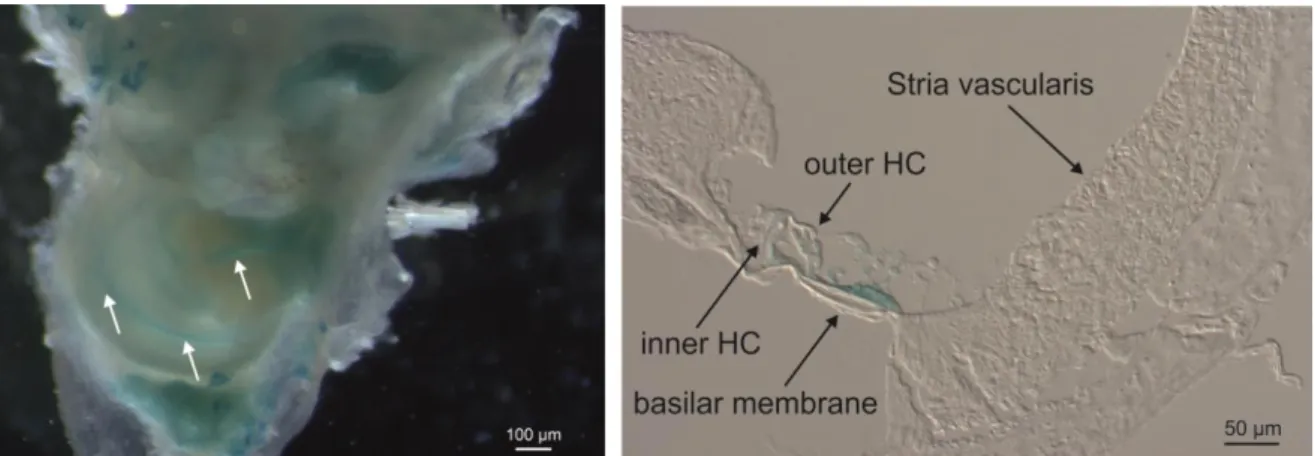
Figure 4. Kir5.1 promotor-driven X-Gal staining of mouse cochlea.
Blue staining indicates promotor-driven X-gal staining in transgenic Kir5.1-/- mice carrying the LacZ gene within the gene locus. The spiral shaped blue color of positive cells was observed in the turns of Kir5.1-/- cochlea (white arrows on the left). Positive staining on a cochlea section was visible in the organ of Corti and hair cells (black arrows on the right). Abbreviation: HC, hair cells; X-Gal, 5-bromo- 4-chloro-3-indolyl-β-D-galactopyranoside; LacZ, lactose operon.
Expression of Kir5.1 in the kidney is mainly found in the distal nephron segments that consists of TAL, DCT and CNT/CCD where it co-localizes with Kir4.1. Due to the nature of Kir5.1, it most likely associates with Kir4.1. Heteromeric Kir4.1-Kir5.1 have been shown to have pH sensitivity in the physiological intracellular range (pKa ~ 7.45). Kir5.1 seems to
-15-
confer an enhanced sensibility to intracellular pH to Kir4.1-Kir5.1 channels.36,39,55,59 The pH sensitivity of Kir4.1-Kir5.1 heteromeric channels in the physiological pH range has led to the assumption that Kir4.1-Kir5.1 might be a good candidate for CO2 chemoreception. The pKa of 7.4 might allow those channels to regulate membrane potentials in response to modest changes in pCO2. Cui et al. discovered that in the oocyte expression system heteromeric Kir4.1-Kir5.1 channels were modulated by changes in pCO2 leading to changes of the membrane voltage. Therefore, the cells could detect states of hyper- and hypocapnia and translate it into a modified membrane excitabiliy. However, the precise contribution of Kir4.1-Kir5.1 to chemosensation is still a matter of debate, since the loss of Kir5.1 appeared to be compensated thereby leaving the ventilatory responses intact.60–62
In Kir5.1-/- mice, it was observed that in the absence of Kir5.1 the remaining K+ channel present in DCT had a conductance of ~25pS instead of ~50pS observed in control animals.
This smaller single channel conductance is typical for homomeric Kir4.1 channels indicating that the physiological K+ channels in mouse DCTs are heteromeric Kir4.1-Kir5.1 channel complexes. Co-expression of Kir4.1 and Kir5.1 subunits lead to the formation of a 40-60 pS single channel K+ conductance and a lower open probability (Po ~ 0.4) when compared with homomeric Kir4.1. 50,51,54 The expression of heteromers in the basolateral membrane of the distal tubule was suggested to be involved in the recycling of the K+ across the basolateral membrane, necessary for maintenance of the activity of Na+/K+-ATPase.31,50,51,53,59 Deletion of KCNJ16 in mice led to a renal phenotype characterized by hypokalemia, hyperchloremic metabolic acidosis with hypercalciuria.54 The authors concluded that disruption of KCNJ16 induces a severe renal phenotype that, apart from hypokalemia, is the opposite of the phenotype seen in EAST/SeSAME syndrome. This phenotype was attributed to increased mean basolateral K+ conductance mediated by the remaining homomeric Kir4.1 channels in the distal nephron.54,60,61,63
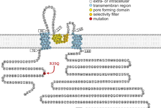
For KCNJ16, unlike KCNJ10, no mutations have yet been associated with human pathologies. However, our collaborator Prof. Dr. Konrad Martin in Münster identified a patient, who suffers from renal salt wasting, hypokalemia and acidosis and has the c.104G>A mutation (NCBI Reference Sequence: NM_001291625.1) within the KCNJ16 gene (unpublished data). This mutation leads to the missense mutation p.Arg35Gln (R35Q) in KCNJ16 (UniProt: Q9NPI9), replacing the charged aliphatic arginine (R) to the neutral and polar amino acid glutamine (Q) (Figure 5). This mutation is located at the N-terminal region of the protein.
-16-
Figure 5. Schematic topology of human Kir5.1 (UniProt Q9NPI9).
Kir5.1 is an inwardly rectifying K+ channel containing two transmembrane regions and a pore-forming region in between the two regions. The missense mutation p.Arg35Gln (R35Q) found in one patient with hypokalemic acidosis is located in the N-terminal region of the protein.
-17-
2. Objectives
Kir5.1 (KCNJ16) potassium channels are strongly expressed in the kidney. Previous work by Paulais and co-workers suggested that Kir5.1 together with Kir4.1 forms a basolateral K+ conductance in distal convoluted tubules (DCT).54 Moreover, these authors provided evidence that genetic inactivation of Kir5.1 leads to disturbed renal salt handling and hypokalemia. Surprisingly and in contrast to the alkalosis observed after inactivation of Kir4.1, knockout mice for Kir5.1 displayed metabolic acidosis. So far, no human disease has been linked to mutations of Kir5.1. Our collaborator Prof. Konrad, Münster, identified in a patient with hypokalemia and acidosis a homozygous missense mutation of Kir5.1 (R35Q) and he asked our group in Regensburg to test if this Kir5.1 mutant can be disease-causing or if it is only a rare variant.
Therefore, my thesis aimed at testing the hypothesis if i) the mutation R35Q leads to impaired channel function and ii) inactivation of Kir5.1 disturbs salt transport in the kidney, especially in the DCT, thereby creating a renal tubulopathy similar to the one observed in Prof. Konrad's patient.
To investigate the functional consequence of the R35Q mutant, I co-expressed Kir5.1 and Kir4.1 in a heterologous expression system and studied the subcellular localization of Kir5.1 and the electrophysiological properties in those transfected cells.
In the second part of my thesis, I studied the consequences of the Kir5.1 gene inactivation in mice in vivo with the focus on renal salt handling, pH homeostasis, K+ induced Ca2+
signaling in DCT cells and respiratory control.
With these sets of experiments, I hoped to gain insights into the pathophysiology of a potentially novel human disease and to establish - or falsify - a genotype/phenotype correlation.
-18-
3. Materials and Methods
3.1 Material
3.1.1 Instruments
Instruments Manufacturer
Blood Gas Analyzer pHOx plusM Nova Biomedical, Rödermark, Germany Cryostar NX70 Thermo Fisher Scientific, Dreieich, Germany Cryostat Leica CM3050 Leica, Wetzlar, Germany
EPC 10 Amplifier Patch-Clamp HEKA Elektronik Dr. Schulze GmbH, Pfalz, Germany
Gas mixer for Isoflurane-Vaporizer MFI Föhr Medical Instruments GmbH, Seeheim, Germany
Incubator Heraeus Instruments, Osteode, Germany
Inverted Microscope Axiovert 10 Zeiss, Jena, Germany Inverted Microscope Axiovert 200 Zeiss, Jena, Germany Inverted Microscope Observer Z.1 Zeiss, Jena, Germany
Ion Chromatography ICS-1600 Dionex Thermo Fisher Scientific, Dreieich, Germany Isoflurane-Vaporizer MFI Föhr Medical Instruments GmbH,
Seeheim, Germany
Laminar Flow Thermo Fisher Scientific, Dreieich, Germany
Light-Cycler LC480 Roche, Mannheim, Germany
Microcentrifuge Hettich, Tuttlingen, Germany
Microtome (Rotary microtome RM2165) Leica, Wetzlar, Germany Millicell® ERS-2 Voltohmmeter Merck, Darmstadt, Germany
Mini Trans-Blot Cell Bio-Rad, Munich, Germany
Mini-PROTEAN Tetra Vertical
Electrophoresis Cell Bio-Rad, Munich, Germany
NanoDrop 2000c Spectrophotometer Thermo Fisher Scientific, Dreieich, Germany NOVOstar Microplate reader BMG Labtech, Ortenberg, Germany
NOVOstar Microplate reader BMG Labtech, Ortenberg, Germany
O2 sensor Oxydig Dräger, Ultra Medical GmbH,
Saarbruecken-Gersweiler, Germany
Paraffin incubator Modell 300, Memmert, Schwabach,
Germany
PatchStar Micromanipulator Scientifica, United Kingdom
Perfusor Precidor 902681, Infors AG, Bottmingen,
Switzerland
Peristaltic perfusion system PP52 Multichannel systems MCS GmbH, Reutlingen, Germany
pH electrode Schott Geräte, Mainz, Germany
Plethysmography chamber EMKA Technologies Company, USA
PowerLab 4/30 ADInstruments Ltd., United Kingdom
PowerPac Basic Power Supply Bio-Rad, Munich, Germany
Roller pump Ismatec SA., Zurich, Switzerland
Scale (EK-600) A&D Instruments Ltd, Tokio, Japan Sound-attenuated chamber Industrial Acoustics, Niederkrüchten,
Germany
-19-
Surgery instruments FST, Bad Oeynhausen, Germany
Thermomixer 5436 Eppendorf GmbH, Hamburg, Germany
Warm plate for mouse operations Dr. J. Barhanin, Nizza, France Warm plate for paraffin HI 1220, Leica, Wetzlar, Germany
Water bath Modell W13, Haake, Karlsruhe, Germany
3.1.2 Laboratory Material
Products Manufacturer
0.5 ml Insulin-syringe
BD Micro-Fine+, BD Consumer Healthcare, Heidelberg, Germany
96-well plate Nuclon Surface, Nunc A/S, Roskilde,
Denmark 96-well Polymerase chain reaction (PCR)
Plate, white Nerbe Plus GmbH, Winsen/Luhe, Germany
Blood gas capillary Hirschmann Labor geräte GmbH & Co. KG, Eberstadt, Germany
Cell culture flasks (T-25, T-75) Nuclon Surface, Nunc A/S, Roskilde, Denmark
Cell Culture Inserts, 0,4µm PCF, 12mm
diameter Merck Millipore, Darmstadt, Germany
Cell culture plates (6-well, 12-well, 24- well)
Nuclon Surface, Nunc A/S, Roskilde, Denmark
Cell scraper TPP Techno Plastic Products AG,
Trasadingen, Switzerland
Coverslips A. Hartenstein GmbH, Würzburg, Germany
DePeX mounting medium Serva Electrophoresis GmbH, Heidelberg, Germany
Epoxy embedding medium Sigma, Taufkirchen, Germany Eppendorf microplestle for 1,2 – 2 mL
tubes A. Hartenstein GmbH, Würzburg, Germany
Eppendorf Tubes (0,2 ml, 0,5 ml, 1,5 ml, 2
ml) Eppendorf AG, Hamburg, Germany
Falcon tubes (15 ml, 50 ml) Greiner bio-one, Frickenhausen, Germany
Filter paper GE Healthcare Life Sciences,
Buckinhamshire, UK GeneRuler™ DNA Ladder (50 bp, 100
bp, 1 kb)
MBI Fermentas GmbH, St. Leon-Rot, Germany
Latex gloves Kimberly-Clark, Roswell, USA
Light-Cycler Capillaries Roche, Mannheim, Germany Millex-GP Syringe Filter Unit, 0.22 µm,
polyethersulfone, 33 mm, gamma sterilized
Merck Millipore, Darmstadt, Germany Millicell® Cell Culture Inserts, 0,4 µm Merck, Darmstadt, Germany
Mortar Rosenthal Technik, Tostedt, Germany
Nanosep Centrifugal VWR International, Darmstadt, Germany Non-fluorescent Glycergel mounting
medium
DakoCytomation, Dakato North America Inc., Carpinteria, USA
Pasteur pipettes Brand, Wertheim, Germany
Pestle Rosenthal Technik, Tostedt, Germany
-20-
Petri dishes (p35, p60, p100 mm) Nuclon Surface, Nunc A/S, Roskilde, Denmark
Pipette tips (10 µl, 200 µl, 1000 µl) Sarstedt, Nümbrecht, Germany Polysine microscope adhesion slides Kindler, Freiburg, Germany Polyvinylidene difluoride (PVDF)
membrane GE Healthcare GmbH, Freiburg, Germany
Serological Pipettes Nerbe Plus GmbH, Winsen/Luhe, Germany Tissue cassettes for paraffin blocks A. Hartenstein GmbH, Würzburg, Germany Tissue Tek OCT-Medium Sakura Finetek Europe B.V., Zoeterwoude,
Netherlands
3.1.3 Substances
Products Manufacturer
0.9% NaCl isotonic solution B. Braun Melsungen AG, Melsungen, Germany
2-Methylbutane Carl Roth GmbH + Co. KG, Karlsruhe,
Germany
2x Laemmli Sample Buffer Bio-Rad, Munich, Germany 4x Laemmli Sample Buffer Bio-Rad, Munich, Germany Acetic acid (glacial) Merck, Darmstadt, Germany
Agarose AppliChem, Darmstadt, Germany
Ammonium acetate Sigma, Taufkirchen, Germany
Ammonium persulfate Sigma, Taufkirchen, Germany
ATP-Na2 (Adenosine 5′-triphosphate
disodium salt) Sigma, Taufkirchen, Germany
BaCl2 Sigma, Taufkirchen, Germany
Bradford Protein Assay Bio-Rad, Munich, Germany BSA (Albumin from bovine serum) Sigma, Taufkirchen, Germany
CaCl2 Carl Roth GmbH + Co. KG, Karlsruhe,
Germany
Ca-Gluconate monohydrate Carl Roth GmbH + Co. KG, Karlsruhe, D
Chloroform Sigma, Taufkirchen, Germany
Citric acid monohydrate Sigma, Taufkirchen, Germany
Creatinine Sigma, Taufkirchen, Germany
D-Mannitol Fluka Chemie GmbH, Buchs, CH
D-Potassium gluconate Sigma, Taufkirchen, Germany
EDTA Sigma, Taufkirchen, Germany
EGTA Sigma, Taufkirchen, Germany
Eosin Y Abcam, Cambridge, UK
Ethanol J. T. Baker, Deventer, Netherlands
FCS (Fetal Calf Serum) Gibco Cell Culture Systems - Invitrogen, Karlsruhe, Germany
Fura-2 AM Invitrogen, ThermoFisher, Darmstadt,
Germany
Glucose Merck, Darmstadt, Germany
Glutaraldehyde solution Sigma, Taufkirchen, Germany
Glycerol Sigma, Taufkirchen, Germany
Glycine Merck, Darmstadt, Germany
-21-
HCl Merck, Darmstadt, Germany
Heparin Liquemin N 25000, 5 ml, Roche, Mannheim,
Germany
HEPES AppliChem, Darmstadt, Germany
IGEPAL CA-630 Sigma, Taufkirchen, Germany
Isoflurane Baxter Deutschland GmbH,
Unterschleißheim, Germany
Isopropanol Merck, Darmstadt, Germany
K2HPO4 Merck, Darmstadt, Germany
K3[Fe(CN)6] Merck, Darmstadt, Germany
K4[Fe(CN)6] Sigma, Taufkirchen, Germany
KCl Merck, Darmstadt, Germany
Ketamine 100mg/mL Sigma, Taufkirchen, Germany
KH2PO4 •3H2O Merck, Darmstadt, Germany
L-Glutamine, 200 mM (100x) Gibco Cell Culture Systems - Invitrogen, Karlsruhe, Germany
Mayer’s hemalun solution Carl Roth GmbH & Co. KG, Karlsruhe, Germany
MEM alpha medium Gibco Cell Culture Systems - Invitrogen, Karlsruhe, Germany
Methanol Merck, Darmstadt, Germany
MgCl2 •6H2O Merck, Darmstadt, Germany
MgSO4 •7H2O Merck, Darmstadt, Germany
Na2HPO4 •2H2O Merck, Darmstadt, Germany
NaCl Merck, Darmstadt, Germany
NaH2PO4 Merck, Darmstadt, Germany
NaH2PO4 •H2O Merck, Darmstadt, Germany
Nonfat-dried milk AppliChem GmbH, Darmstadt, Germany
Opti-MEM Gibco Cell Culture Systems - Invitrogen,
Karlsruhe, Germany
Opti-MEM™, Reduced Serum Medium Thermo Fisher Scientific, Dreieich, Germany
Paraffin Paraplast-Plus Paraffin, Sherwood, St.
Louis, USA
Paraformaldehyde Merck, Darmstadt, Germany
Penicillin-Streptomycin solution
1000 I.E./ml Pen G, 10000 µg/ml Strep.- Sulfat, Gibco Cell Culture Systems - Invitrogen, Karlsruhe, Germany
Periodic acid Thermo Fisher Scientific, Dreieich, Germany
Picric acid Sigma, Taufkirchen, Germany
Polyethylene glycol Sorbitan Monolaurate
(Tween 20) Sigma, Taufkirchen, Germany
Precision Plus Protein™ Dual Color
Standards Bio-Rad, Munich, Germany
Protease Inhibitor Cocktail Set III, EDTA-
Free Merck, Darmstadt, Germany
Rotiphorese® Gel 30 (37,5:1) (30% Acrylamide)
Carl Roth GmbH + Co. KG, Karlsruhe, Germany
Schiff’s reagent Sigma, Taufkirchen, Germany
SDS (dodecyl sulfate) Merck, Darmstadt, Germany
Sodium acetate Merck, Darmstadt, Germany
-22-
Sodium azide Sigma, Taufkirchen, Germany
Sodium citrate dehydrate Merck, Darmstadt, Germany
Sodium deoxycholate Sigma, Taufkirchen, Germany
Sodium Pyruvate, 100 mM solution Sigma, Taufkirchen, Germany
Sucrose Merck, Darmstadt, Germany
TEMED 99%, for electrophoresis Carl Roth GmbH & Co. KG, Karlsruhe, Germany
Tris Hydrochloride Merck, Darmstadt, Germany
Triton X-100 Sigma, Taufkirchen, Germany
TRIzol™ Reagent Thermo Fisher Scientific, Dreieich, Germany Trypsin-EDTA (10x) solution
0,5% Trypsin, 5,3 mM EDTA, Gibco Cell Culture Systems - Invitrogen, Karlsruhe, Germany
Western Blotting Luminol Reagent Santa Cruz, Heidelberg, Germany X-Gal (5-Bromo-4-chloro-3-indolyl-β-D-
galacto-pyranoside) Sigma, Taufkirchen, Germany
Xylol Merck, Darmstadt, Germany
α-Ketoglutarate (α-Ketoglutaric acid) Sigma, Taufkirchen, D
β-Mercaptoethanol Merck, Darmstadt, Germany
3.1.4 Enzyme, Kits
Products Manufacturer
Collagenase Typ II (from Clostridium
histolyticum) Sigma, Taufkirchen, D
DNAse I Roche Diagnostics GmbH, Mannheim,
Germany
dNTP-Mix MBI Fermentas GmbH, St. Leon-Rot,
Germany
M-MLV Reverse Transkriptase Promega, Madison, Wisconsin, USA M-MLV Reverse Transkriptase Puffer (5x) Promega, Madison, Wisconsin, USA Protease (from Streptomyces griseus) Sigma, Taufkirchen, D
Random Hexamer Primer (100 µM) MBI Fermentas GmbH, St. Leon-Rot, Germany
REDTaq® ReadyMix™ PCR Reaction Mix Sigma, Taufkirchen, Germany
RNAse Inhibitor Peqlab Biotechnologie GMBH, Erlangen,
Germany
RNeasy mini kit Quiagen, Hilden, Germany
SYBR® Green PCR Kit Quiagen, Hilden, Germany
Quick Ligase New England Biolabs, Frankfurt, Germany
Trypsin inhibitor Roche Diagnostics GmbH, Mannheim,
Germany
Lipofectamine Thermo Fisher Scientific, Dreieich, Germany
Gel extraction Kit QIAGEN GmbH, Hilden, Germany
NucleoSpin Plasmid Macherey-Nagel GmbH & Co. KG, Düren, Germany
NucleoBond Xtra MIDI Macherey-Nagel GmbH & Co. KG, Düren, Germany
RNeasy Mini Kit QIAGEN GmbH, Hilden, Germany
-23-
3.1.5 Software
Product Manufacturer
ImageJ V1.37c Wayne Rasband, NIH, USA
Labchart® Reader V7.1 ADInstruments, Ltd, Oxford, United Kingdom
IOX v1.8 EMKA Technologies EMKA Technologies Company, USA
Origin 2017 OriginLab Corporation, Northampton, USA
ZEN Pro V2.3 Zeiss, Jena, Germany
Patch Master V2x65 HEKA Elektronik Dr. Schulze GmbH, Pfalz, Germany
Corel draw X8 Corel corporation, Ottawa, Canada
3.1.6 Oligonucleotide
All oligonucleotides were prepared by Invitrogen, Karlsruhe, Germany.
All primers were prepared for Mus musculus, unless otherwise mentioned.
Genotyping of KCNJ16 mice
m = mouse
Gene Primer Sequence Annealing-T.
Kcnj10 (Kir4.1) sense AGTCTTGGCCCTGCCTGT
55 ˚C antisense TTAGCGACCGACGTCATCT
Kcnj16 (kir5.1) sense TACCGCTGTGTCACCGAAGA
62 ˚C antisense GCCAAGGCTGCTCCAATGAT
Kcnj15 (Kir4.2) sense CCCACTGGTGAAGCATACCAA
62 ˚C antisense CGATGACGGTTGTCCACAAGT
Lrp2 (Megalin) sense CAGTCAGTGGCCAAGAATG
57 ˚C antisense CAGTCTCTGGTCCCATCAC
Nphs2 (Podocin) sense CCATCTGGTTCTGCATAAAGG
57 ˚C antisense CCAGGACCTTTGGCTCTTC
Calb1 (Calbindin- D28K)
sense CACAGACCTCATGCTGAAAC
57 ˚C antisense GGTAGTAACCTGGCCATCTC
Slc12a3 (NCC) sense GCTGACCTGCATTCATTCCT
57 ˚C antisense GACCTTGCCGTCCATCAAAG
Slc12a1 (NKCC2) sense TCTGCATCTGCTGCGAAG
61 ˚C antisense GGACACCGTCCCTGAAGC
MCOLN (Mucolipin)
sense AGATGAGGCGGAAACTCAAGT
61 ˚C antisense CCATTCGATCCATGTAGCCCT
Actb (β-Actin) sense CCACCGATCCACACAGAGTACTT
57 ˚C antisense GACAGGATGCAGAAGGAGATTACTG
mKcnj16 wildtype sense CCTCCAGAGCATGCCATCGC
63 ˚C antisense GATCGCTTAATAGGTCTCCG
mKcnj16 knockout sense GCAGCGCATCGCCTTCTATC
60 ˚C antisense TACAAACTCTACTGCCAAAGC