1
Characterization of CDK5RAP2 as a novel regulator in the Hippo signaling pathway
Inaugural-Dissertation zur
Erlangung des Doktorgrades
der Mathematisch-Naturwissenschaftlichen Fakultät der Universität zu Köln
vorgelegt von
Salil Kalarikkal Sukumaran aus Kerala, India
Köln, 2014
2 Vorsitzender: Prof. Dr. H. Arndt
1. Berichterstatter: Prof. Dr. A. A. Noegel 2. Berichterstatter: Prof. Dr. P. Nürnberg
Tag der mündlichen Prüfung: 17 October 2014
3 This research work was carried out from October 2011 to August 2014 at the Centre for Biochemistry, Institute of Biochemistry I, Medical Faculty, University of Cologne, Cologne, Germany, under the supervision of Prof. Dr. Angelika A. Noegel.
Die vorliegende Arbeit wurde in der Zeit von October 2011 bis August 2014 unter der Anleitung von Prof. Dr. Angelika A. Noegel im Institut für Biochemie I der Medizinischen Fakultät der Universität zu Köln angefertigt.
4 Acknowledgements
I would first like to express my deepest gratitude and appreciation to Prof. Dr. Angelika Noegel for the opportunity given to me to carry out this research work in the Institute of Biochemistry I. Without her guidance, endless advices and persistent help, this study would not have been possible. I have been amazingly fortunate to have an advisor who gave me freedom to explore on my own and at the same time the guidance to recover when my steps faltered.
Mrs. Rosemarie Blau-Wasser’s insightful comments and constructive criticisms at different stages of my research were thought provoking and they helped me focus my ideas. I am grateful to her for incorporating me into the D. discoideum field of studies and holding me to high research standards.
I also extend my heartfelt thanks to Prof. Dr. Michael Schleicher (LMU München) and Prof.
Dr. Bernhard Schermer (CECAD, Köln) for their timely support in the provision of reagents during this study. Their presence was inevitable.
I also take the opportunity to thank Dr.Doris Birker and Prof. Carrien Niessen for the moral support, encouragement and kind considerations provided towards my PhD career. I would thankfully remember CECAD, Köln and UniKlinik, Köln for the funding and financial assistance during my PhD studies.
It is such a great honor to be part of an academic research organization, where the members are amenable. Thanking you for providing a very warm environment. To be specific, my appreciation also goes to all members of Lab. 14 and of the Institute of Biochemistry I which includes Napoleon (alias Napo), Maria Marco, Sarah, Sonja, Vivek, Ping Li, Atul and Ilknur.
I cannot forget Mr. Berthold Gassen for all the great antibodies he provided during the course of this study. Mrs. Maria Stumpf is not left out for their technical support with the southern and phosphorylation experiments. Members of the other laboratories in the Biochemistry Institute are too numerous to mention who were in one way or the other of assistance to me during the research are also appreciated.
Most importantly, none of this would have been possible without the love and patience of my family. My immediate family, to whom this work is dedicated to, has been a constant source of love, support, concern and strength all these years. I want to thank members of my family especially my parents, my grandmother and my elder sister and her husband, who engineered
5 my trip to Germany from India for this course. Thank you for allowing me to follow my ambitions throughout my childhood. My extended family members Mr & MrsVenugopalan, Keerthy, and Harikrishnan have always been understanding. They have endlessly helped to move on through my research work. No words can express the contributions of my wife, Vidya Salil, for her tolerance, endless encouragement, care and assistance. I thank her for moving along with me to a country, to be on my side and for the delicious homely food. My son Unnikuttan, who was born during this study, also added new life to this project. I would forget all my strain and stress when I am with him. Many friends have helped me stay happy through this study period. Their support and care helped me to overcome setbacks and stay focused. I am grateful to the Indian families who helped me to adjust and adapt in a new country. I would not forget to thank my friends, Mr. and Mrs. Ram Kumar and Mr and Mrs.
Ganapathi, who are very much, like family members to me.
Above all, I give almighty God for all the blessings that he has given me, including the opportunity to finish this study. Thank you Lord for everything.
6
Table of contents
Abbreviations 10
1. Introduction 12
1.1 Centrosome and centriole biology 13
1.2 Autosomal recessive primary microcephaly 15
1.3 Cyclin Dependent Kinase 5 regulatory subunit-associated protein 2 18
1.3.1 Physiological functions of CDK5RAP2 19
1.4 Hippo signaling pathway 20
1.4.1 Hippo signaling in mammalian organ size determination 22
1.4.2 Hippo signaling at the centrosome 23
1.5 Dictyostelium discoideum as a model organism 24
1.6. Aim of this study 26
2. Results 27
2.1 Identification and characterization of the pericentriolar matrix protein CEP161,
the D. discoideum ortholog of CDK5RAP2 27
2.1.1 Identification of CEP161 as an interacting partner of CP250 27
2.1.2 CEP161 domain and sequence alignment 28
2.1.3 CEP161 is a centrosomal protein 29
2.1.4 CEP161 constructs and truncated proteins 30
2.1.5 Localisation of CEP161 truncated proteins 32
2.2 CEP161 mutant cells are viable, but impaired in growth and development 33 2.2.1 CEP161 regulates nuclear number and centrosome positioning 33 2.2.2 The nuclear-centrosome ratio is altered in the mutants 34
2.2.3 CEP161 regulates size 35
2.2.4 Role of CEP161 for growth 36
7
2.2.5 CEP161 affects development 38
2.2.6 CEP161 affects unicellular motility and polarity 41
2.2.7 CEP161 affects directed migration of slugs 42
2.3 Identification of CEP161 as an interacting partner of the Hippo homolog-
Hrk-Svk 44
2.4 Interaction of CDK5RAP2 with the mammalian Hippo homolog MST1 47 2.4.1 The N-terminal part of CDK5RAP2 is sufficient for targeting to the centrosome 47
2.4.2 Overexpression of Myc-tagged CDK5RAP2 49
2.4.3 CDK5RAP2 interacts with Hippo homolog MST1 49
2.4.4 CDK5RAP2 colocalises with MST1 51
2.5 CDK5RAP2 is a novel regulator of Hippo signaling 52
2.5.1 TAZ interacts with the N-terminal part of CDK5RAP2 53 2.5.2 Ectopic expression of CDK5RAP2 downregulates the expression of TAZ 54 2.5.3 CDK5RAP2 does not influence the interaction of TAZ with 14-3-3 57
3. Discussion 60
4. Materials and methods 66
4.1 Kits 66
4.2 Enzymes, antibodies and antibiotics 66
4.2.1 Enzymes for molecular biology 66
4.2.2 Primary Antibodies 67
4.2.3 Secondary Antibodies 67
4.2.4 Antibiotics 68
4. 3 Media and Buffers 68
4.3.1 Buffers and Solutions 68
4.3.2 Bacteria medium and agar plates 69
4.3.3 Yeast medium 70
8
4.4 Media and buffers for Dictyostelium cultures 72
4.5 Bacteria, D. discoideum, and yeast strains 73
4.6 Oliognoucleotides 73
4.7 Dictyostelium vector construction and transfection 74 4.8 Cloning of CEP161 genomic DNA and expression of recombinant proteins 74
4.9 Growth and development 75
4.10 Generation of Antibodies 75
4.11 Immunofluorescence analysis for D.discoideum cells 76 4.12 Pull down and immunoprecipitation assays for D.discoideum 77 4.13 Analysis of D. discoideum nuclear and centrosome abnormalities 77
4.14 D. discoideum cell adhesion assay 77
4.15 D. discoideum cell migration studies 78
4.16 Phototaxis assay 78
4.17 Yeast Two-Hybrid Interaction 79
4.18 Mammalian cell culture, constructs and transfection 79
4.19 Immunofluorescence 80
4.20 Immunoprecipitation 80
4.21 Fractionation of cytoplasmic and nuclear proteins 82
4.22 Protein extraction from mammalian cells 82
4.23 Western blotting 83
4.24 Luciferase assay 84
4.25 RNA isolation and cDNA generation for quantitative RT-PCR analysis 85
4.26 Mass spectrometry analysis 86
4.27 Miscellaneous experiments 86
4.27.1 D.discoideum cell size 86
4.27.2 Protein sequence alignment 86
9
5. Summary 87
6. Reference 89
7. Erklärung 99
8. Curriculum vitae 100
10 Abbreviations
cAMP cyclic-Adenosinemonophosphate Dd Dictyostelium discoideum
DNA Deoxyribonucleic acid
EDTA Ethylenediaminetetraacetic acid GST Glutathion-S-Transferase
IPTG Isopropyl-thio-galactoside kDa Kilo Dalton
min Minute mM Millimolar
M Molar
NIH National Institutes for Health PAGE Polyacrylamide Gel electrophoresis
PCR Polymerase chain reaction PMSF Phenylmethylsulfonyl fluoride Q-RTPCR Quantitative real time PCR
SDS Sodium dodecyl sulphate TAE Tris-Acetate-EDTA-Buffer
Tris Tris(hydroxymethyl)aminomethane
11 TRITC Tetramethylrhodamine isothiocyanate
v/v volume per volume
X-Gal 5-bromo-4-chloro-3-indolyl-β-D-Galactopyranoside
12 1. Introduction
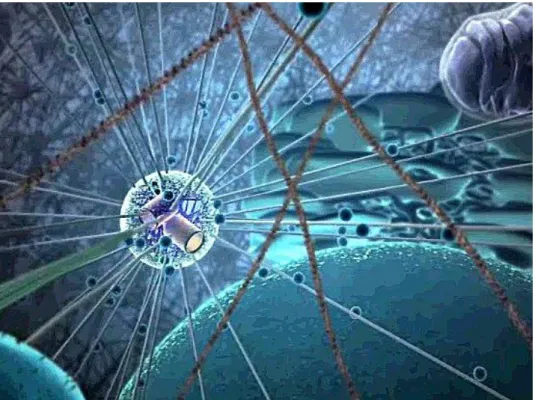
The centrosome is the main microtubule organizing center (MTOC) and plays an important role in mitotic spindle orientation and genome stability (Bornens, 2012). It nucleates, anchors, and organizes microtubules. In animal cells it comprises a pair of centrioles and a surrounding pericentriolar matrix (PCM) (Fig. 1). The PCM is a key structure of the centrosome and is responsible for microtubule nucleation and anchoring. The major components of the PMC are large coiled coil proteins like pericentrin and proteins of the AKAP450 family that provide docking sites for γ-tubulin ring complexes and regulatory molecules. They allow the centrosome to function as MTOC and to carry out its regulatory functions during cell cycle transitions, cellular responses to stress, and organization of signal transduction pathways (Keryer et al., 2003; Doxsey et al., 2005).
Figure 1. An artistic view of the MTOC with its star pattern of microtubules radiating from the cylindrical centrioles to all parts of the cell. Source: Studiodaily, New York.
13 1.1 Centrosome and centriole biology
The centrosome was considered as the organ coordinating karyokinesis and cytokinesis. The absence of the centrosome in an unfertilized egg was seen as an efficient way to avoid parthenogenetic development (Bornens, 2012). The centrosome morphology varies largely in protozoa, algae, and fungi. In Dictyostelium discoideum it is a nucleus associated body consisting of a box-shaped core surrounded by the corona, an amorphous matrix functionally equivalent to the PCM (Ueda et al., 1999). The centrosome of animal cells is a cytoplasmic organelle formed around a core structure made of two microtubule-based cylinders of defined length and diameter, the centrioles, with a highly conserved nine fold radial symmetry (Bobinnec et al., 1998). The centriole pair comprises of a mature mother centriole and a young daughter centriole. It displays structural and functional asymmetry due to the generational difference between each member of the pair: The old, fully mature mother centriole is distinguished by two sets of nine appendages at its distal end whereas the young, immature, daughter centriole, assembled during the previous cell cycle, is about 80% the length of the mother centriole (Piel et al., 2000). Both centrioles are connected together by a linker comprising of various centrosomal proteins, the major ones being C-Nap1 and Rootletin. More recently, CDK5RAP2 was identified as a putative linker component which surrounds the centrioles (Graser et al., 2007).
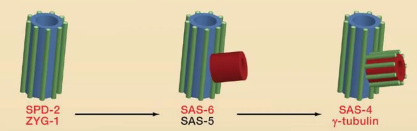
Studies on early C. elegans embryos with genetic and RNA interference based approaches gave major insight into molecular mechanisms of centriole biogenesis (Leidel et al., 2005;
Leidel and Gönczy, 2003; O’Connell et al., 2001). These studies revealed an ordered assembly pathway that involves the products of just five essential genes, termed zyg-1 (Zygote defective-1), spd-2 (Spindle defective-2), and three Spindle assembly genes sas-4, sas-5, and sas-6 (Delattre et al., 2006; Pelletier et al., 2006). SAS-4, SAS-5, SAS-6, and SPD- 2 are coiled-coil proteins, whereas ZYG-1 is a protein kinase. Shortly after fertilization of the
14 egg, SPD-2 is recruited to the paternal centrioles, which then allows the centriolar recruitment of ZYG-1. Next, a complex comprising SAS-5 and SAS-6 is recruited, which leads to the formation of a “central tube” that is closely associated with the original centriole. The central tube encompasses the highly conserved nine fold radial symmetry of the centriole/basal body (Kitagawa et al., 2011). In most species, the centriole is organized around a cartwheel that comprises a central hub of 25 nm in diameter from which nine spokes radiate outward and connect to nine microtubule blades (Strnad and Gönczy, 2008). The major molecular architect of the central hub of the cartwheel is found to be SAS-6, the oligomerisation properties of which help in self-assembling to form 9 radiating spokes (Kitagawa et al., 2011). Finally, SAS-4 facilitates the assembly of microtubules onto the periphery of this tube, resulting in the formation of a procentriole (Pelletier et al., 2006) (Fig. 2).
Figure 2. Centriole duplication in C. elegans embryos. SPD-2 recruits the protein kinase ZYG-1 to mother centrioles, which then recruits a complex of SAS-6 and SAS-5. This promotes the formation of a central tube (red) at right angles to the mother centriole. SAS-6 and SAS-5 then recruit SAS-4, which allows the centriolar microtubules (green) to associate with the central tube, thus forming the procentriole. The protein γ-tubulin is also required to convert the structure in to a procentriole. The proteins highlighted in red all have functional orthologs implicated in centriole duplication in other species (adapted from (Nigg and Raff, 2009).
Importantly, the significance of these findings is not limited to C. elegans. Although an ortholog of SAS-5 awaits definitive identification, SPD-2, SAS-4, and SAS-6 clearly have
15 orthologs in human cells termed Cep192 (Andersen et al., 2003), CPAP/HsSAS-4 (Hung et al., 2000), and HsSAS-6 (Leidel et al., 2005), respectively. Curiously, ZYG-1 does not have obvious structural orthologs outside of nematodes, but the available evidence suggests that PLK4 plays a functionally analogous role in human cells (Bettencourt-Dias et al., 2011;
Habedanck et al., 2005).
In the last decade, the molecular composition of the isolated centrosome in the human and fly has revealed a number of proteins conserved across eukaryotes (Azimzadeh and Marshall, 2010; Carvalho-Santos et al., 2011). For the protein inventory of the Dicytostelium centrosome more than 70 new candidates have been identified (Reinders et al., 2006). In higher vertebrates, sequence database searches resulted in the identification of more than 2,000 peptides representing more than 500 proteins in the peak centrosome fraction (Andersen et al., 2003). Functional genomics in nematodes and flies has identified a small set of conserved proteins required for the initiation of centriole/basal body assembly and for centrosome reproduction.
1.2 Autosomal recessive primary microcephaly
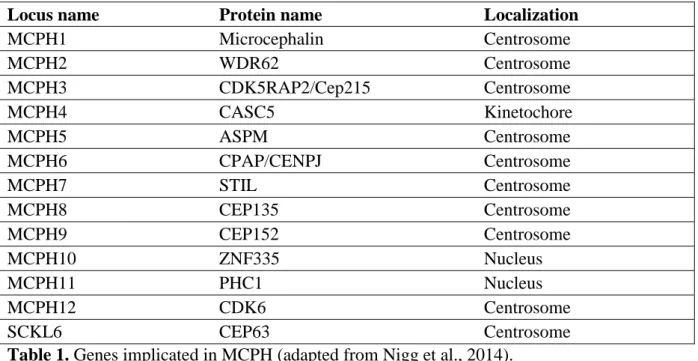
The cellular regulation by centrosomes has been well studied. Increasing evidence indicates that this structure may be well designed for the organization of multiprotein scaffolds that can anchor a diversity of activities ranging from protein complexes involved in microtubule nucleation to multicomponent pathways in cellular regulation (Doxsey et al., 2005). The centrosome is an indispensable component of the cell-cycle machinery of eukaryotic cells, and perturbation of core centrosomal or centrosome-associated proteins is linked to cell-cycle misregulation and cancer (Badano et al., 2005). The structural complexity of the centrosome is reflected in its biochemical composition with a large number of proteins localized either in the centrioles or in the centrosomal matrix among which several are disease gene products
16 (Nigg and Raff, 2009; Kobayashi and Dynlacht, 2011). Aberrations in several centrosomal proteins are also reported in primary microcephaly (Table 1).
Locus name Protein name Localization
MCPH1 Microcephalin Centrosome
MCPH2 WDR62 Centrosome
MCPH3 CDK5RAP2/Cep215 Centrosome
MCPH4 CASC5 Kinetochore
MCPH5 ASPM Centrosome
MCPH6 CPAP/CENPJ Centrosome
MCPH7 STIL Centrosome
MCPH8 CEP135 Centrosome
MCPH9 CEP152 Centrosome
MCPH10 ZNF335 Nucleus
MCPH11 PHC1 Nucleus
MCPH12 CDK6 Centrosome
SCKL6 CEP63 Centrosome
Table 1. Genes implicated in MCPH (adapted from Nigg et al., 2014).
Autosomal recessive primary microcephaly, often shortened to MCPH which stands for
"microcephaly primary hereditary", is a condition in which infants are born with a very small head and a small brain (Fig. 3). The term "microcephaly" comes from the Greek word for
"small head". The MCPH-associated genes described to date have been implicated in cell division and cell cycle regulation and many of them have been localized to the centrosome (Kaindl, 2014).
17 Figure 3. MRI images of a patient at 15 months (on the left) compared to a normal age and gender matched child (on the right) demonstrating cranio-facial disproportion characteristic of microcephaly (adapted from Pagnamenta et al., 2012).
MCPH is a rare neurodevelopmental disorder that results in severe microcephaly at birth with pronounced reduction in brain volume, particularly of the neocortex, simplified cortical gyration and intellectual disability (Issa et al., 2013b). Mutations in MCPH genes reduce the population of neurons in each of the six layers of the cerebral cortex during development, but there is little or no overt structural abnormalities other than reduced thickness of the cerebral cortex (Megraw et al., 2011). Patients with mutations in MCPH loci show moderate to severe microcephaly. Some of the notable phenotypes of microcephaly patients are decrease in size of head and brain volume (Bond et al., 2003, 2005), sensorineural hearing loss (Pagnamenta et al., 2012), short stature, seizures (Darvish et al., 2010) and mental retardation (Neitzel et al., 2002), but no motor deficit.
Mutations in the ASPM (Abnormal spindle-like microcephaly associated) gene is the most frequent cause of MCPH. Analysis of mouse and zebrafish mutants for Aspm demonstrates that Aspm is crucial for maintaining a cleavage plane orientation that allows symmetric, proliferative divisions of neuro-epithelial cells during brain development. Failure to do so leads to microcephaly disease condition (Pulvers et al., 2010; Fish et al., 2006; Novorol et al., 2013). Cdk5rap2an/an mutant mice (Hertwig's anemia mouse, in which an inversion of the exon 4 of Cdk5rap2 gene leading to the deletion of a large part of the gamma-tubulin ring complex binding domain) showed a dramatic reduction in brain size and thin superficial cerebral cortex layers and rarely survived beyond 1 week of age. The Cdk5rap2 mutants exhibited a significant decrease in the ratio of brain to body weight as compared with controls (Lizarraga et al., 2010). It has been reported that CDK5RAP2 shares homology at the amino terminus with several eukaryotic proteins, including Schizosaccharomyces pombe Mto1p (also known as Mbo1p and Mod20p) and Pcp1p, and Drosophila melanogaster centrosomin (Cnn), which
18 have been shown to function in the recruitment of γ-tubulin to MTOCs through interacting with γTuRCs (Fong et al., 2008). Mutation in the CDK5RAP2 homolog in Drosophila, Cnn, led to abnormalities in asymmetric division in the larval brain (Wakefield et al., 2001).
Recently, cerebral organoid cultures with a mutation in Cdk5rap2 led to the observation of smaller embryoid bodies, which when subjected to neural induction failed to develop further (Lancaster et al., 2013). The centrosomal localization of the MCPH-associated proteins, particularly during the cell cycle, underlines the role of the centrosome in the etiology of this disease (Megraw et al., 2011).
1.3 Cyclin Dependent Kinase 5 regulatory subunit-associated protein 2 (CDK5RAP2) The human CDK5RAP2 gene is located on chromosome 9, has 38 exons spanning a region of 191 kb, and the deduced open reading frame encompasses 6130 bp encoding 1893 amino acids. So far four isoforms produced by alternative splicing have been reported: Isoform 1, the full-length form (UniProt ID: Q96SN8-1); Isoform 2, missing residues 702–733 at exon 19 (UniProt ID: Q96SN8-2); Isoform 3, missing residues 1009–1049 at exon 23 (UniProt ID:
Q96SN8-3); Isoform 4, missing residues 1576–1654 at exon 32 (UniProt ID: Q96SN8-4) (Kraemer et al., 2011). To date, four different mutations have been identified in CDK5RAP2:
a nonsense mutation in exon 4 (c.246T > A, p.Y82X), an A to G transition in intron 26 (c.4005-15A > G, p. R1334SfsX5) introducing a new splice acceptor site, a frame shift and a premature stop codon, a nonsense mutation in exon 8 (c.700G > T, p.E234X) and a nonsense mutation, c.4441C > T (p.Arg1481) introducing a premature stop codon (Bond et al., 2005;
Hassan et al., 2007; Pagnamenta et al., 2012b; Issa et al., 2013b).
19 Figure 3. Schematic representation of the domain structure of CDK5RAP2. γTuRC:
Gamma-Tubulin ring complex, SMC: Structural maintenance of chromosomes, EB1: End binding 1, CDK5R1: Cyclin dependent kinase 5 activator 1, CM2: Cnn Motif 2 (picture modified from Issa et al. 2013b).
The human CDK5RAP2 protein (NP_060719.4) contains a predicted N-terminal interaction site for the gamma tubulin ring complex (γTuRC) (Fong et al., 2008), two structural maintenance of chromosome (SMC) domains, a Ser-rich motif for interaction with plus-end tubulin binding protein EB1 (Fong et al., 2009), an interaction site for Cyclin dependent kinase 5 activator 1 (CDK5R1) (Wang et al., 2000) and Pericentrin (Buchman et al., 2010), and a CM2 (Cnn Motif 2) domain which is responsible for sequestering it to the centrosome and the Golgi apparatus which has a pericentrosomal location. The CM2-like sequence of CDK5RAP2 exhibits Ca2+-independent calmodulin binding activity (Wang et al., 2010).
1.3.1 Physiological functions of CDK5RAP2
CDK5RAP2 is a pericentriolar structural component that functions in γTuRC attachment and therefore in the microtubule organizing function of the centrosome (Fong et al., 2008). The EB1 domain of CDK5RAP2 interacts with the growing microtubule tips and hence regulates microtubule dynamics (Fong et al., 2009). Furthermore, CDK5RAP2 also regulates mitotic spindle positioning, asymmetric centrosome inheritance, DNA damage signaling (Barr et al., 2010) and also has spindle checkpoint function (Zhang et al., 2009b). In addition, CDK5RAP2 is reported to restrict centriole replication by maintaining centriole engagement and cohesion (Barrera et al., 2010). Besides, CDK5RAP2 regulates chromosome segregation in neuronal progenitors (Lizarraga et al., 2010). In conjunction with further in vitro studies, CDK5RAP2 function specifically contributes to centrosome asymmetry (Conduit and Raff,
20 2010), centriole replication control and primary cilium formation (Barrera et al., 2010), kinetochore attachment to spindles and checkpoint control (Zhang et al., 2009b), binding to the dynactin complex (Lee and Rhee, 2010), cell-cycle regulation and cell-cycle exit (Buchman et al., 2010), microtubule plus-end dynamics (Fong et al., 2009), mitotic spindle formation, function and orientation (Lizarraga et al., 2010).
From the human phenotype and animal models, it has become clear that CDK5RAP2 has an influence on brain size regulation during fetal development (Issa et al., 2013a). Nonsense and splice mutations in CDK5RAP2 result in truncated, non-functional proteins in microcephaly patients (Issa et al., 2013b). Some of the notable patient phenotypes resulting from CDK5RAP2 mutations are sensorineural hearing loss, intellectual disability and a reduced occipital frontal head circumference (Pagnamenta et al., 2012; Issa et al., 2013; Tan et al., 2014). Whether mutations in CDK5RAP2 and the decrease in neuronal cell density are associated with altered signal transduction pathways is not known.
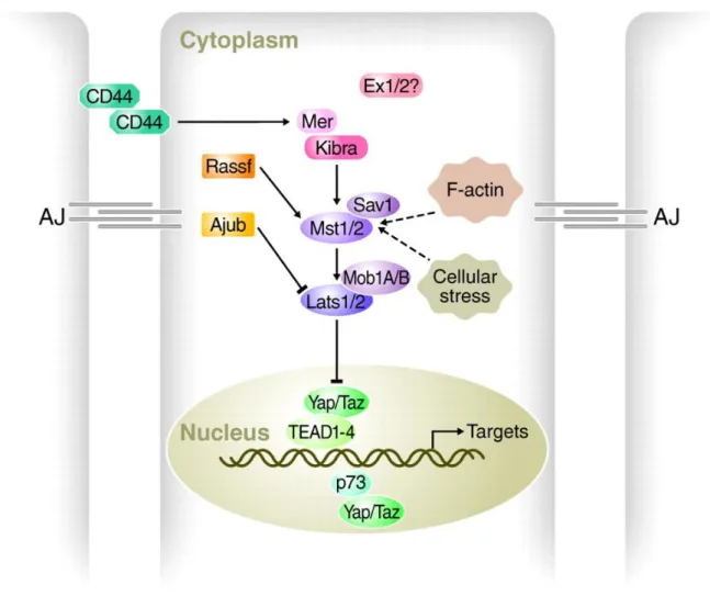
1.4 Hippo signaling pathway
The Hippo signaling pathway is a tumor-suppressive pathway and is inactive at low cell density (Mori et al., 2014). It primarily affects the number of cells produced and has only minor effects on tissue patterning (Harvey et al., 2003) and is known as a key regulator of organ growth and tissue size in Drosophila and mouse (Udan et al., 2003; Lee et al., 2010; Lu et al., 2010; Halder and Johnson, 2011). At the center of the Hippo pathway is a core kinase cassette that consists of a pair of related serine/threonine kinases, mammalian STE20-like protein kinase 1 (MST1 and MST2) which are homologues of D. melanogaster Hippo (HPO), and large tumour suppressor 1 (LATS1) and LATS2 together with the adaptor proteins Salvador homologue 1 (SAV1) and MOB kinase activator 1A (MOB1A) and MOB1B (Harvey et al., 2003; Udan et al., 2003; Pantalacci et al., 2003; Tapon et al., 2002; Justice et al., 1995; Kango-Singh, 2002). These proteins limit tissue growth by facilitating LATS1- and
21 LATS2-dependent phosphorylation of the homologous oncoproteins Yes-associated protein (YAP) and Transcriptional co-activator with PDZ-binding motif (TAZ) (Huang et al., 2005) (Fig. 4).
Figure 4. Schematic of the Hippo pathway in mammals. Cells (outlined in grey, nuclei in green) are shown with adherens junctions (AJ). Hippo pathway components in vertebrates are shown in various colors, with pointed and blunt arrowheads indicating activating and inhibitory interactions, respectively. Continuous lines indicate direct interactions, whereas dashed lines indicate unknown mechanisms (adapted from Halder and Johnson, 2011).
Phosphorylation of YAP and TAZ represses their activity by creating binding sites for the protein 14-3-3. The name 14-3-3 was initially coined by Moore and Perez in 1967 with regard to the particular elution and migration pattern of these proteins on DEAE-cellulose chromatography and starch-gel electrophoresis. The 14-3-3 proteins eluted in the 14th fraction of bovine brain homogenate and were found on positions 3.3 of subsequent electrophoresis
22 (Moore and Perez., 1967). The interaction of YAP/TAZ with 14-3-3 causes them to accumulate in the cytoplasm and that also stimulates their ubiquitin-mediated proteolysis (Zhao et al., 2007; Dong et al., 2007). YAP and TAZ and their D. melanogaster counterpart Yorkie (YKI) do not contain intrinsic DNA-binding domains but instead bind to the promoters of target genes through interaction with transcription factors. They promote tissue growth and cell viability by regulating the activity of different families of transcription factors, including Transcriptional enhancer factor domain (TEAD) and Similar to mothers against decapentaplegic (SMAD) family members (Hong et al., 2005). TEADs seem to be key mediators of growth and the tumorigenic potential of YAP, TAZ and YKI, but the genetic program that these factors regulate to drive tissue growth is not well defined (Hong et al., 2005).
The Hippo pathway is conserved throughout evolution and key kinase components have also been detected in D. discoideum (Rohlfs et al., 2007; Artemenko et al., 2012). In Drosophila, loss - of - function mutant clones for any of the genes Warts, Salvador, Hippo and Mats lead to a strong tissue overgrowth phenotype characterized by increased proliferation and diminished cell death.
1.4.1 Hippo signaling in mammalian organ size determination
In mammals, the first studies that connected Hippo signaling to organ size control employed a Yap overexpression strategy that mimicked pathway inactivation and showed that the induction of Yap expression in the adult mouse liver led to a dramatic three- to fourfold increase in liver mass as a result of increased cell numbers (Dong et al., 2007). Remarkably, when Yap overexpression was terminated, the liver rapidly reverted to its normal size, suggesting that intrinsic size control mechanisms were then activated, presumably to reduce cell numbers through an apoptotic process (Dong et al., 2007). These studies infer that Hippo signaling is crucial for regulating the size of the mammalian liver, and that it acts through a
23 mechanism that integrates global organ size control signals with the regulation of Yap.
However, although the effect of Yap overexpression on liver size is dramatic, whether Hippo signaling is required to maintain liver size was not addressed by these studies. These findings show that Hippo signaling functions in regulating the size of the liver, despite this it appears that Hippo signaling, at least the one mediated by the Mst1 and Mst2 kinases, does not regulate the size or growth of other mammalian tissues to the same degree (Song et al., 2010).
1.4.2 Hippo signaling at the centrosome
The regulation of centrosomal biology by the Hippo components is presently a topic of intense research. The components of Hippo signaling pathway regulate the association of Nima-related Kinase 2 (NEK2) with the centrosome (Mardin et al., 2010). Nek2 is the direct substrate for phosphorylating the centrosomal linker proteins C-Nap1 (centrosomal Nek2- associated protein 1) and Rootletin which causes their dissociation from centrosomes and enables centrosome separation (Fry et al., 1998; Mayor et al., 2002; Bahe et al., 2005). It has also been demonstrated that Mst2 phosphorylates Nek2A, thereby recruiting Nek2A to centrosomes and promoting phosphorylation and displacement of centrosomal linker proteins.
Additionally, it was shown that the hSav1–Mst1/Mst2–Nek2A pathway cooperates with forces provided by the kinesin motor Eg5 to allow centrosome separation and bipolar spindle formation (Mardin et al., 2010). Strikingly, certain Hippo pathway components, including the Lats and Mst1/Mst2 kinases and the scaffolding protein hSav1, localize to centrosomes (Guo et al., 2007). In addition, Mst1 was reported to control centrosome duplication (Hergovich et al., 2009). Another important member of the hippo pathway, hMOB1, also plays a role in centrosome duplication (Hergovich et al., 2009) and localises to the centrosomes (Wilmeth et al., 2010). LATS2 was also observed at the centrosome throughout cell cycle and was associated with Ajuba during mitosis which regulates spindle organization through recruitment of γ-tubulin to centrosomes (Abe et al., 2006).
24 1.5 Dictyostelium discoideum as a model organism
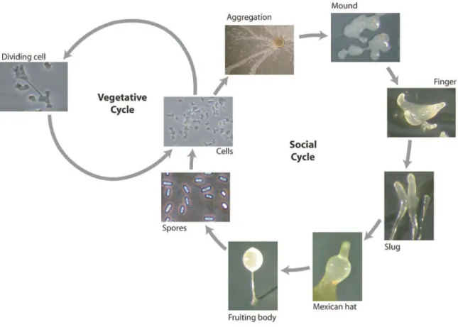
D. discoideum amoebae belong to the monophyletic group of mycetozoans that stands close to animals and fungi. Dictyostelium is a simple eukaryote that lives in the soil and feeds on bacteria by phagocytosis. The life cycle of Dictyostelium consists of growth and developmental phases. The individual cells divide by binary fission as long as food is present, however, when bacteria are exhausted, starvation triggers a process of chemotaxis driven by cyclic-AMP (cAMP) (Willard and Devreotes, 2006; Janetopoulos and Firtel, 2008). The cAMP signaling stimulates the single cell amoebae to aggregate to form multicellular structures leading to the development of fruiting body consisting of a stalk and a spore head filled with spores, and the release of the spores (Fig. 5).
Figure 5. D. discoideum life cycle. Representative pictures of vegetative and developmental stages are shown (picture adapted from Calvo-Garrido et al., 2010).
25 In the wild, Dictyostelium amoeba feed on soil bacteria by phagocytosis but most laboratory strains are also able to grow in liquid axenic media by macropinocytosis. It is important to point out that cells are haploid throughout these vegetative and developmental cycles and this facilitates the generation of knockout strains (Calvo-Garrido et al., 2010). Despite its simplicity, Dictyostelium shows striking similarities with higher eukaryotes in many biological aspects including chemotaxis (Van Haastert and Veltman, 2007), developmental signaling pathways (Strmecki et al., 2005), the response to bacterial infections (Steinert and Heuner, 2005), the response to therapeutic drugs (Alexander et al., 2006) and programmed cell death including autophagic cell death (Giusti et al., 2009). Dictyostelium is an alternative model organism for centrosome research in addition to yeast and animal cells. With the elucidation of morphological changes and dynamics of centrosome duplication, the establishment of a centrosome isolation protocol, and the identification of many centrosomal components, there is a solid basis for understanding the biogenesis and function of this organelle (Gräf et al., 2004).
The Dictyostelium genome has been fully sequenced (Eichinger et al., 2005) and well- annotated data are publicly available (http://dictybase.org/). Moreover, it is amenable to a wide range of molecular genetic techniques including the generation of mutants by homologous recombination and random genetic screens, that have facilitated the comparative genomics to identify relevant genes conserved in the human genome (Torija et al., 2006).
Thus, NIH has cited Dictyostelium discoideum as a non-mammalian model organism for biomedical research (http://www.nih.gov/science/models/).
26 1.6 Aim of this study.
The aim of this study is to characterise the D.discoideum CEP161 protein which we initially identified as a binding partner of CP250, a novel centrosomal protein with important roles in growth and development (Blau-Wasser et al., 2009). CEP161 was characterised based on following features:
1. Homology to proteins from other species 2. Subcellular localization.
3. Analysis of CEP161 in various physiological functions of the cell ranging from size, nuclear number, centrosome-nuclear distance and ratio, growth, development, motility to multicellular migration.
4. Its ability to interact with Dd Hippo related kinase-severin kinase.
The final part of the study was focused on the interaction of human CDK5RAP2 with MST1 and TAZ and hence to characterize the role of CDK5RAP2 in the Hippo signaling pathway.
27 2. Results
2.1 Identification and characterization of the pericentriolar matrix protein CEP161, the D. discoideum ortholog of CDK5RAP2
2.1.1 Identification of CEP161 as an interacting partner of CP250
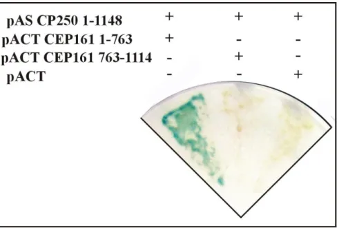
We identified CEP161 as interaction partner of the centrosomal protein CP250 in immunoprecipitation experiments using GFP-CP250, a component of the pericentriolar matrix, followed by mass spectrometry analysis (Blau-Wasser et al., 2009). We then expressed different parts of CEP161 as GST fusion proteins to identify its binding site for CP250 and found that we could pull down GFP-CP250 from whole cell lysates with amino terminal sequences 1-763 (data not shown). A direct interaction of the proteins was shown by yeast-two-hybrid experiments in which the N-terminal sequences of CEP161 (residues 1-763) interacted with residues 1-1148 of CP250 (Fig. 6).
Figure 6. N-terminal part of CEP161 interacts with CP250. Yeast-2-hybrid analysis showing the direct interaction of pAS-CP250 (residues 1-1148) with pACT-CEP161 (residues 1-763), whereas pACT-CEP161 (residues 763-1114) did not show any interaction. The Y2H analysis for pAS-CP250 (residues 1-1148) with empty vector pACT was used as a negative control.
28 2.1.2 CEP161 domain and sequence alignment
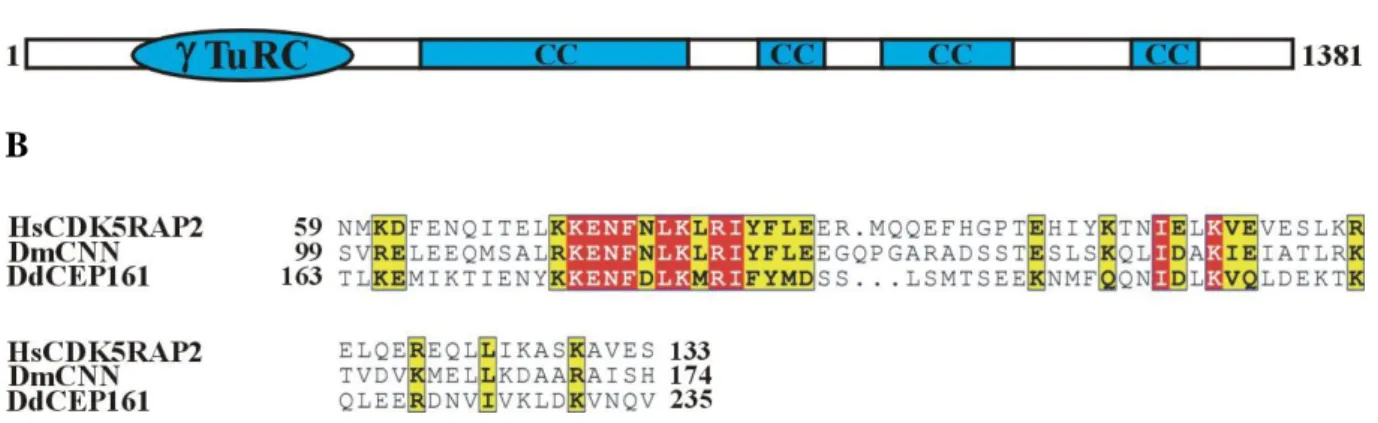
The gene encoding CEP161 (DDB_G0282851) is located on chromosome 3 and has 2 exons.
The open reading frame comprises 4146 base pairs and codes for a protein of 1381 aa with a molecular mass of 161,600. The protein was named CEP161 based on its molecular mass and location (see below). The BLAST prediction program revealed an N-terminal -Tubulin ring complex (-TuRc) domain (residues 99-174); the SMART prediction tool indicated the presence of four coiled-coil domains in the protein (Fig. 7A). The -TuRc domain (pfam07989) is present in several microtubule associated proteins which also include CDK5RAP2. The highest conservation is in a consensus ten amino acid motif as shown for the human, Drosophila and D. discoideum protein (Fig. 7B).
A
B
Figure 7. CEP161 domain and sequence alignment. A. CEP161 domain structure. B.
CDK5RAP2 -TuRc domain sequence alignment and the protein accession numbers for the Human (Hs) (NP_060719), Drosophila melanogaster (Dm) (NP_725298.1) and D.
discoideum protein (Dd) (DDB_G0282851). The numbers indicate the amino acid position of the -TuRc domain in the respective proteins. Colour code: Red background shows the residues which are strictly conserved in the column, yellow background shows the residues which are conserved within a group but not conserved from one group to the other.
29 2.1.3 CEP161 is a centrosomal protein.
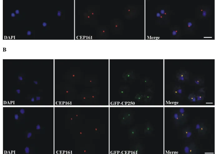
To determine the subcellular localisation of CEP161, we generated monoclonal antibodies against a recombinant polypeptide (CEP161-D1, residues 1-763). mAb K83-632-4 showed in immunofluorescence studies a bright punctate staining near the nucleus suggestive of the centrosome (Fig. 8A). The centrosomal localization was confirmed by labeling GFP-CP250 knock-in cells where the antibody staining coincided with the GFP positive centrosome. GFP tagged CEP161 also was present in a single dot near the nucleus and was recognized by mAb K83-632-4 (Fig. 8B).
A
B
Figure 8: CEP161 as a novel centrosomal protein in D. discoideum. A. Localisation of CEP161 to the centrosomes in AX2 cells. The CEP161 genomic DNA sequence was cloned into pBsrN2 to produce CEP161 proteins with GFP located at the N-terminus. B. Cellular localisation of CEP161 to the centrosome in GFP-CP250 cells and in GFP-CEP161 cells.
30 CP250 was expressed as GFP-fusion rotein, CEP161 was detected with mAb K83-632-4, nuclei were stained with DAPI (Scale bar, 10 µm).
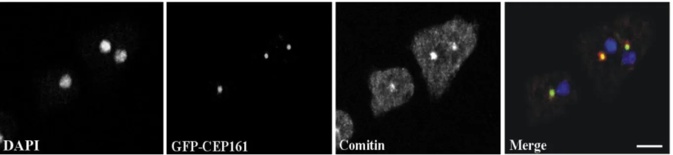
The centrosome and the Golgi apparatus co-localize in the vicinity of the nucleus. When we stained GFP-CEP161 cells with mAb190-340-8 for the Golgi marker comitin, we found CEP161 in the center of the Golgi apparatus (Fig. 9).
Figure 9: CEP161 localises to the Golgi-complex. CEP161 is a novel centrosomal protein in D. discoideum which also localises with the Golgi-complex as revealed by comitin staining with mAb 190-340-8, nuclei were stained with DAPI (Scale bar, 10 µm).
2.1.4 CEP161 constructs and truncated proteins
To identify the region of CEP161 that mediates the centrosome association of CEP161, we generated shortened proteins (GFP-CEP161-D1: designated D1, residues 1-1114; GFP- CEP161-D2: designated D2, residues 1-763; and GFP-CEP161-D3: designated D3, residues 763-1114) (Fig. 10A). Expression and molecular weights of the GFP tagged proteins were analysed by SDS-PAGE (Fig. 10B).
A
31 B
Figure 10. CEP161 constructs and protein expression. A. Overview of N-terminally GFP tagged CEP161 proteins. The position of the amino acids is given. B. Western blot for the GFP tagged proteins stably expressed in AX2 cells. Alpha-tubulin was used as the loading control. GFP fusion proteins were recognized by mAb K3-184-2, α-tubulin by YL1/2.
The expression levels varied for the GFP tagged proteins. GFP-CEP161 was present in lower amounts than endogenous CEP161 whereas GFP-D1 levels were 1.8 fold higher than those of CEP161 (Fig. 11A, B).
A
32 B
Figure 11. Quantification of CEP161. A. Western blot for endogenous CEP161 and GFP tagged proteins detected with CEP161 polyclonal antibodies. The arrow indicates the endogenous CEP161. B. Quantification of western blots for endogenous CEP161 and GFP tagged proteins.
2.1.5 Localisation of CEP161 truncated proteins
In immunofluorescence analysis the D1 and D2 proteins behaved like a centrosomal protein and appeared as a single dot near the nucleus. In contrast, the D3 protein was present throughout the nucleus although the prediction programs did not reveal nuclear localization signals or any DNA binding domain. We conclude that amino acids 1-763 are sufficient for localization of CEP161 to the centrosome (Fig. 12).
Figure 12. Subcellular localisation of GFP tagged proteins. For GFP-CEP161-D3 the centrosome was detected with mAb K68-332-3.Nuclei were stained with DAPI (Scale bar, 5 µm).
33 Based on its overall domain structure, the presence of the conserved -TuRc and the localization at the centrosome we presume that CEP161 is the D. discoideum ortholog of CDK5RAP2.
2.2 CEP161 mutant cells are viable, but impaired in growth and development 2.2.1 CEP161 regulates nuclear number and centrosome positioning
Isolation of a knockout mutant has not been successful. Therefore we overexpressed the GFP tagged versions of CEP161 full length and truncated proteins in AX2 cells in order to analyse the role of CEP161 for cell organization, growth and development. First we determined the nuclei number of the cells to understand the role of CEP161 in cell division and cell cycle.
The D1, D2 and D3 cells were mostly mononucleated with their centrosome located very close to the nucleus in a distance of < 500 nm. By contrast, CEP161 affected the centrosome nucleus distance and approximately 40% of the CEP161 cells had the centrosome far away from the nucleus with a distance > 900 nm (Fig. 13A, B). We also observed that the nuclei- centrosome ratio was aberrant in a low percentage of the D1 and D2 expressors.
A
34 B
Figure 13. CEP161 regulates nuclear number and centrosomal position. A. Number of nuclei per cell. B. Percentage of cells with nucleus-centrosome distance as indicated. A total of 600 cells were counted for AX2 and mutant strains for each of the experiments shown in A, B.
2.2.2 The nuclear-centrosome ratio is altered in the mutants
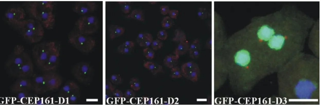
Furthermore, 0.6% of the D1 cells had two centrosomes per nucleus. For D2 cells this number increased to 0.74%, and in 0.24% of the cells we observed 3 and more centrosomes per nucleus (Fig. 14A, B). This was not observed for all other strains.
A
35 B
Figure 14: CEP161 regulates centrosomal number. A. Percentage of cells with nuclear- centrosome ratio. A total of 600 cells were counted for AX2 and mutant strains. D. Confocal images for D1 and D2 cells with abnormal distribution of nucleus-centrosome ratio. Nuclei were stained with DAPI, centrosomes were detected with the GFP signal of the GFP tagged D1 and D2 proteins. Scale bar, 5 µm.
2.2.3 CEP161 regulates cell size
Ectopic expression of CEP161 led to an increase in cell size with approximately 70% of the cells having a diameter greater than 12 µm. Interestingly, D1 (55%), D2 (52%) and D3 (70%) cells showed a decrease in the size of the cells and had a diameter below 10 µm (Fig. 15).
Figure 15. CEP161 regulates size. Cell size of the AX2 and CEP161 mutant strains in micrometers.
36 2.2.4 Role of CEP161 for growth
To examine the role of CEP161 for growth we assessed the phenotype of AX2 and CEP161 mutants on a bacteria lawn by measuring the increase in the diameter of the colony over time.
Colony growth was significantly reduced for all mutants except for D3 which showed increased growth (Fig. 16A, B). Such a behavior could be due to reduced phagocytosis or to altered motility. Hence we assayed the phagocytic capability following yeast particle uptake.
We found that fewer CEP161, D1 and D2 cells had ingested one or more yeast particles after 15 min than AX2 cells. D3 was similar to AX2 (Fig. 16C). In suspension culture the growth was reduced for the CEP161 and the D3 mutants, the D1 and D2 mutants showed a faster growth until day 5 after which there was a sharp decrease in the growth of the cells (Fig.
16D).
A
B
37 C
D
Figure 16: CEP161 affects growth. A. Growth on K. aerogenes. The increase of the plaque size was measured over days. B. Phenotype of AX2 and CEP161 mutants grown on a lawn of Klebsiella. The pictures were taken at day 6 after inoculation. C. Phagocytosis assay. TRITC labeled yeast were added to the cells. After 15 min incubation time the cells were fixed and the number of ingested yeast counted. D. Growth in shaking culture. The cell numbers were determined every 24 hours.
38 2.2.5 CEP161 affects development
When D. discoideum cells starve, they enter a developmental cycle that ends with the formation of fruiting bodies. The CEP161, D1 and D2 cells plated on phosphate agar aggregated and formed multicellular structures, the D3 cells could only form loose aggregates that did not develop into fruiting bodies. There was also marked increase in the size of the fruiting bodies for D1 and D2 mutants, both with regard to the length of the stalks and the size of the fruiting bodies, whereas the CEP161 fruiting bodies were not significantly different from those of AX2 (Fig. 17).
Figure 17. Development of wild type and mutant strains on phosphate agar plates.
Pictures taken at 24 hours after starvation in phosphate agar plates. Scale bar 500 µm.
Development was also studied on a plastic surface with cells kept in Soerensen phosphate buffer. Under these conditions cells can form streams and aggregate. Further development does not take place. We observed that stream formation occurred slightly later for CEP161, D1 and D2 (9 h) as compared to AX2 (8 h). For the D3 cells we noted a severe delay in the streaming and aggregation behavior as they formed streams only after 28 hours (Fig. 18A).
We then analyzed the expression levels of contact site A (csA) glycoprotein when cells were
39 starved in shaken suspension. csA gene expression is developmentally regulated and starts during aggregation reaching a peak at the tight aggregate stage. The protein is a cell adhesion molecule which enables cells to form EDTA stable cell-cell contacts during aggregation (Faix et al., 1990). In the experiment shown csA was detected in D1 at t6 and the amounts strongly increased at t8 and t10. In AX2, CEP161and D2 csA was present at t8 and the level increased at t10. In D3 csA was also seen at t8 and t10, however, the amounts were lower. At the t24 time point, when tight aggregates had formed in all strains, the csA signal was identical in all strains (Fig. 18B). We conclude that ectopic expression of CEP161 does not significantly alter development, whereas D1 and D2 lead to premature development and also to an increased size of the fruiting bodies and D3 has an inhibitory effect.
A
40 B
Figure 18. Streaming of AX2 and mutant cells. A. Stream formation on a plastic surface under phosphate buffer. The time points indicate the occurrence of stream formation. Scale bar, 250 µm. B. Time-dependent expression of csA. Cells were collected during development in shaking suspension at the indicated time points (in hours) and the lysates analyzed by SDS- PAGE and western blot. csA was detected by mAb33-294, mAb 47-16-1 detected α-actinin which was used as loading control. Since the α-actinin blot was similar for all strains, only the α-actinin blot for AX2 is shown.
Streaming and aggregation of the cells depends on the adhesion of the cells to the plastic substratum. Upon testing we observed that the mutants exhibited an altered adhesion and bound more strongly to the surface than AX2. This could account for the altered streaming behavior (Fig. 19).
41 Figure 19. Cell-substrate adhesion. Percentage of cells detached from the surface in 24 well plates after shaking at 200 rpm for 1 hour.
2.2.6 CEP161 affects unicellular motility and polarity
Cell motility is an important feature during growth and development of D. discoideum.
Dictyostelium cells exhibit an amoeboid type of cell motility. They deform very quickly and translocate via rapidly alternating cycles of pseudopod extension and pseudopod retraction in response to external signals which are dependent on changes in the actin cytoskeleton. To assess the role of CEP161 and mutant proteins for chemotactic motility during aggregation we analyzed the motile behaviour of aggregation competent cells and followed the motility of cells with time-lapse video microscopy. Mutant cells D1, D2 and D3 consistently displayed a reduced speed (~5-8 versus ~16 µm/min for AX2) (Fig. 20A and Table 2). AX2 cells were highly elongated and extend pseudopods mainly in the direction of the chemoattractant. The mutants displayed a change in morphology and had a more rounded shape in response to cAMP signaling (Fig. 20B). In the parameters analysed, directionality did not differ between AX2 and mutants CEP161, D1and D2 although D3 mutant showed a decrease, whereas the persistence was significantly reduced in the mutants D1, D2 and D3 (Table 2) suggesting that CEP161 of D. discoideum plays an important role in chemotaxis as it affects motility and polarity.
A
42 B
Figure 20. CEP161 affects cell motility and polarity. A. Chemotaxis assay. Analysis of speed during migration of aggregation-competent wild type and mutant cells towards the chemoattractant cAMP. B. Images showing migrating aggregation competent cells of AX2, CEP161 and D3 cells (scale bar, 50 µm).
Table 2
Parameter AX2 CEP161 D1 D2 D3
Speed(µm/min) 16.9 +/- 6.2 15.0 +/- 3.2 7.5 +/- 2.9** 5.0 +/- 2.1** 5.4 +/- 1.3***
Persistence (µm/min-deg)
7.2 +/- 4.2 5.4 +/- 1.7** 2.8 +/- 1.3** 2.2 +/- 0.5** 1.8 +/- 0.8***
Directionality 0.75 +/- 0.20 0.6 +/- 0.21 0.7 +/- 0.20 0.6 +/- 0.21 0.5 +/- 0.18**
Table 2. Analysis of chemotactic cell motility of AX2 and mutants. Time-lapse image series were captured and stored on a computer hard drive at 30-second intervals. Images were taken at a magnification of 10X every 6 s. The DIAS software was used to trace individual cells along image series and calculate motility parameters. Speed refers to the speed of the cell's centroid movement along the total path; directionality indicates migration straightness;
direction change refers to the number and frequency of turns; persistence is an estimation of movement in the direction of the path. Values are mean ± standard deviation of >30 cells from three or more independent experiments. The symbol * indicates the significance of the P- Value (*<0.5, **<0.01, ***<0.001).
2.2.7 CEP161 affects the directed migration of slugs
During multicellular development cells differentiate into prespore and prestalk cells in the aggregate, undergo a morphogenesis and can form a slug which can sense light and migrate in a straight path towards the light (Wallraff and Wallraff, 1997). Slugs are polar with a tip at the
43 anterior end consisting of prestalk cells, sensing the light that helps to control their migration and phototactic turning. AX2 and CEP161 slugs migrated in a nearly straight path towards the light source. D1, D2 and D3 deviated from this path and migrated in an angle towards light.
Also, the path length appeared shorter in particular for D1 and D2 (Fig. 21A, B).
A
B
Figure 21. CEP161 affects the directed migration of slugs. A. Phototaxis assay. Slugs and slug trails were transferred to nitrocellulose filters and stained with amido black. The red
44 arrow indicates the source of light. B. Angle of deviation of the cells when migrating towards light. Number of slugs analysed n=4. The symbol * indicates the significance of the P-Value (*<0.5, **<0.01, ***<0.001).
2.3 Identification of CEP161 as an interacting partner of the Hippo homolog Hrk-Svk To identify interaction partners of CEP161 we carried out immunoprecipitation experiments using cells expressing GFP tagged CEP161. The samples were analysed by SDS-PAGE and the bands were cut out and further analysed by mass spectrometry. The list of interaction partners of CEP161 from mass spectrometry data is shown in appendix I. Among the identified proteins we found severin kinase SvkA (DDB_G0286359) as a potential interacting partner. SvkA was described as a homolog of human MST3, MST4 and YSK1 kinases (Rohlfs et al., 2007). Our detailed sequence analysis showed that it is related to Hippo of Drosophila (25.1% identity, 35.7% similarity) and Hippo related Stk3 (MST1) of human (34.7% identity; 52.0% similarity). Particularly high homology was observed in the serine/threonine protein kinase domain of the proteins (Fig. 22).
Figure 22. Sequence alignment for the serine/threonine protein kinase domain of human (Hs) MST2 (NP_006272.2), Drosophila (Dm) Hippo (NP_611427.1) and Dictyostelium (Dd) Hrk- Svk (DDB_G0286359). The numbers indicate the position of the STK domain in the respective proteins. Colour code: Red background shows the residues which are strictly conserved, yellow background shows similar residues.
45 Given the significance of SvkA to our study and its relation to Hippo, we have renamed it as Hippo related kinase-Svk (Hrk-Svk). To further establish Hrk-Svk as an interacting partner for CEP161, we performed GST pull down assays with GST-CEP161-D2 and GST-CEP161- D3 proteins. The D2 protein encompasses the -TuRC domain and a coiled coil domain, D3 encodes two coiled coil domains (Fig. 23A). Hrk-Svk interacted with the D2 and also the D3 proteins but not with GST. The precipitate was probed with Hrk-Svk polyclonal antibodies (Fig. 23B).
A
B
Figure 23. Interaction of CEP161 with Hrk-Svk. A. Schematic of the GST tagged CEP161 polypeptides. B. CEP161 interacts with Hrk-Svk in a pull down assay. The blot was probed with pAb for Hrk-Svk. The Ponceau S stained membrane is shown below.
46 The interaction was further investigated by using GST fusion proteins of Hrk-Svk for pulling down CEP161. Only the polypetide encompassing the serine threonine kinase domain (STKc) interacted with CEP161 locating the binding site in this domain (Fig. 24A, B).
A
B
Figure 24. Interaction of CEP161 with the kinase domain of Hrk-Svk. A. Schematic of GST tagged constructs for Hvk-Svk polypeptides. B. Pull down assay showing the interaction of Hrk-Svk-E1 interacts with CEP161. The blot was probed with pAb CEP161.The Ponceau S stained membrane is shown below. GST-1-323 containing the kinase domain was very sensitive to proteolysis.
To investigate the localization of Hrk-Svk we expressed it as GFP tagged protein. Previously a cytosolic localization and enrichment at the centrosome was reported for GFP-Hrk-Svk (Rohlfs et al., 2007). When we stained GFP-Hrk-Svk expressing cells for CEP161 using mAb K83-632-4 we detected CEP161 staining in the center of the strongly GFP positive region
47 near the nucleus showing a yellow puncta in the overlay which indicates a colocalization (Fig.
25).
Figure 25. CEP161 colocalizes with GFP tagged Hrk-Svk in AX2 cells. CEP161 was detected with mAb K83-632-4. Nuclei were stained with DAPI. The boxed area is enlarged at the right. Scale bar, 5 µm.
2.4 Interaction of CDK5RAP2 with the mammalian Hippo homolog MST1
We extended our studies to the mammalian system to probe whether there is a similar interaction between components of the Hippo signaling pathway and CDK5RAP2. We used Myc tagged CDK5RAP2 and generated N-terminally truncated constructs C1 (residues 1-580) and C2 (residues 1-1271) also as Myc tagged polypeptides and expressed them in HeLa cells (Fig. 26).
Figure 26. Schematic of Myc tagged constructs of CDK5RAP2. The amino acid residues and the domains are indicated.
2.4.1 The N-terminal part of CDK5RAP2 is sufficient for targeting to the centrosome The Myc tagged constructs were expressed in HeLa cells and their localisation studied. Myc- CDK5RAP2 as well as the two truncated proteins was present in a dot near the nucleus which
48 colocalized with the centrosomal marker Pericentrin in immunofluorescence analysis (Fig.
27).
Figure 27. Immunofluorescence analysis of HeLa cells expressing Myc tagged CDK5RAP2 proteins. Myc was recognized by mAb 9E10, the centrosomal marker Pericentrin by pAb pericentrin, Nuclei was stained with DAPI. The boxed area is enlarged at the right. Scale bar, 10 µm.
The results suggest that the first 580 amino acids which contain the -TuRc domain of CDK5RAP2 are sufficient for targeting the centrosome. In some of the C1 and C2 expressing cells (0.8%) we noted an abnormal nucleus centrosome ratio (shown for Myc-CDK5RAP2-C1 with three centrosomes and Myc-CDK5RAP2-C2 with two centrosomes in Fig. 27) resembling the data obtained for DdCEP161-D1 and -D2 (Fig. 14A). The nucleus also often displayed an irregular shape in the cells expressing Myc-CDK5RAP2-C1 and -C2.
49 2.4.2 Overexpression of Myc tagged CDK5RAP2
Further experiments were carried out with HEK293T cells ectopically expressing Myc- CDK5RAP2. The amounts of the Myc tagged protein were 15-fold higher than the one of the endogenous CDK5RAP2 (Fig. 28A, B).
A
B
Figure 28. Expression of Myc-CDK5RAP2 in HEK293T cells. A. In the western blot, CDK5RAP2 was detected with pAb CDK5RAP2, Myc tagged protein with mAb 9E10, GAPDH with mAb GAPDH. B. Quantification of the Myc-CDK5RAP2 in wildtype and Myc- CDK5RAP2 transfected cells. Signals were analysed using ImageJ. For loading control we used GAPDH levels.
2.4.3 CDK5RAP2 interacts with the Hippo homolog MST1
The Hippo signaling pathway is highly conserved. Whereas most of the Hippo pathway components were identified in the fruit fly (Drosophila melanogaster) using mosaic genetic
50 screens, orthologs to these components have subsequently been found in mammals. The core component of the Hippo signaling is Hippo in Drosophila or MST1 in Humans. To understand the role of CDK5RAP2 in the Hippo signaling pathway, we tested whether CDK5RAP2 interacts with MST1, the ortholog of the Dictyostelium Hrk-Svk. We co- transfected HEK293T cells with Myc-CDK5RAP2 and GFP-MST1 and precipitated GFP- MST1 using GFP-trap beads. In the precipitate we detected Myc-CDK5RAP2. In immunoprecipitation experiments where we used Myc-CDK5RAP2-C1 and -C2, GFP-MST1 coprecipitated only with CDK5RAP2-C2 indicating that the binding site is located between residues 580 and 1271 which encompass the EB1 domain (Fig. 29).
Figure 29. CDK5RAP2 interacts with MST1. HEK293T cells were transiently transfected with the GFP tagged MST1 and Myc tagged CDK5RAP2 and the GFP-fusion protein precipitated using GFP-trap beads. The precipitate (IP) was probed for the presence of Myc tagged CDK5RAP2 proteins. Analysis was with mAb 9E10 to detected Myc tagged proteins and mAb mAb K3-184-2 to detect GFP.
51 2.4.4 CDK5RAP2 colocalises with MST1
The interaction was further analysed by immunofluorescence analysis. HeLa cells transfected with Myc-CDK5RAP2 and GFP-MST1 were stained for the Myc tag with mAb 9E10 and Pericentrin as a centrosomal marker. GFP-MST1 was present in the cytosol and showed a dot like staining near the nucleus. This dot colocalized with the signal for Myc-CDK5RAP2 and was recognized by Pericentrin specific antibodies. The immunofluorescence analysis was also done with HeLa cells transfected with Myc-CDK5RAP2-C2 and GFP-MST1. A sharp puncta staining near the nucleus was observed for Myc staining. It colocalised with the GFP-MST1 signal which was further recognized by Pericentrin antibodies (Fig. 30A, B). We did not observe a co-localisation of the C1 protein with MST1 (data not shown).
A
B
Figure 30. Immunofluorescence analysis to show the localization of CDK5RAP2 and MST1. A. Colocalisation of Myc-CDK5RAP2 with GFP-MST1 in HeLa cells. The inset shows a magnified image of CDK5RAP2 colocalising with MST1 at the centrosome. The boxed area is enlarged at the right. B Colocalisation of Myc-CDK5RAP2-C2 with GFP- MST1 in HeLa cells. Myc was detected with mAb 9E10, Pericentrin with pAb anti- pericentrin. Size bar, 10 µm.
52 2.5 CDK5RAP2 is a novel regulator of Hippo signaling
Next we examined the functional consequences of the MST1-CDK5RAP2 interaction on Hippo signaling using an established GAL4-TEAD luciferase reporter system, the repression of which reflects activity of the Hippo pathway (Lei et al., 2008; Tian et al., 2010). For the luciferase reporter assay, HEK293T cells were seeded in 24-well plates. A mixture of 5×
upstream activating sequence (UAS)-luciferase reporter, Renilla, and the indicated plasmids were cotransfected. Twenty-four hours after cells were transfected, they were lysed, and luciferase activity was measured using a dual-luciferase reporter assay system (catalog no.
E1960; Promega) following the manufacturer's instructions. The luciferase activity was measured by a luminometer (model TD-20/20). Transfection efficiency was normalized to thymidine kinase-driven Renilla luciferase activity.
The exogenous TAZ increased GAL4-TEAD activity. The positive control of TAZ expression alone expectedly showed a dramatic increase in the luciferase activity, which was normalized to 100%. For negative control, TAZ and LATS was co-transfected, since LATS is a potent inhibitor of TAZ in the Hippo signaling pathway. Inhibition of TAZ by LATS led to a decrease in the relative luciferase activity to 80%. Coexpression of CDK5RAP2 with TAZ augmented TEAD4 transcriptional activity to 121% (P-Value < 0.05), in comparison with TAZ expression alone. The expression of CDK5RAP2 without TAZ was not sufficient to induce TEAD activation (Fig. 31). It behaved like the “no plasmid” control. Although CDK5RAP2 was only able to induce a minor activation of TAZ, it points to the fact that CDK5RAP2 could have an impact on the Hippo signaling pathway.
53 Figure 31. Luciferase assay. CDK5RAP2 regulates the activity of the transcriptional coactivators TAZ. Plasmids encoding the indicated proteins or empty pcDNA6 vector were cotransfected in HEK293T cells together with the TEAD reporter plasmids. Coexpression of CDK5RAP2 increased the TAZ-dependent signaling to 121% (n = 4; P < 0.05).
2.5.1 TAZ interacts with the N-terminal part of CDK5RAP2
To further understand the role of CDK5RAP2 in the Hippo signaling pathway, we analysed for a potential interaction of TAZ with CDK5RAP2 using Flag-trap beads. We also tested if the CDK5RAP2 truncated proteins C1 and C2 could also interact with TAZ, which would inturn help us to recognize the interacting residues of CDK5RAP2 with TAZ. The coimmunoprecipitation showed that Flag-TAZ bound to all three proteins indicating that the N-terminal part of Myc-CDK5RAP2 encompassing residues 1-580 is sufficient (Fig. 32). As negative control Flag-hnRNPF, which is a ribonuclear protein, was employed.
54 Figure 32. Co-immunoprecipitation assays to study the interaction of CDK5RAP2 with TAZ. Myc tagged CDK5RAP2 full length, and the C1 and the C2 proteins were tested.
HEK293T cells were transiently cotransfected with GFP tagged MST1 and Myc tagged CDK5RAP2 proteins. After immunoprecipitation (IP) with GFP trap beads, the resulting blots were probed with mAb 9E10 to reveal Myc, mAb K3-184-2 to reveal GFP and pAb anti-Flag to reveal Flag.
2.5.2 Ectopic expression of CDK5RAP2 downregulates the expression of TAZ
Since there appeared to be an interaction of TAZ with CDK5RAP2, we wondered whether CDK5RAP2 has an impact on TAZ levels. We first quantified the amount of Flag-TAZ in whole cell lysates with wild type untransfected cells as the negative control and Flag-TAZ transfected cells as the positive control. We consistently observed a drastic reduction in the expression levels of TAZ upon cotransfection with plasmid DNA encoding Myc-CDK5RAP2 although the amounts of plasmid DNA were identical (Fig. 34A and B).
55 A
B
Figure 33. Ectopic expression of CDK5RAP2 affects the levelsof TAZ. A.Whole cell lysate for wild type (WT) HEK293T cells and HEK293T cells ectopically expressing Flag- TAZ, and cells ectopically expressing Flag-TAZ and Myc-CDK5RAP2. The blot was probed with pAb anti-Flag to reveal Flag, mAb 9E10 to reveal Myc and mAb GAPDH to reveal GAPDH . B. Quantification of Flag-TAZ from the whole cell lysate for HEK293T cells ectopically expressing Flag-TAZ, and cells ectopically expressing Flag-TAZ and Myc- CDK5RAP2. Signals were analysed using ImageJ. For loading control we used GAPDH levels. (P-value < 0.001).