The HSP47 - Procollagen Interaction:
Mechanism of pH-Dependent Client Release and Development of Antifibrotic Inhibitors
Inaugural-Dissertation
zur Erlangung des Doktorgrades der Mathematisch-Naturwissenschaftlichen
Fakültät der Universität zu Köln
Vorgelegt von
Sinan Öcal
aus Istanbul
Köln, September 2018
Gutachter: Prof. Dr. Ulrich Baumann
Institut für Biochemie / Universität zu Köln Prof. Dr. Karsten Niefind
Institut für Biochemie / Universität zu Köln
Prüfungsvorsitzender: Prof. Dr. Eric von Elert
Institut für Zoologie / Universität zu Köln
Tag der mündlichen Prüfung: 13. 11. 2017
Die Arbeiten und Experimente zur vorliegenden Dissertation wurden zwischen Oktober 2012 und
September 2017 unter Betreuung von Prof. Dr. Ulrich Baumann am Institut für Biochemie der
Universität zu Köln, Otto-Fischer Str. 12-14, D-50674 Köln, Deutschland, durchgeführt.
Teile dieser Arbeit wurden bereits veröffentlicht:
Oecal S, Socher E, Uthoff M, et al. The pH-dependent Client Release from the Collagen-specific Chaperone HSP47 Is Triggered by a Tandem Histidine Pair. The Journal of Biological Chemistry.
2016;291(24):12612-12626.
Table of Contents
Zusammenfassung ... i
Abstract ... iii
1. Introduction ... 1
1.1 Collagen ... 1
1.1.1 The Collagen Superfamily: An Overview ... 1
1.1.2 Composition and Classification of Collagens ... 2
Fibril Forming Collagens ... 3
1.1.3 Structure and Stability of the Collagenous Domain ... 4
The Polyproline Helix Type II ... 4
The Collagen Triple-helix ... 5
Structural Aspects of Triple-helix Stability ... 5
1.1.4 Collagen Biosynthesis ... 9
Hydroxylation ... 9
Glycosylation ... 10
Cis-trans isomerization ... 10
Folding of collagens ... 10
Trafficking, processing and secretion ... 11
1.2 Heat Shock Protein 47 ... 14
1.2.1 An Introduction to HSP47 ... 14
1.2.2 Serpins and Structure of HSP47 ... 15
1.2.3 HSP47 in Collagen Biosynthesis ... 17
1.2.4 Molecular details of the HSP47 - Collagen Interaction ... 18
1.3 Thesis Aims ... 20
Molecular details of the pH-dependent client-release ... 20
Identifying small organic molecule inhibitors of the HSP47 - collagen interaction ... 21
2. Material and Methods ... 22
2.1 Material ... 22
2.1.1 Chemicals ... 22
2.1.2 Proteins ... 22
2.1.3 Buffers and Media ... 23
2.1.4 Synthetic Peptides... 24
2.1.5 Synthetic DNA Oligonucleotides ... 24
2.1.6 E. coli Strains ... 24
2.1.7 Software ... 24
2.2 Nucleic Acid Methods ... 24
2.2.1 Isolation & Purification of DNA ... 24
2.2.2 Spectrometric Determination of DNA concentration ... 25
2.2.3 Agarose Gel Electrophoresis ... 25
2.2.4 Polymerase Chain Reaction & Site-directed Mutagenesis ... 26
2.2.5 Restriction Endonuclease Digestion of DNA & Ligation ... 27
2.2.6 DNA Sequencing ... 28
2.3 Cell Culture Methods ... 28
2.3.1 Transformation of DNA ... 28
2.3.2 Protein Expression in E. coli ... 28
2.3.3 Cell lysis ... 29
2.4 Protein Biochemistry Methods ... 29
2.4.1 Chromatographic Methods ... 29
2.4.2 SDS-PAGE ... 31
2.4.3 Western Blotting & Immunodetection ... 32
2.4.4 Biotinylation of Proteins ... 33
2.4.5 Spectrometric Determination of Protein concentration ... 33
2.4.6 Colorimetric Protein Assays ... 33
2.4.7 Differential Scanning Fluorimetry ... 34
2.4.8 Circular Dichroism Spectroscopy ... 34
2.4.9 Isothermal Titration Calorimetry ... 35
2.4.10 Dynamic Light Scattering ... 35
2.4.11 Biolayer Interferometry ... 36
2.4.12 Fluorescence Spectroscopy & Homogenous Time-Resolved FRET ... 36
2.4.13 Protein Crystallization ... 39
3. Results ... 40
3.1 Protein Production and Characterization ... 40
3.1.1 Proteins and Peptides used in this Work ... 40
HSP47 Constructs ... 40
Collagen Model Peptides ... 40
3.1.2 Heterologous Protein Expression in E. coli ... 40
3.1.3 Protein Purification ... 41
3.1.4 Protein Quality Control ... 42
Differential Scanning Fluorimetry ... 42
Dynamic Light Scattering ... 44
3.2 HSP47 Lacks the Serpin-Typical Hyperthermostable Conformation ... 44
3.3 Characterization of the HSP47 - Collagen Interaction ... 48
3.3.1 Establishing Biolayer Interferometry Experiments ... 48
3.3.2 Characterization of the HSP47 - Collagen Interaction Using BLI ... 49
Kinetic parameters and K
Dof the HSP47 - collagen interaction ... 49
HSP47 client-affinity decreases with pH ... 50
3.3.3 Stoichiometry of the HSP47 - Collagen Interaction ... 51
3.4 Molecular details of the pH-dependent client release ... 52
3.4.1 Systematic Analysis of Histidine Residues in HSP47 ... 52
Phylogenetic analysis ... 52
Molecular dynamics simulations ... 54
3.4.2 Role of HSP47 Histidines in pH-Dependent Client Release ... 55
Histidine Scanning Mutagenesis ... 55
Non-interface histidines are not involved in client-release ... 56
His215 is important for collagen binding ... 57
The mutation H238N abolishes collagen binding ... 58
The collagen binding of mutant H273N + H274N is less sensitive to a decrease in pH ... 60
Influence of charged residues at positions 273 and 274 ... 62
The residue in position 273 fine-tunes pH-dependency of client-release in many organisms 63 There is no evidence for conformational changes accompanying client-release ... 64
3.5 High-Throughput Screening for Inhibitors of the HSP47 – Collagen Interaction ... 64
3.5.1 Design principles ... 64
3.5.2 Characterization of the Assay ... 66
Overview ... 66
Determination of optimal analyte concentration ... 66
Signal specificity ... 67
Signal stability ... 68
Assay response to inhibition ... 69
3.5.4 High-throughput Screening ... 70
Statistics based assessment of high-throughput suitability ... 70
Pilot Screen ... 70
IC
50-Determination and Counter-screening ... 72
3.5.5 Experimental Hit-validation ... 72
Hit validation with Biolayer Interferometry ... 72
3.6 Crystallization of HSP47 ... 75
4. Discussion ... 76
4.1 HSP47 function does not require conformational rearrangements ... 76
4.2 Molecular Details of Client Release ... 77
4.2.1 On Data Quality in BLI Experiments ... 77
4.2.2 Characterization of the Histidine Mutants ... 79
The Role of His215 in Collagen Binding ... 79
The Role of His273 and His274 in Collagen Binding ... 80
Evolution of the XH-motif ... 81
4.3 Open Questions regarding the HSP47 - Collagen Interaction ... 82
4.3.1 The Influence of Chain-register on HSP47 Binding is Unknown ... 82
Limitations of Homotrimeric CMPs ... 82
Synthetic Heterotrimeric CMPs ... 83
Heterologous Expression of Heterotrimeric CMPs ... 83
4.4 HSP47 as a Therapeutic Drug Target ... 84
5. Literature ... 86
Appendix ... I A.1 Figures ... I
A.2 Tables ... III
List of Abbreviations ... IX
List of Figures ... XII
List of Tables... XIV
i
Zusammenfassung
Kollagene sind eine äußerst vielseitige Superfamilie von Proteinen, welche primär als strukturgebende
Hauptkomponente der Extrazellulären Matrix (EM) von Metazoen bekannt sind. Aufgrund ihrer
zentralen Rolle beim Aufbau und der Dynamik der EM sind Kollagene an einer Vielzahl an
unterschiedlichen Prozessen wie z.B. der Zelladhäsion, Zellmigration, Angiogenese, Morphogenese,
Immunantwort oder Krebsprogression beteiligt. Die Biosynthese dieser großen, stark post-translational
modifizierten Proteine wird durch eine fein aufeinander abgestimmte Maschinerie bewerkstelligt. Die
Komplexität dieser Maschinerie äußert sich durch ihre Anfälligkeit gegenüber Störungen, welche oft
zur Entstehung von schwerwiegenden Defekten und Krankheiten führen. Das molekulare Chaperon von
Kollagen, Hitzeschockprotein 47 (HSP47), spielt hierbei eine entscheidende Rolle und gewährleistet
sowohl die fehlerfreie Synthese und den Transport von Kollagenen als auch den Schutz des
Endoplasmatischen Retikulums (ER) vor durch Aggregation und Akkumulation von Kollagenen
bedingtem Stress. Obwohl HSP47 seit mehr als zwei Jahrzehnten Forschungsgegenstand ist, sind viele,
teilweise grundlegende Aspekte seiner Funktion immer noch unbekannt; es ist z.B. unbekannt, welchen
Vorteil die Serinproteasestruktur für die Proteinfunktion bringt, oder ob alle Kollagentypen zum
Substratrepertoire gehören. Die kürzlich gelöste Kristallstruktur von HSP47 im Komplex mit einem
Kollagenmodellpeptid hat der Beantwortung der offenen Fragen neuen Anstoß gegeben. Eine betrifft
den Mechanismus der Substratfreisetzung: untypisch für Chaperone wird die Funktion von HSP47 nicht
durch Austausch oder Hydrolyse von Nukleotiden reguliert, sondern durch die graduelle Abnahme des
pH-Wertes im sekretorischen Weg. Die molekularen Details dieser pH-induzierten Substratfreisetzung
sind ungeklärt und wurden im ersten Teil der vorliegenden Arbeit untersucht. Ausgehend von der
Kristallstruktur und Phylogenetischen Daten wurden gezielt HSP47-Punktmutanten generiert und deren
Bindung an Kollagenmodellpeptide mittels Biolayerinterferometrie kinetisch quantifiziert. Besonderes
Interesse galt hierbei den 14 Histidinresten von HSP47, welche Aufgrund Ihrer Fähigkeit, bei
physiologischen pH-Werten Protonen aufnehmen zu können, als potentielle Auslöser einer
Substratfreisetzung in Betracht kamen. Eine systematische Analyse dieser Aminosäuren enthüllte dass
His273 und His274, am Rande der Interaktionsfläche gelegen, großen Einfluss auf die pH-Sensitivität
der HSP47 - Kollagen Interaktion ausüben. Andere Histidinreste wurden ebenfalls als wichtige
Komponenten der Interaktion ausgemacht, wie z.B. His238, welches bei der korrekten Ausrichtung von
für die Bindung essentiellen Aminosäureseitenketten eine Rolle spielt. Versuche, HSP47 bei leicht
saurem Milieu zu Kristallisieren waren Aufgrund der verminderten Stabilität des Proteins unter solchen
Bedingungen nicht erfolgreich; somit steht eine umfassende, strukturelle Erklärung des
Substratfreisetzungsmechanismus noch aus.
ii
In den letzten Jahren haben mehrere Studien gezeigt, dass Genablation von HSP47 mittels siRNA zu
einer deutlichen Verbesserung bis hin zur Aufhebung von fibrotischen Krankheiten führen kann. HSP47
wurde dadurch als ein vielversprechendes Ziel für die Entwicklung antifibrotischer Medikamente
erkannt. Vor diesem Hintergrund wurde in dieser Arbeit ein auf Fluoreszenz basierender Assay
entwickelt, welcher mittels eines Hochdurchsatzverfahens zur Identifizierung neuartiger Inhibitoren des
HSP47 - Kollagen Komplexes herangezogen werden kann. Der Assay wurde genutzt, um aus einer
40.000 Chemikalien umfassenden Substanzbibliothek heraus 4 potentielle Inhibitorkandidaten zu
identifizieren, von welchen mindestens eine Substanz in Validationsexperimenten vielversprechende
Ergebnisse lieferte.
iii
Abstract
Collagens are a multifaceted superfamily of proteins which constitute the principal structural component of the extracellular matrix (ECM) of metazoan organisms. Intimately tied to ECM architecture and dynamics, collagens are involved in a multitude of processes such as cell adhesion and migration, angiogenesis, morphogenesis, the immune response and cancer progression. The biosynthesis of these large and complex molecules is reliant on a finely tuned machinery, disruptions of which is often causative to severe disease. Heat-shock protein 47 (HSP47), the molecular chaperone of collagen, plays a central role in ensuring proper processing and trafficking of collagens as well as the protection of the endoplasmatic reticulum (ER) from stress induced by aggregation and accumulation of its client. Although HSP47 has been first described more than 20 years ago, critical aspects of its function are still shrouded in mystery, ranging from why it is a member of the serpin superfamily of proteins to whether its client repertoire includes all types of collagen. The rather recently solved crystal structure of HSP47 in complex with a collagen model peptide has provided a new impetus for answering the many open questions. One of these is how client-release is achieved: untypical for chaperones, HSP47 function is not coupled to nucleotide hydrolysis or exchange, but governed by the gradual decrease in pH along the secretory pathway. The mechanism by which the pH-shift induces client release is unclear. In this work, it was investigated whether this process is based on conformational re-arrangements, more subtle distortions of the binding site or electrostatic repulsion.
Of particular interest were the 14 histidine residues in HSP47, which have long been considered as potential trigger residues, since their imidazole side-chains can serve as a proton acceptor at physiological pH. Systematic analysis of these histidines in context of this thesis has revealed that His273 and His274, located at the fringe of the binding interface, exert considerable influence on the pH-sensitivity of the HSP47 - collagen complex. Other histidines have also been found to be important for the interaction; most notably, His238 was shown to be an essential actor in the pre-arrangement of key residues in a client-binding competent conformation.
Over the recent years, studies using gene ablation via siRNA have shown that interfering with the
HSP47 - collagen complex can resolve a variety of fibrotic diseases. HSP47 has thus enjoyed increasing
attention as a potential target for anti-fibrotic drugs. In light of this, one part of this thesis has focused
on developing a fluorescence based, high-throughput screening compatible assay to be utilized for the
identification of novel inhibitors of the HSP47 - collagen interaction. Interrogation of a compound
library using the assay has yielded 4 potential inhibitor candidates, at least one of which having shown
promising results in initial validation studies.
1
1. Introduction
1.1 Collagen
1.1.1 The Collagen Superfamily: An Overview
Collagens are a remarkably diverse superfamily of proteins which are best known for their prominent role in the architecture of the extracellular matrix (ECM) of animals. Collagen is the most abundant protein in vertebrates, constituting almost a third of total protein mass - a consequence of the ubiquity of the extracellular matrix and the markedly low turnover of many collagens, some of which being practically permanent, as for instance collagens in adult cartilaginous tissue
1. By and large the principal function of collagens is to provide structural integrity and scaffolding to the extracellular space.
Collagens fulfil this role via assembling into various superstructures with extraordinary mechanical properties: the elastic modulus of a single collagen I fibril from rat tail, for instance, has been determined to be in the range of 1-10 GPa, coming close to that of typical metals and showcasing formidable tensile strength at a much lower density
2. Besides these well-characterized structural roles, collagens are also involved in a multitude of dynamic phenomena such as cellular adhesion, migration or chemotaxis.
Products of collagen processing are often important biochemical effectors: endostatin, a C-terminal domain of multiplexin collagens, has been shown to inhibit angiogenesis upon proteolytic release and is currently in clinical trials as an anti-tumour agent
3,4, while the C-propeptide of collagen type I is believed to play a key regulatory role both in fibrillogenesis and collagen biosynthesis
5. Similarly, membrane collagens are known not only to function as cell surface receptors but also serve as a reservoir for effectors in the form of shed ectodomains. Another example for the multifaceted nature of collagens is collagen type VI, which besides forming beaded filaments, an important structural element of the ECM, is also capable of disrupting bacterial membranes and as such involved in innate host defense of the ECM
6.
Despite its omnipresence and biochemical significance, the definition of what is a collagen and what is not is still blurry at best. Three features are widely considered to be hallmarks of collagens
7:
(i) The presence of at least one so-called collagenous domain, characterized by a triple- helical structure comprised of three separate polypeptide chains with repeating triplets of Gly-Xaa-Yaa (Xaa, Yaa = any amino acid),
(ii) Localization in the extracellular space, and (iii) Participation in the assembly of superstructures.
These hallmarks are not always sufficient to fully characterize collagens, though, since there are
exceptions aplenty: membrane collagens, for example, do not assemble into superstructures and are
2 strictly seen components of the cell membrane. The triple-helical structural motif, on the other hand, is also encountered in other proteins such as adiponectin, the asymetric form of acetylcholinesterase, C1q, macrophage scavenger receptors (MARCO), ficolins, collectins and many others
7. Emilins and emu proteins fulfil all three criteria of being collagens, but are not (yet) classified as such
8.
Collagens and related molecules are not exclusive to vertebrates and have been discovered in many different multicellular organisms, including primordial forms of life such as Sponges
9and Hydra
10, or rather extraordinary ones, such as the annelid Alvinella pompeiana, one of the most heat-tolerant metazoan known to date
11. A single, 54 bp long sequence is believed to be ancestral to fibrillar collagens, which subsequently arose via multiple duplications of this basic genetic unit
10,12. Such collagen-related structural motifs (CSM), although rare, are also found in unicellular eukaryotes and bacteria
13; the streptococcal cell surface proteins Scl1 and Scl2, for instance, contain extended Gly-Xaa- Yaa repeats in their amino acid sequences
10, as does the exosporium filament BclA of Bacillus anthracis
14,15. An intact collagen gene has even been discovered in the viral shrimp pathogen White Spot Bacilliform Virus
16. It is quite possible that prokaryotic and viral collagen genes are the result of horizontal gene transfer and that the appearance of collagens is tied to the emergence of a hypothetical ancestor of metazoans
9; this would imply that the acquisition and retention of such genes by unicellular organisms provides advantages in the interaction with multicellular hosts.
1.1.2 Composition and Classification of Collagens
Collagens are multidomain proteins comprised of three individual polypeptide chains, called collagen α-chains. The primary structure of α-chains characteristically includes extended regions consisting of repeating Gly-Xaa-Yaa triplets, where Xaa is frequently proline (28%) and Yaa (2S,4R)-4- hydroxyproline (38%)
17. As will be discussed in more detail below, these proline-rich regions are responsible for the formation of the distinctive collagen triple-helix. In collagens, extensive triple- helical stretches, called collagenous domains, are typically interspersed with short non-collagenous (i.e.
non triple-helical) domains, referred to as interruptions, and flanked by globular N- and C-terminal domains. A testament to the complexity of collagens, the identity and processing of these N- and C- terminal domains can vary greatly between different types of collagens.
As of today, 28 types of collagens (designated with Roman numerals I-XXVIII) with a total of 46 unique* α-chains have been characterized in humans (The “novel” collagen α-chain XXIX α1 turned out to be identical to collagen VI α5). For most collagen types, only one α-chain is known; others have multiple unique α-chains (up to 6 for collagen IV, and collagen VI in some mammals) which are distinguished via Arabic numerals. Since some collagens have several isoforms with differing chain compositions and others even form hybrid structures with α-chains of different collagens, the exact definition of a collagen molecule requires the specification of all three incorporated α-chains (e.g.
*α1(II) and α3(XI) share the same sequence but diverge in posttranslational processing and cross-linking18.
3 [α1(I)]
3for homotrimeric collagen type I and [α1(I)]
2, α2(I)] for heterotrimeric collagen type I).
Collagens are commonly classified according to their domain organization and/or the superstructures they form. The subfamilies include fibril forming collagens (I, II, III, V, XI, XXIV and XXVII), fibril associated collagens with interrupted triple-helices (FACIT) (IX, XII, XIV, XVI, XIX, XX, XXI, XXII), membrane associated collagens with interrupted triple-helices (MACIT) (XIII, XVII, XXIII, XXV), collagens with multiple triple-helical domains with interruptions (Multiplexins) (XV, XVIII), and network forming collagens (IV, VIII, X). Furthermore, there are collagens forming beaded filaments (collagen VI) and anchoring fibrils (collagen VII) as well as some which do not quite fit into any category (XXVI, XXVIII). Since a detailed discussion of the differences in structure and biosynthesis between these classes of collagens would be beyond the scope of this work, the following sections will focus on fibril forming collagens, which are the best characterized and have been subject to study in this work.
Fibril Forming Collagens
Fibrillar collagens are the most abundant in terms of total protein mass: collagen type I alone comprises more than 90% of all collagen in the body. The subfamily encompasses collagens of type I, II and III (type A clade, also called major fibrillar collagens in regard to their quantity), collagens type V and XI (type B clade, minor fibrillar collagens) and finally the novel collagens type XXIV and XXVII (type C clade)
19. The latter are rather similar to collagens found in invertebrates, being shorter and containing imperfections/interruptions in their triple-helical domain
20.
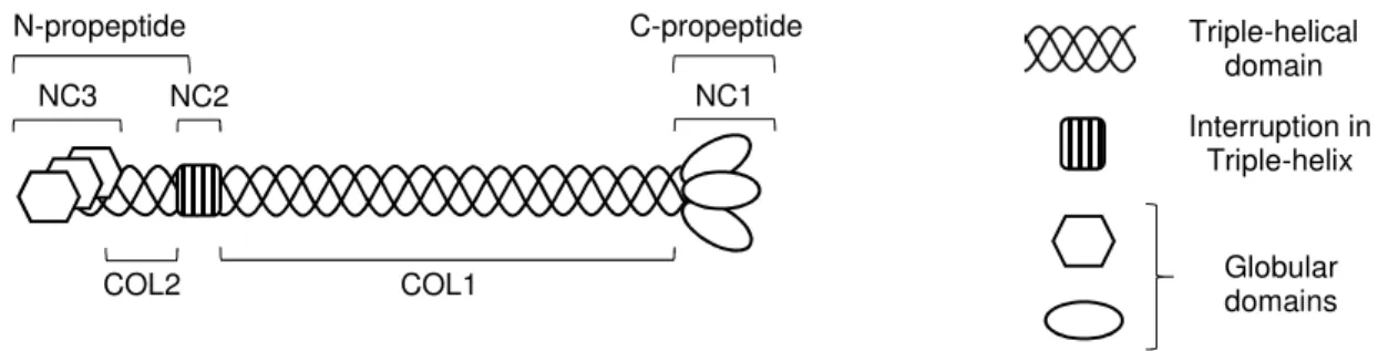
Figure 1.1 shows the domain organization of a prototypical fibrillar collagen. Collagenous domains (COL) and non-collagenous domains (NC) are numbered proceeding from C- to N-terminus (this is consistent for all fibrillar collagens, but there are cases in which the numbering is reversed, such as MACIT collagen type XIII). Fibrillar collagens of the same clade typically share N-terminal NC domains: these are homologous to von Willebrand factor type C (vWFC, also called chordin-like cysteine rich repeats) in members of clade A and to thrombospondin-1 N-terminus-like domain (TSPN)
Figure 1.1│Domain organization of a prototypical fibrillar collagen. An extended and in most cases uninterrupted collagenous domain (COL1) is flanked N- and C-terminally by non-collagenous domains (NC2 and NC1, respectively). Proteolytic cleavage within the NC2 region results in release of the N-propeptide, consisting of a short collagenous domain (COL2) and a globular domain (NC3), usually TSPN or vWFC. The C-propeptide is synonymous with the NC1 domain.
NC1
COL1 NC2
COL2 NC3 N-propeptide
Globular domains Interruption in
Triple-helix Triple-helical
domain C-propeptide
4 in members of clades B and C
19. Unsurprisingly for collagens, there are exceptions: the N-terminal NC domain seems to have been deleted in case of α2(I), and α2(V) harbors a cysteine-rich repeat even though it belongs to clade B. The C-terminal NC1 domain, however, is highly conserved among fibrillar collagens and is important for trimerization and correct registration during collagen maturation.
1.1.3 Structure and Stability of the Collagenous Domain The Polyproline Helix Type II
The PPII helix is a much neglected secondary structure element that is not only encountered in fibrillar proteins such as collagen, but is also widely dispersed in natively folded globular proteins, most often over short stretches of 4 or 5 amino acids and frequently preceding/transitioning into α-helices, β-sheets, 3
10-helices or reverse turns
21. Analysis of protein structures deposited to the Protein Data Bank archive (PDB) of the Research Collaboratory for Structural Bioinformatics (RCSB) shows that around 2% of amino acids are incorporated into PPII helices
22. The PPII helix is particularly important as the structural motif which is recognized by the abundant proline recognition domains such as SH3, WW or EVH1
23,24. The PPII helical conformation is furthermore believed to be dominant in unfolded proteins
25, short polypeptides and regions of proteins previously assumed unstructured
26. Raman spectroscopic studies also suggest that the PPII helix is an important transitory element in protein folding and denaturation, having been observed during the melting of α-helices in peptides
27as well as the transformation of an α-helix in native human lysozyme into a β-sheet strand involved in amyloidogenesis
28.
The PPII helix is an elongated, left handed helix encompassing 3 residues per turn with a helical pitch of 3.1 Å per residue. In its ideal form, it is defined by the backbone dihedral angles φ= -75°, ψ= 145°
and ω = 180° (all-trans conformation). As the name suggests, proline residues have a high propensity to form PPII helices. This is a consequence of the pyrrolidine ring restricting φ to a region suitable for PPII helix formation (-75 ± 15°), thus leading to a decreased loss of conformational entropy upon assuming the ordered structure. It should be noted though that the above mentioned angles correspond to Ramachandran regions which are populated by all amino acids, meaning that proline residues are not an absolute requirement for PPII helix formation.
The PPII helix lacks the backbone hydrogen bonds characteristic of the other secondary structure elements, and the factors governing its stability have long been disputed. One important aspect are steric interactions and restrictions: the region of φ and ψ populated by amino acids in a PPII helix characteristically lacks unfavourable steric interactions
29, as would be intuitive considering the elongated, well-spaced structure of the helix with all side chains pointing away from the helical axis.
Furthermore, steric effects alone have been sufficient to drive PPII helix formation in Monte Carlo
simulations, with the important interactions having been pinpointed to occur between the pyrrolidine
moiety of proline residues and the backbone of the preceding amino acid
30. Another factor contributing
to the stability is the high degree of solvation, especially of the backbone carbonyl and amide groups,
5 which are aligned perpendicular to the helical axis and highly exposed. This is underlined, inter alia, by the correlation observed between solvent-accessible surface area and PPII formation propensity
31. The side chains of the amino acids are also observed to influence the stability of the helix: the strong tendency of Gln residues to form PPII helices, for instance, has been explained with intermolecular hydrogen bonds formed between the amide hydrogen of the side-chain and the backbone carbonyl of the C-terminally neighboring amino acid.
32Another stabilizing effect yet comes in the form of a n → π* interaction, in which overlap between the non-bonding orbital of a carbonyl-oxygen with the anti- bonding π* orbital of the carbonyl moiety of a C-terminally neighbouring residue leads to a stabilization of an estimated 0.7 kcal/mol via electron delocalization
33,34.
The Collagen Triple-helix
The molecular structure of this defining element of collagens has been extensively characterized in the past - the classical collagen model peptide [(Pro-Pro-Gly)
10]
3has even made it aboard the space shuttle Discovery, where crystals were grown under micro-gravity
35, which back on earth resulted in structure determination with a respectable 1.3Å resolution and an R-factor of 0.18
36. The collagen triple-helix is comprised of three parallel PPII helices, supercoiled along a common axis to form a right handed triple helix with a helical pitch ranging from 7/2 for proline-rich regions to 10/3 for proline-poor regions
17. The α-chains are axially staggered by one amino acid and thus can be distinguished as leading (+0), middle (+1) and trailing (+2) strands in order of decreasing C-terminal overhang, as first defined by Emsley et al
37. The triple-helix is stabilized by periodic hydrogen bonds between the amide-nitrogen of glycine residues and the amide-oxygen of residues in position Xaa on the strand with -1 stagger. The estimated contribution of each hydrogen bond to the Gibbs free energy of trimerization is between -1.4 to -1.8 kcal
17.
The observed tight packing of the triple-helix explains the necessity for the staggered assembly of α- chains as well as the strict requirement for glycine residues in every third position: the side-chains of residues in these positions are oriented such that any amino acid other than glycine would experience steric clashes with the neighbouring strand with +1 stagger.
Structural Aspects of Triple-helix Stability
The collagen triple helix is not stable at body-temperature
38, which is rather surprising considering its
significance as a structural element in the extracellular matrix. This meta-stability underlines that
collagens, although often being regarded merely as rigid scaffolding proteins, are remarkably dynamic
and able to utilize their structural “imperfections” for biological activity. Transient structural
perturbations in the collagen triple-helix are indeed essential for many processes such as recognition of
collagens by matrix metalloproteases (MMPs) or the binding of heparin to the collagenous domain of
acetylcholinesterase, determining its anchoring location
39.
6 Although the interaction interface of triple-helix forming PPII helices is dominated by the polypeptide backbone, triple-helix stability is greatly influenced by α-chain sequence. As is often the case with polymeric molecules, the loss of conformational entropy upon adopting an ordered structure is significant for collagen α-chains. This underlies the fact that the stability is intrinsically governed by the propensity of α-chains to pre-organize and adopt a PPII helical conformation, and further of the PPII helices to intertwine to form a triple-helix.
The strict requirement for glycine residues at every third position, as explained above based on the crystal structure, is also showcased by the fact that many diseases, such osteogenesis imperfecta (OI) or epidermolysis bullosa, are linked to substitutions of these key residues in collagen α-chains
40,41. While the disruptive impact of such mutations on triple-helix stability is also influenced by the identity of adjacent amino acids and is for instance attenuated in proline-rich regions
42, they can also have more indirect effects: during collagen biosynthesis, re-nucleation of the triple helix beyond interruptions induced by glycine substitutions occurs after a certain delay, during which the collagen α-chains are overmodified. Such overmodifications alter triple-helix as well as fibril stability and can be detected in patients suffering from OI induced by such glycine substitutions
43. This is further substantiated by the observation that glycine mutations closer to the C-terminus are causative to more severe forms of OI:
triple-helix formation proceeds from the C- towards the N-terminus (see 1.1.4), and thus a larger proportion of the molecule is exposed to overmodification during the presumably identical delay
44. The influence of amino acids in positions Xaa and Yaa on triple-helix stability has been thoroughly characterized in the past. Fortunately, natural collagens only contain relatively few of the theoretically possible different triplets (more than 400), which somewhat limited the effort to map the triple-helix formation propensities of amino acids. The effect of amino acid substitutions on thermal stability is usually assessed using host-guest peptides. These consist of a variable region (guest), which is flanked N- and C- terminally by trimeric regions of (GPP)
nor (GPO)
ntriplets (host). The T
mof peptides is commonly determined using circular dichroism spectropolarimetry, a valuable tool for distinguishing between and quantifying monomeric and triple-helical content. Persikov et al.
45have studied substitutions at either the Xaa or the Yaa site and made the following observations:
(i) Charged residues generally show the least destabilizing effect at either position. This might indicate the presence of polar interactions of the sidechains with neighbouring α-chains. Arginine, for instance, is capable of forming a hydrogen bond with the backbone-carbonyl of an adjacent chain, stabilizing the triple helix
46.
(ii) Glycine and hydrophobic residues show the largest destabilizing effect at either
position. This is most probably due to the conformational restrictions imposed by the
PPII helix geometry in case of the former, which enjoys access to a large area in φ, ψ-
7 space; the hydrophobic residues, on the other hand, are possibly too bulky and block solvent access to the backbone of neighbouring strands.
(iii) The enthalpic contributions (ΔH°) of Pro (in position Xaa) as well as Hyp (in position Yaa) to triple-helix formation were noted to be among the lowest of all amino acids, supporting the idea that entropic aspects and preorganization are critical to triple-helix stability. This was further corroborated by the medium correlation observed between the propensities of PPII helix and triple-helix formation.
(iv) Disregarding GPP and GPO, the most stable triplets were GEO and GPR for substitutions at position Xaa and Yaa, respectively. Complementary studies where both positions were substituted simultaneously have identified GER as the most stable triplet lacking Pro and Hyp, which concurs with the above findings. It was furthermore observed that triplet stability correlates positively with its occurrence in natural collagen.
47Proline hydroxylation is known to exert profound influence on the stability of collagens, and the
incidence of proline hydroxylation in different organisms clearly correlates with the average
environmental temperature these experience
48. The influence of hydroxylation on thermal stability is
dependent on location and stereochemistry: the vast majority of hydroxyprolines are typically found in
the Yaa position and with (2S,4R) configuration, with exceptions being very rare (collagen type IV
incorporates (2S,3R)-hydroxyproline in both positions)
49,50. Due to the supercoiling of the PPII helices,
residues in positions Xaa and Yaa become distinguishable in terms of solvent exposed area as well as
preferred main-chain dihedral angles. Experimentally determined
36φ, ψ and ω values for proline
residues in Xaa or Yaa position of a triple helical α-chain are contrasted with those of an ideal PPII
helix in table 1.1. These differences, especially in φ , lead to a position dependent discrimination against
different pyrrazolidine pucker conformations of proline and derivative residues. The dihedral angles of
the Xaa position favour the Cγ-endo (also called “down”) pucker, which is slightly preferred in prolines,
while in the Yaa position the Cγ-exo (“up”) pucker is preferred, which is the predominant form in
hydroxyprolines due to the gauche effect of the electron withdrawing hydroxyl group (Fig 1.2)
51,52. The
gauche effect describes the tendency of molecules to adopt a conformation with a dihedral angle of ±
60° between vicinal polar bonds, which in case of hydroxyproline allows for a larger overlap between
8 Table 1.1 │ Comparison of backbone dihedral angles between the PPII helix and the collagen triple-helix.
φ
/ °
ψ/ °
ω/ °
Xaa (Triple-helix) -74.5 164.3 176.0
Yaa (Triple-helix) -60.1 152.4 175.4
Xaa (PPII-helix) -75 145 180
Yaa (PPII-helix) -75 145 180
the Cδ-H σ and Cγ-O σ* orbitals and thus leads to increased stabilization via hyperconjugation. Studies with a range of proline derivatives have supported the view that hydroxylation influences stability primarily via this stereoelectronic effect, and that the participation of the hydroxyl-group in H
2O networks, long believed to be the major contributor to stability, is secondary to this
53,54,55.
Figure 1.2
│Gauche effect and preferred pucker conformation in 4S-hydroxyproline.The Newman projection (proximal atom: C
γ, distal atom: C
δ) of hydroxyproline conformers is shown in the top. The gauche effect, in this case attributable to hyperconjugation between the Cδ-H σ and Cγ-O σ* orbitals, leads to a preference of the Cγ-exo pucker over the Cγ-endo pucker, despite increased steric clash of the 4-hydroxyl group with the vicinal amide group. The Cγ-exo pucker corresponding to this conformation is thus favored in 4S-hydroxyproline.
One requirement for the formation of the collagen triple-helix is an all-trans configuration of α-chain
peptide bonds. Proline residues, though, induce a high population of cis-configuration in peptide bonds
with N-terminally preceding amino acids. This is primarily due to the steric clash between Cα of the N-
terminally adjacent amino acid and C δ of proline in trans-configuration, increasing its energy state to
become similar to that of the cis-configuration. The incorporation of hydroxyproline, however, affects
both the thermodynamics and kinetics of the cis-trans isomerisation: it shifts the ratio towards the trans-
configuration due to the large n → π * overlap seen in the Cγ-exo pucker
56and increases the isomerization
rate via weakening of the neighboring amide resonance
57.
9 1.1.4 Collagen Biosynthesis
Like all classical secretory proteins, procollagen α-chains are co-translationally translocated into the lumen of the rough ER. The nascent polypeptide chains are immediately subjected to a range of post- translational modifications and interactions with molecular chaperones, which orchestrate the proper processing, folding and trafficking of the maturing collagens. As is evident from the deleterious impact of α-chain overmodification on the stability and downstream processing of collagens, the regulation of the trimerization of unfolded chains is crucial, since the tightly packed triple-helix renders the α-chains inert towards further enzymatic modification. One consistent observation with collagen modifying enzymes is that they often have a secondary chaperone function. This is a sensible solution considering the sheer abundancy and size of the intrinsically meta-stable and aggregation-prone collagens. Many collagen modifying enzymes are not exclusive to collagens; furthermore, they often act as part of heterocomplexes with more than one function. While the complex P3H1/CRTAP/CypB, for instance, is responsible for (3S) hydroxylation of prolines (vide infra), defects in any member of the complex not only abolish hydroxylation but also lead to a general overmodification of collagens, suggesting that it fulfils a range of chaperoning roles that are not yet fully understood
58. One proposed function of this complex is to transiently stabilize junctions between triple-helical and unfolded regions until other chaperones such as HSP47 can take over
59. Lysine hydroxylases similarly act as member of a multifunctional complex together with FK506-binding protein 65 (FKBP65), 78kDa Glucose-regulated protein (GRP78, or BiP) and HSP47 (vide infra)
60, suggesting a complex interplay between the proteins.
Hydroxylation
The three isoforms of proline-4-hydroxylase (P4H) catalyse the oxidation of proline to (2S,4R)- hydroxyproline. The enzyme acts as part of a heterotetramer, in which two molecules P4H associate with two molecules of protein disulphide isomerase (PDI). The latter is not involved in the hydroxylation, but is required for the solubility and ER retention of P4H
61. The catalysed reaction is stereospecific and occurs at the Yaa position of Gly-Xaa-Yaa triplets; it requires Fe
2+as cofactor and O
2as well as α-ketoglutarate as cosubstrates. The latter is oxidatively decarboxylated to yield succinate and CO
2. Ascorbate, while not involved in proline hydroxylation per se, is required as a reducing agent for the regeneration of Fe
2+. P4H also seems to act as a chaperone, since it can still associate with hydroxylated α-chains, albeit at a lower affinity than with its natural substrate
62.
Proline-3-hydroxylases catalyse the oxidation of proline residues in Gly-Pro-Hyp sequences to (2S,3S)- hydroxyproline. P3H forms a heterotrimer with cartilage-associated protein (CRTAP), which shares homology with P3H but lacks the monooxygenase domain, and the peptidyl-prolyl cis-trans isomerase cyclophilin B (CypB)
59.
Lysine hydroxylases are luminally oriented, peripheral membrane proteins which catalyse the
hydroxylation of lysine residues in Gly-Xaa-Lys sequences. 5-hydroxylysine is a pre-requisite for O-
10 glycosylation and fiber-crosslinking during later stages of collagen maturation
63. The active enzyme, which requires the same cofactor and cosubstrates as P4H and P3H, is a homodimer which forms with the aid of the peptidyl-prolyl cis-trans isomerase FKBP65
64. Latest research has suggested that GRP78 and HSP47 are also involved in the chaperone complex, with the former, an ER homologue of HSP70, acting as a scaffold for the complex and the latter acting as a negative regulator for lysine hydroxylation
60,65. Lysine hydroxylation occurs both throughout the collagenous domain (catalysed by LH1) as well as telopeptide domains (LH2), and its extent varies strongly between different collagen types and tissues
63.
Glycosylation
A number of hydroxylysine residues are further modified via O-glycosylation. This involves the glycosyltransferases GLT25D1 and GLT25D2, which catalyse the attachment of β -galactose, and LH3, which (primarily) catalyses the attachment of α-glucose
58. The biological significance of these O-linked sugars has not been fully elucidated yet; one study indicates that they are, among other roles, involved in recognition by the endocytic collagen receptor uPARAP/Endo180
66.
Collagens are also subjected to N-glycosylation with mannose-rich oligosachharides in their telopeptide domains
67. The consensus sequence for recognition by ER-resident glycosyltransferases in collagens, Asn-Ile-Thr, is highly conserved within fibrillar collagens; collagens lacking N-glycosylation, however, surprisingly do not show any abnormalities in assembly, secretion or deposition
68. The discovery that endocytosis of cleaved collagen type I C-propeptides occurs via the mannose-receptor
69renders it likely that N-glycolysation is utilized in recognition and clearance of cleaved proopeptides.
Cis-trans isomerization
The isomerization of cis peptide bonds to trans, which is the rate limiting step in triple-helix formation
70, is catalysed by the peptidyl-prolyl cis-trans isomerases (PPI) CypB, FKBP22 and FKBP65
58. CypB is believed to be the major catalyst in triple-helix formation; in addition to the above mentioned complex with P3H and CRTAP it also interacts with other collagen modifying enzymes and chaperones such as PDI, Calnexin/Calreticulin, LH1 and HSP47
58. Data on the role of FKBP22 is scarce, but it has been reported to be involved in the processing of collagens type III, VI and X
71. In addition to its cis-trans isomerase activity, FKBP65 also displays properties of a chaperone, since it can bind unfolded as well as triple-helical collagen
72; furthermore, it is a positive regulator for lysyl hydroxylation
73,74.
Folding of collagens
Chain selection, trimerization and proper registration of fibrillar collagens is steered by their C-terminal
propeptides. The folding of these propeptides is aided by rER-residing general chaperones such as
GRP78, PDI, Calnexin/Calreticulin and CypB
58; furthermore, these domains undergo enzymatic
modifications such as LH2 catalysed lysine hydroxylation and PDI catalysed formation of intra- and
11 interchain disulphide bonds. Although this covalent cross-linking stabilizes tertiary structure of the subunits and prevents dissociation of the trimer, it has been shown that collagen type IV mutants lacking critical cysteine residues are still capable of producing mature collagen
75. It thus seems plausible that the disulphide bridges rather serve to impose structure than stabilize it. Besides its thiol-shuffling function, PDI also serves as a chaperone and associates to nascent collagen α-chains, preventing aggregation and improper trimerization
62. The C-propeptides are believed to be closely associated to the rER membrane, facilitating trimerization due to the higher probability of a trimolecular binding event in two-dimensional space
76.Upon assembly of three C-terminal propeptides, triple-helix formation is initiated by prolyl-4-hydroxylation in at least two Gly-Xaa-Pro triplet repeats localized at the C-terminal end of the collagenous domain
77, and proceeds towards the N-terminus in what is often called a zipper-like fashion
78.
It should be mentioned that this folding process is not uniform for all collagens: in the ectodomains of membrane collagens type XIII and XVII, for example, trimerization proceeds in the opposite direction
7; furthermore, in FACIT collagen type IX, the NC2 domain was identified as being responsible for chain selection and trimerization of neighboring collagenous domains
79.
Trafficking, processing and secretion
Collagens are very large proteins with a length often exceeding 300 nm, while vesicles budding from
the ER membrane typically have a diameter of 60 - 80 nm. The transport of procollagen from the rER
to the Golgi apparatus thus requires special vesicles shaped to accommodate rigid, rod-like cargo of
such size. These large vesicles share the coat-protein Complex 2 (COPII) coat associated with ER
anterograde transport vesicles, but the principles governing their formation and cargo selection are not
fully understood. The ubiquitin ligase CUL3-KLHL12 has been reported promote assembly of large
vesicles via monoubiquitinylation of SEC31, a component of the outer COPII coat
80. Loading of the
cargo vesicle with collagen is mediated by transmembrane protein transport and Golgi organization
protein 1 (TANGO1), an ER exit-site residing protein operating in complex with cutaneous T-cell
lymphoma-associated antigen 5 (cTAGE5). TANGO1 interacts with the COPII inner coat components
Sec23A and Sec24C, and furthermore with the guanine nucleotide-exchange factor Sec12, thus possibly
modulating the Sar1 GTPase cycle which initiates vesicle formation
81,82. In this way TANGO1
resembles Sedlin, another protein associated with secretion of large proteins, which also acts via
regulation of nucleotide exchange in Sar1. Recent experiments have revealed that the SH3-domain of
TANGO1 barely recognizes collagens on its own and that the binding is mediated by HSP47
83. The
importance of TANGO1 for collagen shuttling is corroborated by experiments in which knockout of
TANGO1 in chondrocytes, fibroblasts, endothelial and mural cells was observed to hamper secretion
of collagen types I, II, III, IV, VII and IX
84.
12
In the early stages of the secretory pathway (ERGIC and Golgi apparatus), gradually decreasing pH
induces full dissociation of HSP47 from procollagen, while members of the “a disintegrin and
metalloproteinase with thrombospondin motifs” (ADAMTS) family of proteases cleave off the N-
terminal prodomains. The truncated procollagen is then transported to the extracellular space via large
Golgi-to-plasma membrane carriers (GPCs) which originate from the trans-Golgi
85. During this transit,
the C-terminal prodomains are removed by members of the tolloid family of metalloproteinases such
as bone morphogenic protein 1 (BMP1) or tolloid-like 1 (TLL1) to yield tropocollagen consisting only
of a collagenous domain
86. This results in a marked decrease in solubility (up to five orders of
magnitude) and promotes lateral aggregation and assembly into fibrils
87. While it is understood that
fibrillogenesis is initiated by the removal of the C-propeptides, there is uncertainty concerning the exact
location of it. In the Kadler model of fibrillogenesis, tropocollagen generation and its subsequent lateral
association takes place in GPCs, while in the Birk model these steps occur extracellularly in a cavernous
invagination of the plasma membrane
87. Fibrillogenesis is nucleated by collagens V and XI
88, regulated
by small leucine-rich proteoglycans (SLRPs) such as decorin, biglycan, lumican or fibromodulin
87and
aided by adaptor proteins such as the TSPN family member cartilage oligomeric matrix protein
(COMP). Assembly of tropocollagen typically occurs in a staggered, head-to-tail fashion, the former
resulting in the characteristic banded pattern seen in collagen fibrils
89. The fibrils are stabilized by intra-
as well as inter-molecular crosslinking between allysine and/or hydroxyallysine residues, generated by
lysyl oxidases (LOXs) via oxidative deamination of specific lysine and hydroxylysine residues,
respectively. Lateral and longitudinal growth leads to formation of mature collagen fibers, which in turn
also form increasingly complex structures such as parallel bundles or basket waves
90.
13 Figure 1.3
│Schematic overview of collagen biosynthesis. The assembly of a prototypical fibrillar collagen molecule is depicted up to fibril formation. Post-translational processing is grouped into 14 chronologically arranged steps which are indicated by yellow circles. An overview of these steps and the involved enzymes and chaperones is provided in the legend.
Step Description Associated Proteins / Complexes
1 Proline hydroxylation [P3H1/CRTAP/CypB], [P4H/PDI]
2 Lysine hydroxylation [LH2/FKBP65/GRP74/HSP47]
3 Peptidyl-prolyl cis-trans isomerisation CypB, FKBP22, FKBP65
4 Glycosylation GLT25D1, GLT25D2, LH3
5 Folding of C-Propeptide and disulphide bond formation PDI, GRP78, Calnexin, Calreticulin, CypB 6 Triple-helix formation and progression -
7 HSP47 binding to triple-helix -
8 Folding of N-Propeptide PDI, GRP78, Calnexin, Calreticulin, CypB 9 Packaging and anterograde transport to Golgi body [TANGO1/cTAGE/Sec12/Sec23A/Sec24C], HSP47
10 HSP47 dissociation -
11 N-Propeptide cleavage ADAMTS
12 Retrograde transport of HSP47 KDEL-receptors
13 C-Propeptide cleavage BMP1, TLL1
14 Fibril formation and crosslinking SLRPs, Comp, LOXs