Poised enhancers are key cis- regulatory elements during ESC
differentiation whose activity is facilitated by Polycomb repressive
complex 2
Inaugural-Dissertation zur
Erlangung des Doktorgrades
der Mathematisch-Naturwissenschaftlichen Fakultät der Universität zu Köln
vorgelegt von Sara de la Cruz Molina aus Tordesillas (Spanien)
Köln 2017
The Doctoral Thesis “Poised enhancers are key cis-regulatory elements during ESC differentiation whose activity is facilitated by Polycomb repressive complex 2” was performed at the Center for Molecular Medicine of Cologne in collaboration with the Cluster of Excellence of Cologne, both institutions belong to the University of Cologne, from October 2013 to June 2017.
Berichterstatter:
Prof. Dr. Björn Schumacher Prof. Dr. Mirka Uhlirova Prof. Dr. Siegfried Roth
Tag der mündlichen Prüfung: 05.10.17
Para mis padres y hermano
Acknowledgements
First of all, I would like to thank Alvaro Rada-Iglesias for the excellent supervision and support during my PhD-thesis and for giving me the opportunity to work on this amazing project. Furthermore, I would like to thank him for his enthusiasm, his motivating discussions and encouragement. I greatly appreciate his dedication.
I would like to thank Prof. Dr. Björn Schumacher, Prof. Dr. Mirka Uhlirova and Prof. Dr. Roth for agreeing to build my thesis committee.
Of course a big “thanks” to all the former and present members of the AG Rada-Iglesias for the nice time in the lab. Special thanks to Patricia for her good advises inside and outside lab, Christina for the uncountable colony work and Magdalena for her scientific and personal care.
Moreover, I would like to thank AG Papantonis for their help with the 4Cs, especially to Akis for his suggestions and advices, Lili for her patience and support and Theo for the technical recommendations.
Thanks to the Mensa lunch group for making every day more fun.
This list wouldn’t be completed without my expat colleagues, the ones who are still working and to the ones who have already finished. I am especially thankful to Marta for her constant support, to Borja for being a great friend in every sense, to Laura for the scientific and not scientific discussions and to Lara for being up to do any plan in any moment. Thanks also to Alexander for the translations.
Thanks a lot to my parents for their support, love and patience. To Carmen for being the first one who wanted to read my paper. And finally, I would like to especially thank Iñigo, who supported me before I started my PhD. When things were going wrong; I always thought in the strength, kindness and generosity he had with me, and these nice feelings helped me to go on.
TABLE OF CONTENTS
Abstract ... ..6
Zusammenfassung ... ..8
1-Introduction ... 10
1.1-In vitro modeling of in vivo embryogenesis ... 10
1.2-Enhancers regulate cell type transitions ... 11
1.2.1- Identification and characterization of enhancers ... 12
1.2.2- Enhancer states ... 15
1.2.3- Mechanism of action of enhancers ... 16
1.2.4- Poised enhancers during pluripotency and differentiation ... 18
1.3-Polycomb group proteins (PcG) ... 19
1.3.1- Role of PcG in gene regulation ... 21
1.3.2- PcG in pluripotency and differentiation ... 22
1.3.3- Recruitment of PcG ... 22
1.3.4- PcG as major organizers of nuclear architecture ... 24
2- Aim ... 28
3- Material and methods ... 29
3.1 – Equipment ... 29
3.2- Chemicals and reagents ... 30
3.2.1- Chemicals ... 30
3.2.2- Buffers and solutions ... 31
3.2.3- Kits and commercial assays ... 33
3.2.4- Cell culture reagents ... 33
3.2.5- Molecular biology reagents ... 35
3.2.6- Bacterial culture reagents ... 36
3.3 -Cell culture procedures ... 36
3.3.1- Cell lines ... 36
3.3.2- Culture of mESCs in serum plus LIF conditions ... 37
3.3.3- Culture of mESCs in “2i” conditions ... 37
3.3.4- Differentiation into Anterior Neural Progenitors (AntNPCs) ... 37
3.3.5- Differentiation into mesodermal progenitors (Mes) ... 38
3.4 Molecular biology methods ... 38
3.4.1- Genomic DNA isolation ... 38
3.4.2- RNA isolation ... 38
3.4.3- cDNA synthesis ... 39
3.5- Immunological methods ... 39
3.5.1- ChIP (Chromatin Immunoprecipitation) ... 39
3.5.2- Sequential ChIP ... 40
3.5.3- Western blot ... 41
3.5.4- Immunofluorescence staining ... 41
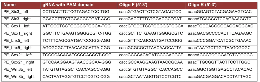
3.6 - Genetic deletions using clustered regulatory interspaced short palindromic repeats (CRISPR-Cas9) ... 43
3.6.1. Design of guide RNA (sgRNA) ... 43
3.6.2. Generation of guide RNA (sgRNA) Cas9 vector ... 45
3.6.3 Miniprep ... 45
3.6.4 Transfection of mESCs ... 45
3.7- Circular chromatin conformation capture (4C)………... 48
3.7.1- 4C library generation ... 48
3.7.2- 4C –seq ... 49
3.7.3- 3C validation of 4C-seq results. ... 50
3.8 - Enhancer reporter assays ... 54
3.9- Polymerase Chain Reaction (PCR) ... 54
3.9.1- RT-qPCR ... 54
3.9.2- ChIP-qPCR ... 57
3.9.3- Colony- PCR ... 59
3.9.4- Clonal genotyping PCR ... 60
3.9.5- 4C –PCR ... 60
3.9.6- 3C-PCR validation of 4C ... 62
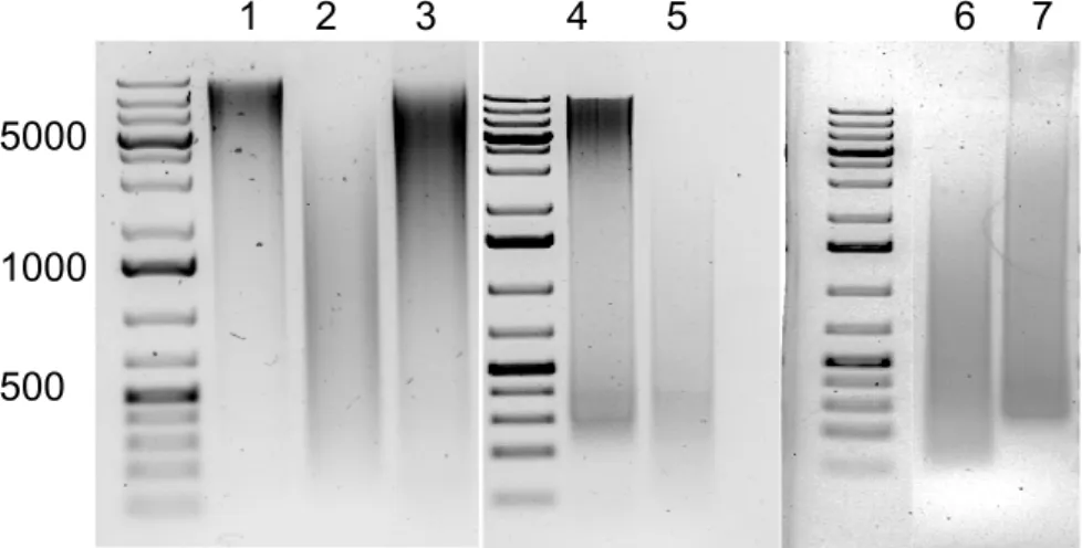
3.9.7- Agarose gel electrophoresis ... 62
3.10 - Statistical Analysis ... 63
3.10.1- RT-qPCR statistical analysis ... 63
3.10.2- ChIP-qPCR statistical analysis ... 63
3.10.3- RNA-seq analysis ... 63
3.10.4- ChIP-seq analysis ... 63
3.10.5- 4C-seq analysis ... 64
3.10.6- Public datasets and additional bioinformatics analysis 64 3.10.7 -Data and Software Availability ... 65
3.10.8– Software and algorithms ... 65
4- Results ... 66
4.1.- Identification and characterization of Poised Enhancers (PEs) in mESCs ... 66
4.1.1- PEs signature in pluripotency ... 67 4.1.2- Distinctive features of mESCs primed, poised and active
enhancers ... 69 4.2- Establishment of a differentiation model for mESCs ... 73 4.3 – Identification of PEs that become active during anterior
neural differentiation ... 75 4.3.1- Functional annotation and characterization of PoiAct
enhancers ... 76 4.4- Identification of putative target genes of selected PEs based on physical interactions ... 79 4.5- PEs are essential for the proper induction of anterior neural
genes ... 80 4.5.1- Additional functional characterization of PE Lhx5 (-109) ... 87 4.5.2- PEs control the expression of the lncRNAs divergently transcribed from the promoters of major anterior neural genes ... 90
4.6- PEs control the induction of anterior neural genes in a
non-redundant manner involving the recruitment of RNA PolII to promoter regions ... 91
4.6.1- PEs are non redundant regulatory elements. ... 91 4.6.2- PEs facilitate the recruitment of the RNA pol II complex to the promoters of their target genes ... 93 4.7. PRC2 as topological facilitator of PEs regulatory activity ... 95 4.7.1- PEs-target gene contacts are PRC2 dependent ... 95 4.7.2- Poised enhancer regulatory activity is facilitated by PRC2 98 4.7.3- H3K27me3 deposition is enabled by the intrinsic genetic features of the poised enhancer regions ... 106
4.8-. Identification of Zic2 as a potential activator of PEs during
AntNPCs differentiation ... 110
5- Discussion ... 112
5.1- Poised enhancers are regulatory sequences associated with the pluripotent cellular state ... 112
5.2- PEs are essential and non-redundant regulatory elements during the induction of major anterior regulatory genes ... 113
5.3- Poised enhancers as part of the “default” induction of anterior neural identity ... 114
5.4- PcG proteins as mediators of short and long-range regulatory interactions ... 115
5.5- PcG complexes as facilitators of gene induction during mESC differentiation ... 116
6- Perspectives ... 120
6.1- In vivo relevance of PEs ... 120
6.2- Mechanism of activation of PEs ... 121
7- References ... 124
Figure index ... 145
Table index ... 148
List of abbreviations ... 151
Eidesstattliche Erklärung ... 154
Abstract
The developmental transitions occurring during embryogenesis involve spatial and temporal changes in gene expression patterns, which are largely dependent on a group of regulatory elements known as enhancers. Enhancers are short DNA sequences able to positively control the expression of their target genes in a distance and orientation independent manner. Previous studies uncovered a unique set of enhancers in pluripotent embryonic stem cells (ESCs), named “Poised Enhancers”. Poised enhancers are marked by repressive histone marks, like the trimethylation of histone three lysine 27, which is deposited by Polycomb repressive complex 2 (PRC2), and also are bound by co-activators like P300. The fact that poised enhancers display both activating and repressing features as well as evidences indicating that they are associated with genes involved in early organogenesis led us to suggest that these regulatory elements are already bookmarked in ESCs and thus primed for their future activation once the differentiation process starts. However, the functional relevance of poised enhancers and the role of their unique chromatin features remain unknown.
To gain insights into these major questions, first, poised enhancers were identified in mouse embryonic stem cells (mESCs) and their activation was evaluated during the establishment of anterior neural identity. Second, using CRISPR/Cas9 technology, poised enhancer candidates were deleted in mESCs, which were then differentiated into anterior neural progenitors. In general, the poised enhancer deletions resulted in severely reduced induction of the poised enhancer’s target genes. Furthermore, circularized chromosome conformation capture coupled to sequencingexperiments revealed that poised enhancers and their target genes physically interacted already in ESCs, thus preceding the activation of poised enhancers and their target genes.
Interestingly, these poised enhancer-target gene interactions observed in mESCs were found to be PRC2 dependent. Additionally, it was demonstrated that while PRC2 was not necessary to maintain poised enhancers in an inactive state in mESCs, was required for the induction of the poised enhancers’ target genes upon anterior neural differentiation. Finally, poised
enhancers were found to frequently reside within a high CpG-dinucleotide genomic context that can directly mediate the recruitment of PRC2.
Overall, these findings demonstrate that poised enhancers are essential for the proper expression of the anterior neural developmental program. Importantly, our work illuminates an unexpected function for PRC2 in promoting neural induction. Our data demonstrates that both poised enhancers and their target genes display intrinsic sequence features that can directly mediate PRC2 recruitment. Consequently, PRC2 can bring poised enhancers and their targets into physical proximity already in mESCs, thus providing a permissive regulatory topology that we propose can facilitate the future induction of mayor anterior neural genes.
Zusammenfassung
Die Entwicklungsphasen während der Embryogenese werden durch räumliche und zeitliche Veränderungen in den Genexpressionsmustern bestimmt, die stark von bestimmten Enhancern reguliert werden. Enhancer sind kurze DNA Sequenzen, die in der Lage sind die Expression von Genen unabhängig von Distanz und Orientierung positiv zu beeinflussen. Einige Studien haben bereits eine spezielle Gruppe von Enhancern in pluripotenten embryonischen Stammzellen (ESC) identifiziert, die "Poised Enhancers" (PE) genannt werden.
PE sind durch repressive Histone-Markierungen, wie z.B., die Trimethylatisierung von Histone-3-Lysine-27 (H3K27me3) gekennzeichnet, welche durch den Polycomb-Repressive-Complex-2 (PRC2) angebracht werden. Gleichzeitig werden sie aber auch durch Co-Aktivatoren wie P300 markiert. Da die PE‘s gleichzeitig aktivierende und repressive Eigenschaften aufweisen und mit bestimmten Genen assoziiert sind, die in der frühen Embryogenese aktiv sind, liegt die Vermutung nahe, dass sie in den ESC vormarkiert werden, um für eine bevorstehende Aktivierung bei einer anstehenden Differenzierung bereit zu sein. Die funktionelle Relevanz der PE‘s und die Rolle ihrer einzigartigen Chromatin Eigenschaften sind bisher noch ungeklärt.
Um diese wichtigen Fragen beantworten zu können, wurden in dieser Arbeit zuerst geeignete PE-Kandidaten in Maus-ESC‘s (mESC‘s) identifiziert und ihre Aktivierung während der frühen neuronalen Entwicklung evaluiert. Zweitens, wurden exemplarisch einige PE-Kandidaten mit Hilfe des CRISPR-Cas9- Systems in mESC entfernt, welche anschließend in anteriore neuronale Vorläuferzellen differenziert wurden (AntNPC). Alle hier erzielten Deletionen der PE führten zu einer stark reduzierten Induktion der PE-Zielgene in den AntNPC. Darüber hinaus zeigten 4C-seq-Experimente, dass PE und ihre Zielgene nicht nur in AntNPC, sondern auch in ESC, d.h. vor der Aktivierung der PE und ihrer Zielgene physikalisch interagieren. Interessanterweise konnte auch nachgewiesen werden, dass die Interaktionen der PE mit ihren Zielgenen von PRC2 abhängig sind. Außerdem wurde hier festgestellt, dass PRC2 nicht
notwendig ist, um die PE‘s in den mESC inaktiv zu halten, jedoch für die Aktivierung des PE-Zielgens während der AntNPC Differenzierung notwendig ist. Anschließend wurde gezeigt, dass PE‘s oft mit einem hohen CpG- dinucleotide Genkontext auftreten, die mit der Rekrutierung von PRC2 in Verbindung stehen.
Zusammenfassend zeigen die hier gezeigten Ergebnisse, dass die PE‘s essenziell für eine normale Expression des vorangehenden neuralen Entwicklungsprogramms sind. Die Daten demonstrieren auch, dass beide, die PE und ihre Zielgene, intrinsische Sequenzeigenschaften besitzen, welche die PRC2-Rekrutierung beeinflussen. Dementsprechend kann PRC2 die PE‘s und ihre Zielgene bereits in mESC in physikalische Nähe zueinander bringen und so eine permissive regulatorische Topologie bereitstellen, welche die zukünftige Induktion der anterioren neuronalen Vorläufergene erlaubt.
1- INTRODUCTION
1.1- In vitro modeling of in vivo embryogenesis
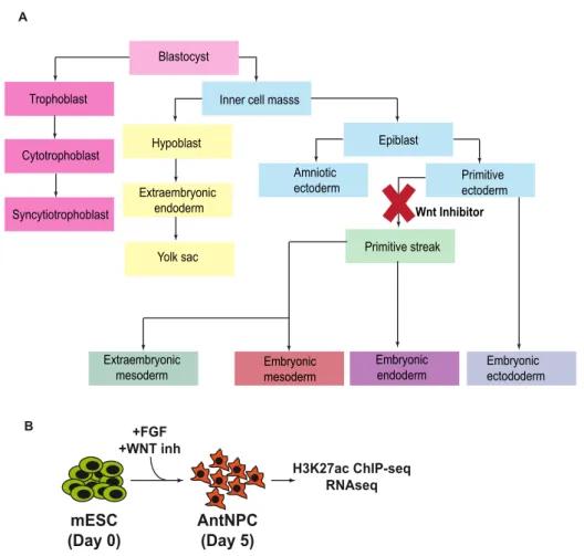
Embryogenesis is the process of cell division and cellular differentiation of the embryo from the one-cell state or zygote to an adult being. In order to study this process, ESCs derived from the epiblast of mammalian embryos can be used as an in vitro highly tractable model. ESCs possess two fundamental characteristics: self-renewal and pluripotency. Self-renewal is the capability of replication of a cell into the same cell state and pluripotency is the ability of a single cell to generate all the cell lineages. In the case of mouse embryonic stem cells (mESCs), pluripotency can be captured in culture under different conditions (see methods 3.32 and 3.33) and each condition represents a different pluripotency state. Naïve pluripotency, which characterizes in vivo the epiblast cells of the mouse pre-implantational blastocyst, can be recapitulated in vitro by the addition of MEK and GSK3 inhibitors (i.e. 2i conditions) (Hackett and Surani 2014). Formative pluripotency represents a more advance stage distinctive of the post-implantaton epiblast, represented in vitro by epiblast-like cells (EpiLC) (Hayashi et al. 2011). Finally, as development proceeds and gastrulation starts, the epiblast cells aquire a primed pluripotent state in which somatic lineage specifiers start to be expressed and that is recapitulated in vitro by epiblast stem cells (EpiSC) (Smith 2017; Kalkan and Smith 2014).
Stem cell fate is determined by the equilibrium between self-renewal and differentiation signals. ESCs are able to respond to the signaling environment and either maintain the core pluripotency program or to exit the pluripotent state when differentiation signals appear. To initiate differentiation, ESCs have to escape from self-renewal, the transcriptional networks providing stem cell identity need to be extinguised and dififferentiation towards a specific lineage has to be specified. ESC differentiation involves the silencing of Nanog, Pou5F1 and other major pluripotency regulators. Moreover, lineage specific transcription factors (TFs) need to be induced in order to initiate the expression of a specific differentiation program (Young 2011). Ultimately, all these
processes are controlled at the chromatin level where transcriptional regulation is mediated by cis-regulatory elements like enhancers, promoters, silencers and insulators (Calo and Wysocka 2013; Signolet and Hendrich 2015).
Enhancers in particular, play a key role during the exit of pluripotency and subsequent differentiation by controlling the specific set of genes that any given cell type expresses (de Laat and Duboule 2013).
Lastly, during the exit of pluripotency, Polycomb group proteins (PcG) play also an important role. These proteins can act as transcriptional activators in certain cellular and genomic contexts as well as mediators of both short and long- range genomic interactions that contribute to global genome architecture (Bantignies et al. 2011; Creppe et al. 2014; Denholtz et al. 2013; Entrevan et al. 2016; Joshi et al. 2015; Kondo et al. 2014; Kondo et al. 2016; Lanzuolo et al. 2007; Morey et al. 2015; Schoenfelder et al. 2015).
This work is focused on the role of a unique group of enhancers, called poised enhancers as well as the function of PRC2 during mESCs differentiation.
1.2- Enhancers regulate cell type transitions
Enhancers are cis-regulatory elements that positively control gene expression over long distances and in an orientation independent manner (Spitz and Furlong 2012). They are compact DNA sequences (200-500 bp) that act as a binding platforms for TFs (Figure 1.1) and play an important role in driving cell- type specific gene expression (Bulger and Groudine 2011; Yáñez-Cuna et al.
2013). TFs can bind to enhancers in a combinatorial and modular manner, allowing the establishment of different spatiotemporal gene expression programs (Figure 1.1) (Spitz and Furlong 2012). The importance of enhancers during embryogenesis is well illustrated in several studies where deletions of specific enhancers in animal models cause developmental abnormalities and affects gene expression patterns (Sagai et al. 2005).
Enhancer 1
TF TF
Target gene
H3K4me1 H3K4me1
H3K27ac H3K27ac
Enhancer 2 Target gene
expression pattern
Insulators/
TAD borders
Architectural proteins Silencer
Activator
Positive regulation Negative regulation Tissue specific TF Signaling effector TF TF TF
Enhancer
Figure 1.1: Genetic and epigenetic features of enhancers. Enhancers are able to confer cell type and developmental-stage specific spatiotemporal gene expression patterns by serving as integrating platforms of different types of developmental information (tissue-specific and signaling-dependent TFs, epigenetic modifications, recruitment of co-activators and architectural proteins). The regulatory influence of enhancers on the expression of nearby genes is delimited by insultators. (Adapted from Noonan, 2009).
1.2.1- Identification and characterization of enhancers
Despite their relevance, enhancers were difficult to identify due to the fact that they can drive transcription in a distance and orientation independent manner with respect to their putative genes, they do not present a stereotypic sequence composition and, in addition, they are not placed at a fixed position or distance with respect to their target gene promoters. However, this dramatically changed with the emergence of epigenomic approaches that based on the presence of particular chromatin signatures, allowed the global
identification of enhancers in a sequence conservation and cell type independent manner. Recent advances in genomic techniques like chromatin immunoprecipitation coupled to sequencing (ChIP-seq), (see methods 3.5.1) have enabled the global mapping of TFs and histone modification binding profiles. Importantly, enhancers can be identified using a variety of chromatin features, including the presence of certain histone marks, binding of transcription factors or chromatin accessibility (Figure 1.1) (Boroviak et al.
2015; Kalkan and Smith 2014; Ying et al. 2008; Buenrostro et al. 2015; Menno P. Creyghton et al. 2010; Heintzman et al. 2009; Rada-Iglesias et al. 2011).
Classically, the target genes of enhancers have been inferred based on correlative observations like proximity. However, enhancers can control the expression of genes located at great distances, which tipically involves the physical proximity between enhancers and their target genes (Calo and Wysocka 2013). Therefore, enhancers do not necessarily control the expression of their nearest genes. Chromatin Conformation Capture (3C) technologies and their derivatives allow evaluating the physical interaction between different genomic loci (de Wit and de Laat 2012; Duan et al. 2012;
Simonis, Kooren, and de Laat 2007; Splinter et al. 2012; van de Werken et al.
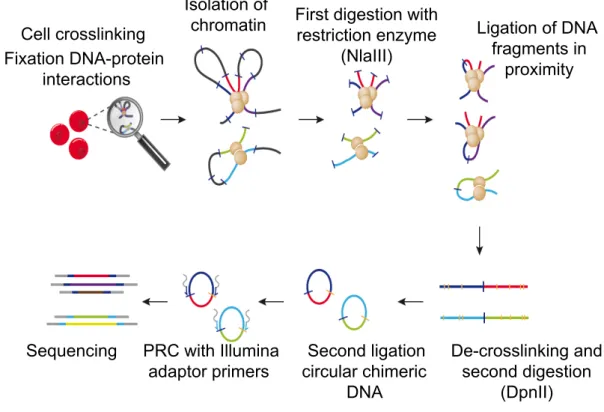
2012). For example, by using Circular Chromatin Conformation Capture coupled to sequencing (4C-seq), the genes that physically interact with the enhancers of interest and that are likely to represent their target genes can be identified (Figure 1.2).
Cell crosslinking! Fixation DNA-protein
interactions
Isolation of
chromatin First digestion with restriction enzyme
(NlaIII)
Ligation of DNA fragments in
proximity
De-crosslinking and second digestion
(DpnII) Second ligation
circular chimeric DNA PRC with Illumina
adaptor primers Sequencing
Figure 1.2: 4C-seq methodology (adapted from Stadhouders et al. 2013). Cells are crosslinked in order to fix the interactions between DNA and proteins. After nuclei isolation, chromatin is digested and then ligated in order to evaluate the contacts of DNA fragments that are in proximity. Following reverse crosslinking, a second round of digestion-ligation is performed. Finally chimeric circular DNA molecules are obtained representing the contacts between different loci which can be amplified by inverse PCR using primers located within a locus of interest (bait or viewpoint) and subsequently sequenced (see methods 3.7).
Enhancers were originally characterized using transient reporter gene assays in cultured cell lines, in which enhancer activity is tested in an exogenous and artificial genomic context (Banerji, Rusconi, and Schaffner 1981; Bulger and Groudine 2011). Since then, new methodologies, such as CRISPR-Cas9, have arised to characterize and determine the function of enhancers within their endogenous genetic and chromatin context. Briefly, this technique consists of gRNA molecules that guide the Cas9 nuclease to specific loci to introduce double-stranded DNA breaks at the desired location. Consequently, these DNA breaks can be repaired by non-homologous end-joining repair (NHEJ), which can result in the deletion of the sequence of interest (e.g. enhancer), whose function can then be evaluated.
1.2.2- Enhancer states
These epigenomic methods also revealed that enhancers can exist in various regulatory states (e.g. active, primed, poised), which can be distinguished based on unique chromatin signatures and that display distinct regulatory properties (Table 1.1) (Calo and Wysocka 2013; Menno P. Creyghton et al.
2010; Rada-Iglesias et al. 2011; Zentner et al. 2011).
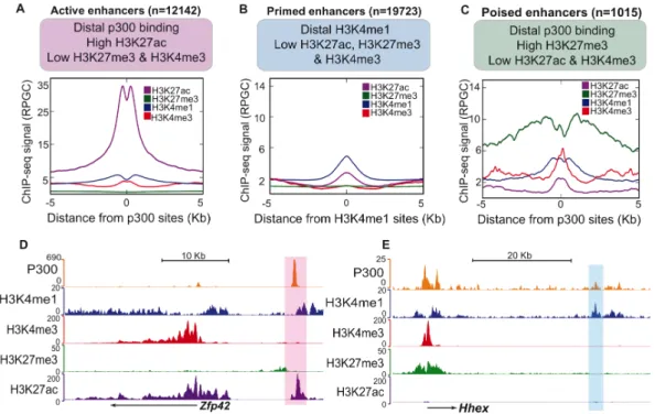
Active enhancers present low nucleosomal density, are bound by TFs and co- activators (e.g. p300/CBP, CHD7, BRG1), are flanked by nucleosomes marked with histone three lysine four monomethylation (H3K4me1) and histone three lysine 27 acetylation (H3K27ac) and they are associated with highly expressed genes (Menno P. Creyghton et al. 2010; Rada-Iglesias et al. 2011; Zentner et al. 2011).
Primed enhancers are marked with H3K4me1 but not H3K27ac. They are linked to genes that exhibit moderate expression levels and are implicated in a broad range of biological processes (Menno P. Creyghton et al. 2010; Zentner, et al. 2011).
Poised enhancers (PEs) were defined originally in human and mouse embryonic stem cells (hESC, mESC) as a small group of highly conserved regulatory elements. Like active enhancers, they are also bound by TFs and co-activators (p300/CBP, CHD7, BRG1) and display low nucleosomal density.
However, in contrast to active enhancers, they are not marked with H3K27ac but with histone three lysine 27 trimethylation (H3K27me3) instead, a repressive histone modification mediated by Polycomb Repressive Complex two (PRC2) (Rada-Iglesias et al. 2011; Zentner et al. 2011). Importantly, PEs are in proximity of genes with major cellular identity and developmental functions that are inactive in ESC and become expressed upon somatic differentiation. Since PEs are already bound in ESC by co-activators like P300, it has been proposed that they represent developmental enhancers that are bookmarked for their future activation once the appropriate differentiation signals are received (Rada-Iglesias et al. 2011; Spitz and Furlong 2012).
Additionally, it has also been proposed that the presence of H3K27me3 and PRC2 might keep PEs in an inactive state, preventing their spurious activation until the relevant differentiation cues become available (Tie et al. 2016).
Table 1.1: Types of enhancers and their epigenetic features
1.2.3- Mechanism of action of enhancers
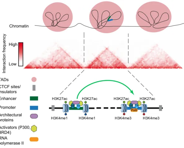
Since the discovery of enhancers, the dominant model to explain their mechanism of action involves the physical interaction of enhancers with their target promoters through the formation of chromatin loops. This “looping model” (Bulger and Groudine 2011; Calo and Wysocka 2013; de Laat and Duboule 2013; Vernimmen and Bickmore 2015) has received extensive experimental support from chromosome conformation capture related technologies (e.g. 4C-seq, Hi-C, ChIA-PET) that enable the detection of physical interactions between distally located loci. Interestingly, most enhancer-promoter interactions occur within Topologically Associated Domains (TADs), which represent self-interacting Mb-scale domains that physically restrain enhancer activity to genes located within the same TAD (Figure 1.3) (Dixon et al. 2012). Although enhancer-promoter communication was originally proposed to occur once the target genes become active (Vernimmen et al.
Properties Active enhancers
Primed enhancers
Poised enhancers
DNAse I
sensitivity
Yes Not reported Yes
H3K4me1 Yes Yes Yes
H3K27ac Yes No In differentiated
cells
H3K27me3 No No In ESC
Activator TFs Yes No Yes
Associated genes
High expressed genes
Non specific Developmental regulators
2007), recent observations indicate that enhancer-promoter contacts can precede gene activation (Ghavi-Helm et al. 2014; Jin et al. 2013). At least in some cases, these pre-formed contacts might confer a permissive chromatin topology that poises genes for future induction (de Laat and Duboule 2013).
Interaction frequency
Low High
Chromatin
TADs CTCF sites/
Insulators Enhancer
Promoter TF
H3K4me1 H3K4me1 H3K27ac H3K27ac
TF
H3K4me3 H3K4me3 H3K27ac H3K27ac
TF TF PolII
Architectural proteins
Activators (P300, BRD4)
RNA
polymerase II
Figure 1.3: Chromatin landscape and mechanism of action of enhancers. The three-dimensional nuclear configuration of mammalian genomes is organized in topological associated domains (TADs). A TAD represents a region of DNA where physical interactions occur relatively frequently, whereas interactions across a TAD boundary occur relatively infrequently. Enhancer-promoter interactions occur within the same TAD, and they are facilitated by the numerous TFs, activators and architectural proteins that are associated to these regions. Figure adapted from (Dixon et al. 2012).
The activation of gene promoters requires that many transcriptional components come together to assemble the pre-initiation complex (PIC), initiate transcription, overcome the paused RNA polymerase II (PolII) and eventually drive transcriptional elongation. By forming loops, enhancers come in close proximity with their target promoters and facilitate the aforementioned process by increasing the concentration of TFs and co-activators around the
promoter regions. Some of these factors include architectural proteins such as cohesin as well as mediator complexes and other factors (e.g. P300, BRD4) involved in releasing paused PolII and initiation of transcriptional elongation (Heinz et al. 2015) (Figure 1.3). Interestingly, previous studies have demonstrated that removal of the promoter does not affect the recruitment of the PolII to the enhancer region, while the enhancer deletion does affect the binding of PolII to the promoter (Vernimmen et al. 2007). It is worth mention that enhancers are also transcribed (enhancer RNAs or eRNAs), although at low levels, and their expression correlates positively with the mRNA levels at their target genes (Vernimmen and Bickmore 2015).
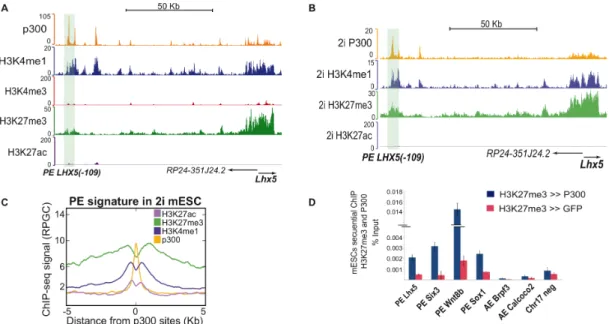
It has been described that PEs already communicate with their putative target genes in undifferentiated ESC (Schoenfelder et al. 2015). However, the molecular basis and functional relevance of these preformed PE-promoter contacts is currently unknown. Similarly, it is not clear yet if and how PEs control the expression of their target genes once activated.
1.2.4- Poised enhancers during pluripotency and differentiation
Previous reports (M. P. Creyghton et al. 2010; Rada-Iglesias et al. 2011) have determined that poised enhancers are developmental regulatory elements that are found in a silent but primed state in undifferentiated ESC. As mentioned above, one major distinctive feature of PEs is that they are bound by PRC2 and, hence, marked with H3K27me3. Upon ESC differentiation, PEs get activated, lose H3K27me3 and gain H3K27ac (Rada-Iglesias et al. 2011;
Zentner et al. 2011). These two histone modifications are mutually exclusive, implying that, despite being bound by co-activators with histone acetyl- transferase (HAT) activity (e.g. P300, CBP) (Rada-Iglesias et al. 2011), the presence of H3K27me3 at PEs might avoid the accumulation of H3K27ac and the spurious activation of these regulatory elements (Pasini et al. 2010;
Reynolds et al. 2012; Tie et al. 2016). Interestingly, this data is in concordance with the classically view of Polycomb group proteins as major epigenetic repressors playing important roles in differentiation and maintenance of cellular identity (Di Croce and Helin 2013). Additionally, some studies reported that PEs were also found in non-pluripotent cells and therefore, their relevance was
questioned since they were proposed to display a repressed rather than poised state or to even act as silencers (Bonn et al. 2012; Entrevan et al. 2016; Kondo et al. 2016; Spitz and Furlong 2012). However, in these and other studies the identification of PEs in ESC was based on the presence of H3K4me1 and/or H3K27me3 but the binding of TFs/co-activators was not required (Bonn et al.
2012; Menno P. Creyghton et al. 2010; Schoenfelder et al. 2015).
Due to the association of PEs with major cell identity and developmental regulators, it has been previously suggested that PEs might play important roles during the execution of somatic differentiation programs (Heintzman et al.
2009; Bulger and Groudine 2011; Rada-Iglesias et al. 2012). However, we still do not know which mechanisms lead to lineage-specific activation of poised enhancers. Most importantly, the functional relevance of PEs is only supported by correlative observations, including reporter assays in zebrafish embryos that demonstrated that human poised enhancers sequences can act as developmental enhancers in vivo (Rada-Iglesias et al. 2011).
In conclusion, the importance of PEs for the induction of their cognate genes and the execution of somatic differentiation programs has not been formally demonstrated. Correspondingly, it is also unknown if and how the PEs chromatin features actually facilitate the future activation of the PE target genes. (Rada-Iglesias et al. 2011; Zentner et al. 2011). Therefore, additional research is needed in order to elucidate these important questions.
1.3- Polycomb group proteins
Polycomb group proteins (PcG) are a large, conserved and diverse family of epigenetic regulators of transcription. Historically they have been considered as repressors of homeotic (HOX) genes, but more recently it has been revealed that they have broader roles in stem-cell identity, differentiation and disease (Chittock et al. 2017; Di Croce and Helin 2013; Pasini et al. 2010;
Schoenfelder et al. 2015).
PcG proteins are part of transcriptional-repressive complexes, termed Polycomb Repressive Complexes (PRCs). Two major PRCs have been identified in most metazoan species, PRC1 and PRC2. In mammals, the canonical PRC1 contains two core subunits, RING1 which monoubiquitinates histone H2A on lysine 119 (H2AK119ub) and PcG ring finger protein (PCGF), which interacts with specific binding partners. PRC1 also contains a chromobox protein (CBX) which binds the H3K27me3 mark and a Polyhomeotic (PH) homolog protein (PHC). PHC proteins might play an important role in higher order chromatin organization by forming long-range contacts between remote PRC-bound sites in the genome (Figure 1.4) (Chittock et al. 2017; Di Croce and Helin 2013; Entrevan et al.2016).
Canonical PRC1 PRC2
RING1 CBX PCGF PHC
SUZ12 EED
EZH1/2 RbAp48
- Ubiquitination of H2AK119 - Binding to H3K27me3 - Chromatin compactation - Blocking RNA PolII elongation
- Chromatin compactation - Methylation of H3K27
JARID2
Figure 1.4: Composition of Polycomb group proteins. PcG consist of two main complexes, PRC1 and PRC2. PRC1 contains RING1, PHC, CBX and PCGF subunits (light-blue) and is responsible of ubiquitinilation of H2AK119, recognition of H3K27me3, chromating compactation and PolII inhibition. PRC2 consist of Suz12, EED and EZH the core subunit responsible of the methylation of H3K27. In addition, PRC2 also contains RbAP48, a protein which stabilizes the whole complex and Jarid2, a cofactor that facilitates the recruitment of PRC2 to its genomic targets (Entrevan et al. 2016; Blackledge, Rose, and Klose 2015; Di Croce and Helin 2013;
Sanulli et al. 2015; Chittock et al. 2017).
The PRC2 core complex has three main proteins: EZH1/2, EED and SUZ12 The SET (enhancer of zeste) domain-containing proteins responsible of methylation of H3K27, EZH1 and EZH2, are mutually exclusive and differentially expressed in proliferating and non dividing tissues (Entrevan et al.
2016). EED (Embryonic ectoderm development) binds to H3K27me3 and contributes to the propagation of this repressive mark. SUZ12 (suppressor of zeste) is also required for the histone methyltransferase catalytic activity of the PRC2 complex. In addition, PRC2 also contains other subunits such as RbAp48, which stabilizes the complex, and JARID2, a co-factor which recruits PRC2 to its genomic targets (Figure 1.4) (Chittock et al. 2017; Di Croce and Helin 2013; Entrevan et al. 2016; Peng et al. 2009).
1.3.1- Role of PcG in gene regulation
Polycomb proteins are present at repressed genes with developmental functions. PcG repressive function entails chromatin compaction, mediated by PRC1, block of HATs (Histone Acetyltransferases) and interference with RNA Polymerase two (PolII) activity. PRC1 chromatin compaction activity involves the ligase activity of RING1 (Bantignies et al. 2011; Francis 2004) as well as the function of other PRC1 canonical subunits (e.g. CBX, PH) which can lead to the formation of subnuclear compartments, known as Polycomb bodies (Creppe et al. 2014; Schoenfelder et al. 2015, 1; Isono et al. 2005).
On the other hand, PcG binding at gene promoters can interfere with transcription by “holding” PolII at the transcription start sites (TSS). This results in a “paused” PolII, unable to elongate. In agreement with this function, the deletion of RING1 results in PolII phosphorylation at serine2 (S2) and increased transcriptional elongation (Stock et al. 2007) Moreover, this PolII pausing function of PcG complexes might also explain the existence of so called bivalent promoters, which are enriched in H3K27me3 and histone three lysine four (H3K4me3). These bivalent promoters are typical of developmental
genes and particularly abundant in ESC where they are proposed to bookmark genes for subsequent activation upon differentiation.
1.3.2- PcG in pluripotency and differentiation
PcG proteins are essential for embryonic development and, mice lacking EZH2, EED or SUZ12 die soon after implantation (Faust et al. 1998; O’Carroll et al. 2001; Pasini et al. 2004). On the other hand, PRC2 activity is not required for mESCs self renewal although it appears necessary for proper differentiation. Nevertheless, EED-/- mESCs, which display very low PRC2 activity and H3K27me3 levels, are able to contribute to all embryonic tissues when injected into mouse blastocyst, indicating that PRC2 null mESCs are to some extent pluripotent (Chamberlain et al. 2008).
On the contrary, the role of PRC1 in ESC self-renewal and differentiation is not as clear due to the large variety of PRC1 complexes and associated subunits.
It has been reported that, with the exception of RING1B, the deletion of most PRC1 subunits leads to late developmental defects during mouse embryogenesis. However, embryos with deletion of at least two PRC1 subunits (two different ring finger subunits (Akasaka et al. 2001) or ZFP144 ring finger protein and RING1B (Isono et al. 2005)) failed to pass midgestation.
Taken together, the data suggest that in vitro, PcG proteins are not required for mESC self renewal but can affect pluripotency and differentiation potential (Riising et al. 2014).
1.3.3- Recruitment of PcG
Previous studies reported that the recruitment of canonical PRC1 complexes depends on the presence of the H3K27me3 mark deposited by PRC2, which is recognized by the chromodomain of the CBX subunits (Min et al. 2003). On the other hand, other studies described that PRC1 complexes are targeted to CpG islands (CGIs) by the KDM2B histone demethylase. PRC1 then catalyzes H2A ubiquitination, which in turn can recruit PRC2 (Blackledge et al. 2014).
Although PcG recruitment mechanisms are not fully understood, CGIs have
recently emerged as potential Polycomb response elements (PREs) in vertebrates, similar to those previously described in Drosophila (Entrevan et al.
2016). Different mechanisms have been suggested for the recruitment of PRC2 to CGIs. JARID2, a PRC2 co-factor, has a DNA binding domain that preferentially binds to CpG-rich sequences and could thus help in the recruitment of PRC2 to CpG islands (CGIs). Accordingly, the loss of JARID2 leads to a decrease in PRC2 binding to their target genes (Di Croce and Helin 2013; Peng et al. 2009). More recently, non-canonical PRC1 complexes containing RYBP (RING1A and YY1 binding protein) and KDM2B were shown to bind to CGIs in the absence of H3K27me3 (van den Boom et al. 2016). For instance, the histone lysine demethylase KDM2B, has been reported to specifically recognize non-methylated DNA in CGIs and recruit PRC1 (Farcas et al. 2012). RYBP and its mouse homolog YAF2 are responsible for PcG recruitment to DNA, which is mediated by YY1 TF DNA binding (Basu et al.
2014).
Recent studies indicate that the transcriptional status at CGIs can determine whether PcG complexes are recruited to their target genes, since global inhibition of transcription lead to ectopic recruitment of PcG proteins to all CGIs (Riising et al. 2014). In addition to CGI, sequence-specific TFs like SNAIL, REST or PLFZ can regulate the recruitment of PRC2 to their target genes although this does not seem to be a general mechanism for PcG recruitment, and TFs may only transiently associate with PcG (Dietrich et al. 2012). Lastly, several studies propose that noncoding RNAs have a role in gene silencing and PcG-protein recruitment. RNA-immunoprecipitation techniques have identified several thousand of RNAs associated with PRC2 and furthermore, it has been demonstrated that the long noncoding Xist transcript can interact with PRC2 during the inactivation of the X chromosome (Plath et al. 2003).
However, most recently, RNA binding to PcG proteins has been suggested to facilitate the eviction rather than the recruitment of these complexes to their chromatin targets (Aranda et al. 2015).
In summary, different models of recruitment of PcG proteins have been proposed, although most likely several of the suggested mechanisms might act
in a combinatorial manner to confer robustness to the binding of these important epigenetic repressors to their cognate genomic targets.
1.3.4- PcG as major organizers of nuclear architecture
It is known that cell fate transitions are accompanied by alterations in chromatin structure. The action of PcG on chromatin leads not only to compaction, but also to specific 3D chromatin folding (Bantignies et al. 2011).
Due to the appearance of novel approaches to study genome nuclear organization, such as 3C and derivative techniques (e.g. 4C-seq, Hi-C), it is now possible to elucidate the topological organization of chromatin within the nucleus.
PcG are starting to emerge as major organizers of nuclear architecture. For example, it has been demonstrated that the repressive chromatin hubs found at the homeotic bithorax complex (BX-C) locus in Drosophila are composed of multiple chromatin loops. In these loops, all major interacting elements, including core promoters and PREs, are bound by PcG. As a result, a topologically complex structure referred to as a PcG body is formed and required for the silencing of BX-C (Lanzuolo et al. 2007). Furthermore, it has been also shown that switches in the BX-C transcriptional status are accompanied by major rearrangements in the high order chromatin structures (Lanzuolo et al. 2007). On the other hand, in mECSs it has been revealed that EED, one of the PRC2 subunits, is required for the maintenance of interactions between PcG regions separated by tens of megabases or even located on different chromosomes (Denholtz et al. 2013). These interactions were reduced when mESCs were grown under naïve pluripotency conditions, most likely due the lower genomic levels of PcG complexes and associated histone marks (e.g. H3K27me3) present under those conditions (Tee et al. 2014).
Similarly, it has been recently reported that RING1 depletion in mESC leads to the lost of long-range interactions between PcG target genes, while interactions between active pluripotency genes remained unaffected (Schoenfelder et al. 2015). Moreover, PcG-bound promoters also interacted with poised enhancers marked by H3K4me1 and H3K27me3. Remarkably,
loss of RING1 led to activation of poised enhancers, whereas poised enhancer–promoter contacts were overall maintained. Lastly, it has been also proposed that during mouse brain development, the Meis2 gene promoter is initially associated with a silencing element via RING1B. Interestingly, this PcG-bound element topologicaly facilitates the subsequent interaction between Meis2 and a specific enhancer, ultimately leading to gene activation. When RING1B is not present, the enhancer can no longer contact the promoter region, resulting in reduced Meis2 expression (Kondo et al. 2014).
Since TADs were discovered, research efforts have been invested in understanding the link between chromatin 3D structure and gene expression.
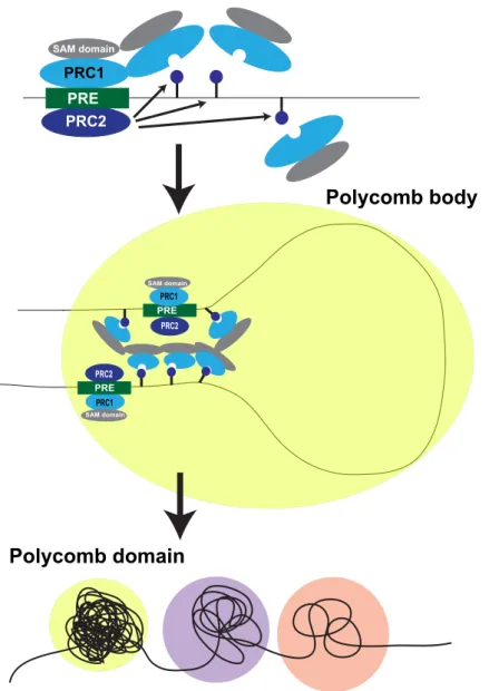
In Drosophila, TADs can be broadly classified into active, inactive and PcG- repressed states. PcG TADs are the most compact, show the highest inter- TAD interactions and display minimal contacts with other domains. On the other hand, active and transcriptionally inactive TADs show higher levels of inter-domain mixing. It has been illustrated that PH, a component of Polycomb PRC1, plays an important role in these interactions and without PH, PcG TADs are lost (Boettiger et al. 2016). In agrrement with this study, it was shown that the disruption of the polymerization activity of the PH sterile alpha motif (SAM) domain leads to a dispersion of PcG clusters and chromatin interactions.
Moreover, similar findings were recently reported in mouse cells (Isono et al.
2013). Overall, these recent reports indicate that PH SAM domains may mediate the organization of PcG proteins into Polycomb bodies (Figure 1.5) (Wani et al. 2016).
SAM domain
PRC1 PRC2 PRE
SAM domain
PRC2 PRE PRC1
SAM domain
PRC1 PRE PRC2
Polycomb body
Polycomb domain
Figure 1.5: Proposed model for Polyhomeotic (PH) SAM domains in the formation of Polycomb bodies. PcG complexes are recruited to their target sites via PREs and PcG polymerization occurs through SAM-mediated interactions. PcG- mediated looping might involve the recognition of H3K27me3 (dark blue lollipop) by PRC1 and the PRC1 polymerization mediated by PH SAM domains. These PRC1/PRC2 clusters correspond to microscopically visible subnuclear domains known as Polycomb bodies. These Polycomb bodies or Polycomb domains (yellow circle) are significantly more compacted than transcriptionally inactive (purple) or active (pink) domains. Adapted from (Entrevan et al. 2016).
The role of PRC2 in regulating global nuclear architecture is still not very clear.
As it was mentioned previously, the loss of EED in mESCs leads to reduced interactions between Polycomb-bound regions (Denholtz et al. 2013; Joshi et al. 2015). However, it was also reported that the loss of PRC2 did not affect TAD boundaries either globally or across the X chromosome (Nora et al.
2012). Nevertheless, it has been demonstrated that induced recruitment of
EZH2 to a particular locus can lead to relocation into a new nuclear compartment occupied by other Polycomb-bound regions. This study suggests that specific nuclear subcompartments, including PcG domains, are formed as a consequence of affinities between chromatin-associated proteins or modifications (Wijchers et al. 2016).
In summary, PcG proteins can mediate looping interactions between regulatory elements, define global nuclear architecture and ultimately regulate gene expression. However, the role of PcG upon PEs activation and lineage commitment needs to be further characterized.
2- AIM
The knowledge about the establishment of gene expression programs during mammalian developmental transitions is far from being completed. We hypothesize that poised enhancers are important regulatory elements that, in ESCs, display a silent but primed state sustained by PcG. Moreover, we also hypothesize that, once activated, poised enhancers might be important for the induction of their target genes during the earliest steps of somatic differentiation. Therefore, the major goals of this project are to examine the functional and developmental relevance of poised enhancers and to investigate how PcG influences poised enhancer’s regulatory activity.
In order to fulfill these objectives, we first characterized poised enhancer candidates with well-defined activation dynamics during mESCs differentiation into anterior neural progenitors. Secondly, we identifed the putative target genes of poised enhancers based on their physical interactions. Then, we engineered poised enhancer deletions to assess the functional importance of these regulatory elements during mESCs differentiation. Lastly, we examined the role of PcG, specially the PRC2 complex as a regulator of poised enhancers activity and function.
3. MATERIAL AND METHODS 3.1 – Equipment
Table 3.1: Equipment
Equipment Company Catalog number
Bacteria Incubator Infors HT Ecotron s00120638
Cell culture Incubator Sanyo 8070263
Microscopes
Inverted microscope Leica DMILED 376977 Fluorescent microscope Olympus IX2-UCB 9A01123 Microscope ECLIPSE TS100 Nikon Kurian’s Lab Electrophoresis
Chambers Biorad 1704502 and 1704406
Power supply Biorad 041BR110323
Western Blot
Chamber Biorad 1658004
Power supply Biorad 043BR500041
Centrifuges
Cell culture Hermle 311001101
Big 4° for falcon tubes Hermle 31130026
Small 4° for eppendorf tubes Hermle 61150064 For bacterial liquid culture Labscience 7.601.314.101
Cell culture bath Memmert 325741
Tissue Culture hood Kojair 22198
Bioruptor plus Diagenode Brng130125
PRC Thermal cycler
q-PCR LightCycler 480 II Roche 5662
c1000 Touch Biorad ct024292
Electic balance Kern WB1251009
Regular balance Kern WB1300143
Spectrophotometer Nanodrop Thermo Scientific F673
pH meter Sartorius 29053099
Automated cell counter Biorad 508BR05586
Shaker Skyline 12DE117
Thermo Block Ditabis 980052301
ChemiDoc MP Biorad 731br01726
3.2- Chemicals and reagents
3.2.1- ChemicalsTable 3.2: Chemicals
Reagent Company Catalog number
2-Propanol Roth 9866.5
25:24:1 Phenol- Chlorophorm Isoamyl
Alcohol Sigma Aldrich P2069
4-(2-hydroxyethyl)-1-
piperazineethanesulfonic acid
(HEPES) Roth HN78.2
30% Acrylamide/ Bis solution Sigma Aldrich 161-0156 Adenosine triphosphate (ATP) Sigma Aldrich A2383
Agarose Life Technologies 16500100
Bovine Serum Albumina (BSA) Roth, Germany 3737.2
Bromophenol Blue Sigma Aldrich B0126
Chloroform Sigma Aldrich 366919
DAPI Sigma Aldrich D9542-5MG
Dithiothreitol (DTT) Roth 6908.3
Ethanol Roth 5054.3
ethylendiaminetetraacetic acid (EDTA) Roth 8043.1 ethylene glycol-bis(β-aminoethyl
ether)-N,N,N',N'-tetraacetic acid) EGTA Roth 3054.2 Formaldehide solution 37% Sigma Aldrich 252549
Glycerin Sigma Aldrich 68898
Gycerol Roth 3783.2
Hydrochloric acid (HCl) Roth 281.1
Litium Chloride (LiCl) Roth 3739.2
Magnesium Chloride (MgCl2) Roth KK36.2
Methanol Sigma Aldrich 494437
Mounting medium Southern Biotech 0100-01
N-Lauroylsarcosine Sigma Aldrich 61743
Na-Deoxycholate Sigma Aldrich D678
Non-Fat Milk Hartestein CM35
NP-40 Sigma Aldrich I3021
Parafolmaldehide Sigma Aldrich 158127
Phosphate buffered saline (PBS) Sigma Aldrich D8537
Potasium Chloride (KCl) Roth HN02.3
Sodium Acetate (C2H3NaO2) Roth 6773.2
Sodium Chloride (NaCl) Roth 3957.2
Sodium dodecyl sulfate (SDS) Roth 183.3
Triton X-100 Roth 3051.4
Trizma Base Sigma Aldrich T1503
Trizol (Tripure) Roche 11667157001
Tween-20 Roth 9127.2
3.2.2- Buffers and solutions Table 3.3: Buffers
Buffer/ Solution Composition
Blocking Solution ChIP PBS (1x)
0.5% BSA (w/v)
Lysis Buffer 1 (LB1) 50 mM Hepes (pH 7,5)
140 mM NaCl 1 mM EDTA 10% glycerol 0.5% NP-40 0.25% TX-100 dH20
Lysis Buffer 2 (LB2) 10 mM Tris pH 8
200 mM NaCl 1 mM EDTA 0.5 mM EGTA dH20
Lysis Buffer 3 (LB3) 10 mM Tris pH 8
100 mM NaCl 1 mM EDTA 0.5 mM EGTA
0.1% Na-Deoxycholate 0.5% N-lauroylsarcosine dH20
RIPA Wash Buffer for ChIP 50 mM HepespH 7,5 500 mM LiCl
1 mM EDTA 1% NP-40
0.7% Na-Deoxycholate dH2O
Elution Buffer 50 mM Tris pH 8
10 mM EDTA 1% SDS dH2O
Dilution Buffer (Sequential ChIP) 20 mMTris-HCl pH 8 2 mMEDTA
1% Triton X-100 150 mM NaCl dH2O
RIPA Wash Buffer for Western Blot 50 mM Tris, pH 8.0 150 mM NaCl 1.0 % NP-40
0.5% sodium deoxycholate 0.1% SDS
dH2O
Laemmli buffer 375 mM Tris pH 6.8
60% glycerol 600 mM DTT
0.06% bromphenol blue 12% SDS
dH2O
Running buffer 25 mM Tris pH 8,3
190 mM glycine 0.1% SDS dH2O
Transfer buffer 25mM Tris
190 mM glycine 20% Methanol 0.1 % SDS dH2O
PBST washing buffer PBS
1% Tween Blocking Solution for Western Blot 5% milk
1% PBST
Permeabilizing solution IF 0.3% Triton X-100 dH2O
Lysis Buffer for 4C 50 mM Tris–HCl pH 7.5
150 mM NaCl 5 mM EDTA 0.5% NP-40 1% TX-100
1X protease inhibitors dH2O
Ligation Buffer for 4C 50 mM Tris-HCl pH 7.6 10 mM MgCl2
1 mM ATP 1mM DTT dH2O
TE Buffer 10 mM Tris pH 8.0 1 mM EDTA
dH2O
TAE buffer (1X) 40mM Tris pH 8.6
20mM Acetate 1mM EDTA dH2O 3.2.3- Kits and commercial assays
Table 3.4: Kits
Kits Company Catalog number
Innuprep RNA mini kit Analytic Jena 845-KS-2040250 ProtoScript II First Strand cDNA
Synthesis Kit NEB E6560L
PCR purification column QIAgen 28104
SuperScript® VILO™ cDNA Synthesis
Kit Thermo Fisher
11754-050 Nucleospin Plasmid MiniPrep, 250 rxn Macherey-Nagel 740588-250 3.2.4 – Cell culture reagents
Table 3.5: Cell culture medium
Medium Components Company Catalog
number Serum +LIF 15% Fetal Bovine Serum
(FBS) Life
Technologies 16141-061 1 % Antimycotic/antibiotic Hyclone SV30079.01 0.02% Beta-
mercaptoethanol 55 mM
Life
Technologies
21985-023
1 %Glutamax Life
Technologies
35050-038
1 % MEM NEAA Life
Technologies
11140-035 Knock-out DMEM Life
Technologies
10829-018
LIF N/A N/A
Freezing medium Serum plus LIF (2x)
40% Serum plus LIF medium
40% FBS Life
Technologies
16141-061
10% DMSO Sigma Aldrich D2650
N2B27 50% DMEM/F-12 Invitrogen 21041-025
0.5 % N2 suplement Invitrogen 17502-048
25 µg/m Insulin Sigma Aldrich I-1882 1% penicillin-
streptomycin Invitrogen 15070
2 mM l-glutamine Invitrogen 25030-081 50% Neurobasal medium Invitrogen 12348-017 1 % B27 suplement Invitrogen 12587-010 0.1 mM β-
mercaptoethanol
Sigma Aldrich M6250
2i medium N2B27 medium
3 µM CHIR 99021 Amsbio 1677-5
0.4 µM PD 325901 Miltenyi Biotech
130-103- 923
LIF Housemade
0.3 % BSA Invitrogen 15260-037
Freezing medium
2i (1x) 50% N2B27
40% FBS Invitrogen 16141-061
10% DMSO Sigma Aldrich D2650
Tryple Wash
medium DMEM/F-12 Invitrogen 21041-025
10 % BSA Invitrogen 15260-037
Mesodermal precursors basal medium
75% IMDM Thermo
Scientific
21980032
25% Ham’s F12 Thermo
Scientific
11765054
2mM GlutaMax Life
Technologies
35050-038 0.5X B27 supplement Invitrogen 12587-010 0.5X N2 supplement Invitrogen 17502-048 50 µg/ml Ascorbic acid Pan Biotech P04-0070K 4.5x10⁻⁴ M
Monothioglycerol
Sigma Aldrich M6145 0.05 % BSA Invitrogen 15260-037 Mesodermal
precursors differentiation medium
Basal mesodermal medium
5 ng/ml human VEGF Pan Biotech P04-0070K 4 ng/ml human Activin A Peprotech 120-14-10 0.2 ng/ml human BMP4 Miltenyi
Biotech
130-098- 786
Table 3.6: Cell culture reagents
Reagent Company Catalog number
Trypsine Thermo Fisher 25200072
Tryple Express Thermo Fisher 12604-021
Xav939 Sigma Aldrich x3004
Puromycin Thermo Fisher A2856,0025
Optimem medium for transfection Thermo Fisher 51985026 X-tremeGene transfection reagent Roche 6366236001
b-FGF Peprotech 100-18B
Neomycin AppliChem A6798,0020
3.2.5 – Molecular biology reagents Table 3.7: Molecular biology reagents
Reagent Company Catalog number
Proteinase K Sigma Aldrich P2308
RNAse A Peqlab 12-RA-03
Turbo DNAse Thermo Fischer AM1907M
Proteinase Inhibitors Roche 5892953001
Glycogen Peqlab 37-1810
Lumi-Light Western Blotting Substrate
Roche 1.2015196E
QuickExtract DNA Extraction Solution
Biozym QE09050
TRIzol® Reagent /tripure Roche 11667157001
dNTPS Promega U1515
SYBR® Safe DNA gel stain Invitrogen S33102
Dynabeads Protein G Thermo Fisher 1004D
Protease inhibitor cocktail Roche 05892953001
GeneRuler 1 kb Plus DNA Ladder,
75-20,000 bp Peqlab 25-2240
Table 3.8: Enzymes
Enzyme Company Catalog number
NlaIII NEB R0125L
DpnII NEB R0543M
BsaI NEB R0539L
T4-Ligase (1U/µl) Invitrogen 15224-041
T4-Ligase NEB M0202M
Expand long template PCR system Roche 11681842001 Platinum Taq polymerase Life technologies 10966034
ORA qPCR Green ROX L Mix HighQu QPD0105
One Taq DNA polymerase NEB M0509S
3.2.6 – Bacterial culture reagents Table 3.9: Bacteria strains
Bacteria strains Source Identifier CopyCutter EPI40
Electrocompetent E. coli
Epicentre
Biotechnologies
C400EL10
Top10 E.Coli Kurian Lab N/A
Table 3.10: Plasmids
Plasmid Source Identifier
PB enhancer GFP Neo Buecker et al., 2014 N/A pX330-U6-Chimeric_BB-
CBh-hSpCas9 Addgene 42230
pX330-hCas9-long- chimeric-grna-g2p
Kurian Lab N/A
Super PiggyBac Transposase
System Biosciences PB210PA-1
3.3 -Cell culture procedures
Cell culture work was performed under sterile conditions in order to avoid contamination. Sterile conditions were guaranteed by laminar flow cell culture hoods as well as sterile solutions, materials and medium supplemented with antibiotics and antimycotics. Cells were kept in an incubator at 37° in a humid atmosphere containing five per cent CO2.
3.3.1- Cell lines
For this study, we used three different types of mESCs lines and we generated seven PE knockout lines:
Table 3.11: mESC lines used in culture
Cell line Reference WT (E14) mESC Wysocka Lab
EED-/- mESC Schoeftner et al., 2006 SUZ12-/- mESC Pasini et al., 2007 PE Lhx5 (-109)-/- N/A
PE Six3 (-133)+/- N/A PE Six3 (-133)-/- N/A
PE Wnt8b (+21)+/- N/A PE Sox1 (+35)-/- N/A PE Sox21 (+3,5)-/- N/A
3.3.2- Culture of mESCs in serum plus LIF conditions
WT (E14) mESC, EED-/- mESC (Schoeftner et al. 2006) and mESC lines with PE deletions were grown on gelatin-coated plates using Knock-out DMEM (KO-DMEM, Life Technologies) supplemented with 15% FBS (Life Technologies) and LIF in order to promote a primed pluripotency state (Table 3.5).
3.3.3- Culture of mESCs in “2i” conditions
For inducing the “naïve ground state of pluripotency” (see introduction 1.1), WT (E14) mESC were grown in 2i plus LIF medium, as it has been previously reported (Respuela et al. 2016). To reach this state, mESCs growing in serum plus LIF were disaggregated using Tryple Express and washed with Tryple wash medium (Table 3.5). Afterwards, cells were splitted 1:8-1:10 and cultured in N2B27 medium supplemented with MEK inhibitor PD0325901 at 0.4 µM, GSK3 inhibitor CHIR99021 at 3 µM, and LIF in cell culture dishes pre-treated with gelatin during at least four passages.
3.3.4- Differentiation into Anterior Neural Progenitors (AntNPCs)
E14 mESC were plated at a density of 10000 cells/cm2 on gelatin-coated plates and grown for three days in N2B27 medium supplemented with 10 ng/ml b-Fgf (Table 3.5) without serum or LIF, following a previously described protocol (Gouti et al. 2014) with slight modifications. Subsequently, cells were grown for another two days in N2B27 medium without b-Fgf (D3–D5).
Moreover, to improve the homogeneity of the differentiation, from D2-D5 the N2B27 medium was also supplemented with 5µM Xav939, a potent WNT inhibitor (see results 4.2) (Matsuda and Kondoh 2014).
3.3.5- Differentiation into mesodermal progenitors
E14 mESCs were plated at 75.000 cells/ml in a mixture of 75% IMDM (and 25% Ham’s F12, supplemented with GlutaMax, B27, N2, Ascorbic acid 4.5x10- 4 M, Monothioglycerol and BSA in order to produce embryonic bodies. After 48h, embryonic bodies were dissociated and re-aggregated for two days in the presence of 5 ng/ml human VEGF, 4 ng/ml human Activin A and 0.2 ng/ml human BMP4 following the instructions of a reported protocol (Wamstad et al., 2012).
3.4- Molecular biology methods
3.4.1- Genomic DNA isolationDNA was isolated using QuickExtract DNA Extraction Solution 1.0 (Table 3.7) or an already established isolation protocol. Briefly, in this protocol, cells were suspended at 107 cells /ml in 10 mM Tris (pH 8.0), 10 mM EDTA. Then SDS and Proteinase K were added to a final concentration of 0.5% and 200 µg/ml, respectively. The mixture was incubated at 55 °C for 2 h. After that, NaCl was added to a final concentration of 0.2 M. The extraction was done twice with equal volumes of phenol/chloroform/isoamyl alcohol (25:24:1) and once with chloroform. RNase A was added to a final concentration of 25 µg/ml and incubated for 1 h at 37 °C. Another extraction with phenol/ chloroform/isoamyl alcohol (25:24:1) was done followed by a second one with only chloroform.
DNA was precipitated with 1.5 volumes of ethanol followed by a centrifugation at 10000g for 5 minutes to pellet the DNA. The DNA pellet was washed with 70% ethanol twice and centrifuged at 10000g for 5 minutes. Finally the DNA pellet was resuspended in water.
3.4.2- RNA isolation
Total RNA was isolated using innuprep RNA mini kit (Table 3.4) according to the manufacturer’s instructions or Tripure reagent (Table 3.7). For the Tripure isolation, this reactive was added directly to the wells after washing with PBS (500 µL reagent per 1x10⁶ cells), following a two minutes incubation at RT.
Afterwards, cells were resuspended, transferred to an eppendorf and a five minutes additional incubation at RT was performed to ensure complete
dissociation of nucleoprotein complexes. Then, chloroform was added (100 µL per 500 µL Tripure), followed by vigorous mixing and a posterior RT incubation during 10 min. Samples were centrifuged subsequently at 12.000g for 15 minutes at four degrees and the upper aqueous phase was isolated. RNA was precipitated by adding isopropanol to the aqueous phase (250 µL isopropanol per 500 µL Tripure), followed by intensive mixing and posterior incubation at RT during 10 min. Subsequently, samples were centrifuged at 7500g for 5 min at four degrees and pellet representing RNA was resuspended in RNAse free water. RNA concentration was measured by using Qubit fluorometer.
3.4.3- cDNA synthesis
To perform expression analyses, RNA was reversely transcribed into cDNA since RNA cannot serve as a template for RT-qPCR, cDNA was generated using ProtoScript II First Strand cDNA Synthesis Kit (Table 3.4). Exclusive transcription of mRNAs was warranted using an oligo-dT Primer binding to the mRNA specific poly-A tail.
3.5- Immunological methods
3.5.1- ChIP (Chromatin Immunoprecipitation)
A previously described protocol (Boyer et al. 2005) was followed with slight modifications. Briefly, 50 million of cells for a P300 ChIP or 10 million for histones ChIP were crosslinked with 1% formaldehyde for 10 minutes at RT and then quenched with 0,125M glycine for another 10 min. Posteriorly, cells were rinsed with PBS and resuspended sequentially in three different lysis buffers (Table 3.3) to isolate chromatin. Chromatin was then sonicated for 18 cycles (30 sec on 45 sec off) for P300 ChIP and 23 cycles for histones ChIP using the Bioruptor Plus. After sonication the material was centrifuged during 10 min at 16000g and 4°C. Afterwards, the chromatin from the supernatant was divided in different aliquots that were incubated overnight at 4 °C with 3 µg antibody for histones and 10 µg for P300 (Table 3.12). One of the aliquots was not incubated with the antibody, representing total input control for the ChIP reactions. Next day 100 µl of protein G magnetic beads were added to the