• Keine Ergebnisse gefunden

Troxerutin protects hippocampal neurons against amyloid beta-induced oxidative stress and apoptosis

N/A
N/A
Protected

Academic year: 2021

Aktie "Troxerutin protects hippocampal neurons against amyloid beta-induced oxidative stress and apoptosis"

Copied!
9
0
0

Wird geladen.... (Jetzt Volltext ansehen)

Volltext

(1)

Original article:

TROXERUTIN PROTECTS HIPPOCAMPAL NEURONS AGAINST AMYLOID BETA-INDUCED OXIDATIVE STRESS AND APOPTOSIS Fereshteh Farajdokht

1

, Mohammad Amani

2

, Fariba Mirzaei Bavil

2

, Alireza Alihemmati

2

, Gisou Mohaddes

2

, Shirin Babri

1*

1

Neurosciences Research Center (NSRC), Tabriz University of Medical Sciences, Tabriz, Iran

2

Drug Applied Research Center of Tabriz University of Medical Sciences, Tabriz, Iran

* Corresponding author: Dr. Shirin Babri, Neurosciences Research Center (NSRC), Tabriz University of Medical Sciences, Tabriz, Iran.

Tel./Fax number: +98-41-33364664; E-mail: shirinb46@yahoo.com

http://dx.doi.org/10.17179/excli2017-526

This is an Open Access article distributed under the terms of the Creative Commons Attribution License (http://creativecommons.org/licenses/by/4.0/).

ABSTRACT

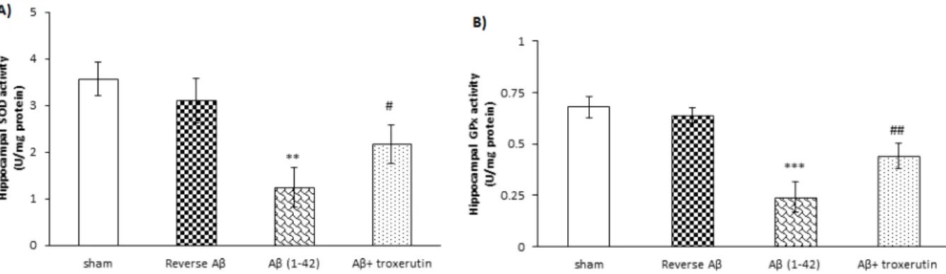
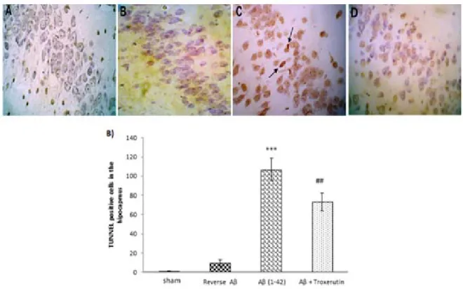
Alzheimer’s disease (AD) is an age-related neurodegenerative disease linked with increased production and/or deposition of amyloid-beta (Aβ) in the brain. The aim of the present study was to investigate the possible neuro- protective effect of troxerutin on an animal model of Alzheimer's disease. Alzheimer model was induced by a single dose intracerebroventricular (ICV) injection of Aβ 1–42 (5 nmol/5 µl). Thereafter, troxerutin (300 mg/kg) was gavaged for 14 days. The hippocampal malondialdehyde (MDA) levels and enzymatic activities of superoxide dismutase (SOD), glutathione peroxidase (GPx), and acetylcholinesterase (AChE) were measured using enzyme- linked immunosorbent assay (ELISA) method. In addition, the number of apoptotic cells in the dentate gyrus (DG) was assessed by TUNEL kit. The results showed that ICV microinjection of Aβ 1-42 increased MDA levels, re- duced SOD and GPx, and increased AChE activities in the hippocampus. Chronic administration of troxerutin significantly attenuated MDA levels and AChE activity and increased SOD and GPx activities in the hippocampus.

Moreover, the number of apoptotic cells was decreased by troxerutin treatment. Taken together, our study demon- strated that troxerutin could increase the resistance of hippocampal neurons against apoptosis, at least in part, by diminishing the activity of AChE and oxidative stress. Therefore, troxerutin may have beneficial effects in the management of Alzheimer's disease.

Keywords: Alzheimer’s disease, amyloid beta, acetylcholinesterase, oxidative stress

INTRODUCTION

Alzheimer’s disease (AD) is a neuro- degenerative disease characterized by pro- gressive loss of memory and cognitive func- tion (Butterfield and Boyd-Kimball, 2004).

AD is the most common cause of dementia in the people over 65 years which imposes a sig- nificant economic burden on families and so- ciety, and remarkably decreases the quality of life (Takizawa et al., 2015).

Two main neurological hallmarks in AD are extracellular senile plaques and intracellu- lar neurofibrillary tangles in the brain regions critical for learning and memory, the hippo- campus and other cortices, resulting in the loss of neurons and synapses (Blennow and Hampel, 2003; Giannakopoulos et al., 2003;

Hardy and Selkoe, 2002). Although, the exact

mechanism of AD remains unclear, it seems

that alterations in the production and pro-

cessing of Aβ leads to accumulation of Aβ

(2)

plaques in the neuronal space of the brain (Bloom, 2014; Mayeux and Stern, 2012).

Other proposed mechanisms associated with AD are mitochondrial dysfunction and oxida- tive stress (Pohanka, 2014; Wang et al., 2014), impairment of cholinergic transmis- sion (Kumar and Singh, 2015), neuro-inflam- mation (Morales et al., 2014), and glutamate neurotoxicity (Rudy et al., 2015).

Oxidative stress is defined as an imbal- ance between pro-oxidant stress and anti-oxi- dant defense which may lead to tissue injury (Halliwell and Gutteridge, 1999). Previous studies support the vulnerability of the central nervous system (CNS) to oxidative stress pos- sibly due to large rate of oxygen consumption, the richness of iron, high level of polyunsatu- rated fatty acids, and low levels of antioxi- dants (Butterfield et al., 2001; Paula et al., 2005). Recent studies have highlighted the importance of oxidative processes in the path- ogenesis of AD (Cioanca et al., 2015; E Abdel Moneim, 2015). Although the initiating events are still unknown, it has been proposed that oxidative damage is involved in the initi- ation of AD and is the first apparent sign in progression of AD (Arimon et al., 2015;

Wang et al., 2014). Antioxidant enzymes in- cluding superoxide dismutase (SOD), thiore- doxin, glutathione peroxidase (GPx), gluta- thione reductase (GR), and catalase (CAT) form important protective mechanism against reactive oxygen species (ROS) (Birben et al., 2012; Pohanka, 2014). Previous studies have shown that the activities of antioxidant en- zymes are diminished, whereas the levels of oxidative stress markers are elevated in the brain of AD patients (Arimon et al., 2015;

Krstic and Knuesel, 2013).

Acetylcholine (ACh) and cholinergic sys- tem are essential for regulation of learning and memory processes (Papandreou et al., 2011). Previous studies showed that accumu- lation of Aβ reduced ACh levels in the AD brain through increasing the expression of AChE (Perry et al., 1992). Moreover, AChE has the capability to augment Aβ deposition and fibril formation (Chacón et al., 2003). Un-

der normal conditions AChE is not an apop- tosis initiator; however overexpression of AChE increases the sensitivity of cells to apoptosis (Zhang and Greenberg, 2012). Alt- hough several agents such as cholinesterase inhibitors (Parsons et al., 2013), M1 musca- rinic receptor agonists (Jiang et al., 2014), and some of phosphodiesterases (Fiorito et al., 2013) are used to relieve symptoms of AD, most of these drugs are toxic and have numer- ous side effects.

Moreover, there is an inverse relationship between oxidative stress and Aβ levels.

Persson et al. indicated that oxidative stress increased production and accumulation of Aβ, which in turn increased ROS production and mitochondrial dysfunction (Persson et al., 2014). Several studies found that diminished number of neurons and synapses due to neu- ronal apoptosis in the cerebral cortex and hip- pocampus is the main reason of cognitive im- pairment of AD (Morishima et al., 2001;

Scheff et al., 2006). Since oxidative stress is a part of normal aging and starts very early in the disease progression, preventive therapies using antioxidants still hold great promise (Chakrabarti et al., 2014).

Troxerutin, derivative of natural bioflavo- noid rutin, is found in tea, coffee, cereals, and a variety of fruits and vegetables. Troxerutin possess biological properties such as antioxi- dant (Panat et al., 2016) and anti-inflamma- tory effects (Fan et al., 2009). Previously we demonstrated that oral administration of troxerutin improved synaptic failure (Babri et al., 2014) and learning and memory impair- ments induced by ICV injection of Aβ (Babri et al., 2012).

The aim of the present study was to inves- tigate the effect of troxerutin on the hippo- campal activity of AChE and oxidative status, and the number of neuronal apoptotic cells in the DG in Aβ 1-42-induced AD model in rats.

MATERIALS AND METHODS Animals

Sixty four adult male Wistar rats about 14

weeks old, weighing 300 to 350 g were ob-

tained from Pasteur Institute of Iran and kept

(3)

at standard conditions four per cage, 22–

24 °C, 12 h light–dark cycle, and free access to food and water. All experiments were per- formed in agreement with guidelines of the Tabriz University of Medical Sciences for care and use of laboratory animals. After one week of habituation animals were randomly allocated into the following groups (n=12 per each group):

i) Sham operated

ii) Reverse Aβ 42–1 (Bachem, Switzerland) iii) Aβ 1-42 (Bachem, Switzerland)

iv) Aβ 1-42 + troxerutin (Merck, Germany).

Surgical procedures

In order to perform stereotaxic surgery, animals were deeply anesthetized by an intra- peritoneal (i.p) injection of ketamine (80 mg/

kg) and xylazine (12 mg/kg) and placed on a stereotaxic instrument (Stoelting Co., Illinois, USA). The scalp was incised and a small hole was drilled at a proper location according to the Paxinos and Watson rat brain atlas (Pax- inos, 2007). Aβ (5 nmol/5 µl), reverse Aβ (5 nmol/5 µl), or saline (5 µl) were injected into the right lateral ventricle (AP: −0.8, ML:

1.6 and DV: 3.5 mm below dura) using a Hamilton micro syringe during 5 min. Needle was left in the place for 5 min before it was slowly withdrawn. Animals in the Aβ + troxerutin group received troxerutin (300 mg/

kg P.O for 14 days) one hour before injection of Aβ (Babri et al., 2012) and continued daily for 14 days.

Assessment of hippocampal MDA levels and enzymatic activities of SOD, GPx and AChE

At the end of experiments, rats were deeply anesthetized with 80 mg/kg sodium pentobarbital and sacrificed by decapitation, then hippocampal tissues were immediately removed. All samples were kept at -80 °C for later analysis. Samples were homogenized in 1.15 % KCl solution and centrifuged at 1000 rpm for 1 min at 4 °C for acquiring the supernatant. The supernatants were used for determination of MDA levels, and activities of SOD, GPx, and AChE. Hippocampal MDA

level was measured using the thiobarbituric acid reactive substances (TBARS) method at 535 nm with a UV spectrophotometer (Kaya et al., 2004). SOD, GPx, and AChE activities were measured using the commercial rat-spe- cific ELISA kits (Randox Crumlin, UK) ac- cording to the manufacturer’s protocols and expressed as U/mg protein and nmol/mg pro- tein in tissue homogenate.

Histological study

Following deep anesthesia (80 mg/kg so- dium pentobarbital) animals were perfused transcardially through the ascending aorta with 10–20 ml saline followed by 200 ml of 4 % paraformaldehyde. The brain tissue was removed and post fixed in the same solution, then processed for histological assay. Paraffin embedded brain tissue was cut in 10 μm cor- onal sections using a microtome. Brain sec- tions were stained with TUNEL staining kit for determination of apoptotic cells in the dentate gyrus (DG) according to the manufac- turer’s instructions. The numbers of apoptotic cells were counted by a blind person to the treatments using a light microscope (Nikon, Tokyo, Japan) at final magnification 400×. At least average TUNEL-positive cells of eight sequential brain sections from each animal were used for analysis.

Statistical analysis

Data were expressed as mean ± standard error of means (S.E.M.). Data were analyzed using SPSS (version 16) with One-way ANOVA followed by Tukey post-hoc test.

Data of the histological changes were ana- lyzed by the Kruskal-Wallis test followed by the post hoc Mann-Whitney test. Significance was assessed at the p<0.05 level.

RESULTS

Troxerutin attenuated hippocampal MDA levels

To investigate the effect of chronic

troxerutin treatment on oxidative stress, hip-

pocampal MDA level was measured (Figure

1). Our results demonstrated that the level of

MDA, an indicator of lipid peroxidation, in

(4)

the hippocampus was significantly (p<0.001) increased by Aβ 1-42 administration, while administration of reverse Aβ 42-1 had no sig- nificant effect on MDA levels. On the other hand, treatment with troxerutin significantly (p<0.05) decreased MDA levels compared with Aβ treated animals.

Figure 1: Effect of troxerutin on the hippocampal malondialdehyde (MDA) levels. Data are ex- pressed as mean ± SEM for n=8 animals per group. *** p<0.001 vs. sham group and

#

p<0.05 vs. Aβ group.

Troxerutin enhanced antioxidant enzyme activities in the hippocampus

The results revealed that Aβ 1-42 admin- istration significantly decreased the activities of SOD (p<0.01) and GPx (p<0.001), indica- tors of antioxidant defense, in the hippocam- pus (Figure 2A and 2B, respectively). How- ever, reverse Aβ 42-1 had no significant ef- fects on the hippocampal SOD and GPx activ- ities. Conversely, SOD (p<0.05) and GPx

(p<0.01) activities were significantly in- creased in the chronic troxerutin treated group as compared to the Aβ-received group.

Troxerutin reduced the activity of AChE in the hippocampus

In the current study, hippocampal activity of AChE, as a cholinergic marker, was also assessed. The one-way ANOVA analysis re- vealed that Aβ 1-42 administration induced a significant (p<0.001) increase in the AChE activity (Figure 3). Nevertheless, treatment with troxerutin remarkably (p<0.01) attenu- ated the hippocampal AChE levels. Reverse Aβ treatment did not significantly affect the hippocampal AChE levels.

Figure 3: Effect of troxerutin treatment on the hip- pocampal acetylcholinesterase (AChE) activity.

Values are expressed as the mean ± SEM for n=8 animals per group: ***p<0.001 vs. sham,

##

p<0.01 vs. Aβ group.

Figure 2: Effect of troxerutin on (A) superoxide dismutase (SOD) and (B) glutathione reductase (GPx) activities in the hippocampus. Data are expressed as mean ± SEM for n=8 animals per group.**p<0.01,

*** p<0.001 vs. the sham group and

#

p<0.05,

##

p<0.01 vs. the Aβ group.

(5)

Troxerutin reduced the numbers of TUNEL-positive cells in the dentate gyrus

Figure 4A shows the morphological fea- tures of TUNEL-stained hippocampal sec- tions. Histological study demonstrated that DG neurons were almost intact in the sham group; however, Aβ 1-42 administration in- creased neuronal damage. Intriguingly, troxerutin treatment could decrease neuronal apoptosis induced by Aβ in rats.

The results of Kruskal-Wallis analysis showed that the number of TUNEL-positive cells in the DG were significantly (p<0.001) increased in the Aβ-received group (Figure 4B). In contrast, neurons were significantly (p<0.01) preserved in the troxerutin-treated group and sparse TUNEL-positive cells were found in the DG region of the hippocampus.

No significant difference was observed be- tween the sham and reverse Aβ-treated groups.

DISCUSSION

The present study showed that ICV injec- tion of Aβ 1-42 increased hippocampal MDA and AChE levels and attenuated antioxidant enzymes activities (SOD and GPx). On the other hand, chronic troxerutin treatment for 14 days significantly reduced MDA levels and AChE activity, and improved enzymatic antioxidant defense in the hippocampus.

Moreover, troxerutin showed a neuroprotec- tive effect and reduced the number of TUNEL-positive cells in the DG.

Emerging evidence suggests that oxida- tive damage plays a causal role in the patho- genesis of AD (E Abdel Moneim, 2015). Prin- cipal manifestation of oxidative stress in the CNS is lipid peroxidation occurring in the early phase of AD (Mattson, 2004; Qin et al., 2009). Lipid peroxidation, in part, accounts

Figure 4: Troxerutin prevents Aβ (1-42)-induced apoptosis in the dentate gyrus (DG). (A) TUNEL stain-

ing was used to identify apoptotic nuclei in response to Aβ administration (×400) [A: sham; B: Reverse

Aβ; C: Aβ (1-42); D: Aβ + troxerutin]. Central injection of Aβ induced neuronal apoptosis (black arrows)

in the DG (B) TUNEL-positive cell counts. Following β-amyloid injection, an increased number of apop-

totic cells were found in the dentate gyrus. Values are expressed as the mean ± SEM (n=4): ***p<0.001

vs. sham group,

##

p<0.01 vs. Aβ group.

(6)

for apoptosis and neurodegeneration in the AD brain. It has also been revealed that Aβ 1–

42 can lead to lipid peroxidation and neuronal apoptosis (Butterfield et al., 2001; Ivins et al., 1999).

In view of the fact that oxidative stress and impaired cholinergic system play a path- ogenic role in AD, we investigated the effects of troxerutin on oxidative status in the hippo- campus. Central injection of Aβ 1-42 pro- vokes several impairments including oxida- tive stress (Bagheri et al., 2011), cholinergic dysfunction (Olariu et al., 2001), and neu- ronal apoptosis (Ivins et al., 1999; Ruan et al., 2010) possibly through induction of protein oxidation and lipid peroxidation. In line with other studies, our results demonstrated that Aβ 1-42 increased oxidative stress in the hip- pocampus which was confirmed by dimin- ished enzymatic antioxidant defense and in- creased MDA levels, end product of lipid pe- roxidation (Butterfield and Boyd-Kimball, 2004; Cioanca et al., 2013, 2015; Turunc Bay- rakdar et al., 2014). Nevertheless, we found that administration of troxerutin (300 mg/kg) could significantly reverse MDA levels and enhance enzymatic antioxidant defense against Aβ 1-42 in the hippocampus.

Acetylcholine, which involves in learning and memory processes, is degraded by AChE (Papandreou et al., 2011). In the AD brain, cholinergic activity decreases possibly due to increased activity of AChE around β-amyloid plaques (Moran et al., 1993). It is well known that increased AChE activity within and around amyloid plaques increases cytotoxi- city by promoting the aggregation of amyloid beta-peptides into fibrils which is more toxic than Aβ fibrils (Chacón et al., 2003; Inestrosa et al., 2008; Reyes et al., 2004). Previous studies have also revealed that hyperactivity of AChE leads to memory deficit, and AChE inhibitors are effectively used for relieving symptoms of AD in rodents (Ballard et al., 2005; Giacobini, 2004). In the present study, Aβ noticeably increased the hippocampal AChE activity, which was in accordance with previous study (Xu et al., 2017). Neverthe-

less, chronic troxerutin administration effec- tively decreased AChE activity in the hippo- campus induced by Aβ. Similarly, previous study has shown that troxerutin inhibits activ- ity of AChE in the basal forebrain, hippocam- pus, and frontal cortex of D-galactose-treated mice (Lu et al., 2010). Furthermore, it has been shown that oxidative stress is related to the brain AChE activity (Inestrosa et al., 2008). Therefore, the reduction of the hippo- campal AChE activity suggests that troxerutin is capable of improving memory impairment and oxidative stress induced by Aβ 1-42.

The present study demonstrated that Aβ 1- 42 administration increased the number of TUNEL-positive cells in the DG. In support of our study, extensive evidence shows that accumulation of Aβ triggers neuronal apopto- sis in the hippocampus, especially in the DG, which results in neuronal loss (Kadowaki et al., 2005; Obulesu and Lakshmi, 2014;

Shimohama, 2000; Stadelmann et al., 1999;

Yu et al., 2006). On the other hand, several studies indicated a protective role for troxeru- tin in different tissues through inhibition of the oxidative stress markers (Elangovan and Pari, 2013; Fan et al., 2009; Lu et al., 2010;

Zhang et al., 2009). Our results further showed that troxerutin reduced the TUNEL- positive cell counts in the DG indicating a marked inhibitory effect on cell apoptosis.

Similarly, Lu et al. showed that troxerutin in- hibited endoplasmic reticulum stress-induced apoptosis in the hippocampus of mice (Lu et al., 2011). Therefore, troxerutin might reverse Aβ-induced neuronal loss through decreasing lipid peroxidation end product (MDA), inhib- iting AChE activity, and enhancing the enzy- matic antioxidant defense in the hippocam- pus.

Overall the findings of the present study

revealed that troxerutin attenuates Aβ 1-42-

induced deleterious effects in the hippocam-

pus of rats. This is the first study showing the

neuroprotective potential of troxerutin against

Aβ 1-42-induced Alzheimer's disease possi-

bly through its anti-apoptotic, antioxidant,

and AChE inhibitory effects in the hippocam-

pus.

(7)

Acknowledgments

This work was financially supported by grant No. 89-60-12 from the Neurosciences Research Center (NSRC) at Tabriz University of Medical Sciences.

Conflict of interest

None of the authors has any conflict of in- terest.

REFERENCES

Arimon M, Takeda S, Post KL, Svirsky S, Hyman BT, Berezovska O. Oxidative stress and lipid peroxidation are upstream of amyloid pathology. Neurobiol Dis.

2015;84:109-19.

Babri S, Amani M, Mohaddes G, Alihemmati A, Ebrahimi H. Protective effects of troxerutin on β-amy- loid (1-42)-induced impairments of spatial learning and memory in rats. Neurophysiology. 2012;44:387- 93.

Babri S, Mohaddes G, Feizi I, Mohammadnia A, Nia- pour A, Alihemmati A, Amani M. Effect of troxerutin on synaptic plasticity of hippocampal dentate gyrus neurons in a β-amyloid model of Alzheimer׳s disease:

An electrophysiological study. Eur J Pharmacol. 2014;

732:19-25.

Bagheri M, Joghataei M-T, Mohseni S, Roghani M.

Genistein ameliorates learning and memory deficits in amyloid β (1–40) rat model of Alzheimer’s disease.

Neurobiol Learn Mem. 2011;95:270-6.

Ballard CG, Greig NH, Guillozet-Bongaarts AL, Enz A, Darvesh S. Cholinesterases: roles in the brain during health and disease. Curr Alzheimer Res. 2005;2:307- 18.

Birben E, Sahiner UM, Sackesen C, Erzurum S, Kalayci O. Oxidative stress and antioxidant defense.

World Allergy Organ J. 2012;5:9-19.

Blennow K, Hampel H. CSF markers for incipient Alz- heimer's disease. Lancet Neurol. 2003;2:605-13.

Bloom GS. Amyloid-β and tau: the trigger and bullet in Alzheimer disease pathogenesis. JAMA Neurol.

2014;71:505-8.

Butterfield DA, Boyd-Kimball D. Amyloid β-pep- tide(1-42) contributes to the oxidative stress and neu- rodegeneration found in Alzheimer disease brain.

Brain Pathol. 2004;14:426-32.

Butterfield DA, Drake J, Pocernich C, Castegna A. Ev- idence of oxidative damage in Alzheimer's disease brain: central role for amyloid β-peptide. Trends Mol Med. 2001;7:548-54.

Chacón MA, Reyes AE, Inestrosa NC. Acetylcholines- terase induces neuronal cell loss, astrocyte hypertrophy and behavioral deficits in mammalian hippocampus. J Neurochem. 2003;87:195-204.

Chakrabarti S, Munshi S, Banerjee K, Thakurta IG, Sinha M, Bagh MB. Mitochondrial dysfunction during brain aging: role of oxidative stress and modulation by antioxidant supplementation. Aging Dis. 2014;2:242- 56.

Cioanca O, Hritcu L, Mihasan M, Hancianu M. Cogni- tive-enhancing and antioxidant activities of inhaled co- riander volatile oil in amyloid β (1–42) rat model of Alzheimer's disease. Physiol Behav. 2013;120:193- 202.

Cioanca O, Hancianu M, Mihasan M, Hritcu L. Anti- acetylcholinesterase and antioxidant activities of in- haled juniper oil on amyloid beta (1–42)-induced oxi- dative stress in the rat hippocampus. Neurochem Res.

2015;40:952-60.

E Abdel Moneim A. Oxidant/antioxidant imbalance and the risk of Alzheimer's disease. Curr Alzheimer Res. 2015;12:335-49.

Elangovan P, Pari L. Ameliorating effects of troxerutin on nickel-induced oxidative stress in rats. Redox Rep.

2013;18:224-32.

Fan S-h, Zhang Z-f, Zheng Y-l, Lu J, Wu D-m, Shan Q, et al. Troxerutin protects the mouse kidney from d- galactose-caused injury through anti-inflammation and anti-oxidation. Int Immunopharmacol. 2009;9:91-6.

Fiorito J, Saeed F, Zhang H, Staniszewski A, Feng Y, Francis YI, et al. Synthesis of quinoline derivatives:

discovery of a potent and selective phosphodiesterase 5 inhibitor for the treatment of Alzheimer's disease. Eur J Med Chem. 2013;60:285-94.

Giacobini E. Cholinesterase inhibitors: new roles and therapeutic alternatives. Pharmacol Res. 2004;50:433- 40.

Giannakopoulos P, Herrmann F, Bussiere T, Bouras C, Kövari E, Perl D, et al. Tangle and neuron numbers, but not amyloid load, predict cognitive status in Alz- heimer’s disease. Neurology. 2003;60:1495-500.

Halliwell B, Gutteridge J. Free radicals in biology and

medicine. New York: Oxford Univ. Press, 1999.

(8)

Hardy J, Selkoe DJ. The amyloid hypothesis of Alzhei- mer's disease: progress and problems on the road to therapeutics. Science. 2002;297:353-6.

Inestrosa NC, Dinamarca MC, Alvarez A. Amyloid–

cholinesterase interactions. FEBS J. 2008;275:625-32.

Ivins KJ, Thornton PL, Rohn TT, Cotman CW. Neu- ronal apoptosis induced by β-amyloid is mediated by caspase-8. Neurobiol Dis. 1999;6:440-9.

Jiang S, Li Y, Zhang C, Zhao Y, Bu G, Xu H, et al. M1 muscarinic acetylcholine receptor in Alzheimer’s dis- ease. Neurosci Bull. 2014;30:295-307.

Kadowaki H, Nishitoh H, Urano F, Sadamitsu C, Matsuzawa A, Takeda K, et al. Amyloid [beta] induces neuronal cell death through ROS-mediated ASK1 acti- vation. Cell Death Diff. 2005;12:19-24.

Kaya H, Sezik M, Ozkaya O, Dittrich R, Siebzehnrubl E, Wildt L. Lipid peroxidation at various estradiol con- centrations in human circulation during ovarian stimu- lation with exogenous gonadotropins. Horm Metab.

Res. 2004;36:693-5.

Krstic D, Knuesel I. Deciphering the mechanism un- derlying late-onset Alzheimer disease. Nat Rev Neurol.

2013;9:25-34.

Kumar A, Singh A. A review on Alzheimer's disease pathophysiology and its management: an update. Phar- macol Rep. 2015;67:195-203.

Lu J, Wu D-m, Hu B, Cheng W, Zheng Y-l, Zhang Z- f, et al. Chronic administration of troxerutin protects mouse brain against d-galactose-induced impairment of cholinergic system. Neurobiol Learn Mem. 2010;93:

157-64.

Lu J, Wu D-m, Zheng Z-h, Zheng Y-l, Hu B, Zhang Z- f. Troxerutin protects against high cholesterol-induced cognitive deficits in mice. Brain. 2011;134:783-97.

Mattson MP. Pathways towards and away from Alzhei- mer's disease. Nature. 2004;430:631-9.

Mayeux R, Stern Y. Epidemiology of Alzheimer Dis- ease. Cold Spring Harb Perspect Med. 2012;2:

a006320. DOI: 10.1101/cshperspect.a006239 a006239 Morales I, Guzmán-Martínez L, Cerda-Troncoso C, Farías GA, Maccioni RB. Neuroinflammation in the pathogenesis of Alzheimer’s disease. A rational frame- work for the search of novel therapeutic approaches.

Front Cell Neurosci. 2014;8:112.

Moran M, Mufson E, Gomez-Ramos P. Colocalization of cholinesterases with β amyloid protein in aged and Alzheimer's brains. Acta Neuropathol. 1993;85:362-9.

Morishima Y, Gotoh Y, Zieg J, Barrett T, Takano H, Flavell R, et al. β-Amyloid induces neuronal apoptosis via a mechanism that involves the c-Jun N-terminal ki- nase pathway and the induction of Fas ligand. J Neuro- sci. 2001;21:7551-60.

Obulesu M, Lakshmi MJ. Apoptosis in Alzheimer’s disease: an understanding of the physiology, pathology and therapeutic avenues. Neurochem Res. 2014;39:

2301-12.

Olariu A, Tran M, Yamada K, Mizuno M, Hefco V, Nabeshima T. Memory deficits and increased emotion- ality induced by β-amyloid (25–35) are correlated with the reduced acetylcholine release and altered phorbol dibutyrate binding in the hippocampus. J Neural Transm. 2001;108:1065-79.

Panat NA, Maurya DK, Ghaskadbi SS, Sandur SK.

Troxerutin, a plant flavonoid, protects cells against ox- idative stress-induced cell death through radical scav- enging mechanism. Food Chem. 2016;194:32-45.

Papandreou MA, Tsachaki M, Efthimiopoulos S, Cordopatis P, Lamari FN, Margarity M. Memory en- hancing effects of saffron in aged mice are correlated with antioxidant protection. Behav Brain Res. 2011;

219:197-204.

Parsons CG, Danysz W, Dekundy A, Pulte I. Meman- tine and cholinesterase inhibitors: complementary mechanisms in the treatment of Alzheimer’s disease.

Neurotox Res. 2013;24:358-69.

Paula IM, Kazuhiro H, Quan L, Maria SS, Catarina RO, Gjumrakch A, et al. Oxidative stress: the old en- emy in Alzheimers disease pathophysiology. Curr Alz- heimer Res. 2005;2:403-8.

Paxinos GW. The rat brain in stereotaxic coordinates.

Burlington, MA: Elsevier Inc., 2007.

Perry E, Johnson M, Kerwin J, Piggott M, Court J, Shaw P, et al. Convergent cholinergic activities in ag- ing and Alzheimer's disease. Neurobiol Aging. 1992;

13:393-400.

Persson T, Popescu BO, Cedazo-Minguez A. Oxida- tive stress in Alzheimer’s disease: why did antioxidant therapy fail? Oxid Med Cell Longev. 2014;2014:

427318.

Pohanka M. Alzheimer s disease and oxidative stress:

a review. Curr Med Chem. 2014;21:356-64.

Qin Z-x, Zhu H-y, Hu Y-h. Effects of lysophosphati- dylcholine on β-amyloid-induced neuronal apoptosis.

Acta Pharmacol Sin. 2009;30:388-95.

(9)

Reyes AE, Chacón MA, Dinamarca MC, Cerpa W, Morgan C, Inestrosa NC. Acetylcholinesterase-Aβ complexes are more toxic than Aβ fibrils in rat hippo- campus: effect on rat β-amyloid aggregation, laminin expression, reactive astrocytosis, and neuronal cell loss. Am J Pathol. 2004;164:2163-74.

Ruan C-J, Zhang L, Chen D-H, Li Z, Du G-H, Sun L.

Effects of trans-2, 4-dimethoxystibene against the neu- rotoxicity induced by Aβ 25–35 both in vitro and in vivo. Neurosci Res. 2010;67:209-14.

Rudy CC, Hunsberger HC, Weitzner DS, Reed MN.

The role of the tripartite glutamatergic synapse in the pathophysiology of Alzheimer’s disease. Aging Dis.

2015;6:131-48.

Scheff SW, Price DA, Schmitt FA, Mufson EJ. Hippo- campal synaptic loss in early Alzheimer's disease and mild cognitive impairment. Neurobiol Aging 2006;27:

1372-84.

Shimohama S. Apoptosis in Alzheimer's disease - an update. Apoptosis. 2000;5:9-16.

Stadelmann C, Deckwerth TL, Srinivasan A, Bancher C, Brück W, Jellinger K, et al. Activation of caspase-3 in single neurons and autophagic granules of granu- lovacuolar degeneration in Alzheimer's disease: evi- dence for apoptotic cell death. Am J Pathol. 1999;155:

1459-66.

Takizawa C, Thompson PL, van Walsem A, Faure C, Maier WC. Epidemiological and economic burden of Alzheimer's disease: a systematic literature review of data across Europe and the United States of America. J Alzheimers Dis. 2015;43:1271-84.

Turunc Bayrakdar E, Uyanikgil Y, Kanit L, Koylu E, Yalcin A. Nicotinamide treatment reduces the levels of oxidative stress, apoptosis, and PARP-1 activity in Aβ (1–42)-induced rat model of Alzheimer's disease. Free Radic Res. 2014;48:146-58.

Wang X, Wang W, Li L, Perry G, Lee H-g, Zhu X. Ox- idative stress and mitochondrial dysfunction in Alzhei- mer's disease. Biochim Biophys Acta. 2014;1842:

1240-7.

Xu P, Wang K, Lu C, Dong L, Gao L, Yan M, et al.

Protective effects of linalool against amyloid beta-in- duced cognitive deficits and damages in mice. Life Sci.

2017;174:21-7.

Yu M-S, Suen K-C, Kwok N-S, So K-F, Hugon J, Chuen-Chung Chang R. Beta-amyloid peptides in- duces neuronal apoptosis via a mechanism independent of unfolded protein responses. Apoptosis. 2006;11:

687-700.

Zhang X-J, Greenberg DS. Acetylcholinesterase in- volvement in apoptosis. Front Mol Neurosci. 2012;5:

40.

Zhang Z-f, Fan S-h, Zheng Y-l, Lu J, Wu D-m, Shan Q, et al. Troxerutin protects the mouse liver against ox- idative stress-mediated injury induced by D-galactose.

J Agric Food Chem. 2009;57:7731-6.

Abbildung

Figure 2: Effect of troxerutin on (A) superoxide dismutase (SOD) and (B) glutathione reductase (GPx)  activities in the hippocampus
Figure 4A shows the morphological fea- fea-tures of TUNEL-stained hippocampal  sec-tions

Referenzen

ÄHNLICHE DOKUMENTE

Alzheimer’s disease (AD) is a deleterious neurodegenerative disorder with an increasing prevalence. Currently, there are neither reliable early diagnostic markers nor available

Relative elevation in the levels of iNOS expression by beta amyloid treatment in cortical neurons might indicate the potential role of beta amyloid to induce oxidative stress

Since mitochondria are a primary source of reactive oxygen species (ROS), and the neuronal mitochondria of Mecp2 -/y hippocampus revealed a more oxidizing and

The findings from our study indicate that two metabolites accumulating under ketogenic diet treatment, namely beta-hydroxybutyrate, the predominant ketone body, and decanoic

In addition, AME, 5, and 8 exhibited signifi cant antiproliferative activity against colon carcinoma cells (HCT-116); however, CE and the other examined compounds

co-trimoxazole in comparative trials versus tetracycline alone or tetracycline + streptomycin, respectively, for the treatment of acute brucellosis.. In Brescia Italy Sueri

The aims of the studies described in this thesis were to: (1) use the X/XO-system to induce different/defined levels of oxidative stress in stallion sperm,

When we compared the polyP levels in HOCl, HOBr and HOSCN-treated wild-type PA14 we found that all three treatments elicited a comparable increase in polyP level (Fig. Instead,