Cell-type specific responses to antidepressants – the epigenetic makeup of the glia-neuron
interface
DISSERTATION ZUR ERLANGUNG DES
DOKTORGRADES DER NATURWISSENSCHAFTEN (DR. RER. NAT.) DER FAKULTÄT FÜR BIOLOGIE UND VORKLINISCHE MEDIZIN
DER UNIVERSITÄT REGENSBURG
vorgelegt von Victoria Malik
aus Freising im Jahr
2016
Das Promotionsgesuch wurde eingereicht am 10.06.2016
Die Arbeit wurde angeleitet von
Prof. Dr. Inga D. Neumann und Dr. Barbara Di Benedetto
_____________________________
Unterschrift
Zusammenfassung ... 10
Introduction ... 12
1. 1.1. Major depressive disorder (MDD) ... 12
1.2. Neuronal circuits involved in depression ... 12
1.3. Theories on the etiopathogenesis of depression ... 14
1.3.1. Monoamine hypothesis ... 14
1.3.2. Neurotrophic hypothesis ... 15
1.3.3. Glia cell hypothesis ... 16
1.3.4. Epigenetic hypothesis ... 18
1.4. Astrocytes ... 19
1.5. Lineage of astrocytes ... 19
1.5.1. Characterization ... 20
a. Morphology ... 21
b. Antigenic properties ... 22
c. Receptors ... 22
1.6. Astrocyte activity ... 23
1.7. Functions of astrocytes ... 23
1.7.1. Metabolic support... 23
1.7.2. The blood-brain barrier ... 24
1.7.3. GABA and Glutamate ... 24
1.7.4. Neurotransmission and synapses ... 25
1.8. Astrocytes in depression ... 25
1.8.1. Morphometric evidence ... 26
1.8.2. Antidepressants act on astrocytes ... 27
1.9. Epigenetic mechanism ... 30
1.9.1. DNA methylation ... 31
1.9.2. Posttranslational histonemodification ... 32
a. Acetylation ... 32
b. Methylation ... 33
c. Phosphorylation ... 33
d. Ubiquitination... 33
1.9.3. Histone code hypothesis ... 34
1.10. Epigenetic mechanisms in depression ... 35
1.11. Animal model ... 38
1.11.1. Anxiety-related behavior ... 38
1.11.2. Depressive-like behavior ... 39
1.11.3. Hypothalamic-pituitary-adrenal (HPA) axis and pharmacological intervention ... 39
1.12. Microarray – candidate genes ... 40
1.12.1. Growth differentiation factor 15 (GDF15) ... 41
1.12.2. Ephrin ... 42
a. General features ... 43
b. Implications in CNS pathologies ... 44
1.13. Scope of study ... 46
Material and Methods ... 47
2. 2.1. Animals ... 47
2.2. Drugs ... 47
2.3. Preparation of astrocytes ... 47
2.3.1. Cell culture passaging ... 48
2.3.2. Cultivation ... 48
2.4. Preparation of neurons ... 48
2.5. Preparation of co-cultures ... 49
2.6. Antidepressant treatment ... 49
2.7. Harvesting ... 49
2.8. Immunofluorescence (IF) analysis ... 50
2.8.1. Drug treatment and perfusion ... 50
2.8.2. Immunofluorescent-Immunohistochemistry ... 50
2.8.3. Immunofluorescent-Immunocytochemistry (IF-ICC) ... 52
2.8.4. Confocal microscopy ... 54
2.9. Quantitative analysis ... 55
2.9.1. Histone marks... 55
2.9.2. Statistical analysis ... 59
2.10. Western blot experiments ... 59
2.10.1. Protein expression quantification ... 61
2.11. Short interfering RNAs (siRNA) fot the downregulation of ephrinA1 ... 62
2.11.1. Transfection... 62
2.12. Native chromatin-Immunoprecipitation (ChIP) [151, 210] ... 63
2.12.1. Cell culture ... 63
2.12.2. Preparation of native chromatin ... 63
2.13. GDF15 ... 64
2.13.1. Treatment ... 65
2.13.2. Western blot ... 65
2.14. Immunofluorescent analysis - GDF15 ... 66
2.14.1. Fluorescent in situ hybridization (FISH) on CD1 mice, HAB and NAB ... 66
2.14.2. IF-ICC on primary astrocytes ... 66
2.15. RNAi ... 67
2.16. Fluorescent and morphological analysis of GDF15 ... 68
2.17. Statistical analysis ... 69
Results ... 70
3. 3.1. Histone 3 – Lysine 4 – trimethylation (H3K4me3) ... 70
3.1.1. Global expression of H3K4me3 ... 70
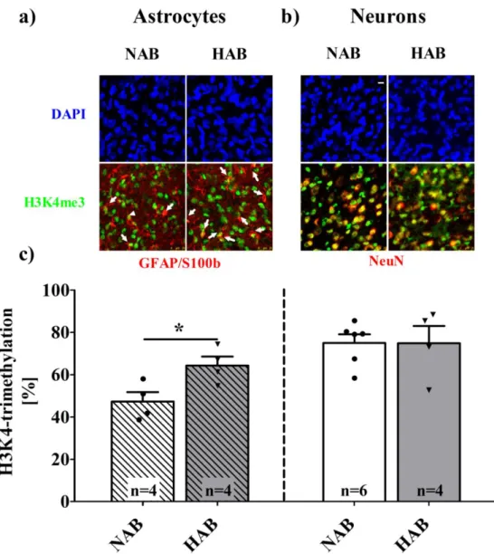
3.1.2. Cell-type specific expression of H3K4me3 ... 70
3.2. H3K4me3 after fluoxetine treatment... 72
3.2.1. Global expression of H3K4me3 after FLX injections in NAB ... 72
3.2.2. Cell-type specific expression of H3K4me3 after FLX injection in NAB ... 72
3.2.3. Global expression of H3K4me3 after FLX injection in HAB... 74
3.2.4. Cell-type specific expression of H3K4me3 after FLX injection in HAB ... 75
3.3. Posttranslational histone modifications (PTM) – protein expression ... 76
3.3.1. H3K4me3 ... 76
3.3.2. Histone 3-lysine 27-trimethylation ... 79
3.3.3. Histone 3-Lysine 27-acetylation (H3K27ac) ... 82
3.4. Global expression of H3K27me3 under baseline conditions ... 85
3.4.1. Cell-type specific expression of H3K27me3 ... 85
3.5. Candidate genes ... 86
3.5.1. Chromatin-immunoprecipitation ... 87
3.5.2. EphrinA1 protein expression ... 87
3.5.3. EphrinA1 knockdown via siRNA... 90
3.6. IF-IHC on ephrinA1 and EphA4 in PFC of untreated NAB and HAB animals ... 90
3.7. EphrinA1 and EphA4 ... 92
3.8. GDF 15 ... 95
(Malik et al., in preparation) ... 95
3.8.1. Microarray ... 95
3.8.2. In situ hybridization -validation of GDF15 probe ... 96
3.9. GDF15 in blood vessels ... 97
3.9.1. GDF 15 expression around blood vessel in HAB ... 98
3.9.2. GDF 15 protein expression ... 100
3.10. In depth analysis of astrocytic process ... 101
3.10.1. Number of processes in primary cortical astrocytes under baseline conditions ... 102
3.10.2. Number of processes in primary cortical astrocytes after treatment with FLX and exogenous GDF15 ... 102
3.10.3. Sprouting of processes in primary cortical astrocytes after treatment with FLX and exogenous GDF15 ... 105
3.10.4. Knockdown of endogenous GDF15 in NAB cells ... 109
Discussion ... 112
4. 4.1. Posttranslational histonemodifications ... 113
4.2. H3K4me3 regulation of ephrinA1 ... 118
4.3. EphrinA1 and its implication it the regulation of synapses ... 120
4.4. Summary and outlook on PTMS and ephrinA1 ... 121
4.5. GDF15 ... 123
Supplementary ... 129
List of abbreviations ... 133
List of figures ... 137
References... 140
Understanding the neurobiological underpinnings of major depressive disorder (MDD) is of utmost importance. Recently, it was shown in post-mortem tissue of MDD patients that the number of glia cells is reduced, accompanied by atrophy of neuronal cells and a decreased volume of the prefrontal cortex (PFC). Following the observation that morphological deficits are present in both cell-types, it is likely that a misregulation in the communication between glia and neuronal cells might be one of the key factors underlying the development of MDD. Furthermore, the coverage of blood vessels by astrocytic endfeet has been shown to be reduced in MDD, implicating a misregulation also at the level of the glia-vasculature interface. A well-established approach to understand these deficits is the investigation of the molecular responses of neuronal cells to antidepressants (AD) in a cell-type specific manner. ADs have been shown to influence astrocytes as well as neurons and have been implicated in the regulation of gene expression via modulation of epigenetic modifications, like posttranslational histonemodifications (PTM).
To understand these molecular changes, I first investigated the expression pattern of several PTMs (H3K4me3, H3K27me3 and H3K27ac) both, on global and cell-type specific levels before and after treatment with fluoxetine (FLX), one of the most commonly used ADs, in high anxiety-related behavior (HAB) rats, serving as a well- established model for comorbid depressive-like behavior. I focused on the PFC, hence this brain area is known to be highly involved in depression in humans and animal models, although not much is known about epigenetic modifications in this area. For examining deeper cell-type specific changes and responses to ADs, I used both in vitro and in vivo models. In the present study I demonstrated that under baseline conditions, HAB rats resumed not only the human loss of glia cells in the PFC, but they also showed a putatively upregulated gene expression indicated by a 2-3 fold increase of H3K4me3, an activating PTM. I further showed that increased activation of H3K4me3 was specific of astrocytes and was targeted by FLX treatment. I additionally demonstrated that H3K27me3, a repressive mark which is supposed to be a counteracting partner of H3K4me3, is downregulated in HAB animals. Moreover, it decreased even further after FLX treatment, whereas H3K27ac, a permissive mark, was also decreased in HAB animals, which suggested that it might be a target of FLX, but after long term treatment. Together these data indicate a unique code of epigenetic modification in HAB rats under baseline
time, an aberrantly high gene expression of ephrinA1, regulated by a high accumulation of H3K4me3 at its promoter, in HAB astrocytes as well as in the PFC of HAB rats, thus suggesting that this might represent a novel target of AD therapy in MDD. In view of the crucial role that the ephrin/Eph system plays at the glia-neuron interface to modulate synaptic transmission, its misregulation may very well lead to the development of depressive symptoms.
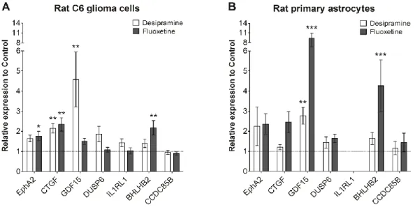
In addition, I identified GDF15, a neurotrophic factor, as a target of AD treatment in vitro and in vivo. My findings indicated that the ADs desipramine and FLX might induce increased expression of GDF15, leading to altered astrocytic morphology both in cell culture and around blood vessels of the PFC. An in-depth analysis revealed that FLX was specifically able to increase the sprouting of new processes as well as the elongation of preexisting ones, after a prolonged treatment in normal cells (derived from non-selectively bred animals, NAB), thereby also rescuing the morphology of the diseased HAB astrocytes, which showed a lack of processes with respect to NAB cells. These data corresponded to the in vivo findings in HAB rats, which show a reduced coverage of blood vessels with astrocytic processes. This deficit could be reversed by AD treatment.
Moreover, I investigated the effects of exogenous GDF15 on astrocytic processes.
Application of GDF15 was able to restore in HAB astrocytes both the number and length of processes in a dose- and time-dependent manner. Taken together, these data suggested that GDF15 might be involved in recovering a proper glia-vasculature interface that is needed for the proper exchanges of substances at the blood-brain-barrier.
Erst kürzlich konnte in postmortalem Gewebe gezeigt werden, dass depressive Patienten eine geringere Anzahl an Gliazellen, neuronale Atrophie sowie ein vermindertes Volumen des präfrontalen Kortex aufweisen. Da morphologische Veränderungen in diesen beiden Zelltypen auftreten ist es sehr wahrscheinlich, dass eine fehlerhafte Kommunikation zwischen Glia- und neuronalen Zellen eine Schlüsselrolle in der Entstehung einer Depression spielen könnte. Des Weiteren konnte gezeigt werden, dass die Blutgefäße in depressiven Patienten zu einem geringeren Ausmaße mit den Fortsätzen von Astrozyten bedeckt sind, was eine Deregulierung in diesem System bedeuten könnte. Ein gängiger Ansatz diese Defizite zu untersuchen, besteht darin die Reaktion dieser Systeme nach Behandlung mit Antidepressiva auf molekularer sowie zellspezifischer Ebene zu untersuchen. Antidepressiva können Astrozyten und Neurone beeinflussen und wurden bereits mit der Regulation von Genexpression mittels epigenetischer Modifikationen, wie beispielsweise posttranslationaler Histonmodifikationen (PTM) in Zusammenhang gebracht. Um diese molekularen Veränderungen zu verstehen, habe ich die Expressionsmuster verschiedener PTMs (H3K4me3, H3K27me3 und H3K27ac), vor und nach Behandlung mit Fluoxetin (FLX), auf globaler sowie zell-spezifischer Ebene untersucht. Als Tiermodell für diese Analyse habe ich high anxiety-related behavior (HAB) Ratten genutzt, die ein bereits gut etabliertes Tiermodel für Depression darstellen.
Ich konzentrierte mich auf den präfrontalen Kortex, da diese Gehirnregion in der Pathophysiologie dieser Erkrankung sehr stark involviert ist, obwohl in diesem Areal bisher nur wenig über epigenetische Veränderungen bekannt ist. Um eine bessere Vergleichbarkeit zu erzielen, nutzte ich sowohl in vitro als auch in vivo Modelle. Mit der vorliegenden Studie konnte ich zeigen, dass HAB Ratten den humanen Phänotyp, welcher eine verringerten Anzahl an Glia Zellen aufweist, widerspiegeln, sowie dass diese Tiere eine vermeintlich gesteigerte Geneexpression haben, welche durch ein 2-3 fach höheres H3K4me3 Vorkommen angedeutet wird. Des Weiteren, konnte ich zeigen, dass die gesteigerte H3K4me3-Aktivität spezifisch in Astrozyten vorkommt und durch FLX Behandlung beeinflusst werden kann. Außerdem konnte ich zeigen, dass H3K27me3, eine repressive PTM, welche zudem vermutlich ein Gegenspieler von H3K4me3 ist, in HAB Ratten unter basalen Bedingungen herunterreguliert ist und durch eine längere FLX Behandlung sogar noch weiter reduziert wird. H3K27ac, eine aktivierende PTM, hingegen ist in HAB Ratten ebenfalls herunterreguliert und scheint durch eine längere FLX
wahrscheinlich durch Antidepressiva verändert werden kann. Im Zuge dieser Studie konnte ich ephrinA1, als ein Gen identifizieren, welches in HAB Astrozyten sowie im präfrontalem Kortex von HAB Ratten aufgrund eines gesteigertem H3K4me3 Vorkommens in dessen Promoter abnorm stark exprimiert wird. Diese Daten weisen darauf hin, dass ephrinA1 ein neues therapeutisches Ziel für die Behandlung einer Depression sein könnte. Eine Fehlregulation des Eph/ephrin Systems, welches maßgeblich an der synaptischen Übertragung zwischen Glia und neuronalen Zellen beteiligt ist, könnte zu den morphologischen Defiziten dieser Schnittstellen beitragen und letztendlich zu depressiven Symptomen führen.
Zudem konnte ich GDF15, einen neurotrophen Faktor, identifizieren, welcher durch Antidepressiva beeinflussbar ist. Meine Ergebnisse deuten darauf hin, dass die Expression von GDF15 in Astrozyten durch die Gabe von Antidepressiva gesteigert werden kann, was zu einer morphologischen Veränderung dieser Zellen führt. Eine detaillierte Analyse hat gezeigt, dass FLX das Entstehen neuer, sowie die Verlängerung bereits bestehender Astrozyten-Fortsätze in „gesunden“ Zellen begünstigt. In HAB Zellen scheint FLX die aberrante Morphologie der Astrozyten so zu verändern, dass sie dem „gesunden“ Phänotyp gleichen. Diese Daten korrespondieren zu den in vivo Experimenten, in denen gezeigt wurde, dass eine verringerter Abdeckung der Blutgefäße mit Astrozyten-Fortsätzen in HAB Ratten durch FLX Behandlung verbessert werden kann. Weiterführend habe ich zudem die Effekte von exogen verabreichtem GDF15 untersucht. Die Behandlung von Astrozyten mit exogenem GDF15 führt dazu, dass neue Fortsätze entstehen und bestehende verlängert werden. Dieser Effekt scheint von der Dosis und der Behandlungsdauer abhängig zu sein. Aufgrund dieser Daten lässt sich spekulieren, dass GDF15 vermutlich daran beteiligt ist die funktionelle Schnittstelle zwischen Gliazellen und den Blutgefäßen wiederherzustellen, um damit einen angemessen Austausch von Substanzen zwischen dem Blutstrom und dem Hirnparenchym zu gewährleisten.
Introduction 1.
1.1. Major depressive disorder (MDD)
According to a recently published new data set by the world health organization (WHO [7]), unipolar disorder, also known as major depressive disorder (MDD), has become the leading cause of disability worldwide. Globally, over 350 million people of all ages are estimated to be affected by depression. MDD is a polygenic and multifactorial disease with a high lifetime prevalence, hence it is considered that one out of three people may suffer from depressive episodes during the life [8-10]. This disease is characterized by core symptoms of depressed mood and anhedonia, often accompanied by changes in body weight, loss of energy or pleasure, disturbances in sleep and cognitive functions [11, 12].
Especially the lack of specific biomarkers which might help to properly define the state of depression makes it very complicated to get a precise diagnosis. Moreover, the high comorbidity with other psychiatric illnesses, especially anxiety-related disorders, complicate a proper diagnosis even further [13]. Patients suffering from MDD also exhibit a markedly reduced lifespan, not necessarily due to concomitant suicide, but rather due to a well-documented increased risk of other life-threatening diseases, such as diabetes or cardiovascular disease [14]. Unfortunately, to date the most reliable criterion to diagnose/
categorize the state and severity of depression is based on questionnaires for the patients (DSM-Ⅴ, 2013), making a definite diagnosis dependent on subjective perceptions rather than on concrete pathophysiological evidences. Therefore, it is of utmost importance to understand the etiology and pathophysiology of MDD, its underlying molecular and additionally cell-type specific mechanisms to distinguish early on time people who might be prone to develop depression and to eventually being able to formulate a clearer diagnosis using specifically identified biomarkers. To approach these questions it is highly important to open new ways to identify novel targets and develop better and faster-acting therapeutics. Unfortunately, some of the major drawbacks in antidepressant (AD) therapy are still the high rate of non-responders (about 50-70%) and the delayed onset of amelioration of symptoms (4-12 weeks [15]).
1.2. Neuronal circuits involved in depression
Several limbic and cortical brain regions have been shown to play crucial roles in depression (see Fig.1, adapted from [2, 3]). These highly interconnected circuits involve brain structures that are important for interpreting and responding to stress and reward as well as cognitive functions [3, 16]. In the simplified scheme, one can see dopaminergic
neurons (blue arrows) projecting from the ventral tegmental area (VTA) to the nucleus accumbens (NAc) and hippocampus (HIPPO), among other areas. The NAc receives glutamatergic inputs (red arrows) from the prefrontal cortex (PFC), the HIPPO and amygdala (AMY) [2, 3, 16]. These interaction between the PFC and NAc are primarily part of the reward system and are implied in MDD [17]. Degeneration or lesions in these areas were found to increase the risk of patients to develop depressive episodes [18], while deep brain stimulation in the NAc and Cg25 (subcallosal cingulate gyrus) of treatment resistant patients successfully reduced depressive symptoms [19, 20]. With regard to these connections, it has been postulated that the PFC can exert regulatory effects on the NAc.
Reciprocal connections between the PFC, especially the medial (m)PFC and AMY, via the uncinate fasciculus (black arrow, a long-range white matter association fiber-tract in the human brain, connecting the orbitofrontal cortex to the anterior temporal lobes through a direct, bidirectional monosynaptic pathway [21]) have been shown to exist in rats and non- human primates [22]. A reduction in the PFC-AMY connectivity has been suggested to play a role in MDD and it normalizes after drug treatment [23]. Another important area is the dorsal raphe nucleus (DR), which is the main site of serotonin (5-HT) synthesis in the brain. It receives glutamatergic inputs from the PFC [24], among which most of the projections terminate onto local GABAergic (γ-Aminobutyric acid) interneurons that inhibit 5-HT neurons, impacting neuronal excitability. Antidepressant treatment with, for example, selective-serotonin-reuptake inhibitors (SSRIs) might interact with these connections. Dysfunctional HIPPO-PFC interactions are known to play a central role in MDD, which is not surprising, considering the central role of the HIPPO in regulation of the hypothalamic-pituitary-adrenal (HPA) axis [25]. The HIPPO is also highly interconnected via glutamatergic projections to the NAc and AMY, which both play important roles in the formation of emotional memory [26]. So far, most studies focused on the HIPPO and NAc, but the PFC represents an emerging target in the pathophysiology of depression, whose implications still need to be explored thoroughly.
1.3. Theories on the etiopathogenesis of depression
Since the discovery of the first ADs [27], many hypotheses have been developed to explain the pathophysiology of depression and the action of ADs. With the emerging of new research fields and advances in the understanding of this multifaceted disorder, the theories were refined and new perspectives were added. Nevertheless, to date, none of them is able to fully explain the development of MDD and how ADs work. Therefore, I will introduce the main hypotheses that developed over the past 60 years, specifically focusing on the most promising ones.
1.3.1. Monoamine hypothesis
The ´monoamine hypothesis´ of depression was the first one to be postulated [28], stating that depression is caused by a decrease in monoaminergic signaling in the brain. This assumption is based on clinical findings of the late 1950s. Back then, two structurally unrelated compounds originally developed for non-psychiatric diseases, namely ipronazid (for tuberculosis) and imipramine (IMI, for schizophrenia) were found to “enhance the mood” in patients treated with these drugs [29]. Both drugs were later proven to affect the monoaminergic system by enhancing the availability of 5-HT and noradrenalin (NA) transmission. Since then, several antidepressants have been developed, but most of them are still designed to intervene with the monoaminergic system, either by inhibiting neuronal re-uptake of 5-HT or NA (e.g. fluoxetine (FLX), a SSRI; reboxetine, a selective noradrenalin reuptake inhibitors (NRI); venlafaxine, a serotonin and noradrenalin reuptake
Fig. 1: Neuronal circuitries involved in depression (adapted from [2, 3])
Abbreviations: PFC = prefrontal cortex, AMY = amygdala, HIPP = hippocampus, NAc= Nucleus accumbens, PVN = paraventricular nucleus of hypothalamus, VTA = ventral tegmental area, HYP = Hypothalamus, DR= dorsal raphe nucleus, LC = locus coereleus. Blue arrows = dopaminergic neurons projecting from VTA, red arrows = glutamatergic projections to NAc, black arrows = grey matter connections, green arrows = GABAergic connections
inhibitor (SNRI)) or by inhibiting their degradation (e.g. phenelzine, a monoamine oxidase A inhibitors (MAOI)) [11]. Although these compounds have been proven to be potent antidepressants, the simplified view that a sole monoamine deficiency is causing depression has been disproven. First of all, such substances increase the availably of monoamines on an acute basis, whereas behavioral amelioration requires several weeks.
Moreover, a depletion of monoamines in animal models is not sufficient to elicit depressive symptoms [30]. Furthermore, illegal drugs like cocaine and amphetamines, which enhance the availability of serotonin, dopamine or norepinephrine do not seem to possess antidepressant effects. These data suggest that a dysregulation of the monoamine system is far from being the only factor which contributes to MDD.
1.3.2. Neurotrophic hypothesis
To target the question why antidepressants require several weeks of treatment before an amelioration of symptoms takes place, it was thought that an enhancement of monoamines in the synaptic cleft might lead to subsequent changes in neuroplasticity. As far as these changes are dependent on transcriptional and translational adaptations they might probably require a longer time scale, thereby explaining the delayed onset of ADs´ action. Together with data from post-mortem tissue and imaging studies, which showed structural alterations in several brain areas (e.g. PFC and HIPPO [31, 32]), this hypothesis proposes that depression might result from a decreased neurotrophic support, which in turn leads to neuronal atrophy (i.e. decreased dendritic arborization and synaptic contacts), a decreased hippocampal neurogenesis and glia cell loss, and that antidepressants are capable to block and/or reverse these deficits [33-35]. Among other neurotrophic factors (e.g. vascular endothelial growth factor, VEGF; insulin-like growth factor -2, IGF-2; fibroblast growth factor-1, FGF-1; glia-cell derived neurotrophic factor, GDNF), the most prominent and best-studied is brain-derived neurotrophic factor (BDNF) [34, 36-40]. Decreased levels of BDNF were found in post-mortem brain tissue as well as in the serum of depressed patients [34, 41]. Decreased levels of BDNF in specific brain areas are found in several animal models of stress-induced depression, thereby substantiating the human results [26, 34, 38]. Infusions of BDNF in relevant brain areas are sufficient to elicit AD-like effects, but these effects seem to be dependent on the respective brain area. While BDNF displays antidepressant properties in the HIPPO [42], it exerts pro-depressive effects in the NAc [43]. Additionally, depletion of BDNF is not sufficient to cause depressive symptoms in male knockout mice [44]. Pinning down the cause of depression to a single neurotrophic factor, like BDNF, might as well be too simplistic. However this theory raises awareness to
incorporate the fact that neurotrophic factors are involved in neuroplastic responses to stress and antidepressant treatment. Numerous studies have shown that stress and antidepressants evoke opposite effects on neurogenesis and neuronal complexity. This might be a possible explanation for the delayed onset of symptomatic improvement.
Neurogenesis occurs in specific brain areas of the adult brain, the subventricular zone (SVZ) that gives rise to neurons of the olfactory bulb and the subgranular zone (SGZ) that generates granular cells of the dentate gyrus [45]. Different types of acute and chronic stress, both psychological and physiological, decrease neurogenesis [46], while several classes of antidepressants, including SSRIs (e.g. FLX) and SNRIs induce it [45, 47].
Nonetheless, inhibiting neurogenesis, via irradiation [47] or genetic manipulations is not sufficient to induce depressive-like behavior in rodents. Although this theory sheds light on some important mechanisms that need to be considered, it can neither explain why antidepressant efficacy varies greatly among patients (high rate of non-responder) nor does it consider differences in pathways between stress-induced depression and genetically determined (“endogenous”) depression.
1.3.3. Glia cell hypothesis
Pioneering work by Rajkowska and colleagues extended the complex topic of depression even further [12, 31, 48]. Their work revealed tremendous glia cell impairment, with regard to both number and size, in post-mortem tissue of MDD patients [31, 49-51], thereby attracting notice to astrocytic abnormalities as a cause and/or consequence of depression. Additionally, several studies observed neuronal atrophy, displayed by smaller cell body sizes and reduced branching [31, 32, 50, 52], rather than complete loss of neurons. These results were substantiated by a study examining the serum of MDD patients for the amount of enolase 2 (a neuron-specific protein). The authors could not detect any difference in its expression, whereas the levels of S100β, an astrocyte specific marker, were elevated [53]. Furthermore, injecting a neuron-specific toxin (ibotenic acid) into the PFC of rodents did not cause any behavioral changes, while ablation of astrocytes (via L- alpha-aminoadipic acid (L-AAA)) was sufficient to induce a depressive-like behavior [54].
These data led to the suggestion that MDD might not primarily be caused by neuronal deficiencies but in first instance by alterations in astrocytes. In fact, astrocytes have been shown to play a pivotal role in synaptic transmission by physically stabilizing synapses and also by actively participating in the exchange of signaling molecules, such as diverse neurotrophic factors and glutamate, between the pre- and post-synaptic terminals. On the one hand, astrocytes enwrapping synapses can clear the synaptic cleft from excess of those
signaling molecules, on the other hand, they supply neurons with diverse neurotransmitters and nutrients, such as glucose [55-57]. In particular, astrocytes play an important role in regulating the release and reuptake of glutamate, which in high concentrations is detrimental for neuronal survival. Dysfunctional astrocytes may contribute to the development of MDD, as their buffering capacities on the glutamate system seem to be compromised, thereby leading to a consequential (not causal) loss of neurons [11, 58]. This hypothesis further suggests that antidepressants may act on the remaining astrocytes by increasing their number and reinstate a proper glia-neuron proportion via enhancing their proliferation through the release of glia-derived trophic factors [59]. Such neurotrophic factors released from astrocytes (e.g. BDNF and GDNF) can signal back to neurons, which in turn are capable to control presynaptic activity through regulation of factors responsible for neuronal survival and maturation [11]. Indeed, those neurotrophic factors play an important role in the neurogenic response to antidepressants. They have been shown to be beneficial in restoring the physiological state of neuronal branching that is reduced under pathological conditions [60, 61]. Antidepressants can also directly act on astrocytes.
Several classes of antidepressants activate specifically the ERK/MAPK (extracellular signal–regulated kinases/mitogen-activated protein kinase) pathway, whose downstream effectors are, among others, GDNF and BDNF [57, 62, 63].
Further supporting the role of glia cells in response to antidepressants is the involvement of the blood brain barrier (BBB) in the uptake of clinically efficient dosages of drugs.
The BBB is most tightly regulated by astrocytic endfeet, which surround and interact with blood vessels, thus controlling the passage of exogenous substances in and out of the brain.
Very recently it has been shown that in MDD patients, blood vessels of the prefrontal cortex (PFC) lack the coverage with Aqp-4 (aquaporin-4)-positive endfeet of astrocytes [12]. A finding that was reproduced by our group in an animal model of depressive-like behavior (see below 1.11) [51]. Furthermore, we could show that Aqp-4 is necessary to mediate antidepressant effects on restoring a basal amount of astrocytic processes in astrocytes derived from the PFC of an animal model for depression. Considering these facts about astrocytes as cause and/or consequence of MDD and their responses to antidepressant treatment, it makes them a highly interesting target to study in order to elucidate molecular underpinnings of depression.
1.3.4. Epigenetic hypothesis
Over the past decade several epigenetic mechanisms have been identified as important effectors of psychiatric diseases [3, 16, 64, 65]. It is known that depression can, at least partially, be inherited (heritability around 40% in familial cases [66]), nevertheless so far only very few genetic variants could be robustly correlated with depression. Additionally, the high discordance rate in monozygotic twins cannot be explained by pure genetic underpinnings. Therefore, the combination of genes with environment came into the spotlight of research. Indeed, epigenetic mechanisms are ideal candidates to study mental diseases that are caused by interaction of genetic factors (predisposition) and environmental exposure. Several lines of evidence have linked alterations in PTMs, DNA methylation and non-coding RNAs to psychiatric diseases. It is proposed that misbalances in such a delicate system of epigenetic modifications can severely impact brain functions.
Various animal models of stress-induced depression and early life experiences demonstrated aberrant histonemodification, leading to changes in gene expression. The glucocorticoid receptor (GR) is one of these candidates, whose expression is altered in offspring derived from low maternal care mothers [67], regulated by DNA methylation.
This effect could be reversed by application of histone-deacetylase inhibitors (HDACi), suggesting a crosstalk between epigenetic mechanisms. HDACi can also exert antidepressant-like effects dependent on the brain area. Besides acetylation, several other PTMs occur to play a role in MDD, foremost methylation of different histones (see below).
For example elevate levels of a specific activating PTM (histone 3-lysine 4-trimethylation, H3K4me3), were found at the synapsin promoter in the PFC of depressed patients [68], whereas histone 3-lysine 27-trimethylation (H3K27me3, repressive) levels are increased at certain BDNF promoters in the PFC and both can be reversed by AD treatment. Research has only begun to understand how epigenetic modifications influence the outcome of MDD and how to target inter-individuals differences. Understanding the interplay of a potential predisposition interacting with environmental circumstances on the development of psychiatric diseases needs more detailed analysis to identify how epigenetic regulation impacts specific genes or gene networks related to depression. Keeping these hypotheses in mind, it seems valid to take a closer look at the different levels and at their functions under physiological and pathological conditions.
1.4. Astrocytes
In the 1850s the term “Nervenkitt” (i.e. “brain glue”) was first introduced by the pathologist Rudolf Virchow. He defined neuroglia cells as small round-shaped cells that fill up the extracellular space as parts of the connective tissue. Although the term neuroglia still persists, to date especially the definition of astrocytes has changed drastically [69].
With the rise of more elaborate techniques than a simple Nissl staining, it is now well accepted that astrocytes are a highly heterogeneous cell population with numerous functions in the brain [70].
Glia cells are considered to be the most abundant cell-type in the mammalian CNS (central nervous system) [71]. A comprehensive study by Pelvig and colleagues [72] quantified the number of neurons and glia cells in the neocortex of adult humans. Their results show that the total number of glia cells ranges between 27.9 billion (females) and 38.9 billion (males), while the total number of neurons only ranks between 21.4 billion (females) to 26.3 billion (males), providing a 1.3-1.5 glia/neuron ratio. With about an average of 75%, oligodendrocytes account for the majority of glia cells, followed by astrocytes (20%) and microglia cell (5%).
1.5. Lineage of astrocytes
Gliogenesis is the generation of astrocytes and oligodendrocytes, which starts in the late embryonic development and continues during the neonatal and postnatal period. The cerebral cortex is one of best documented brain regions for gliogenesis. Astrocytes derive from three different sources. As depicted in Fig. 2, they derive from (1) radial glia cells in the embryonic ventricular zone (VZ), (2) progenitors in the postnatal SVZ and (3) probably another lineage determined by glia-restricted precursors.
Fig. 2: Diagram summarizing different lineages of neuron glia development [69]
Radial glia cells originate from the early transformation of neuroepithelial cells in the VZ.
They have the potential to generate both neurons and astrocytes [73]. After a period of neuronal migration along radial fibers, these cells retract their processes and transform into star-shaped astrocytes during the perinatal period [74]. They can further develop into specialized astrocytes, e.g. Bergmann glia in the cerebellum.
In the early neonatal period radial progenitor cells can generate intermediate progenitors before transforming into astrocytes of the SVZ. These intermediate progenitors migrate into the cortex and differentiate into mature astrocytes or oligodendrocytes [69].
Recent studies suggest the existence of multipotent, bi-potential progenitor [75] cells and possibly glia-restricted progenitors in the neonatal SVZ [76], though these multipotent progenitors need to be further investigated.
1.5.1. Characterization
Astrocytes, originally named after their star-shaped appearance, are defined as process- bearing cells that lack axons and dendrites and are distributed throughout the CNS [69].
But the different astrocytic lineages already indicate that these cells are highly heterogeneous and that it is not easy to distinguish all sub-populations by a single criterion.
For this reason, I will elucidate the most important criteria how astrocytes can be characterized.
a. Morphology
Fig. 3: schematic illustration of protoplasmic and fibrous astrocytes [77]
In general astrocytes can be distinguished by their particular location and morphology.
Protoplasmic astrocytes (Fig. 3) are found in the grey matter and mainly derive from embryonic radial glia cell (to a lesser extent also from intermediate progenitors). These astrocytes are characterized by their numerous, highly branched processes (~50µm length) that envelop synapses. They can also contact blood vessels via their so-called ´perivascular endfeet´.
Fibrous astrocytes (Fig. 3), on the other hand, are mostly found in the white matter. They are predominantly derived from SVZ progenitors. Their characteristic morphology encompasses long (up to 300µm), thin and widely unbranched processes, with endfeet enveloping the nodes of Ranvier [78]. With their processes, astrocytes are capable to infiltrate neuronal networks including synaptic terminals, dendrites and dendritic spines, though the degree of synaptic ensheathment varies greatly among brain areas. In the HIPPO only 57% of synapses have astrocytic processes opposing them, and among those only half surround the synaptic interface [79]. Such an arrangement might allow a certain degree of neurotransmitter spillover between synapses. Whereas in the cerebellum the degree of ensheathment can be as high as 94% in climbing fibers, which are sending excitatory inputs to Purkinje cells [69]. Recent studies have elucidated the organization within mature astrocytes, suggesting that they may consist of hundreds of independent compartments, each capable to interact autonomously with the ensheathed synapse. This high degree of compartmentalization allows them to communicate with synapses in a one- to-one manner which may be necessary hence a single astrocytes can contact over 100,000 synapses at once [80]. This work also led to a reevaluation of astrocytic morphology away
from the star-shaped appearance to a more spongiform one, with dense ramifications of fine processes extending 2-10 µm from the main branches.
b. Antigenic properties
Another commonly used approach to identify astrocytes is via their expression of diverse markers. The major component of their cytoskeletal intermediate filaments is the glial fibrillary acidic protein (GFAP), which is strongly expressed in mature and reactive astrocytes [81]. To date, eight different isoforms of GFAP have been identified to distinguish specific subpopulations of astrocytes during development, aging and disease.
But GFAP is not exclusively expressed in astrocytes. Cells derived from radial glia progenitor cells, like ependymal cells, which do not belong to the astrocytic family, also express GFAP [82]. Consequently, it seems necessary to include other markers in the characterization of astrocytes. S100β is another commonly used marker, which belongs to the family of calcium binding proteins and is predominantly expressed in astrocytes of the grey matter [83]. Other markers which are considered to be exclusively expressed in astrocytes are glutamate transporters, such as the glutamate aspartate transporter-1 (GLAST-1) and the glutamate transporter (GLT-1) (equivalent to human EAAT1 and EAAT2) [84], glycogen granules and glutamine synthase (GS), an enzyme that catalyzes the conversion of ammonia and glutamate into glutamine [85]. In addition some channels, including Kir 4.1, an inwardly rectifying K+ channels and Aqp-4 (water channel) and the most recently identified aldehyde dehydrogenase family, member 1 (AldhL1), represent possible candidates to identify specific astrocytic populations.
c. Receptors
Furthermore, astrocytes express virtually all receptors of the major neurotransmitter systems, including the glutamatergic, GABAergic, serotonergic, dopaminergic, acetylcolinergic and purinergic ones [86]. Astrocytes also express receptors for growth factors, chemokines, steroids and receptors involved in the innate immune response [69]. It is also important to take into account that astrocytes can modify their receptor expression according to the environmental surrounding, e.g. after brain injury astrocytes express EGF (ependymal growth factor) receptors, which can trigger resting astrocytes to become reactive [87]. This high degree of adaptability is very interesting to study the roles of astrocytes in a variety of psychiatric disorders.
1.6. Astrocyte activity
Astrocytes were long considered as non-excitable cells, because their activity cannot be measured by electrophysiological recordings. However, astrocytes exhibit regulated increases in intracellular calcium concentrations that represents a form of excitability [88].
By now, a large body of evidence suggests, that these intracellular oscillations are of functional significance in astrocyte-astrocyte and astrocyte-neuron communication.
Astrocytes respond to synaptic activity with increases in cytosolic Ca2+. These Ca2+
elevations occur as intrinsic oscillations resulting from calcium release of internal stores triggered by glutamate, which is released during neuronal activity and might reach astrocytic receptors through synaptic spillover or ectopic release [89]. Thereby, astrocytes can trigger receptor mediated currents of neighboring cells [71]. This mode of excitability has been implicated in direct roles on synaptic transmission and in the functions at the vasculature [90] as well as in response to antidepressant treatment [91].
1.7. Functions of astrocytes
In the CNS astrocytes display a great variety of functions, which, next to metabolic support, include promoting neuronal maturation and survival, synapse formation, regulating angiogenesis and maintenance of a viable microenvironment for neurons.
1.7.1. Metabolic support
The most accepted functions of astrocytes include the maintenance and support of neuronal development. Astrocytes represent the major source of adhesion molecules and proteins of the extracellular matrix (ECM). Cultured astrocytes can act bi-directionally depending on the surrounding environment and constitution of the ECM, either promoting or inhibiting neurite outgrowth. Growth-promoting factors include laminin, N-cadherin, fibronectin and NCAM (neural cell adhesion molecule). Moreover, they also express inhibitory proteoglycans and synthesize/secrete proteolytic enzymes, like MMPs (matrix metalloproteases) that play important roles in degradation and remodeling of the ECM [92]. In vitro studies have well characterized the growth factors released from astrocytes, including BDNF, FGF, nerve growth factor (NGF), neurotrophin-3 (NT-3) and GDNF. All of them control neuronal maturation, survival and differentiation, some via Ca2+ - dependent pathways.
Due to their close proximity to blood vessels, neuronal axons, perikarya and synapses, astrocytes are in a superior position to take up metabolites (e.g. glucose) and provide them to different neuronal elements in the brain. Compelling evidence demonstrates that
astrocytic glycogen utilization can sustain neuronal activity during hypoglycemia and periods of high neuronal activity [93].
Furthermore, astrocytes can buffer extracellular K+ ions that are released during neuronal activity, but can elicit detrimental effects when present in too high concentrations.
Astrocytes can take up the excessed K+, distribute them through the gap junction-coupled astrocyte syncytium and extrude the ions at sites with lower K+. To exert these regulatory mechanisms astrocytes possess channels and co-transporters (for a passive uptake) as well as Na+/K+ - ATPases for active transport. Passive uptake is mainly mediated by K+ channels (Kir) expressed at their endfeet. Blockade of Kir channels (e.g. Kir4.1) results in depolarization of astrocytes. This in turn leads to impaired K+ buffering, glutamate uptake and thereby possibly to behavioral abnormalities [94, 95].
1.7.2. The blood-brain barrier
The BBB is a specialized system that regulates the penetration of molecules in and out of the brain parenchyma. The major constituents of the BBB are cerebral capillary endothelial cells that form tight junctions and are surrounded by a basal lamina, perivascular pericytes and astrocytic endfeet. Together they form close connections with capillaries, thus making astrocytes the primary target of any molecule entering the brain [96]. During development, astrocytes contribute to the tightening of the BBB and the up-regulation of different transporter mechanisms. Several astrocytic signals that regulate various aspects of BBB properties have been identified, including transforming growth factor-β (TGF-β), GDNF, FGF, interleukin- 6 (IL-6) and Aqp-4 [12, 51]. The release of these molecules opens the paracellular pathway by increasing the permeability of tight junction, indicating that astrocytes are involved in regulating the BBB by cross-talk with endothelial cells, possibly upon Ca2+ and ATP receptor mediated signaling. Interestingly, components of the BBB have been implicated to play a role in depression and in response to antidepressants (see below).
1.7.3. GABA and Glutamate
Astrocytes express a high density of high affinity GABA transporters, which are located near the synaptic cleft, most likely controlling GABA spillover from the cleft either alone or together with neuronal transporters. Just recently, astrocytes were also indicated as GABA-releasing cells. This release is proposed to occur through different pathways: (1) vesicular release, (2) reversal of GABA transporters and (3) non-vesicular channel mediated release [97]. But the identification of the true mechanism still needs to be determined.
Astrocytes also express glutamate transporters, including GLAST (in human EAAT1) and GLT-1 (in human EAAT-2) suggesting active participation in the uptake, metabolism and recycling of glutamate. Glutamate is internalized and subsequently converted within the astrocytes to glutamine via the GS [85]. The newly synthesized glutamine is released by astrocytes, taken up by neurons and can be converted to glutamate or GABA.
The regulation of extracellular levels of these transmitters by astrocytes has raised an avalanche of new studies investigating its function in health and disease.
1.7.4. Neurotransmission and synapses
It is well established that astrocytes sense neuronal activity through activation of ion channels, transporters and receptors, resulting in fast depolarization and/or intracellular calcium increases [98]. Calcium transients can be induced following the activation of different metabotropic receptors. It was shown that astrocytic depolarizations following neuronal stimulation is involved in short-term plasticity [90]. These findings indicate that astrocytes have the capability to process and integrate information in response to neuronal activity.
In fact, with the postulation of the tripartite synapse [90], it became clearer that astrocytes actively participate in the regulation of synapses. Using-time lapse confocal microscopy Haber and colleagues demonstrated that astrocytes can rapidly extend or retract fine processes to and from postsynaptic dendritic spines [99], suggesting a possible involvement in synaptic plasticity [100]. At the synaptic level, astrocytes contribute to the regulation of synaptic and extrasynaptic transmission [101]. In particular they control the level of activation of presynaptic metabotropic glutamate receptors on glutamatergic terminals, thereby regulating the strength of excitatory synapses. Astrocytes seem to be directly involved in dendritic spine formation, shown by a study using astrocyte- conditioned medium, which promotes proliferation of spines [102]. Furthermore, Nishida and Okabe could show that astrocytic motility is essential to stabilize individual dendritic protrusions and their subsequent maturation into spines [103]. Additionally, they could show that manipulations of the ephrin/Eph dependent neuron-astrocytes signaling suggest the involvement of this pathway in astrocyte-dependent stabilization of newly dendritic protrusion.
1.8. Astrocytes in depression
Considering the multiple functions of astrocytes under physiological conditions, a fruitful research field opened, examining possible functions in various neuropathological diseases.
A major branch of these studies is dedicated to the involvement of astrocytes in psychiatric disorders like MDD. In the following section I will focus on the findings implicating astrocytes as a cause and/or consequence in the development of MDD, based on aberrancies from the physiological state.
1.8.1. Morphometric evidence
Groundbreaking work in 1999 by Rajkowska and independently also by Öngür revealed that the observed volumetric changes in post-mortem tissue of MDD patients are most likely not caused by neuronal cell loss, but rather occur due to the loss of glia cells [31, 104]. 3D cell counting approaches identified that neurons are indeed affected in depression, but it is rather a shrinkage or morphological atrophy than a complete loss, as observed in neurodegenerative diseases, like Huntington´s disease. A reduction of glia cell density in the PFC is accompanied by enlargement of glia cell bodies, making this combination a unique patter for MDD, hence typically enlargement of glia cell bodies occurs together with increased glia cell density [31, 104]. The significantly reduced cortical thickness in MDD is possibly related to decreased metabolic support in response to neuronal activity, implying an extremely important function of astrocytes in MDD. These studies were the first to implicate glia cells in the histopathology of depression, opening new insights into the underlying neurobiological mechanisms. Since then, the knowledge about morphological aberrancies in depression greatly enhanced. Using animal models, it became possible to investigate underlying mechanism of neuronal atrophy and glia cell loss. Chronic stress paradigms or application of glucocorticoids decreased the number and length of apical dendrites in hippocampal CA3 pyramidal neurons of tree shrews and reduced neurogenesis [105]. Moreover, adult rats subjected to chronic unpredictable stress (CUS) exhibit a reduction in glia cells in the prelimbic cortex and selective ablation of glia cells, by using astrocyte specific L-AAA, but not neuronal toxins is sufficient to induce depression-like behavior [54]. Together these findings indicate that stress paradigms might provide a cellular basis for the astrocytic impairments seen in MDD patients [106].
In 2011 a post-mortem study investigated further the exact morphological alterations in cortical astrocytes, comparing healthy subjects to depressed suicide completers [52].
Fibrous astrocytes of the Brodmann area 24 in suicide completers were found to be larger and to extend longer and more ramified processes than in matched controls, shedding light on selective cellular changes occurring in the white matter in depression, independently from adjacent protoplasmic astrocytes of the grey matter. These hypertrophic fibrous astrocytes might reflect local inflammatory processes on the white matter, suggesting
neuroinflammatory processes among additional causes of depression [107]. It has been documented that MDD patients have significantly higher levels of circulating pro- inflammatory cytokines and that these cytokines are implicated in stress-induced depressive symptoms [108]. Previous studies demonstrated a significant reduction in the number of astrocytes (in HIPPO) after chronic social stress. This loss of glia cells could be reversed by chronic FLX treatment [109]. Data suggest that antidepressants might enhance gliogenesis. Pronounced gliogenesis has been observed after electroconvulsive therapy in the HIPPO and PFC, as well as after FLX treatment in animals [59, 110].
1.8.2. Antidepressants act on astrocytes
Knowing about the drawbacks of current antidepressant treatment options, several groups independently analyzed transcriptional and translational changes with unbiased approaches to reveal common molecular targets of different antidepressants [111, 112]. Interestingly, they found pronounced up- or down-regulation of astrocyte associated genes like GFAP, Aqp-4 and vimentin, whereas others reported upregulation of connexin 43 (a major component of astrocytic gap junctions) in the PFC upon chronic FLX or clozapine treatment, whereas non-antidepressants (lithium, haloperidol) showed opposite effects [113]. Furthermore, astrocytes release several trophic factors and cytokines which have been hypothesized to be misregulated in depression. For example, stress-induced reduction of GFAP and GDNF expression in the HIPPO could be reversed by clomipramine (a tricyclic antidepressant) in a rodent model of depressive-like behavior [114]. Additionally, other trophic factors are affected by antidepressants. In primary cortical astrocytes, treatment with the SSRIs FLX and paroxetine caused a pronounced up-regulation of BDNF, VEGF and VGF mRNA expression [115], although tricyclic antidepressants (IMI, desipramine) did not affect these factors.
We and other groups have identified the ERK/MAPK pathway as a common target in glial cells for most antidepressants [57, 62, 63]. FLX and several other classes of antidepressants applied to primary rat astrocytes or C6 glioma cells (a model cell line for astrocytes) in vitro targeted the MAPK signaling pathway and induced specific downstream effects on BDNF, GDNF and their respective receptors TrkB and GFRα.
Furthermore, in our lab we could show that reboxetine and norquetiapine (the major metabolite of a recently approved antidepressant) activate simultaneously ERK1 and ERK2, with a subsequent increased release of GDNF [116]. A recent study demonstrated a differential epigenetic status of the GDNF gene that is responsible for susceptibility vs.
resilience to chronic stress [117]; the authors demonstrated that IMI could reverse these
effects, indicating a possible underlying mechanism of an increased GDNF expression after antidepressant treatment. These data led to the suggestion that interfering with the MAPK pathway specifically in astrocytes may allow a faster response to antidepressants.
Given the plethora of divergent receptors expressed on astrocyte, many of which have already been implicated in depression, it became very interesting to investigate their contribution in response to antidepressants. FLX and nortriptyline were shown to block Kir4.1, an astrocytic inwardly rectifying K+ channel, responsible for potassium buffering in astrocytes [94, 118], a highly interesting finding since potassium channels have been implicated in the pathophysiology of mood disorders in human and animal models [119].
Among the receptors expressed on astrocytes, also glutamate and GABA receptors have been implicated in the pathophysiology of mood disorders. Post-mortem microarray studies in the locus coereleus and cortical areas of depressed patients revealed significant changes in the expression of proteins involved in glutamate homeostasis, some of which are highly associated with astrocytes [120]. Central or region-specific injections of dihydrokainic acid, an inhibitor of GLT-1, cause anhedonia and cognitive impairments similar to depressive symptoms [121]. In a stress paradigm the observed depressive-like phenotype, accompanied by impaired glia cell metabolism in the PFC, could be restored by riluzole, a glutamate-modulating drug [122], enhancing the glutamate uptake from the extracellular space (ECS) by astrocytes. Compelling evidence demonstrated that the glutamatergic and GABAergic systems are involved in MDD and in response to antidepressants presumably via activation and/or inhibition of astrocytes.
Astrocytes are excitable by Ca2+ oscillations resulting in the release of glutamate into the ECS, thereby triggering receptor mediated currents in neurons [90]. Two SSRIs (FLX and citalopram) have been shown to elicit calcium signals in the PFC of acute mouse brain slices, even if neuronal signal propagation is inhibited. The intracellular calcium signaling is involved in the regulation of numerous essential functions, therefore it seem highly important to investigate the calcium signaling propagation in astrocytes under pathological conditions.
Astrocytic processes, as part of the tripartite synapse, actively participate in the modulation of synaptic transmission, in response to environmental cues [90, 100]. Synaptic plasticity is one of the most fundamental functions in the CNS, playing a key role in memory processing, specifically short- and long-term memory, and the disruption of underlying mechanisms has been implicated in depression. Post-mortem studies found reduced numbers of synapses in the PFC of depressed patients [123]. Chronic administration of
MAOIs increase the levels of monoamines in the synaptic cleft and can thereby interfere with neurotransmission on different levels, including neurogenesis, neurotrophic factor expression and synapse formation. Chronic FLX treatment has been shown to ´rejuvenile´
the adult brain via the re-opening of the so-called critical period of postnatal development.
In the ocular dominance paradigm, Maya Vetencourt and colleagues demonstrated that FLX is able to induce recovery of visual functions of the deprived eye through reactivation of the neuroplastic program [124]. Similar observations were made in the amygdala, where it has been shown that FLX can change the fate of neuronal circuits, important for fear conditioning. In both studies BDNF was implicated in the effects of FLX [125]. Moreover, there is evidence that chronic FLX treatment can increase spine density and block the effects of chronic stress [126, 127].
Further implications of effects of ADs on astrocytes come from the aforementioned work by Rajkowska and colleagues [12, 128], which demonstrated that the coverage of blood vessels by astrocytic endfeet is significantly reduced in MDD patients. But this reduction was only observed in astrocytic processes immunoreactive for Aqp-4 in the grey matter.
Just recently our group could substantiate these finding in high anxiety-related behavior (HAB) animals, further validating HAB rats as a good model for depression [51].
Aqp-4, as mentioned above is the predominant water channel in the adult CNS and is primarily expressed on the endfeet of astrocytes [129]. It balances K+ -buffering in astrocytes [130] and has been in implicated as a mediator of antidepressant efficacy and as protective factor against corticosterone-induced depressive-like behavior, which is accompanied by an astrocyte pathology [105, 131]. Furthermore, Aqp-4 plays a role in regulating adult neurogenesis [132] and is necessary for FLX induced neurogenesis, thus making Aqp-4 in astrocytes a new target for the mechanism of action of antidepressants.
The decrease in Aqp-4+ -positiveastrocytic endfeet might have many consequences. The decreased number might reduce the effective surface for exchanges with the blood circulation, thereby putatively regulating the uptake of antidepressants from the blood stream. Thus, possibly explaining the slow onset of AD efficacy, as far as first the BBB needs to recover a proper coverage with astrocytic processes before clinically effective dosages of ADs can be transported into the brain parenchyma. Therefore we examined further if FLX causes morphological alterations in our animal model.
Our data [51], together with the observation in humans [31], support the link between cerebrovascular pathology and MDD, providing the first links at the cellular level.
Fig. 4: The nucleosome is the major constituent of chromatin
The nucleosome core consists of 147bp of DNA wrapped around a core octamer consisting of two copies of each histone H2A, H2B, H3 and H4. The nucleosomes are linked via a H1-linker histone.
Taken together, the findings that antidepressants can directly target several astrocytic functions as the regulation and availability of neurotransmitters (GABA, glutamate), the regulation of metabolic homeostasis and are involved in regulation at the level of the BBB and synapses, make them promising target to study mechanisms underlying the effects of ADs.
1.9. Epigenetic mechanism
Originally, the term epigenetics was defined by Conrad Waddington [133] as “the interaction of genes with their environment which brings the phenotype into being”, stating further “the importance of epigenetic modifications as being a mechanism for an adaptive response that can be fixed without waiting for a mutation to occur”.
More recent definitions describe epigenetic modifications as an “ensemble of alterations in gene functions that are heritable through both mitosis and meiosis, but cannot be explained by changes in the DNA sequence itself” [134]. On the molecular level, epigenetic mechanisms represent biochemical modifications of the DNA and histone proteins which are the major constituents of chromatin. Histones (Fig. 4) consist of a basic protein core octamer encompassing two copies of each histone (H2A, H2B, H3 and H4), and an N- terminal histone tail composed of loosely-structured sequences of amino acids. 147bp of DNA are wrapped around the core, typically linked to the next nucleosome via an H1- linker histone.
Posttranslational modifications occur on virtually all amino acids of the N-terminus, controlling the spacing between nucleosomes and degree of condensation, thereby determining access of the transcriptional machinery to the DNA, and ultimately
determining the degree of gene activity. Generally spoken, chromatin consists of a continuum between inactivated, highly condensed heterochromatin, and open, easily accessible, euchromatin (Fig. 5).
These chromatin states determine gene activity in a highly regulated manner by complex biochemical processes. In general, there are three major epigenetic mechanisms that can be clearly distinguished: 1. DNA methylation, 2. non-coding RNA and 3. posttranslational histonemodifications. Only very little is known about non-coding RNAs in psychiatric diseases, therefore I will focus mainly on PTMs with a short overview about DNA methylation.
1.9.1. DNA methylation
DNA methylation occurs throughout the genome, but predominantly at so-called CpG islands (i.e. cytosine-guanine dyads [5]), often found at promoter regions. Due to the covalent binding of methyl-groups at the C5 carbon in cytosine projecting into the major groove of DNA, DNA methylation is thought to be the most stable epigenetic mark [135]
which can persist throughout the lifetime. In the human genome approximately 3% of all cytosines are methylated and this methylation is highly involved in cell differentiation, genetic imprinting, suppression of repetitive elements and X-chromosomal inactivation.
DNA methylation is catalyzed by DNA-methyltransferases (DNMTs) including DNMT1 DNMT2, DNMT3a, and DNMT3b [136], which play very distinct roles. DNMT1 is also known as the ´maintenance methyltransferase´ that perpetuates DNA methylation during replication, whereas DNMT3a and 3b catalyze ´de novo´ methylation at the unmehtylated
heterochromatin, condensed
euchromatin, open
Fig. 5: Highly condensed heterochromatin and easily accessible euchromatin
![Fig. 1: Neuronal circuitries involved in depression (adapted from [2, 3])](https://thumb-eu.123doks.com/thumbv2/1library_info/4128719.1551900/14.892.150.723.104.357/fig-neuronal-circuitries-involved-depression-adapted.webp)
![Fig. 2: Diagram summarizing different lineages of neuron glia development [69]](https://thumb-eu.123doks.com/thumbv2/1library_info/4128719.1551900/20.892.104.768.111.427/fig-diagram-summarizing-different-lineages-neuron-glia-development.webp)
![Fig. 3: schematic illustration of protoplasmic and fibrous astrocytes [77]](https://thumb-eu.123doks.com/thumbv2/1library_info/4128719.1551900/21.892.135.789.107.387/fig-schematic-illustration-protoplasmic-fibrous-astrocytes.webp)
![Fig. 6: Epigenetic marks on histone tails [5]](https://thumb-eu.123doks.com/thumbv2/1library_info/4128719.1551900/34.892.237.668.324.765/fig-epigenetic-marks-histone-tails.webp)