Conformational Transitions of the Ras Protein Involved in Macromolecular Interactions and Modulated by Small Compounds. A Biophysical Approach Using NMR and X-Ray Crystallography
at Ambient and High Pressures
DISSERTATION ZUR ERLANGUNG DES DOKTORGRADES DER NATURWISSENSCHAFTEN (DR. RER. NAT.) DER FAKULTÄT FÜR
BIOLOGIE UND VORKLINISCHE MEDIZIN DER UNIVERSITÄT REGENSBURG
Vorgelegt von Pedro Lopes aus Marinhas, Esposende. Portugal
2018
Das Promotionsgesuch wurde eingereicht am:
Die Arbeit wurde angeleitet von:
Prof Dr. Dr. Hans Robert Kalbitzer
Unterschrift
- This page was deliberately left blank -
i Table of Contents
Acknowledgments ... ix
Summary ... xi
Zusammenfassung ... xiii
1. Introduction 1.1 Ras as a Molecular Switch ... 3
1.1.1 Structural and Biochemical Considerations ... 4
1.1.2 Interaction with GEF’s: the ‘Switch On’ Reaction ... 9
1.1.3 Interaction with Effectors ... 11
1.1.4 Interaction with GAP’s: the ‘Switch Off’ Reaction ... 14
1.1.5 Differential Dynamics of the Switch Mechanism ... 16
1.1.6 Consequences of Ras Mutations ... 17
1.1.7 Partial Loss-of-Function Mutants ... 18
1.1.8 Probing the Bound Nucleotide:
31P NMR spectroscopy ... 18
1.2 Drugging an Undruggable Protein ... 20
1.2.1 General Strategies ... 20
1.2.2 Recent Breakthroughs ... 22
1.2.3 Allosteric Inhibition: the Case of Zn
2+-Cyclen ... 22
1.3 HP Technologies in The Study of Protein Conformation and Dynamics ... 23
1.3.1
31P HP NMR to Study the Nucleotide-bound Ras Proteins ... 25
1.3.2 Rare Interaction States Detected by [
1H-
15N]-HSQC HP NMR ... 26
1.3.3 High Pressure Macromolecular Crystallography (HPMX) ... 27
1.4 Research Goals of This Thesis ... 29
ii
2. Methods
2.1 Material ... 33
2.1.1 Plasmids ... 33
2.1.2 Oligonucleotides ... 33
2.1.3 Bacterial Strains used for DNA Cloning and Amplification ... 34
2.1.4 Bacterial Strains used for Protein Expression ... 34
2.1.5 Media and Antibiotics ... 35
2.1.5.1 Lysogeny Broth (LB) ... 35
2.1.5.2 Terrific Broth (TB) ... 35
2.1.5.3 New Minimal Medium (NMM) ... 36
2.1.6 Chemicals ... 37
2.1.7 Expendable Materials and Common Software ... 38
2.1.8 Main Instrumentation ... 39
2.1.9 Software ... 40
2.2 Methods ... 40
2.2.1 Molecular Biology ... 40
2.2.1.1 Preparation of Chemically Competent Cells ... 40
2.2.1.2 Bacterial Transformation by Heat-Shock ... 42
2.2.1.3 Plasmid Isolation and DNA sequencing ... 43
2.2.1.4 Expression of Unlabelled Ras ... 43
2.2.1.5 Expression of
15N Labelled Ras ... 45
2.2.1.6 Expression of the Effector Protein Raf-RBD ... 46
2.2.1.7 Expression of the GAP Protein NF1 ... 47
2.2.1.8 Expression of the GEF Protein SOS
cat(W729E)... 47
2.2.1.9 Polymerase Chain Reaction (PCR) ... 47
2.2.1.10 Agarose Gel Electrophoresis ... 48
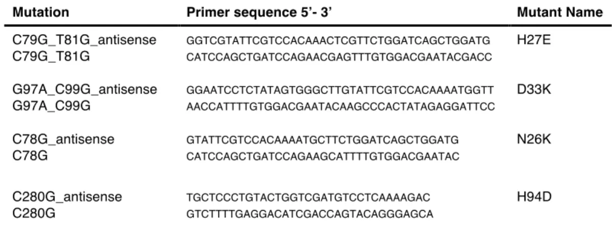
2.2.1.11 Site-Directed Mutagenesis (SDM) ... 49
2.2.2 Protein Biochemistry ... 50
2.2.2.1 Purification of Ras Proteins ... 50
2.2.2.1.1 Cell Lysis ... 50
2.2.2.1.2 Protein Precipitation with (NH
4)
2SO
4... 52
2.2.2.1.3 Ion Exchange Chromatography (IEX) ... 53
iii
2.2.2.1.4 Size Exclusion Chromatography (SEC) ... 57
2.2.2.2 Purification of Raf-RBD and NF1 ... 59
2.2.2.2.1 Cell Lysis ... 59
2.2.2.2.2 Affinity Chromatography: GST-Fusion ... 59
2.2.2.3 Purification of SOS
cat(W729E)... 61
2.2.2.3.1 Cell Lysis ... 61
2.2.2.3.2 Affinity Chromatography: Histidine-Tag Methodology ... 61
2.2.2.4 Nucleotide Exchange Reactions ... 62
2.2.2.4.1 GDP Against GppNHp ... 62
2.2.2.4.2 GDP Against GTP ... 64
2.2.2.5 Polyacrylamide Gel Electrophoresis (SDS-PAGE) ... 64
2.2.2.6 Determination of Protein Concentration ... 66
2.2.2.6.1 The Bradford Method ... 66
2.2.2.6.2 UV-Absorption at 280 nm using Nanodrop™ ... 67
2.2.2.6.3 Analytical High Performance Liquid Chromatography (HPLC) ... 67
2.2.3 Nano Differential Scanning Fluorimetry (nanoDSF) ... 68
2.2.4 Isothermal Calorimetry (ITC) ... 69
2.2.5 NMR Spectroscopy ... 73
2.2.5.1 Sample Preparation. General Considerations ... 73
2.2.5.2 Ambient Pressure and HP
31P NMR Data Acquisition ... 73
2.2.5.3 [
1H-
15N]-HSQC NMR Acquisition, Processing and Evaluation ... 74
2.2.5.4
31P (HP) NMR Processing and Evaluation ... 75
2.2.6 High Pressure Macromolecular Crystallography ... 78
2.2.6.1 Protein Crystallization ... 78
2.2.6.2 Data Collection ... 79
iv
3. results
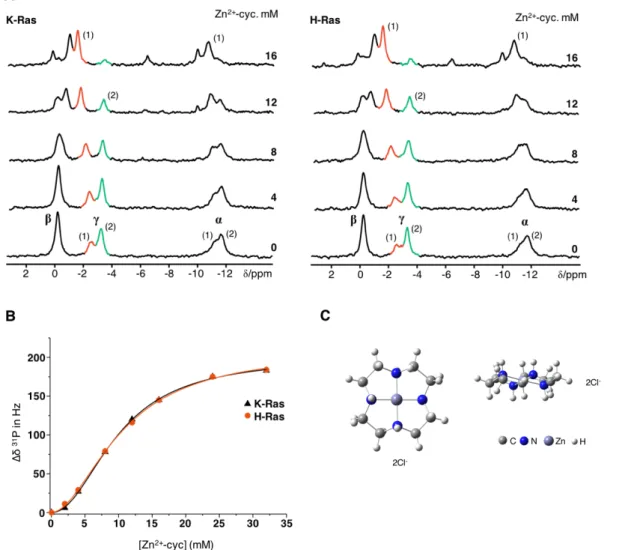
3.1 Dynamics, Equilibrium and Inhibition of FL Ras Studied by
31P NMR ... 85
3.1.1 Comparison of the Conformational Equilibrium in HRas
WTand KRas4b
WT... 85
3.1.2 Modulation of the Conformational Equilibrium in KRas4b
WTby Zn
2+-cyclen . 89 3.1.3 Effect of Raf-RBD on the Displacement of Zn
2+-cyclen From HRas
WT... 91
3.1.4 Conformational Equilibria of KRas
G12D•GTP and KRas
G12V•GTP ... 93
3.1.5 Modulation of the Equilibrium in KRas
G12Dby Zn
2+-cyclen ... 94
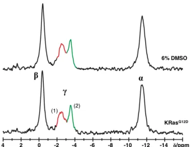
3.2 Inhibition of KRas
G12D(1-188)
•Mg
2+•GppNHp Investigated by
31P NMR and ITC 97 3.2.1 Effect of DMSO ... 98
3.2.2 Compound #16324643 ... 99
3.2.3 Compound #16328098 ... 99
3.2.4 Compound #35127727 ... 99
3.2.5 Compound #35139703 ... 101
3.2.6 Compound #35141449 ... 101
3.2.7 Compound #35145071 ... 102
3.2.8 Compound #35117109 ... 103
3.2.9 Compound #35129755 ... 103
3.2.10 Compound #35129757 ... 106
3.2.11 Compound #35131307 ... 108
3.2.12 Compound #35131308 ... 108
3.2.13 Compound #35135612 ... 109
3.2.14 Compound #35135613 ... 109
3.2.15 Compound #35135624 ... 110
3.2.16 Compound #35135616 ... 111
3.3 High Pressure
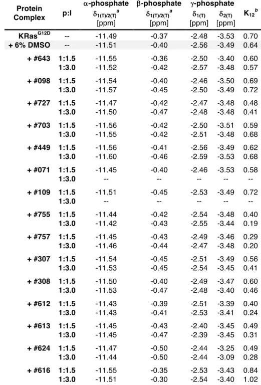
31P NMR Spectroscopy ... 115
3.3.1 Studies on GppNHp ... 115
3.3.2
31P HP NMR on HRas
WT(1-166)•Mg
2+•GppNHp ... 118
3.3.2.1 Measurements Conducted at 278 K ... 118
3.3.2.2 Measurements Conducted at 303 K ... 125
3.3.3
31HP NMR on HRas
T35S(1-166)•Mg
2+•GppNHp ... 127
v
3.3.4
31HP NMR on KRas
G12V(1-188)•Mg
2+•GTP ... 132
3.4 Mutational Analysis of HRas
WT(1-166) studied by
31P NMR and ITC ... 139
3.4.1 Preliminary Considerations about Ras
WT(1-166) ... 140
3.4.1.1 Conformational Equilibria of H, K and NRas
WT•Mg
2+•GppNHp ... 140
3.4.1.2 Titration of HRas
WT•Mg
2+•GppNHp with NF1 Followed by
31P NMR spectroscopy ... 142
3.4.2 Site Directed Mutagenesis ... 147
3.4.2.1 2(T)-to-1(T) Transition: N26K, H94D and A66T ... 147
3.4.2.1.1
31P NMR GppNHp and GTP Spectra. Raf Interaction and GTPase Activity ... 148
3.4.2.2 2(T)-to-1(0) Transition: S39L and E3V ... 153
3.4.2.2.1
31P NMR GppNHp and GTP Spectra. Raf Interaction and GTPase Activity ... 154
3.4.2.3 2(T)-to-3(T) Transition: H27E and D33K ... 157
3.4.2.3.1
31P NMR GppNHp and GTP Spectra. Raf Interaction and GTPase Activity ... 157
3.4.3 Interaction Between HRas
H27E•Mg
2+•GppNHp and Raf-RBD ... 161
3.4.4 Interaction Between HRas
D33K•Mg
2+•GppNHp and Raf-RBD ... 163
3.4.5 Interaction Between HRas
D33K•Mg
2+•GppNHp and NF1 ... 165
3.4.6
31P T
1Longitudinal Relaxation Times of Ras
WT, Ras
T35Aand Ras
D33K... 168
3.4.7
31P HP NMR on HRas
D33K•Mg
2+•GppNHp ... 171
3.4.8 Thermal Unfolding of HRas Proteins Investigated by nanoDSF ... 175
3.4.9 [
1H-
15N]-HSQC NMR on HRas
WTand HRas
D33K... 177
3.4.10
31P NMR Investigations of HRas
G12Pand HRas
G12V/T35S... 180
3.4.10.1
31P NMR GppNHp and GTP Spectra. Raf Interaction and GTPase Activity ... 180
3.4.10.2 Interaction Between HRas
G12P•Mg
2+•GppNHp and Raf-RBD ... 186
3.4.10.3 Interaction Between HRas
G12P•Mg
2+•GppNHp and NF1 ... 188
3.4.10.4 Interaction Between HRas
G12V/T35S•Mg
2+•GppNHp and Raf-RBD . 190
vi
3.5 High Pressure Macromolecular Crystallography (HPMX) ... 193
3.5.1 Crystal Structure of Ras
WT(1-166)•Mg
2+•GppNHp at Ambient Pressure .... 197
3.5.2 Crystal Structure of Ras
WT(1-166)•Mg
2+•GppNHp at High Pressure ... 197
3.5.2.1 Analysis of the Compressibility Curve ... 197
3.5.2.2 Analysis of rmsd and b-factor Values ... 198
3.5.2.2.1 Structure at 270 MPa ... 199
3.5.2.2.2 Structure at 490 MPa ... 200
3.5.2.2.3 Structure at 650 MPa ... 200
3.5.3 Crystal Structure of Ras
D33K(1-166)•Mg
2+•GppNHp at Ambient Pressure .. 205
3.5.4 Crystal Structure of Ras
D33K(1-166)•Mg
2+•GppNHp at High Pressure ... 207
3.5.4.1 Analysis of the Compressibility Curve ... 207
3.5.4.2 Analysis of rmsd and b-factor Values ... 208
3.5.4.2.1 Structure at 200 MPa ... 208
3.5.4.2.2 Structure at 880 MPa ... 209
3.5.4.2.3 Ras
D33Kat 880 MPa vs Ras
WTat 650 MPa ... 209
3.5.5 Crystal Structure of Ras
WT•Mg
2+•GppNHp Soaked with Zn
2+-cyclen ... 213
3.5.5.1 Analysis of the Compressibility Curve ... 213
3.5.5.2 Analysis of the rmsd and b-factor Values ... 214
3.5.5.2.1 Ras Apo vs Ras•Zn
2+-cyclen at Ambient Pressure ... 214
3.5.5.2.2 Structure at 240 MPa ... 214
3.5.5.2.3 Structure at 520 MPa ... 215
4. Discussion 4.1 FL H and KRas. Conformational Equilibria and Inhibition by Zn
2+-cyclen ... 221
4.1.1 Interaction of KRas
WT(1-188)•Mg
2+•GppNHp with Raf-RBD ... 221
4.1.2 Stabilization of State 1(T) by Zn
2+-cyclen Studied by
31P NMR ... 223
4.1.3 Conformational Equilibria of KRas
G12D•GTP and KRas
G12V•GTP ... 223
vii 4.2 Modulation of the Conformational Equilibrium of KRas
G12D(1-188)
•Mg
2+•GppNHp
by Small Compounds ... 225
4.2.1 General Considerations ... 225
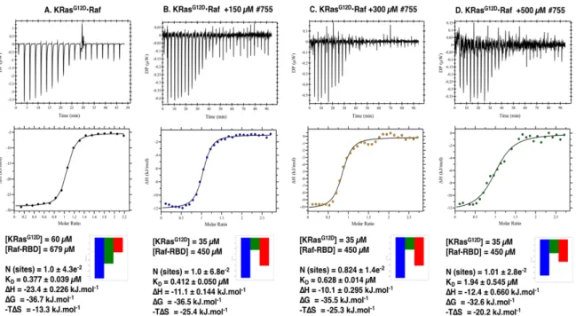
4.2.2 Inhibition of Ras-Raf Interaction by #755 and #757 Followed by ITC ... 226
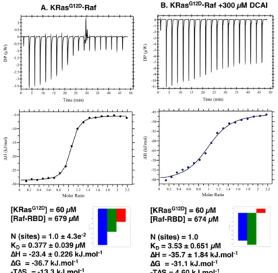
4.2.3 Inhibition of Ras-Raf Interaction by #616 (DCAI) Followed by ITC ... 227
4.3 High Pressure
31P NMR Spectroscopy ... 229
4.3.1 High Pressure
31P NMR on GppNHp ... 229
4.3.1.1 Pressure Effects in Presence and Absence of Mg
2+... 229
4.3.2 High Pressure
31P NMR on Ras Proteins ... 231
4.3.2.1 HP
31P NMR on HRas
WT(1-166)•Mg
2+•GppNHp ... 231
4.3.2.1.1 Pressure Coefficients, Energy and Volume Changes ... 232
4.3.2.2 HP
31P NMR on HRas
T35S(1-166)•Mg
2+•GppNHp ... 233
4.3.2.3 HP
31P NMR on KRas
G12V(1-188)•Mg
2+•GTP ... 234
4.3.2.4 HP
31P NMR on KRas
D33K(1-166)•Mg
2+•GppNHp ... 235
4.3.2.5 General Considerations ... 235
4.4
31P NMR Spectra of H, K and NRas
WT(1-166)
•Mg
2+•GppNHp ... 237
4.5 Interaction of Ras
WT, Ras
G12Pand Ras
D33Kwith NF1 Followed by
31P NMR ... 237
4.5.1 Ras
WT(1-166)•Mg
2+•GppNHp•NF1 ... 237
4.5.2 Ras
G12P(1-166)•Mg
2+•GppNHp•NF1 ... 238
4.5.3 Ras
D33K(1-166)•Mg
2+•GppNHp•NF1 ... 238
4.6 Mutational Studies on HRas
WT(1-166) ... 239
4.6.1 General Considerations ... 239
4.6.2 2(T)-to-1(T) Transition: N26K, A66T and H94D ... 241
4.6.3 2(T)-to-1(0) Transition: E3V and S39L ... 241
4.6.4 2(T)-to-3(T) Transition: H27E and D33K ... 242
4.7 Interaction Between Ras
D33Kand Raf-RBD Followed by
31P NMR. Structural Basis for the Loss of Affinity ... 245
4.8 Thermal Unfolding of Ras Followed by nanoDSF ... 247
viii
4.9 Conformational Dynamics of Ras
G12Pand Ras
G12V/T35S... 247
4.9.1 Considerations About Ras
G12P... 247
4.9.2 Considerations About Ras
G12V/T35S... 248
4.10 High Pressure Macromolecular Crystallography (HPMX) ... 249
4.10.1 HRas
WT(1-166)•Mg
2+•GppNHp ... 249
4.10.2 HRas
D33K(1-166)•Mg
2+•GppNHp ... 251
4.10.3 HRas
WT(1-166)•Mg
2+•GppNHp in Complex with Zn
2+-cyclen ... 253
5 Appendix ... 255
5.1 Figures ... 257
5.2 Tables ... 267
5.3 List of Figures ... 274
5.4 List of Tables ... 280
5.5 Abbreviations Used in this Thesis ... 283
5.6 List of Publications ... 286
6 References ... 289
ix Acknowledgments
First and foremost I would like to thank the Bayerischen Forschungsstiftung for their complete financial support and to Prof. Dr. Dr. Hans Robert Kalbitzer for accepting me in his group, for sharing his ideas, scientific knowledge and for his overall contribution as main supervisor of the entire project.
I would like to thank to Dr. Michael Spoerner, without whom this work would have been impossible to perform. His insightful ideas,
31P NMR expertise and broad knowledge on the Ras field were always detrimental for the project’s well being.
Many thanks to Prof. Dr. Werner Kremer, for all the suggestions, advices and concerns not only with the scientific side but also with the human side, not only with me but with all members of the group.
My deepest gratitude to Dr. Sunilkumar P. Narayanan for helping me with his incredible scientific expertise, particularly for all the teachings in the field of high pressure NMR. More importantly, thank you for your friendship and for guiding me through the ways of life and science.
My deepest gratitude to the high pressure crystallography team that I had the pleasure to work with: Dr’s. Nathalie Colloc´h, Eric Girard, Anne-Claire Dhaussy and Prof. Dr. Thierry Prangé. Your brightness, insightful ideas and seemingly endless wisdom caused my scientific interests to irrevocably entwine with the field of X-ray crystallography. Your expertise and extraordinary technical skills are to me the prototype of what great scientists and the scientific method should always be.
The warmest thank you to Dr. Malte Andrasch for sharing with me his extensive and remarkable knowledge in the fields of protein biochemistry and biophysics. More importantly, thank you for your positive and vibrant energy during many dark moments.
Thank you still for your ongoing help and concern towards the future.
I am grateful to Sabine Laberer for her help in the scope of molecular biology techniques, particularly with DNA technologies. Similarly, I would like to thank Sabine Ruppel, to whom I owe all the expertise I acquired in the field of protein biochemistry and purification techniques. Thank you for your constant effort to teach me, even through the barriers imposed by communicating in a different language.
My deepest thank you to Dominique Quetting, for her help with the expression and
purification of difficult proteins and for the permanent concern in providing the most
organised and efficient laboratorial environment. Above all, thank you for your friendship,
for all the laughs, relaxing coffees together with Sunil and the nice moments we spent
looking for furniture.
x
I want to show my gratitude to Dr. Markus Bech Erlach for his help and know-how with the processing and interpretation of NMR spectra and contagious positive attitude.
From many students that I had the pleasure to meet and work with side-by-side, I leave here my heartfelt thank you to all: Beatta Zablocka, Gani, Margarita Neun, Maria Watzlowik, Silvia Planck, Linda Stabi, Martin Winter, Simon Nimmermehr, Andy, Mathias Karl, Marcell Kaljanac. Thank you, for your eagerness to learn the ways of science, for your questions and for your teachings. Above all, thank you for the awesome moments and stories we shared and for introducing me to the subtle details of another culture and lifestyle.
I am grateful to Claudia Kiesewetter and Marcus Hoering for their prompted availability to help me with the translation of the summary of this thesis. Thank you Claudia for your brightness and lovely friendship and Marcus for all the good laughs and memorable stories.
I am ever so grateful to my brother João Miguel for always being a positive influence in my life and to my parents Manuel Lopes and Maria Celina for their support and concern with my well-being.
I leave here a word of appreciation to all the musicians that accompanied me for the long hours and lonely nights spent at the biochemistry and NMR laboratories. Among many, I am especially grateful to Sirs Mark Knopfler, David Gilmour and Gary Moore for the legacy they gave to all of us. Their music was countless times the only motivation to carry on and it will always be a source of inspiration through my entire life.
Last but by no means least, I leave here my heartfelt deepest gratitude to the most important
person of all: my Carina! Thank you for your constant tender and care, for your patience
and for helping me more times than I can remember. As you know so well my dear, writing
this book has been an exercise of sustained suffering through which I persisted only
because of your relentless support and dedication. It came to realisation only because of
you and therefore it is dedicated to you!
xi Summary
The guanine binding nucleotide protein Ras is a small GTPase that controls central regulatory processes such as cell differentiation, proliferation and apoptosis by alternating between an active, GTP-bound, and an inactive, GDP-bound state. Both, the intrinsic hydrolysis of GTP and the exchange reaction from GDP to GTP are very slow. Two classes of regulatory proteins called GAP’s and GEF’s are necessary to accelerate these two respective reactions. Active Ras interacts also with another class of proteins generally called effectors. Using
31P NMR spectroscopy two conformational states that interconvert in the millisecond time scale could be directly observed. They are named state 1(T) and state 2(T) (T or D, depending whether if Tri- or Di-phosphate is bound) and correspond to the GEF and to the effector recognition states, respectively. Their nature is influenced by specific mutations and by the type of bound nucleotide. It was verified that specific somatic mutations render Ras a very potent oncogene. As consequence, the protein becomes insensitive to inactivation by GAP’s and therefore locked in the active state, leading ultimately to uncontrolled cellular proliferation. Approximately 30% of all human tumours are estimated to harbour mutations in one of the three isoforms (H, K and NRas) at positions 12, 13 or 61, with KRas being the most preponderant one. Due to its key role in malignant transformation, efforts have been made over the years to develop effective inhibitors against a seemingly undruggable protein.
In the framework of this thesis, it was demonstrated that KRas
WTshows an almost identical behaviour to HRas
WTin terms of their intrinsic equilibria. Their
31P NMR spectra is dominated by two conformational states with an equilibrium constant, K
12, of 2.0 measured at the g-phosphate of bound GppNHp. The ability to modulate the equilibrium was further addressed by elucidating the mode of action of Zn
2+-cyclen. It was shown that this compound can selectively recognise and stabilise state 1(T), disrupting the affinity of both isoforms towards effectors such as Raf-RBD. Titration experiments showed a cooperative binding at two different sites with a Hill coefficient of 2 and an apparent dissociation constant of 9.9 mM for both isoforms.
The inhibition of the oncogenic KRas
G12Dby a library of 15 different small compounds was
further investigated.
31P NMR showed that at least two of them (#755 and #757) led to a
significant stabilisation of state 1(T), with more than 70% decrease in the equilibrium
constant. A 75% decrease in the affinity of the Ras-Raf complex in the presence of 300 µM
of each drug was obtained from ITC measurements. The screening showed that the
compounds initially developed for inhibition of Ras bound to GDP are also able to inhibit
xii
Ras in the triphosphate-bound form and are good candidates for further lead optimisation.
High pressure (HP)
31P NMR was conducted in the isolated GppNHp molecule and in several Ras proteins. For all of them, the pressure-dependence of the chemical shifts revealed to be non-linear as given by the obtained positive values of the second order pressure coefficients, B
2. A direct shift of the conformational equilibrium from state 2(T) towards state 1(T), given by the decrease in the equilibrium constant from 1.7 at 0.1 MPa to 0.24 at 250 MPa was observed for Ras
WT. Additional transitions were detected involving the GAP binding and the nucleotide-free states, named 3(T) and 1(0), respectively.
State 3(T) was identified by
31P NMR upon titration of Ras with NF1 and assigned to the resonance line at d= -3.00 ppm in the spectrum of Ras
WT•Mg
2+•GppNHp. At equimolar concentrations saturation was achieved for the g-phosphate but not for the b-phosphate, supporting the existence of conformational differences in the local environment of this phosphate group upon formation of the protein complex.
Using site-directed mutagenesis and
31P NMR spectroscopy, it was verified that several point mutants (H27E, S39L, E3V and D33K) led to a pronounced modification of the conformational equilibrium. The highest effect was observed for Ras
D33K•Mg
2+•GppNHp that is almost completely shifted towards state 2(T) as given by the increase of the equilibrium constant from 1.7 to 11.3. The mutation is located in the effector-loop region of Ras involved in the interaction with different proteins. A 30-fold decreased affinity towards Raf-RBD (K
D= 10.4 µM) comparatively to Ras
WT(K
D= 0.42 µM) and an impaired affinity towards NF1 was observed. The increase of the equilibrium constant from 1.7 to 2.0 in the case of Ras
E3Vrepresents a good example of a typical conformational modulation by an allosteric mechanism, as the mutation is located in a distal site relative to the bound nucleotide.
The crystal structures of Ras
WTand Ras
D33Kwere solved for the first time under high
pressure. Analysis of the unit cell compressibility revealed the existence of a transition zone
for Ras
WTbetween 200 and 270 MPa. A decrease of the main-chain temperature factors
from 20 Å
2at 0.1 MPa to 13.5 Å
2at 650 MPa, together with the increase in the average
rmsd values, gave an insight for the possible stabilisation of non-native conformations at
high pressures. Ras
D33K, on the other hand, is less sensitive to pressure, with the transition
zone occurring around 500 MPa and significant rearrangements at 880 MPa. Typical
pressure-induced modifications involved the helix a
2as part of switch 2, the loop λ
7, helix
a
1at the beginning of switch 1.
xiii Zusammenfassung
Das Guaninnukleotid-bindende Ras Protein ist eine kleine GTPase, die durch den Wechsel zwischen dem aktiven, GTP-gebundenen Zustand und dem inaktiven, GDP-gebundenen Zustand verschiedene regulatorische Prozesse in der Zelle kontrolliert, wie z.B.
Zelldifferenzierung, Zellproliferation und Apoptose. Sowohl die intrinsische Hydrolyse von GTP als auch die Austauschreaktion von GDP zu GTP sind sehr langsam. Um diese Reaktionen zu beschleunigen wird die Hilfe der zwei regulatorischen Proteinklassen GAP’s und GEF’s benötigt. Außerdem interagiert Ras in seinem aktiven Zustand mit einer weiteren Proteinklasse, den sogenannten Effektoren. Mit Hilfe der
31P-NMR-Spektroskopie können die zwei Konformationen, deren Umwandlung nur Millisekunden benötigt, direkt detektiert werden. Die beiden Zustände werden als 1(T) und 2(T) bezeichnet (T bzw. D, je nachdem ob Tri- oder Diphosphat gebunden ist) und entsprechen jeweils dem GEF und dem Effektor- erkennenden Zustand. Beeinflusst werden sie durch spezifische Mutationen und durch die gebundene Nukleotidform. Außerdem ist bekannt, dass spezifische somatische Mutationen Ras zu einem potenten Onkogen machen. Als Folge daraus wird das Protein unempfindlich gegen die Inaktivierung durch GAP’s und verbleibt im aktiven Zustand, der zu einer unkontrollierten Zellteilung führt. In 30% aller menschlichen Tumore liegen Mutationen in einer der drei Ras Isoformen (H, K und NRas) in den Positionen 12, 13 oder 61 vor, von denen KRas am häufigsten betroffen ist. Wegen seiner Schlüsselfunktion bei der Tumorbildung wurden im Laufe der letzten Jahre viele Versuche unternommen, um effektive Inhibitoren gegen das scheinbar unbehandelbare Protein zu finden.
Im Rahmen dieser Arbeit konnte nachgewiesen werden, dass KRas
WTund HRas
WTein nahezu identisches Verhalten in Bezug auf ihre intrinsischen Gleichgewichte besitzen. Ihre
31
P-NMR-Spektren werden von zwei konformationellen Zuständen dominiert, die eine Gleichgewichtskonstante K
12von 2.0 besitzen, die am g-Phosphat des gebundenen GppNHp bestimmt wurde. Die Möglichkeit dieses Gleichgewicht zu beeinflussen wurde durch den Wirkungsmechanismus von Zn
2+-Cyclen verdeutlicht. Es konnte gezeigt werden, dass diese Komponente selektiv den Zustand 1(T) stabilisiert und die Affinität beider Isoformen gegenüber den Effektoren (z.B. Raf-RBD) stört. Titrationsexperimente haben eine kooperative Bindung an zwei verschiedenen Seiten mit einem Hill-Koeffizienten von 2 und einer Dissoziationskonstanten von 9.9 mM für beide Isoformen ergeben.
Die Inhibition von onkogenem KRas
G12Ddurch 15 verschiedene kleine Substanzen wurde
weiter untersucht und es konnte mit Hilfe der
31P-NMR-Spektroskopie gezeigt werden, dass
mindestens zwei dieser Komponenten (#755 und #757) zu einer signifikanten Stabilisierung
xiv
des Zustands 1(T), mit einer Verkleinerung der Gleichgewichtskonstanten von mehr als 70%, führen. Mittels ITC Messungen konnte eine 75%ige Abnahme der Affinität des Ras- Raf Komplexes in der Anwesenheit von 300 µM der entsprechenden Komponente bestimmt werden. Die Experimente zeigten, dass die Komponenten, die ursprünglich für die Inhibition des GDP-gebundenen Ras entwickelt wurden, auch Ras in seiner Triphosphat-gebundenen Form inhibieren können und dementsprechend optimale Kandidaten für eine weitere Optimierung wären.
Das isolierte GppNHp Molekül sowie verschiedene Ras Proteine wurden mittels Hochdruck
31
P-NMR-Spektroskopie untersucht. In allen Fällen konnte eine nicht lineare Abhängigkeit der chemischen Verschiebung anhand der positiven Werte des Druckkoeffizienten zweiter Ordnung (B
2) gezeigt werden. Für Ras
WTkonnte eine direkte Verschiebung des koformationellen Gleichgewichts von Zustand 2(T) zu Zustand 1(T) anhand der Abnahme der Gleichgewichtskonstanten von 1.7 bei 0.1 MPa zu 0.24 bei 250 MPa beobachtet werden. Außerdem wurden zwei zusätzliche Übergänge detektiert, die Zustand 3(T) und 1(0) benannt wurden. Dabei handelt es sich um den GAP-bindenden Zustand und um den Nukleotid freien Zustand.
Der Zustand 3(T) wurde mittels Titrationsexperiment und
31P-NMR-Spektroskopie identifiziert, bei dem NF1 zu Ras titriert wurde und die Resonanzlinie bei d= -3.00 ppm dem Spektrum von Ras
WT•Mg
2+•GppNHp zugeordnet wurde. Bei equimolarer Konzentration wurde eine Sättigung für das g-Phosphat, aber nicht für das b-Phosphat, erzielt. Diese Tatsache unterstützt die Existenz der konformationellen Unterschiede in der lokalen Umgebung dieser Phosphat Gruppe bei der Bildung des Proteinkomplexes.
Mit Hilfe von ortsspezifischer Mutagenese und der
31P-NMR-Spektroskopie konnte
nachgewiesen werden, dass einige Punktmutationen (H27E, S39L, E3V und D33K) zu
einer starken Modifikation des konformationellen Gleichgewichts führen. Der stärkste Effekt
wurde bei Ras
D33K•Mg
2+•GppNHp beobachtet. Hier erfolgt eine nahezu komplette
Verschiebung hin zu Zustand 2(T), wie anhand der Zunahme der Gleichgewichtskonstanten
von 1.7 zu 11.3 zu erkennen ist. Die Mutation befindet sich an der Effektor-Schleifen Region
von Ras, die in die Interaktion mit verschiedenen Proteinen involviert ist. Eine 30-fache
Abnahme der Affinität zu Raf-RBD (K
D= 10.4 µM) im Vergleich mit Ras
WT(K
D= 0.42 µM)
und eine gestörte Affinität zu NF1 konnte beobachtet werden. Die Zunahme der
Gleichgewichtskonstanten von 1.7 zu 2.0 von Ras
E3Vstellt ein gutes Beispiel einer
typischen konformationellen Änderung durch einen allosterischen Mechanismus dar, da
sich die Mutation auf der entfernten Seite des Nukleotid Bindungszentrums befindet.
xv Die Kristallstrukturen von Ras
WTund Ras
D33Kwurden erstmals unter Hochdruck ermittelt.
Die Analyse der Kompressibilität der Einheitszelle zeigte die Existenz einer Übergangszone
für Ras
WTzwischen 200 und 270 MPa. Durch die Abnahme der Hauptketten Temperatur
Faktoren von 20 Å
2bei 0.1 MPa zu 13.5 Å
2bei 650 MPa, zusammen mit der Zunahme der
Durchschnittwerte von rmsd, konnte ein Einblick in die mögliche Stabilisierung von nicht
nativen Konformationen bei Hochdruck erhalten werden. Auf der anderen Seite ist Ras
D33Kweniger sensitiv zu Druck und besitzt seine Übergangszone bei etwa 500 MPa und
signifikante Änderungen treten ab 880 MPa auf. Typische Druck induzierte Modifikationen
involvieren die Helix a
2als Teil des Switch 2, die Schleife λ
7und die Helix a
1am Anfang
des Switch 1.
xvi
- This page was deliberately left blank -
1. Introduction
“No human investigation can be called true science without passing through mathematical tests”
Leonardo Da Vinci
1. Introduction
3 1.1 Ras as a Molecular Switch
This thesis deals with the Ras protein, a prototype member of a large superfamily called guanine nucleotide binding proteins (GNBP’s or G proteins, for short) that act as molecular switches by cycling between an ‘on’ (active) and an ‘off’ (inactive) state, thus controlling diverse regulatory processes of capital importance in the living cells. Ras was identified in 1964 when Jennifer Harvey observed that a preparation of murine leukaemia virus (MLV) induced rat sarcomas in newborn mice [1]. Within the next three years, an additional retrovirus containing a new Ras gene, homologous to the first one, was further discovered by Werner Kirsten [2]. Ten years later, a third gene was identified in human Neuroblastoma cells [3]. The three homologue proteins codified by the three genes were thereafter named as H, K and NRas. Their complex structural dynamics and profound biochemical importance has been unveiled slowly but steadily for more than half a century, up to the present day.
Since the last decade there has been an uprising interest by the scientific community in the chemistry of Ras (particularly KRas) due to its involvement in tumour formation and progression, triggered by an impaired function of the molecular switch mechanism that originates the permanent activation of the protein. A renewed scientific endeavour is taking place at the moment, in an attempt to find truly effective Ras inhibitors capable of drugging a seemingly undruggable protein [4].
The GNBP superfamily can be grouped by sequence homology into five major subgroups.
Incidentally the subgroups also define a more or less similar biological function. The Ras family is a branch that contains the Ras protein itself, together with the three isoforms and is involved in cell proliferation, gene expression, differentiation and apoptosis; the Rho (Ras homologous) controls the dynamics of the cytoskeleton; the Rab (Ras in the brain) and Arf (ADP ribosylation factor) families regulate the vesicular transport and the Ran (Ras in the nucleolus) determines the direction of the nucleocytoplasmic transport and regulates the mitotic spindle organization [5]. Altogether they are the masters of the signalling transduction pathways, controlling virtually all molecular events by which any chemical or physical stimuli is relayed into the nucleus and culminates with an appropriate cellular response (e.g. transcription of specific genes). All members have the ability to bind the small nucleotides guanosine di-phosphate (GDP) and guanosine tri-phosphate (GTP) with high selectivity and affinity (in order of 10 pM), being the binding process strongly dependent of Mg
2+, which is their natural co-factor.
With exception of Ran, all G proteins are posttranslationally modified by addition of lipids
either at their N-terminus by acetylation (myristoylation) or at their C-terminus by prenylation
1. Introduction
4
and palmitoylation [6]. Prenylation takes place by addition of farnesyl or geranyl-geranyl lipidic groups via a stable thioether linkage, catalysed by the enzyme farnesyltransferase.
Palmitoylation takes place through the formation of a reversible thioester linkage and is used to dynamically regulate membrane localization. These modifications serve as important components of their membrane targeting motifs and promote membrane binding.
GNBP’s can be also regulated by non-lipidic modifications such as phosphorylation, nitrosylation, mono- and di-ubiquitination, acetylation and oxidation, all of which alter the protein localization and/or interaction with regulatory molecules. An in-depth coverage of these topics can be found in excellent recent reviews [7-9].
The human Ras superfamily contains 167 proteins, of which 39 belong to the Ras sub- family and comprise a total of 940 structures deposited in the protein data bank (pdb), as of October 2017, either in uncomplexed forms or bound to any regulatory protein or small ligand.
1.1.1 Structural and Biochemical Considerations
The first crystal structure of Ras was solved in 1988 independently by Sung-Hou Kim [10]
and Alfred Wittinghofer [11] with a 2.1 and 2.6 Å resolution, respectively. In the next year, the latter group published a refined structure with a resolution of 1.35 Å that became a representative model of the protein and a starting point for many theoretical and experimental structural studies in the years to come (pdb: 5p21) [12]. All Ras members share the same structural fold, so-called “G domain”, containing the catalytic machinery comprised of 6 b-strands (b
1-b
6) flanked by and 5 a-helices (a
1-a
5) and 10 connecting loops (λ
1-λ
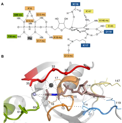
10, Figure 1.1). The G domain contains five fingerprint regions, named from G1 to G5, all of them located in loops: G1, also called the P-loop (aa 10-17; GxxxxGKS, x= any aliphatic amino acid) is a glycine-rich loop that twines around the negatively charged phosphate groups, binding tightly to them through its main chain positively charged N atoms. Lys16 contacts directly the b- and g-phosphates and is crucial for nucleotide binding.
The OH group of Ser17 contacts the b-phosphate and the Mg
2+ion [13]. The region G2,
also called switch 1 (switch 1, aa 30-40 in HRas) or “effector region” since is involved in the
binding of effectors when in the GTP state, is one of the regions that changes conformation
upon nucleotide exchange. It contains a Thr35 that is totally conserved in all members of
the superfamily and crucial for sensing the presence of the g-phosphate of GTP,
establishing a polar contact with it via its NH main chain. The same amino acid also binds
the Mg
2+ion via its OH side chain [14]. The region G3, also called switch 2 (switch 2, aa 59-
70 in HRas) has no conserved sequence besides a small loop at its beginning called the
1. Introduction
5 DxxG motif (aa 57-60) in
which Gly60 forms a main chain H-bond with the g- phosphate. Another important residue in switch 2 is Gln61 that plays a crucial role in the stimulation of GTP hydrolysis (section 1.1.4) [15]. G4 is the N/TKxD motif (aa 116-119), where the Asp119 contacts the nitrogen atoms from the
base with a bifurcated H-bond and Asn116 contacts the C=O group from the purine ring, thus conferring specificity for the guanidinium base. Lys117 stacks along the plane of the base [16]. G5, also called SAK (aa 145-147), is a weakly conserved motif in which the backbone NH groups interact with the C=O moiety from the guanine base. These five structural elements of the G domain are depicted in Figure 1.2, along with key molecular contacts to the nucleotide.
The three Ras isoforms (H, K and NRas) share more than 90% identity across the catalytic domain but differ considerably in the last 25 residues, having less than 15% similarity.
These residues constitute the C-terminal part of the protein and are called the hypervariable region (HVR). They are necessary for interaction with the membrane and play a fundamental role in signalling processes. For technical reasons, most of the biochemical studies were performed over the years with truncated variants of Ras, comprising only the catalytic domain (aa 1-166, 18 kDa). It was demonstrated that the first 166 residues are necessary and sufficient for the biochemical properties of the protein [15, 17]. In fact, to the present date, there are no crystal structures of the full-length (aa 1-189, 21 kDa) wild type Ras deposited in pdb. The only available structure was obtained in 2012 for the mutant KRas4b
G12D•Mg
2+•GDP [18]. FL Ras undergoes crystallization as easy as the truncated variant but attempts to retrieve diffraction data from HVR fail consistently due to the lack of electron density, which portends the high flexibility of this region. In recent years, however, the scientific interest in FL Ras instead of the truncated variant gained much more attention, especially after the discovery that the HVR is capable of acting on the catalytic domain, modulating its action and being largely responsible for the biological differences across the three isoforms [19-21].
Figure 1.1. Topology of the G domain. The diagram represents the order of secondary structure. Helices are shown as cylinders and marked as a1-a5 (helix a3 is slightly distorted) and the b-sheets are shown as arrows and marked as b1-b6. Loops are labelled λ1-λ10. The amino acids flanking the beginning and the end of each element of secondary structure are indicated.
1. Introduction
6
Ras is intrinsically capable of hydrolysing the GTP nucleotide by transferring electrons to a water molecule strategically placed in the catalytic centre that promotes the nucleophilic attack of the g- phosphate (Figure 1.2). The averaged half-life for this process is ca. 25 min.
(Table 3.14). Very often the natural nucleotides need to be replaced by more stable, non-hydrolysable ones, especially when performing long term experiments of several hours or days,
common in NMR
spectroscopy. A frequently used analogue is GppNHp, being structurally identical to GTP except for the oxygen atom that mediates the b- and g-phosphates that
becomes replaced by an NH group. Other analogues include GTPgS and GppCH
2p [22].
Detailed analysis revealed that both GTP and GppNHp bind Ras in the same manner with only slight differences in the bridging NH group, indicating that small local changes can still lead to drastic differences in the dynamics of the catalytic site [23].
The switch mechanism between the active and the inactive states is accompanied by prominent structural changes in the switch 1 and switch 2 regions: in GppNHp-bound HRas, the g-phosphate forms two H-bonds with Gly60 from switch 2 and Thr35 from switch 1. The Mg
2+ion is bidentately coordinated by the non-bridging oxygen atoms of b- and g- phosphates, Thr35, Ser17 (from the P-loop) and two H
2O molecules [24-26]. Quantum mechanical (QM) calculations showed that Mg
2+provides a temporary storage for electrons
Figure 1.2. HRas•Mg2+•GppNHp as the prototype of the G domain:
structural details of the catalytic centre. A. Schematics of the interaction between selected residues and B. their representation in the crystal structure (pdb: 5p21). switch 1 and switch 2 are coloured in red and green, respectively, the P-loop is coloured in orange and the G4 and G5 motifs are coloured in blue and yellow, respectively. The position of important residues is indicated by small spheres (centred at their a- carbon) or explicitly by lines. Q61 is implicated in the hydrolysis of the g- phosphate by interacting transiently with a nearby catalytic water (shown as a light blue sphere). Thr32, whose side chain (not represented) can bind transiently to the g-phosphate is also indicated. Its neighbor, Asp33, is a very important residue in the scope of the work developed in this thesis. The Mg2+ ion is shown as a black sphere. The detail of the amide group of the non-hydrolysable GppNHp nucleotide is shown in blue colour. The nitrogen atom establishes a polar contact with Gly13 from the P-loop. This interaction is absent in the case of the natural nucleotide GTP.
1. Introduction
7 taken from the triphosphate and that after release of the g-phosphate it returns them back to GDP, contributing for the process of hydrolysis [27].
Upon GTP hydrolysis, the b-phosphate has no direct interactions with Thr35 and Gly60;
coupled with the loss of coordination between the Mg
2+ion and Thr35. These alterations promote an extensive remodelling of switch 1 and switch 2, both adopting now a more
‘relaxed’ or open conformation and effectively moving away from the nucleotide, as shown in Figure 1.3. At the same time the helix a
2(aa 66-74, Figure 1.1) undergoes a large rotation together with the unwind of one of its helical turns. This open conformation contributes afterwards for the dissociation of GDP, perpetuating the cycle. Another important difference upon nucleotide exchange is Tyr32 located in switch 1: in GppNHp-bound Ras, Tyr32 is set upwards, pointing to the solvent, whereas in the GDP-bound Ras undergoes a large flip and shifts towards the interaction site. The root mean square deviation (rmsd) between the whole catalytic domains of GppNHp and GDP-bound HRas is 1.58 Å for the backbone
Figure 1.3. The switch mechanism in three dimensions. The Ras protein is in a continuous cycle of activation/ inactivation defined by the permanent exchange between GTP and GDP nucleotides. The crystal structures of the inactive, GDP-bound (blue colour), and active, GTP- bound, (red colour) HRas protein are shown (pdb: 1q21 and 5p21, respectively). The flexible switch 1 and switch 2 regions are coloured in orange and the coordinated Mg2+ ion is shown as a black sphere. The two common mutation sites in cancer (G12 and G13) lie close to the nucleotide and are represented as grey spheres. The totally conserved Q61 is shown as an orange sphere and can be used here to visually understand the movement of the switch regions as the protein cycles between GDP and GTP. The activation mechanism is catalysed by GEF’s and the intrinsically slow hydrolysis of GTP is catalysed by GAP’s. In the active form, Ras interacts with effectors to trigger downstream signaling events.
1. Introduction
8
atoms. If the switch regions are excluded, the rmsd is only 0.66 Å [28, 29].
Ras is an incomplete enzyme: its intrinsic rate of GTP hydrolysis (also called GTPase activity) is too slow for many cellular processes and needs to be catalysed by a group of proteins called GTPase activating proteins (GAP’s). Similarly, the release of GDP is also intrinsically very slow and needs to be catalysed by guanine nucleotide exchange factors (GEF’s) [30, 31].
Ras is capable of initiating more than 10 different signalling cascades (Figure 1.4) that control cell differentiation, proliferation or apoptosis by transducing extracellular ligand- mediated stimuli to the nucleolus. The general mechanism for transduction can be described as follows: upon recognition of the external stimuli, different receptors (tyrosine kinases, RTK’s, G-protein-coupled receptors, etc.) hetero-dimerize and auto-phosphorylate each other. In the case of the common RTK receptor, additional adaptor proteins called Grb2 or Shc bind to one of its domains called SH2. The binding promotes the recruitment of GEF’s to the membrane, which, in turn, will activate the Ras protein by stimulating the exchange of GDP to GTP. Active Ras can now interact with its multiple downstream targets generally called effectors (from more than 10 known effectors of Ras, the most representative ones within the framework of this thesis, are Raf-RBD, Ral-GDS, PI3K and Byr2 [32, 33]). Each effector will activate specific kinase proteins that will start a series of kinase chain reactions through which the signal is relayed down, reaching the cellular nucleus and culminating with the transcription of genes that will confer an appropriate response to the original stimuli. One of the most common signalling cascades of Ras is the
Figure 1.4. Schematics of the main Ras signaling pathways. From more than 10 known signaling cascades under direct control of Ras, the MAPK (Ras/Raf/MEK/ERK) is the best understood. For sake of simplicity the full names for the abbreviations in the scheme are not given here. The reader is submitted to the original review [34] from where this scheme was adapted.
1. Introduction
9 mitogen-activated protein kinase (MAPK) pathway that uses the protein Raf as effector and two different MAP kinases: MEK and ERK.
Phosphorylated ERK translocates to the nucleus and triggers gene transcription [34]. The signalling cascade is eventually inactivated by negative feedback from the nucleus after gene transcription, which leads to the hydrolysis of g-phosphate of GTP-bound Ras, catalysed by GAP’s, as the protein returns to the original (inactive) GDP-bound state [35].
1.1.2 Interaction with GEF’s: the ‘Switch On’ Reaction
The direct consequence of the picomolar affinity between Ras and GDP/GTP is that nucleotide dissociation is not compatible with the time scale of most cellular processes. The activation of Ras needs to be catalysed by GEF’s, a large class of multidomain enzymes capable of accelerating the exchange reaction by 3 to 4 orders of magnitude [36, 37]. The GEF-catalysed reaction is an intricate multistep process whose kinetics have been described in detail only for the HRas•Cdc25 complex. The affinity of binding between the two proteins (K
D) was found to be 4.6 nM and the maximal acceleration by Cdc25 of the rate of nucleotide dissociation was estimated to be more than 10
5-fold [36]. In general, Ras has a similar affinity for both, GDP and GTP, and GEF’s do not favour the re-binding of either nucleotide. The direction of the reaction is dictated instead by the concentrations of the free nucleotides in the cell at a given time. Since normally the GTP concentration is 10- fold higher than GDP, the resulting product of the catalytic reaction is Ras loaded with GTP [38].
GEF’s can be grouped in two different classes. The first one comprises the RasGEF’s which
are activated by second messengers like Ca
2+, calmodulin or diacylglycerol [39]. The
second class is represented by the son-of-sevenless (SOS). Human SOS1 is a very large
multiprotein of ~1330 residues (152.4 kDa) containing at least 4 different domains, from
which the catalytic domain (aa 551-1050, abbreviated as SOS
cat, 36 kDa) is the one involved
in Ras binding and catalysis [40, 41]. SOS
catcomprises the Ras exchanger motif (Rem, aa
551-750), the Cdc25 motif (aa 751-1050) and a C-terminal region (aa 1051-1333) that
provides a docking site for the adaptor protein Grb2 (Figure 1.4) [42]. Previous
investigations have shown that the Rem motif is responsible for the stability of the whole
catalytic domain and that the Cdc25 motif is necessary and sufficient for nucleotide
exchange [36, 43]. The first crystal structure of the nucleotide free HRas•SOS
catcomplex
was solved in 1998 [44] and shows Ras bonded to the Cdc25 motif, with no direct contacts
to Rem (Figure 1.5A). The primary contact regions are the P-loop, helix a
1and the switch
regions. The most significant conformational change in HRas is due to the insertion of an
1. Introduction
10
helical hairpin composed of helices a
Hand a
Ifrom Cdc25 that penetrates through the catalytic pocket and projects switch 1 away from the nucleotide binding site (Figure 1.5B). The hairpin introduces in the nucleotide binding site a hydrophobic side chain (Leu938), which blocks magnesium binding by interacting with Ser17 from Ras, and an acidic side chain (Glu942), which overlaps with the site where the a-phosphate of the nucleotide would otherwise be bound. At the same time, switch 2 is held very tightly
by SOS and constitutes the heart of the interface between the two proteins. A cluster of three residues with hydrophobic side chains from Ras, Tyr64, Met67 and Tyr71, is buried into the hydrophobic core of SOS. Surrounding this hydrophobic triad is an array of polar interactions between the two proteins making almost all side chains of switch 2 coordinated to SOS (for example, Ala59 occupies the previous position of Mg
2+ion and the Glu62 side chain interacts with both, the NH group from Gly60 and the side chain of Lys16). As consequence, the conformation of switch 2 is very well defined in the complex, contrary to the typical poorly ordered conformation in the nucleotide-bound forms of Ras. In summary, switch 1 is pushed away from its normal position whereas switch 2 is pulled towards the nucleotide binding site [31, 44]. From these structural insights, the mechanism of nucleotide association/ dissociation was proposed to be as follows: in the dissociation process, the phosphate moieties of GDP are released first after binding of SOS, and then the base and ribose moieties are released. In contrast, the association process takes place by binding first the base and the ribose moieties of GTP and only then the phosphate groups.
Significant conformational changes of the switch regions are thought to occur to rebuild the
Figure 1.5. Structural insights on the Ras•SOS complex (pdb: 1bkd). A.
Surface and cartoon representation of nucleotide free Ras (coloured in grey) bound to the catalytic domain of SOS (coloured in blue). The Rem, N-terminal motif (coloured in light purple) is also shown. The hairpin formed by the helixes aH and aI from SOS is indicated. B. Details of the binding interface between the two proteins. Switch 2 is ‘clamped’ by SOS through interaction with hydrophobic residues and switch 1 is projected away from the vicinity of the nucleotide upon insertion of the helical hairpin. Adapted from [44].
1. Introduction
11 binding sites for the phosphate and Mg
2+and subsequently to displace SOS [31, 37].
It is worth mention that a new binding site of Ras to SOS
catwas found in 2003. This site is located between the Rem and the Cdc25 motifs and is in a distal position relative to the binding site on the Cdc25 motif [45]. Interestingly, the solved crystal structure is of a ternary complex HRas•GppNHp•SOS
cat:Ras(nucleotide-free) (pdb: 1nvw), where the nucleotide- bound Ras is located in the newly discovered distal site and the nucleotide-free Ras is located at the ‘usual’ place, at the Cdc25 domain. The implications of a second biding site are thought to be related to a mechanism of feedback activation of SOS: the binding at the distal site has a profound influence in the conformation of the Rem motif, resulting in its rotation by about 10° relative to Cdc25. In turn, this rotation echoes the interactions between the helical hairpin and switch 1 of nucleotide-free Ras in the active site [45, 46].
1.1.3 Interaction with Effectors
In the active state, GTP-bound Ras relays signals to the downstream targets by direct association with different proteins generally called “effectors”. There are more than 10 different known families of effectors capable of interacting with the Ras subfamily alone and many more that bind to other GTPases [47-49]. All known effectors differ in their function and surprisingly show no homology in their structure despite having a common region of
~100 amino acids usually named ‘Ras binding domain’ (RBD) that was found to be necessary and sufficient for Ras recognition. RBD binds preferentially Ras-GTP with typical dissociation constants between 0.01 and 3 µM [48, 50, 51] in contrast with Ras-GDP, whose apparent K
Dvalues are in the upper µM range, corresponding to an averaged 1000-fold decreased affinity [52]. The most representative and well-studied effector of Ras is the Raf protein (from rapid accelerated fibrosarcoma), expressed in all mammals as three paralogues (ARaf, BRaf and CRaf. CRaf is sometimes called Raf1 and is the variant used in the work presented in this thesis) [35]. Raf is a 72 kDa serine-threonine kinase involved in the signalling of the MAPK pathway composed of three conserved main regions (Figure 1.6): the RBD located in its N-terminal segment (aa 55-132, 9.4 kDa), presenting a ubiquitin- like architecture (bbabbab), followed by a C-kinase homologous (C1 or CRD) domain (aa 133-184), which is a specialized zinc finger, rich in cysteines and stabilized by two zinc ions.
Both domains (RBD and CRD) act as a single unit to negatively regulate the activity of the
protein [53]. They are labelled together as belonging to the CR1 region (conserved region
1). Between this auto inhibitory CR1 region and the catalytic domain (CR3) there is a hinge
region, named CR2 (aa 185-349), whose sequence is rich in serine amino acids despite
being poorly conserved across related Raf genes. CR2 acts as a natural hinge between the
1. Introduction
12
rigid auto inhibitory (CR1) and catalytic (CR3) domains enabling complex movements and profound conformational rearrangements within the molecule [54]. This hinge contains a small island of amino acids that are responsible for
recognition of a protein named 14-3-3 when a critical Ser259 (in CRaf) becomes phosphorylated. The C-terminal part of Raf, labelled as CR3 (aa 350-648), holds the kinase domain and is responsible for its catalytic activity [55]. In many biophysical investigations it is a common procedure to express the RBD domain of Raf alone and use it in subsequent kinetic and/or thermodynamic Ras-binding experiments. The RBD’s of different effectors share very little homology: comparative studies show that no particular sequence pattern is recognizable between Raf, AF6, Ral-GDS or Ral-GEF, which envisages the idea that the C1 domain is a structural module that appears to have been “shuffled around” in the course of evolution [56, 57]. Rather interestingly, their non-similarity constitutes the basis for the description of Ras as a master switch of the cell, capable of adapting its surface and interacting with a such wide conformational variety of effectors [58]. Despite of the observed differences, the topology of RBD is identical for many effectors [59]. The first structural insights for the interaction between Ras and Raf were discovered in 1996 by Nassar and co-workers, who solved a 2.0 Å crystal structure of the complex between the HRas homologue Rap1A bound to GppNHp and Raf-RBD (pdb: 1gua) [60]. Rap1A is highly homologous to Ras with more than 57% similarity and shares the same effector interface.
The complex revealed that Raf-RBD interacts mainly with switch 1, in good agreement with previous experiments indicating that specific mutations in HRas switch 1 (Y32F, P34S/G T35V, E37A and D38A/N, among others) significantly impaired the association to Raf-RBD [61, 62]. The binding interface is thus formed by the antiparallel co-alignment of RBD b
2and Rap1A b
2within the switch 1 region. Detailed analysis showed that the crucial contacts stem from strong/weak salt bridges formed between Rap1A
E31-Raf
K84, Rap1A
D33-Raf
K84/R73, Rap1A
E37-Raf
R59/R67and Rap1A
D38-Raf
R89. Subsequent molecular dynamics (MD) investigations using a model structure of HRas
D33A•Mg
2+•GTP•Raf-RBD showed that Asp33 establishes one of the strongest salt bridges at the interaction surface and that a mutation at this position severely impairs the binding between the two proteins [63].
The first crystal structure of HRas
WT•Mg
2+•GppNHp bound to Raf-RBD was solved only in
Figure 1.6. General architecture of effector proteins representing the auto inhibitory domain (CR1), where Ras binds at the RBD site, the hinge region (CR2), and the kinase, catalytic, domain (CR3).