Impact of Baz/PAR-3
phosphorylation by aPKC on cell polarity
DISSERTATION ZUR ERLANGUNG DES DOKTORGRADES DER
NATURWISSENSCHAFTEN (DR.RER. NAT.) DER FAKULTÄT FÜR BIOLOGIE UND
VORKLINISCHE MEDIZIN DER UNIVERSITÄT REGENSBURG
vorgelegt von
Sabine Ursula Katharina Feicht
aus
Burglengenfeld
im Jahr 2017
Das Promotionsgesuch wurde eingereicht am:
10.03.2017
Die Arbeit wurde angeleitet von:
Junior Prof. Dr. med. vet. Dr. rer. nat. Michael Krahn
Unterschrift:
Table of contents
1 Zusammenfassung ... 6
2 Summary ... 7
3 Introduction ... 8
3.1 Cell polarity ... 8
3.2 Epithelial cell polarity in the model organisms Drosophila melanogaster ... 9
3.3 The PAR complex ... 13
3.4 Bazooka/PAR-3 ... 16
3.5 The serine/theronine kinase aPKC ... 18
3.6 The adaptor protein PAR-6 ... 19
3.7 The Crb complex ... 20
3.8 The embryonic development of Drosophila melanogaster ... 21
3.9 Research objectives ... 23
4 Material & Methods ... 24
4.1 Material ... 24
4.1.1 Reagents ... 24
4.1.2 Solutions and buffer ... 26
4.1.3 Media and agarose plates ... 29
4.1.4 Oligonucleotides ... 31
4.1.5 Plasmids ... 34
4.1.6 Commercial kits ... 34
4.1.7 Antibodies ... 35
4.1.8 Enzymes ... 37
4.1.9 Marker ... 39
4.1.10 Fly lines ... 39
4.1.11 Cell lines ... 40
4.1.12 Bacterial strains ... 41
4.1.13 Disposables ... 41
4.1.14 Devices ... 42
4.1.15 Data bases and software ... 44
4.2 Methods ... 45
4.2.1 Molecular biology methods ... 45
4.2.1.1 Polymerase chain reaction (PCR) ... 45
4.2.1.1.1 Mutagenesis PCR ... 46
4.2.1.2 Agarose gel electrophoresis ... 47
4.2.1.3 Molecular cloning ... 47
4.2.1.4 Subcloning via Entry/GatewayTM technology ... 48
4.2.1.5 Transformation of chemical competent E. coli cells ... 50
4.2.1.6 Isolation of plasmid DNA via alkaline lysis ... 50
4.2.1.6.1 Mini Preparation ... 50
4.2.1.6.2 Midi Preparation ... 51
4.2.1.7 Analysis of plasmid DNA via analytical digestion ... 52
4.2.2 Biochemical methods ... 52
4.2.2.1 Protein extraction from Drosophila embryos ... 52
4.2.2.2 Protein extraction from Drosophila Schneider cells ... 53
4.2.2.3 Measurement of protein concentration ... 53
4.2.2.4 Co-immunoprecipitation ... 54
4.2.2.5 SDS-PAGE ... 54
4.2.2.6 Western blot ... 55
4.2.2.7 Protein purification ... 56
4.2.2.8 In vitro kinase assay ... 57
4.2.3 Drosophila cell culture ... 58
4.2.3.1 Transfection with FuGene®HD Transfection Reagent ... 58
4.2.4 Fly genetics ... 59
4.2.4.1 Drosophila melanogaster breeding conditions ... 59
4.2.4.2 The PhiC31 integrase system ... 60
4.2.4.3 The dominant female sterile technique ... 61
4.2.4.4 The UAS-GAL4 system ... 63
4.2.4.5 Lethality test ... 64
4.2.5 Histology ... 64
4.2.5.1 Fixation and immunostaining of Drosophila embryos ... 64
4.2.5.2 Detection of apoptosis in Drosophila embryos ... 65
4.2.5.3 Confocal microscopy ... 66
4.2.5.4 Cuticle preparation of Drosophila embryos ... 66
5 Results ... 67
5.1 Localization of aPKC is shifted in early development of Drosophila embryonic epithelia ... 67
5.2 Baz PDZ domains and aPKC-binding region are involved in aPKC binding . 68
5.3 Baz/PAR-3PDZ2-3 domain is phosphorylated by aPKC at multiple residues .... 70
5.4 The impact of aPKC-mediated phosphorylation of BazPDZ2-3 in vivo ... 75
5.5 Phosphorylation of BazPDZ2-3 is important for aPKC binding and protein stability ... 77
5.6 Phosphorylation of BazPDZ2-3 by aPKC is important for early Drosophila embryonic development ... 79
5.7 aPKC mediated phosphorylation of BazPDZ2-3 is crucial for early Drosophila embryonic development due to a lack of Crb expression ... 86
5.8 Phosphorylation of BazPDZ2-3 by aPKC is important to restrict basolateral PAR-1 activity ... 88
5.9 The impact of kinase activities on the phosphorylation of BazPDZ2-3 ... 90
5.10The impact of PAR-6 on the kinase activity of aPKC ... 93
6 Discussion ... 99
6.1 Structural and functional analysis of the BazPDZ2-3 phosphorylation by aPKC100 6.2 The relevance of BazPDZ2-3 phosphorylation in early epithelial development104 6.3 The impact of kinase activity on the establishment of early epithelial polarity107 7 References ... 110
8 Appendix ... 119
8.1 List of Figures ... 119
8.2 List of Tables ... 120
8.3 Abbreviations ... 121
9 Acknowledgements ... 123
6
1 Zusammenfassung
Die apikal-basale Zellpolarisierung und die Bildung von Zell-Zell Verbindungen sind fundamentale Mechanismen in der epithelialen Entwicklung, um Abgrenzungen festzulegen sowie einen gerichteten Transport und die mechanische Integrität des Gewebes sicherzustellen. Hierbei ist der PAR-Komplex, welcher aus den multimodularen Gerüstproteinen Bazooka (Baz, PAR-3 in Säugern) und PAR-6, sowie der atypischen Proteinkinase C (aPKC) besteht, von wesentlicher Bedeutung. Die Proteine in diesem Komplex agieren in einer spezifischen Hierarchie, bei der Baz die Schlüsselrolle in der korrekten subzellulären Lokalisation anderer apikaler Marker spielt. Später in der epithelialen Entwicklung wirken das Gerüstprotein Baz mit dem Transmembranprotein Crb redundant zusammen, um die apikale Region der epithelialen Zelle zu bestimmen und die Bildung apikaler Zellverbindungen durch die Rekrutierung von aPKC zu dirigieren. Demgegenüber wird die Formierung des aktiven PAR Komplexes auch durch die basolaterale Kinase PAR-1 ausbalanciert. Allerdings ist die genaue Positionierung von Baz durch aPKC an die apikalen Zellverbindungen noch nicht gänzlich verstanden.
In der vorliegenden Studie wurde eine genaue Untersuchung der Interaktion zwischen aPKC und Baz/PAR-3 vorgenommen. aPKC kann mit der PDZ2-3 Domäne und dem aPKC-Binde Motiv von Baz interagieren. Weiterhin konnten fünf Serine/Threonine in BazPDZ2-3 und entsprechend sieben Serine/Threonine im Säugerhomolog PAR-3PDZ2-3
identifiziert werden, welche von aPKC phosphoryliert werden. Eine veränderte Baz- PDZ Phosphorylierung beeinträchtigt die Akkumulation von Baz/aPKC an die apikalen Zellverbindungen und stört die AJ-Bildung während der späten Zellularisierung/frühen Gastrulation in der Drosophila Epidermis. Diese Defekte können durch eine ektopische Crb-Expression oder durch aktiviertes aPKC sowie durch Inhibierung von PAR-1 gerettet werden. Folglich ist die PDZ-Phosphorylierung von Baz durch aPKC essentiell für die Rekruitierung und Aktivierung von aPKC in Abwesenheit von Crb, um entsprechend die basolaterale PAR-1-Aktivität zu regulieren und dadurch eine korrekte apikal-basale Polarisierung sowie AJ-Bildung der epithelialen Zelle zu gewährleisten.
Da PAR-6 ein putatives PDZ-Binde Motiv (PBM) aufweist, stellte sich die Frage, ob dieses Motiv wichtig für die PAR-6 Lokalisierung und Funktion ist. Experimente in dieser Studie geben einen ersten Hinweis darauf, dass das PDZ-Binde Motiv eine gewisse Rolle bei der richtigen Funktionalität von PAR-6 spielt.
7
2 Summary
The apical-basal polarization and the formation of cell-cell junctions are important key steps in the development of epithelia to ensure barrier formation, directed transport and mechanical integrity of the tissue. In this context the PAR complex, which consists of the multi modular scaffold proteins Bazooka (Baz, PAR-3 in mammals), PAR-6 and the atypical protein kinase C (aPKC), is of essential significance. The proteins in this complex operate in a specific hierarchy, in which Baz plays a key role in the proper subcellular localization of other apical markers.
Later in epithelial development, the scaffolding protein Baz and the transmembrane protein Crumbs (Crb) function to some extent redundantly to determine the apical domain of epithelial cells and further direct the formation of apical junctions by recruiting aPKC. On the other hand, the formation of the active PAR complex is counterbalanced by the basolateral kinase PAR-1. However, it is still not entirely understood, how aPKC functions in positioning Baz at the apical junctions.
In this study the interaction between aPKC and Baz/PAR-3 was characterized in more detail. aPKC can interact with the PDZ2-3 domain and the aPKC-binding motif of Baz. Furthermore, five serines/threonines within the BazPDZ2-3 region and seven serines/threonines of the mammalian homolog PAR-3PDZ2-3, respectively, could be identified to be phosphorylated by aPKC. Impaired Baz PDZ-phosphorylation disrupts apical junctional accumulation of Baz/aPKC and AJ formation during late cellularization/early gastrulation in the Drosophila epidermis. These defects can be rescued by ectopic expression of Crb or activated aPKC as well as by inhibition of PAR-1. Thus PDZ-phosphorylation of Baz by aPKC is essential to recruit and activate aPKC in the absence of Crb in order to restrict basolateral PAR-1 activity to ensure correct apical-basal polarization and AJ formation of epithelial cells.
As PAR-6 has a supposed PDZ-binding motif (PBM), we questioned if this PBM is important for PAR-6 localization and function. Experiments made in this study give a first hint to a certain role of the PDZ-binding motif in the correct function of PAR-6.
8
3 Introduction
3.1 Cell polarity
Cell polarity is a fundamental mechanism from single cells to multicellular organisms in order to establish and maintain epithelial barrier formation, directed three-dimensional growth and movement as well as communication between tissues over long distances like between neurons. The prerequisite of polarity consists in a difference of architecture, composition and function within a cell. Hence a polarized cell comprises an asymmetric distribution of cell components, which is achieved by defined spatial and temporal localization and interactions of key polarity proteins and other molecules like lipids in a complex network. The mechanisms controlling these complex interactions are fundamentally important for the establishment and maintenance of epithelial tissues up to complex body plans and are therefore highly conserved throughout evolution.
Cell polarity can be classified into four general concepts according to their orientation and temporal appearance: the transient polarity like in directed migrating cells, the planar polarity during tissue formation, the anterior-posterior polarity in one-cell embryos in oocytes of the worm Caenorhabditis elegans and the fruit fly Drosophila melanogaster and finally the apical-basal polarity found in epithelial cells (Figure 1) (Chen and Zhang, 2013).
Initial cues for a cell to start polarization can be either from internal or external origin. For example a contact to another cell or the extracellular matrix can lead an epithelial cell to form junctions with other cells in order to start the formation of an epithelial tissue (Nelson, 2003).
Model organisms like the fruit fly Drosophila melanogaster are commonly used in many aspects of cell and developmental biology to understand fundamental mechanisms of cell polarity in order to gain deeper insight in processes, such as differentiation, proliferation and morphogenesis. This understanding of complex cellular processes in detail forms the basis of the development of drugs or therapies against many diseases including cancer.
9 Figure 1 General types of cell polarity
The four general concepts of cell polarity are shown in various tissues: the anterior-posterior polarity in one-cell embryos in oocytes of the worm C. elegans (A) and the fly Drosophila (B), the apical- basal polarity in Drosophila epithelial cells (C) and in mammalian cultured epithelial cells (D), transient polarity during directed migrating cells in mammalian primary-cultured astrocytes to the front (E) and the planar cell polarity in a mammalian primary-cultured neuron that specifies one of the immature neurites as an axon (from Suzuki and Ohno 2006).
3.2 Epithelial cell polarity in the model organisms Drosophila melanogaster
One of the most important and thereby easily accessible model organisms for studying cell polarity in vivo is the epithelial tissue with its apical-basal polarization in Drosophila melanogaster. Epithelial cells play key roles throughout evolution by forming physiological and mechanical barriers and controlling tissue architecture (Rodriguez-Boulan and Nelson, 1989).
10 In multicellular organisms, epithelial cells are organized as a layer that subdivides the body into morphologically and physiologically different compartments. The epithelial tissue functions as a diffusion barrier between the inside and outside of the organism, thereby allowing vectorial transport from one side to the other (Chen and Zhang, 2013).
The polarity of epithelial cells is achieved by the presence of distinct apical, lateral and basal domains. The apical region faces the external environment or a lumen, the lateral membrane contacts neighboring cells, whereas the basal domain attaches the interstitial space of the body and the extracellular matrix (ECM) (Chen and Zhang, 2013). As the lateral and the basal domains are excluded from the external environment in contrast to the apical domain, they are often summarized as the basolateral domain. The specific function of these domains results from different protein and lipid compositions. Several protein complexes and lipids act in a coordinated interaction to produce together a cell polarity axis and develop cell-cell junctions in epithelial cells. Moreover the establishment and maintainance of epithelial polarity includes besides cell junctions also cytoskeletal cues and membrane trafficking (Harris and Pfeifer, 2004; Harris and Pfeifer, 2005).
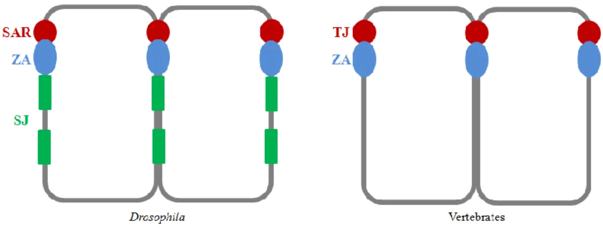
The formation of special cell-cell contact zones is a very important step to establish membrane domains and guarantee close adhesion, which provides barrier function and interaction among the cells in an epithelium. Epithelial cells possess an adhesive belt surrounding the cell just below the apical domain, called the zonula adherens (ZA), where adherens junctions between neighboring cells are formed. Apical to the ZA a specialized plasma membrane microdomain in vertebrates, called the tight junction (TJ), is formed (Feigin and Muthuswamy, 2009; Flores-Benitez and Knust, 2016). The TJs are composed of different protein complexes and are responsible for intercellular sealing (Tsukita et al., 2001). Although the assembly and also the regulation of epithelial cell architecture and function are highly conserved, the Drosophila epithelium exhibits in contrast to vertebrate TJ the so-called subapical region (SAR) apical to the ZA. Nevertheless the Drosophila SAR includes protein complexes that correspond to tight junction proteins in vertebrates and thereby are highly conserved throughout evolution. Additionally Drosophila epithelial cells possess another complex basal to the ZA, the so-called septate junction (SJ), which covers large parts of the lateral domain and acts as the functional equivalent to the
11 vertebrate TJ by forming a paracellular diffusion barrier (Figure 2) (Knust and Bossinger, 2002). Transmembrane proteins, containing extracellular domains within these junctions, mediate interactions between adjacent cells in order to enable interplay with signaling molecules and cytoskeletal proteins (Feigin and Muthuswamy, 2009).
Figure 2 The organization of epithelial cells in comparison between Drosophila and vertebrates
Comparison between epithelial cells of Drosophila (left) and vertebrates (right) shows for both the ZA (blue), surrounding the cells just below the apical domain. In vertebrates TJs (red) apical to the ZA are formed, whereas Drosophila epithelial cells possess the SAR (red) including protein complexes comparable to TJ proteins in vertebrates. Additionally Drosophila epithelial cells possess SJ (green) basal to the ZA (adapted from Knust and Bossinger, 2002).
The initial cues to start epithelial differentiation comprise cell-cell contacts and cell- extracellular matrix contacts, which are mainly mediated by transmembrane adhesion molecules and receptors of the cadherin superfamily and the integrin family, respectively (Drubin et al., 1996; Yeaman et al., 1999; Tepass et al., 2000; Giancotti et al., 1999; Wodarz, 2002). The development of epithelial cells starts with the formation of cadherin-containing cell contacts, the spot adherens junctions (AJ) along the lateral plasma membrane between neighboring cells (Tepass and Hartenstein, 1994; Wodarz, 2002). As the TJs are not emerged at this developmental stage, several components of the TJs colocalize with AJ components. Afterwards AJs comigrate to the apicolateral region thereby establishing the ZA, whereas simultaneously residual components of both AJ and TJ start to arrange and establish the TJ domain (Wodarz, 2002). The establishment of the TJ requires the proper arrangement of the ZA, which in turn is achieved by the transmembrane protein E-
12 cadherin and its associated cytoplasmic proteins α-catenin and β-catenin (Armadillo (Arm) in Drosophila) and furthermore by the formation of special protein complexes apical and basal to the ZA itself (Feigin and Muthuswamy, 2009; Knust and Bossinger, 2002).
In the Drosophila SAR as well as in the vertebrate TJs a highly conserved protein complex can be found, namely the PAR complex, consisting of the PDZ-domain containing protein PAR-3 (Bazooka (Baz) in Drosophila), the atypical protein kinase C (aPKC) and PAR-6, together with Cdc42. Moreover a second very important conserved complex located slightly apical to the AJs is the Crumbs (Crb)/PALS1 (protein associated with Lin7; Stardust (Sdt) in Drosophila)/PATJ (PALS1- associated TJ protein)/Lin7 complex. Crb is a transmembrane protein which binds to the MAGUK (membrane associated guanylate kinase) protein Stardust.
Moreover transmembrane proteins like occludin and members of the claudin family, which span the membrane four times, are localized at the TJ domain as well as the single-span transmembrane proteins JAM (junctional adhesion molecule) (Ebnet et al., 2001). These transmembrane proteins interact directly with cytoplasmic, PDZ domain containing proteins, like the zonula occludens (ZO) proteins 1-3, Baz/PAR-3 and Sdt/PALS1, which function as scaffolds to recruit other cytoskeletal and signaling molecules (Tsukita et al., 2001; Shin et al., 2006; Flores-Benitez and Knust, 2016).
Basally to the ZA, another important protein complex is located at the basolateral membrane domain, called the Discs large/Scribble/Lethal (2) giant larvae complex, which consists of the multi-PDZ and leucine rich-repeat protein Scribble (Scrib), the MAGUK protein Discs large (Dlg) and the WD-40 repeats containing Lethal (2) giant larvae (Lgl) (Feigin and Muthuswamy, 2009). Several additional polarity regulating proteins are located at the basolateral membrane domain, namely the serine-threonine kinase MARK1/2 (PAR-1), the adaptor protein 14-3-3 (PAR-5) and the multifunctional protein kinase LKB1 (PAR-4).
Although most constituents are highly conserved, the Drosophila epithelial cells show some differences in the arrangement of the lateral domain. In the Drosophila SAR, the Crb/Sdt/PATJ complex localizes slightly apical to the PAR complex and the Dlg/Scrib/Lgl complex is localized to the special SJs.
13 Figure 3 Localization of protein complexes in Drosophila and vertebrate epithelial cells
Comparison of the distribution of important protein complexes along the apical to basal axis between epithelial cells of Drosophila (left) and vertebrates (right). In vertebrates, TJ (red) apical to the ZA are formed, whereas Drosophila epithelial cells possess a SAR (red) whereby most of the constituents are highly conserved. Different to the vertebrate TJs the Crb/Sdt/PATJ complex (light red) localizes slightly apical to the PAR-3/PAR-6/aPKC complex (dark red) in the Drosophila SAR. The Dlg/Scrib/Lgl complex is localized to the special SJ (green). All complexes are working in a dynamic and spatial interaction (adapted from Knust and Bossinger, 2002).
3.3 The PAR complex
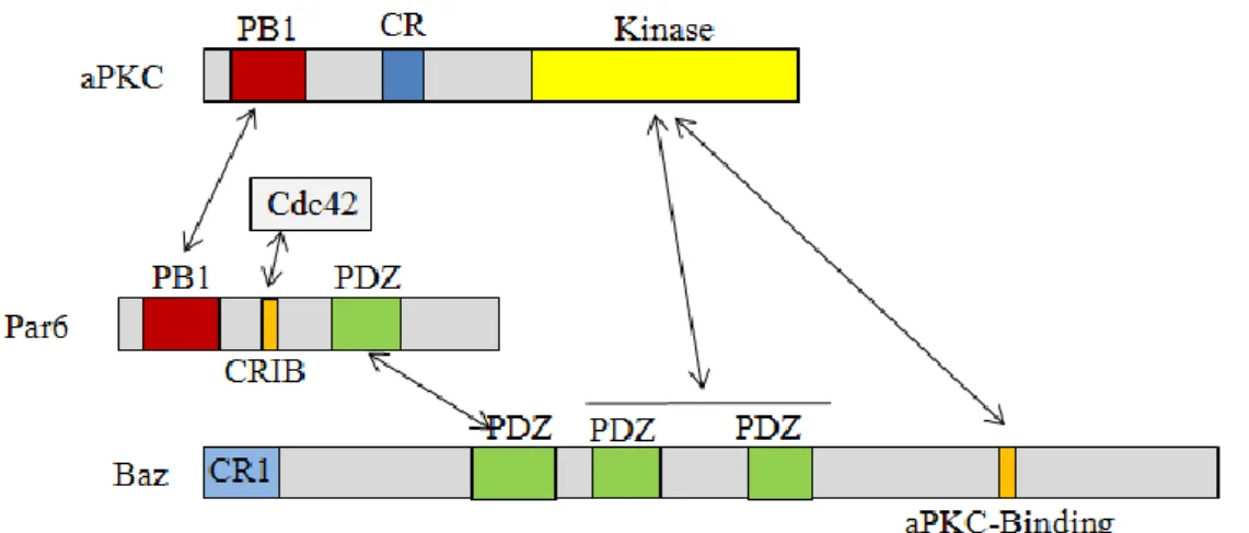
One of the key regulators of apical-basal polarity in epithelial cells is the PAR complex. The PAR complex, consisting of the multi modular scaffold proteins Bazooka (Baz, PAR-3 in mammals), PAR-6 and the atypical protein kinase C (aPKC), contribute to the initial establishment of epithelial cell polarity and it controls the localization of additional complexes functioning further downstream in the regulation of epithelial development (Figure 4). The name “PAR” derives its origin from genetic screens in C. elegans zygotes for genes that establish anterior- posterior polarity and stands for “partitioning-defectiveness of zygotes after mutation of the genes” (Kemphues et al., 1988). The PAR complex was initially found to regulate polarity in early stages of C. elegans and Drosophila embryos (Tabuse et al.,
14 1998), as well as in the asymmetric cell division of Drosophila neuroblasts (Wodarz et al., 1999) and also in mammalian epithelial cells and the axon-dendrite polarity of neurons (Figure 1) (Plant et al., 2003; Lin et al., 2000).
The proteins in this complex operate in a specific hierarchy, in which Baz/PAR-3 plays a key role in the proper subcellular localization of other apical markers (Johnson and Wodarz, 2003). At the onset of epithelial polarization Baz/PAR-3 can associate via PDZ-PDZ domain interaction with the hetero-dimer PAR-6/aPKC (Hung and Kemphues, 1999; Lin et al., 2000; Chen and Zhang, 2013). The PDZ (Post-synaptic-density protein 95 - Discs large - Zonula Occludentes 1) domain is a highly conserved protein-protein interaction module which can occur from single to multiple copies in many cytoplasmic as well as membrane adaptor proteins (Lee and Zheng, 2010). This domain can either interact with proteins via other PDZ domains or with specific carboxy-terminal motifs, the so-called PDZ-binding motif (PBM).
The specific peptide sequence of PBMs is usually located at the very C-terminus of the target protein, nevertheless also internal PBMs have been reported (Javier and Rice, 2011).
The scaffold protein PAR-6 can interact with aPKC through their N-terminal PB1 (phagocyte oxidase/Bem1) domains (Terasawa et al., 2001; Suzuki et al., 2001;
Suzuki et al., 2003). Additionally PAR-6 can interact with the small GTPase Cdc42 via its Semi-CRIB (N-terminal Cdc42/Rac interactive binding) motif (Burbelo et al., 1995). This binding of Cdc42 activates the kinase activity of associated aPKC (Garrard et al., 2003; Joberty et al., 2000; Lin et al., 2000; Peterson et al., 2004).
Cdc42 is a member of the family of Ras-like GTPases and is involved in many pathways connected to cell polarity. Nevertheless it is not jet completely investigated how Cdc42 interacts in detail with the PAR complex. Subsequently activated aPKC can phosphorylate Baz/PAR-3, which in turn dissociates from the PAR-6/aPKC heterodimer (Nagai-Tamai et al., 2002; Horikoshi et al., 2009; Morais-de-Sá et al., 2010).
At a later point in development, Baz/PAR-3 directly binds the scaffolding protein Stardust (Sdt)/PALS-1, which becomes expressed during early gastrulation (Krahn et al., 2010a; Chen and Zhang, 2013). Thereby, the transmembrane protein Crumbs (Crb) is stabilized at the apical junctions by its adaptor Sdt. Upon maturation of epithelia, the scaffolding protein Baz/PAR-3 and the transmembrane protein Crb
15 function to some extent redundantly to determine the apical domain of epithelial cells (Tanentzapf and Tepass, 2003) and further direct the formation of apical junctions by recruiting aPKC.
Figure 4 The PAR complex
PAR-6 can interact with aPKC through their N-terminal PB1domains, whereas the first PDZ domain of Baz can interact with the PDZ domain of PAR-6 and the aPKC-binding domain of Baz with the kinase domain of aPKC. Interaction of PAR-6 with the small GTPase Cdc42 via its Semi-CRIB motif activates the kinase activity of associated aPKC, which in turn leads to phosphorylation of Baz in the PDZ2-3 region and the aPKC-binding domain (adapted from Chen and Zhang, 2013; Johnson and Wodarz, 2003; n.t.s.).
On the other hand, the activity of the PAR complex is counterbalanced by basolateral polarity proteins in order to achieve and maintain distinct cortical domains. aPKC can phosphorylate and thereby inactivate the kinase PAR-1 to prevent it from associating with apical regions (Benton and St. Johnston, 2003a; Chen and Zhang, 2013). On the other way round PAR-1 is able to phosphorylate Baz/PAR-3 at two conserved serines (at position 151 and 1085 in Baz; figure 5) in Drosophila and mammalian epithelial cells and the basal cortex of Drosophila neural stem cells, thereby preventing dimerization of Baz/PAR-3 and providing a movement of Baz/PAR-3 to a more apical position via binding to 14-3-3 proteins (PAR-5 in Drosophila) (Benton and St. Johnston, 2003a; Hurd et al., 2003; Krahn et al., 2009;
McKinley and Harris, 2012). In the beginning of Drosophila epithelial development PAR-1 and Baz show a partial overlap during early cellularization (McKinley and Harris, 2012). However already in late cellularization/early gastrulation PAR-1 and
16 Baz start to segregate to establish a clear apical (Baz) and basal (PAR-1) localization (McKinley and Harris, 2012).
Moreover Lgl can physically interact with the heterodimer aPKC/PAR-6. Thereby Lgl is phosphorylated by aPKC in order to stay at the basolateral membrane (Betschinger et al., 2003; Plant et al., 2003; Chen and Zhang, 2013).
3.4 Bazooka/PAR-3
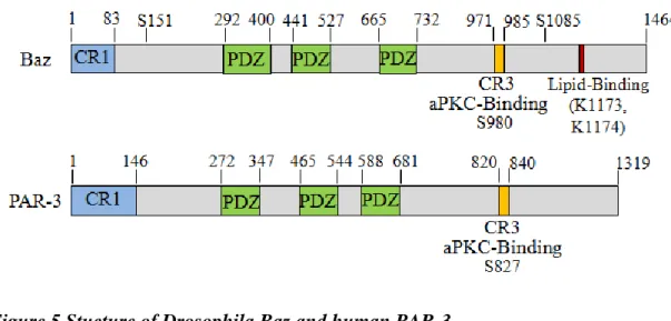
The name “Bazooka” has its origin from the big holes observed in the cuticle of Drosophila baz mutant embryos and was first identified in a screen for embryonic defects (Wieschaus et al., 1984) as a gene product essential for the polarization of the Drosophila embryonic ectoderm (Kuchinke et al., 1998). The PDZ-domain- containing scaffold protein Baz is highly conserved in evolution and has a homolog in worm and mammalian called PAR-3. The Drosophila baz encodes for a large protein with 1464 amino acids whereas the human homologue par-3 has 1319 amino acids. Both show high similarities and possess corresponding domains (Figure 5).
Figure 5 Stucture of Drosophila Baz and human PAR-3
Drosophila Baz and its homologe PAR-3 show high similarities. Both possess an N-terminal conserved region, three PDZ domains and an aPKC-binding motif in the CR3 (data according to NCBI databases, n.t.s.).
17 Baz/PAR-3 contains a conserved N-terminal domain (NTD) or CR1 (conserved region 1), which is able to self-associate (Benton and St. Johnston, 2003a). The CR1 self-association leads to an oligomerization of Baz/PAR-3 molecules in order to establish a molecular basis for an enrichment of the PAR complex and their binding partners (Chen and Zhang, 2013; Benton and St. Johnston, 2003b; Mizuno et al., 2003; Feng et al., 2007).
Baz/PAR-3 displays three highly conserved PDZ domains, whereby the first PDZ domain interacts with the PDZ domain of PAR-6 (Lin et al., 2000; Suzuki et al., 2001) and moreover with several cell adhesion molecules like nectin, p75 and members of the transmembrane protein families JAM (junctional adhesion molecule) (Chan et al., 2006; Ebnet et al., 2003; Itoh et al., 2001; Chen and Zhang, 2013). Baz can interact with aPKC via its PDZ domains 2 and 3 (Wodarz et al., 2000; Rolls et al., 2003). Moreover the Baz/PAR-3 PDZ domains 2 and 3 are described to bind to lipid membranes in order to achieve a localization at the TJs and PDZ 3 interacts with the C-terminal PDZ binding motif (PBM) of the lipid Phosphatase PTEN (Yu and Harris, 2012; Wu et al., 2007; Chen and Zhang, 2013), which has been reported to promote apical polarity in Drosophila (Chartier et al., 2011) and mammalian system (Martin-Belmonte al., 2007).
The C-terminal half of Baz/PAR-3 possesses another interaction domain with aPKC, a short conserved aPKC-binding motif, also annotated as conserved region 3 (CR3), whereby in detail a serine at position 980 for Drosophila Baz and a serine at position 827 in human PAR-3, respectively, are phosphorylated by aPKC (Horikoshi et al., 2009; Izumi et al., 1998; Lin et al., 2000; Morais-de-Sá et al., 2010; Nagai-Tamai et al., 2002). The CR3 of Bazooka starts at position 971 and spans over 14 amino acids, whereas for the vertebrate homologe PAR-3 the CR3 of twenty amino acids, starting at position 820, is reported to interact with the kinase domain of aPKC (Izumi et al., 1998; Tabuse et al., 1998). Sdt binds to Baz at a region which includes the aPKC- binding motif. Therefore a phosphorylation of Baz by aPKC at the serine 980 weakens the interaction of Baz with Sdt, which in turn enables the formation of the Crb-Sdt complex (Krahn et al., 2010a). Baz also exhibits a lipid-binding domain which consists of two lysines at position 1173 and 1174 (Figure 5). Furthermore the C-terminal part of Baz/PAR-3 contains three conserved regions, called 4N1/2/3, which show no recognizable domain composition (Chen and Zhang, 2013).
18 Moreover Baz/PAR-3 binding partners include phosphoinositols (Krahn et al., 2010b), the Rho-kinase (Simões et al., 2010) and AJ proteins Armadillo (Arm in Drosophila; ß-catenin in vertebrates) and Echinoid (Ed) (Wei et al., 2005), which bind to the central PDZ domain region.
3.5 The serine/theronine kinase aPKC
The atypical protein kinase C (aPKC) is a very important component of the PAR complex, as it represents prominently an evolutionary conserved regulator in cellular polarity by phosphorylation of many important polarity proteins including PAR-3, PAR-1, Crb, Lgl, GSK-3ß, MARCKS and LIN5/NuMA (Chen and Zhang, 2013).
PAR-6 operates as a regulatory subunit of aPKC, thereby controlling its position and regulating its kinase activity (Tepass, 2012; Atwood et al., 2007; Graybill, 2012).
This is achieved by a PAR-6 mediated interaction with active, thereby GTP-loaded, Cdc42, which derestrict inhibition of aPKC kinase activity (David et al., 2010;
Atwood et al., 2007). The PAR-6/aPKC heterodimer initially needs to form a complex with Baz/PAR-3 to be recruited to the apical membrane (Harris and Pfeier, 2005; Tepass, 2012). Nevertheless the interaction between the PAR-6/aPKC heterodimer and Baz is weakened by phosphorylation of Baz by aPKC, which in turn allows the interaction between Baz and Sdt in order to achieve the Crb/Sdt/PATJ complex formation (Krahn et al., 2010a). Additionally Baz/PAR-3 is able to act as an inhibitor of the PAR-6/aPKC heterodimer activity, which leads to stabilization of actomyosin networks through a reduced phosphorylation of cytoskeletal regulators by aPKC (Soriano et al., 2016).
Moreover aPKC is also responsible for the regulation of basolateral polarity proteins, particularly by phosphorylation of the basolateral proteins Lgl and PAR-1. This phosphorylation prevents the accumulation of these basolateral proteins at the more apical part of the cell (Betschinger et al., 2003; Hurov et al., 2004; Kusakabe and Nishida, 2004; Plant et al., 2003; Yamanaka et al., 2003; Suzuki et al., 2004).
Besides, aPKC is also able to autophosphorylate although the effects are not known at the moment (Tepass, 2012).
19 The regulation of protein function by aPKC is achieved by the phosphorylation of hydroxyl groups of serine or threonine amino acid residues within a special phosphorylation site on the particular protein. Thereby members of the PKC kinase family, like aPKC possess a consensus phosphorylation site motif, which is conserved from Drosophila to mammals, consisting of amino acid sequences like R/K-X-X-S/T, R/K-X-X-X-S/T, or S/T-X-R-X whereby X indicates any amino acid (Pearson and Kemp, 1991).
3.6 The adaptor protein PAR-6
The regulatory adapter protein of the PAR complex, PAR-6, is composed of a N- terminally positioned PB1 domain, which enables PAR-6 to interact with aPKC in order to form the functionally PAR-6/aPKC heterodimer (Terasawa et al., 2001;
Suzuki et al., 2001; Suzuki et al., 2003), a semi CRIB domain, which can associate with the small GTPase Cdc42 (Burbelo et al., 1995; Garrard et al., 2003; Joberty et al., 2000; Lin et al., 2000; Peterson et al., 2004), and a PDZ domain for interaction with Baz/PAR-3 (Hung and Kemphues, 1999) (Figure 4).
PAR-6 regulates the kinase activity of aPKC in many different cell types of diverse species (Suzuki and Ohno, 2006; Atwood et al., 2007; Tepass, 2012; Graybill, 2012) and furthermore controls the positioning of the PAR-6/aPKC heterodimer to the apical region. The apical positioning is strengthened by the interaction of PAR-6 with the Crb/Sdt/PATJ complex, which thereby weakens the PAR-6-Baz interaction (Morais-de-Sá et al., 2010; Walter and Pichaud, 2010). PAR-6 directly binds the N- terminal region of Sdt and PATJ as well as the PDZ domain of Drosophila PATJ with its N-terminus (Nam and Choi, 2003; Wang et al., 2004; Bulgakova et al., 2008). Additionally PAR-6 can interact with the C-terminus of Crb (Wang et al., 2004). Thus PAR-6 functions as a mediator between the two important apical protein complexes in a polarized cell, the PAR and the Crb complex. Moreover in mammalian epithelial cells the dominant human homologue of Crb, CRB3, can recruit PAR-6 to the apical region in unpolarized cells (Hurd et al., 2003).
20
3.7 The Crb complex
The Crb complex consists of Crb, Sdt/PALS-1, PATJ and Lin7 and is highly conserved from fly to human. The apical polarity protein Crb regulates the apical- basal polarity and integrity in many different tissues among various species together with its intracellular adaptor protein Sdt/PALS-1 (Tepass and Knust, 1993; Tepass et al., 1990; Bulgakova and Knust, 2009).
The transmembrane protein Crb consists of a large extracellular domain and a short intracellular domain (Tepass et al., 1990; Wodarz et al., 1993). Instead of Drosophila, humans possess three Crb genes, CRB1, CRB2 and CRB3. CRB3, which is further subdivided into two isoforms, is broadly expressed in simple epithelial cells in mammals and lacks most of the extracellular domain, but shows in one isoform the highly conserved intracellular tail like in other species (Fan et al., 2007; Shin et al., 2006). Crb can physically interact with its C-terminal ERLI motif with the single PDZ domain of Sdt thereby initiate the formation of the Crb/Sdt complex (Bachmann et al., 2001; Hong et al., 2001). Sdt, which encodes a membrane-associated guanylate kinase (MAGUK) without kinase domain activity (Tepass, 2012), possesses an N-terminal conserved region containing ECR1 and ECR2, which can interact with PAR-6 (Bachmann et al., 2001; Hong et al., 2001;
Tepass, 2012). Furthermore Sdt contains two L27 domains with can bind to PATJ and Lin7, respectively (Bachmann et al., 2004; Bulgakova et al., 2008). PATJ and Lin7 comprise an N-terminal L27 domain itself and additionally four PDZ domains in case of PATJ and two PDZ domains in case of Lin7.
Crb is not expressed in all epithelia in Drosophila, like for example in the intestinal epithelia (Tepass et al., 1990). In some cases, Crb is not required to maintain epithelial polarity, like in late embryonic epidermis or in larval imaginal disc epithelia and functions to some extent redundantly with other apical polarity regulators like Baz (Campbell et al., 2009; Pellikka et al., 2002; Tanentzapf and Tepass, 2003).
21
3.8 The embryonic development of Drosophila melanogaster
The ectodermal epidermis of Drosophila embryos is a good model for studying fundamental processes of cell polarity. The development of Drosophila embryos was subdivided into stages in 1975 by Mary Brownes and revised by Volker Hartenstein and José Campos-Ortega, which became a general reference in Drosophila research (Brownes, 1975; Campos-Ortega and Hartenstein, 1997) (Table 1).
The ectodermal epidermis is established at first by dividing the zygotic nuclei of a fertilized Drosophila egg for thirteen times at the blastoderm stage. The syncytium is established out of 128 nuclei by the first seven synchronous zygotic divisions (Campos-Ortega and Hartenstein, 1997). The following divisions producing pole cells at the posterior pole and continuing to divide until the number of syncytial blastoderm nuclei reaches approximately 5000 (Campos-Ortega and Hartenstein, 1997). Subsequently cellularization takes place to form the somatic cells by multiple invaginations of the plasma membrane around each nucleus, thereby forming definite epithelial cells (Figure 1 C) (Suzuki and Ohno, 2006). This cellular blastoderm stage, before gastrulation occurs is also called stage 5 (Table 1) (Campos-Ortega and Hartenstein, 1997). The transition from late cellularization to early gastrulation is called stage 6 and is a timepoint of special interest for this work as here the PAR complex is recruited to the forming apical junctions. Gastrulation, which causes the formation of germ layers, occurs by the gradual invagination of a consistent midventral cell area, which forms a tubular structure inside the embryo (Campos- Ortega and Hartenstein, 1997). This time span extends over stage 5 until stage 7. In stage 7 the germ band starts to elongate, which proceeds during stage 8 until stage 11 (Campos-Ortega and Hartenstein, 1997). The expression of components of the Crb complex starts also during embryonic stage 8 and therefore is another special point in embryonic epithelial cell polarity development in this study.
22 Table 1 Time table of Drosophila embryonic development (from http://flymove.uni- muenster.de/stages/StgTabelle.html)
From stage 9 onwards the germ band elongation enters its slow phase and ventral and procephalic neuroblasts start to segregate, which are first divide in stage 10 (Campos-Ortega and Hartenstein, 1997). The epidermal segmentation of the embryo becomes visible in stage 11 and the germ band retraction occurs during stage 12 and 13 (Campos-Ortega and Hartenstein, 1997). The dorsal closure and the head involution take place in stage 14 to 15 and in stage 16 and 17 the embryo finally differentiates (Brownes, 1975; Campos-Ortega and Hartenstein, 1997).
23
3.9 Research objectives
Cell polarity is a fundamental mechanism in nature and affects various species in different conditions. Therefore it is fundamentally important to understand how cell polarity is achieved and how polarity proteins work together in specific complexes in order to create a basis of the development of drugs or therapies against many diseases including cancer.
Until now, many aspects of the important polarity determinants Baz/PAR-3 and its interaction in the PAR complex and with other polarity complexes are investigated, although there are still many open questions remained.
Thus the main address of this work was to gain knowledge about the interaction and function of phosphorylation events of Baz/PAR-3 by the kinase aPKC. In detailed structure and function analysis the two regions of Baz which are involved in aPKC binding were investigated. Moreover new phosphorylation sites of Baz and its human homologue PAR-3 for aPKC were identified which were analyzed for Baz in studies in vivo in the fly.
Moreover we had a closer look on the third member of the Par complex, PAR-6. As PAR-6 has a supposed PDZ-binding motif (PBM), a short peptide at the very C- terminus, we questioned if this PB motif is important for PAR-6 binding and functionality. To clarify this issue, we performed a lethality test as well as immunostainings, were we compared PAR-6 wildtype flies with PAR-6ΔPBM flies, which are lacking the supposed PBM.
24
4 Material & Methods 4.1 Material
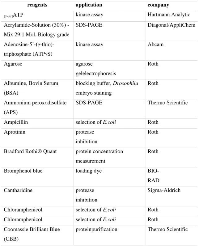
4.1.1 Reagents
Table 2 Reagents
reagents application company
[-32]ATP kinase assay Hartmann Analytic
Acrylamide-Solution (30%) - Mix 29:1 Mol. Biology grade
SDS-PAGE Diagonal/AppliChem
Adenosine-5’-(γ-thio)- triphosphate (ATPγS)
kinase assay Abcam
Agarose agarose
gelelectrophoresis
Roth
Albumine, Bovin Serum (BSA)
blocking buffer, Drosophila embryo staining
Roth
Ammonium peroxodisulfate (APS)
SDS-PAGE Thermo Scientific
Ampicillin selection of E.coli Roth
Aprotinin protease
inhibition
Roth
Bradford Rothi® Quant protein concentration measurement
Roth
Bromphenol blue loading dye BIO-
RAD
Cantharidine protease
inhibition
Sigma-Aldrich
Chloramphenicol selection of E.coli Roth
Chloramphenicol selection of E.coli Roth
Coomassie Brilliant Blue (CBB)
proteinpurification Thermo Scientific
25
1, 4-Dithiothreitol (DTT) SDS-PAGE Roth
4',6-Diamidino-2-
phenylindole dihydrochloride (DAPI)
immunohistology Roth
dNTPs (dATP, dGTP, dCTP, dTTP)
PCR Thermo Scientific
Ethidiumbromide agarose gelelectrophoresis Sigma-Aldrich Ethylendiamintetraacetate
(EDTA)
buffer P1/S1 Sigma- Aldrich
Fetal Bovine Serum (FBS) cell culture gibco® Formaldehyde 37%, v/v fixation of
Drosophila embryos
Roth
FuGENE®HD Transfection Reagent
transfection Promega
Glycine blotting buffer Merck
Isopropyl-β-D-
thiogalactopyranosid (IPTG)
protein purification Roth
Kanamycin selection of E.coli Roth
Lactate Hoyer´s medium Fluca
Leupeptin protease inhibition Roth
Lysozyme protein purification Biomol
Mercaptoethanol, 2- protein npurification Merck Methanol LC/MS ultra Drosophila embryo staining Roth
Methylene blue methylen blue solution Sigma-Aldrich
Mowiol 4-88 immunohistology Merck
N,N,N’,N’-
tetramethylethylenediamine (TEMED)
SDS-PAGE Roth
n-Heptane immunohistology Roth
Oil 10 S VOLTALEF(R) injection for germline transformation
VWR
Penicillin-Streptomycin cell culture Sigma-Aldrich
26
Pepstatin A protease inhibition Roth
Phosphatase inhibitor cocktail
phosphatase inhibition Sigma-Aldrich
Phyenylmethylsulfonylfluori de(PMSF)
protease inhibition Roth
p-Nitrobenzyl mesylate (PNBM)
kinase assay Abcam
Protein G Plus Agarose immunoprecipitation Santa Cruz Protino Glutathione Agarose
4B
protein purification Macherey-Nagel
RNAse A buffer P1/S1 Roth
Schneider´s Drosophila medium
cell culture PAN Biotech
Sodium dodecyl sulfate (SDS) ultrapure
SDS-PAGE Diagonal/AppliChem
Strep-Tactin-Sepharose 90%
Suspension
immunoprecipitation IBA GmbH
Tris Base buffering Sigma-Aldrich
TritonTM X-100 protein purification Serva
Tween® 20 immunohistology Roth
4.1.2 Solutions and buffer
Solutions and buffer were prepared with sterile H2O and, if necessary, subsequently autoclaved.
Table 3 Solutions and buffer
solution/buffer Composition
APS 438 mM APS
Blocking buffer 12,5 ml TBS (20x)
27 2,5 ml
2,5 g 7,5 g ad 250 ml
10% Tween BSA
Skim milk powder H2O
Blotting buffer 6,17 M
399 mM 49,44 mM
Methanol Glycine Tris base
Buffer P1/S1 50 mM
10 mM 100 µg/ml
Tris-HCl EDTA RNase A
Buffer P2/S2 20 mM
3,5 mM
NaOH SDS
Buffer P3/S3 3 M Potassium acetate
Coomassie Brilliant Blue (CBB) solution
15,61 M 46 mM 1,67 M
Methanol
concentrated Coomassie Brilliant Blue
Acetic acid
Embryoglue 50 cm
14 ml
adhesive tape n-heptane
Fixation solution 59,95 mM
440 ml 4,99 M
Formaldehyde PBS
Heptane
GST elution buffer 30mM
50 mM 150 mM
reduced Glutathione Tris-HCl pH 7.5 NaCl
Injection buffer (10x) 5mM
0.1 mM KCl
sodium phosphate, pH 6.8
Kinase buffer 50 mM
10 mM 1 mM
TRIS pH 7.5 MgCl2 DTT LEW washing buffer (300
mM)
300 mM NaCl in PBS LEW washing buffer (2 M) 2 M NaCl
28 in PBS
Loading dye (6x) 3 ml
35 mg 10 ml
30% Glycerol Bromphenol blue H2O
Lysis buffer 1370 µg/ml
10 µg/ml 40 µg/ml 10 µg/ml 5 ml
Pepstatin A Aprotinin PMSF Leupeptin TNT buffer
Methylen blue solution 1 pinch Methylen blue powder in TAE (0,1x)
Mowiol solution 0,4 g/ml
1 g/ml 2%
Mowiol Glycerol
Tris-HCl (0,2M) NB fixing solution 4% (v/v)
0.1 M 0.3% (v/v) 20mM 1mM ad 10 ml
formaldehyde (37%) PIPES (1 M, pH6,9) Triton X-100 (10%) EGTA (0.1 M, pH8.0) MgSO4 (1M)
H2O
PBS (10x) pH 7.4 1,37 mM
27 mM 0,1 M 20 mM
NaCl KCl Na2HPO4
KH2HPO4
PBT 0,1 % Tween 20 in PBS
SDS loading dye (2x) 6,4 ml
1600 µl 1 ml 1600 µl ad 16 ml
10% SDS Glycerol Tris (1M)
Bromphenol blue H2O
SDS running buffer 0,96 M
125 mM 3,46 mM
Glycin Tris SDS
29
Stripping buffer 12,5 M
2,28 M
Methanol Acetic acid
T4 ligase buffer 400 mM
100 mM 100 mM
5 mM
Tris-HCl MgCl2 DTT ATP
TAE (1x) 20 mM
1 mM 40 mM
Tris EDTA AcOH
TBS (20x) 2,74 M
53,66 mM 0,5 M
NaCl KCl Tris-HCl
TBST 50 ml
10 ml ad 1 l
TBS (20x) 10% Tween H2O
TNT buffer 150 mM
50 mM 8 mM
NaCl Tris
Triton X-100
10 % Tween 20 41 mM Tween 20
Washing buffer 300 mM NaCl in PBS
4.1.3 Media and agarose plates
For cultivation of E.coli strains selective culture medium (LB0 medium) or agar plates (LB0 agar plates) with a dilution of the required antibiotic stock concentration were used under sterile conditions. For ampicillin a final concentration of 100 µg/ml was necessary whereas for kanamycin 50 µg/ml and for chloramphenicol 30 µg/ml were used. Agarose plates as well as all of the following media were stored at 4°C and preheated before using.
30 Table 4 Media and agarose plates
medium/agarose plate composition
Apple juice agar plates 1980 ml 60 g 1020 ml 51 g 10%
H2O Agar Apple juice Sugar Nipagin
Freezing medium 45%
45%
10%
Conditioned medium FBS
DMSO Hoyer´s medium (ready to use) 50ml
30g 200g 16 ml
1:1 H2O
Gummi arabicum Chloralhydrat Glycerine Lactate
LB0 medium 1%
0,5%
1%
ad
Tryptone Yeast extract NaCl
H2O
LB0 agar plates 10%
5%
5%
15%
ad
Tryptone Yeast extract NaCl
Agar H2O Schneider´s Drosophila medium
(ready to use) 0,40 g/l
10%
1%
L-Glutamin NaHCO3
FBS
Penicillin-Streptomycin
YTA medium 16%
10%
5%
ad
Tryptone Yeast extract NaCl
H2O
31
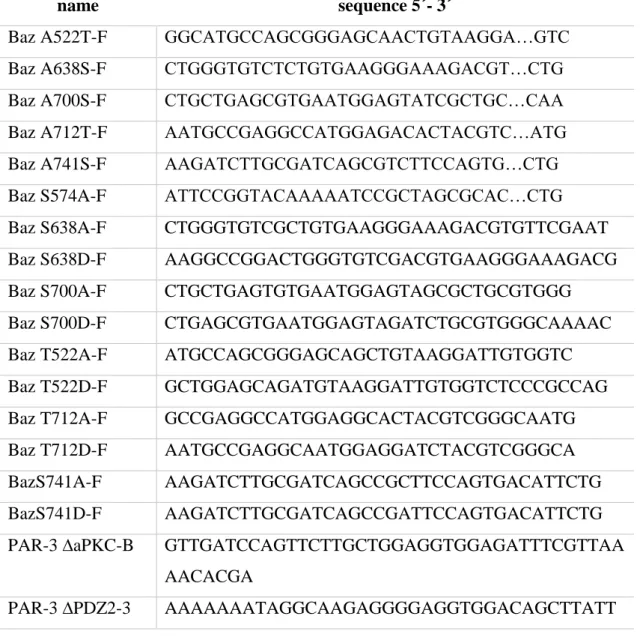
4.1.4 Oligonucleotides
The Oligonucleotides were designed with DNADynamo (BlueTractorSoftware, UK) and subsequently sent to Metabion international AG (Planegg/Steinkirchen, Germany), Microsynth AG (Balgach, Switzerland) or Sigma-Aldrich Chemie GmbH (Munich, Germany) for synthesizing. The freeze-dried oligonucleotides were resolved in autoclaved, de-ionized H2O to a final concentration of 50 pmol/µl. After that, a 10 pM solution was prepared for further use and stored at - 20°C.
Following oligonucleotides listed in table 5 were used to achieve a specific mutation of the destined gene of interest.
Table 5 Oligonucleotides for mutagenesis
name sequence 5´- 3´
Baz A522T-F GGCATGCCAGCGGGAGCAACTGTAAGGA…GTC
Baz A638S-F CTGGGTGTCTCTGTGAAGGGAAAGACGT…CTG
Baz A700S-F CTGCTGAGCGTGAATGGAGTATCGCTGC…CAA
Baz A712T-F AATGCCGAGGCCATGGAGACACTACGTC…ATG
Baz A741S-F AAGATCTTGCGATCAGCGTCTTCCAGTG…CTG
Baz S574A-F ATTCCGGTACAAAAATCCGCTAGCGCAC…CTG
Baz S638A-F CTGGGTGTCGCTGTGAAGGGAAAGACGTGTTCGAAT
Baz S638D-F AAGGCCGGACTGGGTGTCGACGTGAAGGGAAAGACG
Baz S700A-F CTGCTGAGTGTGAATGGAGTAGCGCTGCGTGGG
Baz S700D-F CTGAGCGTGAATGGAGTAGATCTGCGTGGGCAAAAC
Baz T522A-F ATGCCAGCGGGAGCAGCTGTAAGGATTGTGGTC
Baz T522D-F GCTGGAGCAGATGTAAGGATTGTGGTCTCCCGCCAG
Baz T712A-F GCCGAGGCCATGGAGGCACTACGTCGGGCAATG
Baz T712D-F AATGCCGAGGCAATGGAGGATCTACGTCGGGCA
BazS741A-F AAGATCTTGCGATCAGCCGCTTCCAGTGACATTCTG
BazS741D-F AAGATCTTGCGATCAGCCGATTCCAGTGACATTCTG
PAR-3 ∆aPKC-B GTTGATCCAGTTCTTGCTGGAGGTGGAGATTTCGTTAA
AACACGA
PAR-3 ∆PDZ2-3 AAAAAAATAGGCAAGAGGGGAGGTGGACAGCTTATT
32 GTTGCAAGG
PAR-3 A453T-F AGTACGACTGTAAGCAGTGGTTATAACACCAAAAAAA
TA
PAR-3 A534T-F AGCACCAAGATGGAAGGTACCGTGAGCCTACTAGTCT
TT
PAR-3 A604S-F TCAGGATCTGCAGGCCTTGGTGTCTCTGTCAAAGGTA
AC
PAR-3 A661T-F AACCAAGATGCCATGGAGACCCTAAGAAGGTCTATG
PAR-3 S533A-F GAAGTTGTTTCGCTCTTAAGAGCCACCAAGATGGAAG
GA
PAR-3 S604A-F TCTGCAGGACTTGGTGTCGCTGTCAAAGGTAACCGG
PAR-3 S667A-F CTAAGAAGGTCTATGGCCACTGAAGGCAATAAACGA
PAR-3 S685A-F ATTGTTGCGAGGAGAATAGCCAAGTGCAATGAGCTG
PAR-3 T453A-F GTAAGCAGCGGTTATAACGCCAAAAAAATAGGCAAG
PAR-3 T468A-F CAGCTTAAGAAAGGTGCAGAAGGCCTGGGATTCAGC
PAR-3 T468A-F2 ATCCAGCTTAAAAAAGGTGCAGAAGGTT…TTC
PAR-3 T476/481A-F
GGATTCAGCATCGCTTCTAGATGTAGCAATAGGTGGC
PAR-3 T534A-F TCGCTGTTGAGAAGCGCCAAGATGGAAGGTACCGTGA
GC
PAR-3 T661A-F AACCAAGATGCCATGGAGGCCCTAAGAAGGTCTATG
Following oligonucleotides listed in table 6 were used for cloning and sequenzing of destined DNA constructs.
Table 6 Oligonucleotides for cloning and sequenzing
name sequence 5´- 3´
Baz PDZ2-F CACCATGGCCAATACCCGTAAACTGGG
Baz1108-NotI-R GATAAAGCGGCCGCCTACAACGCCTGATGACCA
Baz948-NotI-F GATAAAGCGGCCGCTTTCCAGCAACTTT…CAC
BazPDZ1-3pEntry-F CACCATGGATGGTGAAATGCTGCTGATC
BazPDZ1pEntry-R TCATCTCAACTCTGAGCTCTCCAA
BazPDZ1pGexNotI-F AAAGCGGCCGCTTATGGATGGTGAAATGCTGCTG
33 ATC
BazPDZ1pGexNotI-R GATAAAGCGGCCGCTCATCTCAACTCTG…CAA
BazPDZ2pEntry-F CACCATGGCCAATACCCGTAAACTGGG
BazPDZ2pEntry-R TCACTCCTGCTGGCGGGA
BazPDZ2pGexNotI-R AAAGCGGCCGCTTACTCCTGCTGGCGG
BazPDZ2pGexT436A- F
AAAGTCGACTAGCCAATGCCCGTAAACTGG
BazPDZ2pGexT436No tI-F
AAAGCGGCCGCTTGCCAATACCCGTAAACTGG
BazPDZ3pEntry-R TCACGATCGCAAGATCTTGCGG
BazPDZ3pGexNotI-F AAAGCGGCCGCATGAAGCATGACGGTGACTTG
BazPDZ3pGexNotI-R AAAGCGGCCGCTCAGTCCAGAATGTCACTGGAA
BazPDZ3-R GTGACTGTTACTGTGGTCCAG
GFP-Seq-F CACATGGTCCTGCTGGAG
GFP-Seq-R GTTTACGTCGCCGTCC
GST-Seq-F TGCGTTCCCAAAATTAGTTTG
GST-Seq-R GACGGGCTTGTCTGCTCCCG
M13-F CGTTGTAAAACGACGGCCAG
M13-R CAGGAAACAGCTATGAC
PAR-3-PDZ1-3 NotI R AAAGCGGCCGCTCATCCAGGGGGGCTCCC PAR-3-PDZ1-3 NotI R AAAGCGGCCGCTCATCCAGGGGGGCTCCC
PAR-3-PDZ2 NotI F AAAGCGGCCGCTTGGTTATAACACCAAAAAAAT
PAR-3-PDZ2 NotI F AAAGCGGCCGCTTGGTTATAACACCAAAAAAAT
PAR-3-PDZ2 T453 NotI F
AAAGCGGCCGCTTGGCAAGAGGCTTAATATCCA
PAR-3-PDZ2 T453 NotI F
AAAGCGGCCGCTTGGCAAGAGGCTTAATATCCA
UASp-attB-seq-F GCGTTAGGTCCTGTTCATTG
34
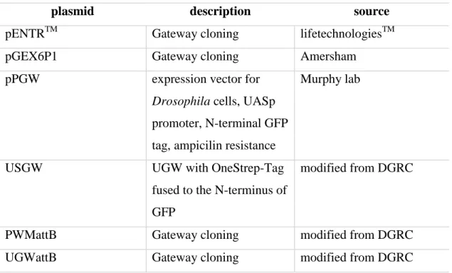
4.1.5 Plasmids
Plasmids were obtained from GE Healthcare Life Sciences (Chalfont St Giles, UK), Thermo Scientific ( Waltham, Massachusetts, USA), Murphy lab (Baltimore, USA) and New England Biolabs (Frankfurt am Main, Germany).
Table 7 Plasmids
plasmid description source
pENTRTM Gateway cloning lifetechnologiesTM
pGEX6P1 Gateway cloning Amersham
pPGW expression vector for
Drosophila cells, UASp promoter, N-terminal GFP tag, ampicilin resistance
Murphy lab
USGW UGW with OneStrep-Tag
fused to the N-terminus of GFP
modified from DGRC
PWMattB Gateway cloning modified from DGRC
UGWattB Gateway cloning modified from DGRC
4.1.6 Commercial kits
The commercial kits shown in table 8 were used according to manufacturer´s instructions.
Table 8 Commercial kits
kit application company
BM Chemiluminescence Western Blotting Substrate
western blotting Roche
In situ Cell Death Detection Kit, TMR red
TUNEL-assay for apoptosis detection
Roche
LR Clonase II Enzyme Mix clonase reaction lifetechnologiesTM
35 Nucleo Bond® PC 100 midi-preparation Magery-Nagel
Nucleo Bond® PC 20 midi-preparation Magery-Nagel Nucleo Spin®Gel and PCR
Clean-Up
PCR purification Magery-Nagel
pEntr/D-Topo Cloning Kit Gateway Cloning lifetechnologiesTM WesternBright ECL HPR
Substrate
western blotting Advansta
4.1.7 Antibodies
The primary antibodies used for western blotting (WB) and immunofluorescence (IF) are shown in table 9 and the corresponding secondary antibodies are listed in table 10.
Table 9 Primary antibodies
target species dilution for IF
dilution for WB
product/clone number
company/source
Actin Mouse 1:1000 sc-47778 Santa Cruz
aPKC (PKCζ)
Rabbit 1:500 1:500 sc-216 Santa Cruz
Baz N-term Rabbit 1:2000 1:2000 Wodarz et al.,
1999 Baz PDZ1-
3
Guinea pig
1:400 1:400 Shahab et al., 2015
Baz pS151 Rabbit 1:100 Krahn et al., 2009
Crb Mouse 1:10 Cq4 DSHB, Tepass et
al., 1990) DE-
Cadherin
Rat 1:20 DCAD2 DSHB, Oda et al.,
1994
Dlg Mouse 1:50 4F3 DSHB, Parnas et
al., 2001
GFP Chicken 1:1000 1:1000 GFP-1020 Aves Lab
36
GFP Mouse 1:500 11814460001 Roche
GFP Mouse 1:500 1:500 sc-9996 Santa Cruz
GFP Mouse 1:500 A11120 Molecular Probes
Mir Rat 1:1000 homemade
PAR-3 Rabbit 1:500 07-330 Millipore
PAR-6 Rat 1:500 homemade
PAR-6 Guinea
pig
1:500 Kim et al., 2009
PAR-6 Rabbit 1:500 homemade
Sdt Mouse 1:10 Bulgakova et al.,
2010
SXL Mouse 1:20 M114 DSHB, Bopp et
al., 1991
α-tubulin Mouse 1:1000 12G10 DSHB
Table 10 Secondary antibodies
antibody dilution product
number
company/origin
Alexa Fluor 488- anti Guinea Pig
1:400 A-11073 Life Technologies
Alexa Fluor 488- anti Rat 1:400 A-11006 Life Technologies Alexa Fluor 488- anti Rabbit 1:400 A-11034 Life Technologies Alexa Fluor 488- anti Mouse 1:400 A-11001 Life Technologies Alexa Fluor 488- anti Chicken 1:400 A-11039 Life Technologies Alexa Fluor 568-anti Guinea Pig 1:400 A-11075 Life Technologies Alexa Fluor 568-anti Rat 1:400 A-11077 Life Technologies Alexa Fluor 568-anti Chicken 1:400 A-11041 Life Technologies Alexa Fluor 647-anti Guinea Pig 1:400 A-21450 Life Technologies Alexa Fluor 647-anti Rat 1:400 A-21247 Life Technologies Alexa Fluor 647-anti Rabbit 1:400 A-21244 Life Technologies
37 Alexa Fluor 647-anti Mouse 1:400 A-21235 Life Technologies Alexa Fluor 647-anti Chicken 1:400 A-21449 Life Technologies HRP-anti Guinea Pig 1:10000 5220-0366 Medac Diagnostics
HRP-anti Rat 1:10000 5220-0284 Medac Diagnostics
HRP-anti Rabbit 1:10000 VWRKDPV
R55HRP
Medac Diagnostics
HRP-anti Mouse 1:10000 VWRKDPV
M55HRP
Medac Diagnostics
HRP-anti Chicken 1:10000 5220-0373 Medac Diagnostics
4.1.8 Enzymes
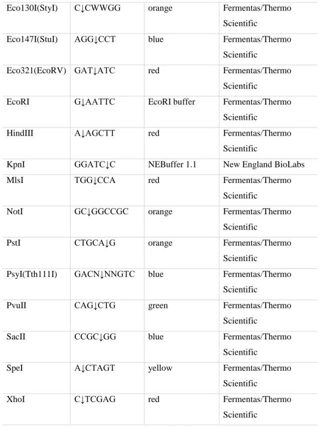
The restriction enzymes are shown in table 11. In table 12 all specific enzymes like polymerases or kinases are listed.
Table 11 Restriction enzymes
enzyme interface buffer company
BamHI G↓GATCC BamHI buffer Fermentas/Thermo
Scientific
BglI GCCNNNN↓NG
GC
orange Fermentas/Thermo
Scientific
BglII A↓GATCT orange Fermentas/Thermo
Scientific
Bpu1102I(BlpI) GC↓TNAGC yellow Fermentas/Thermo
Scientific
BseRI GAGGAG(N)10↓ CutSmart® Buffer New England BioLabs
Bsp119I(BstBI) TT↓CGAA yellow Fermentas/Thermo
Scientific
BtsαI GCAGTGNN↓ CutSmart® Buffer New England BioLabs
DpnI Gm6A↓TC yellow Fermentas/Thermo
Scientific
38
Eco130I(StyI) C↓CWWGG orange Fermentas/Thermo
Scientific
Eco147I(StuI) AGG↓CCT blue Fermentas/Thermo
Scientific
Eco321(EcoRV) GAT↓ATC red Fermentas/Thermo
Scientific
EcoRI G↓AATTC EcoRI buffer Fermentas/Thermo
Scientific
HindIII A↓AGCTT red Fermentas/Thermo
Scientific
KpnI GGATC↓C NEBuffer 1.1 New England BioLabs
MlsI TGG↓CCA red Fermentas/Thermo
Scientific
NotI GC↓GGCCGC orange Fermentas/Thermo
Scientific
PstI CTGCA↓G orange Fermentas/Thermo
Scientific
PsyI(Tth111I) GACN↓NNGTC blue Fermentas/Thermo
Scientific
PvuII CAG↓CTG green Fermentas/Thermo
Scientific
SacII CCGC↓GG blue Fermentas/Thermo
Scientific
SpeI A↓CTAGT yellow Fermentas/Thermo
Scientific
XhoI C↓TCGAG red Fermentas/Thermo
Scientific
Table 12 Specific enzymes
enzyme company
ACCUZYMETM DNA Polymerase Bioline
cAMP-dependent Protein Kinase (PKA), catalytic subunit
New England BioLabs
39 FastAP Thermosensitive Alkanine Phosphatase Thermo Scientific
GST-PKC Sigma-Aldrich
T4 DNA Ligase Fermentas/Thermo Scientific
Taq Polymerase homemade
4.1.9 Marker
Markers were used for quantification analysis in agarosegel electrophoresis as well as SDS-PAGE.
Table 13 Marker
Marker Company
Blue Prestained Protein Standard Broad Range (11-190 kD)
New England Biolabs
GeneRulerTM 1 kb DNA Ladder Thermo Scientific
PageRulerTM Prestained Protein Ladder Thermo Scientific
4.1.10 Fly lines
Fly lines were bought from different sources listed in table 14 or generated using the PhiC31-Integrase system (Ubi::GFP-Baz variants, UASp::aPKCCAAX, UASp::Crb, UASp::Sdt-myc; attP25C=attP40, attP86F=ZH-86Fb and attP65B=VK00033) (Chapter 4.2.4.2). Germ line clones were generated according to the dominant female sterile technique (Chou et al., 1993) (Chapter 4.2.4.3). For generating embryos which express GFP-Baz variants with maternally and zygotically mutant baz815-8 background, virgins of baz815-8 FRT19A / FRT19A, hs::Flp, OvoD1; Ubi::GFP-Baz- variants rescue crosses were mated with hemizygous mutant males expressing Bazwt
or Baz5xA (baz815-8/Y; Ubi::GFP-Bazwt/5xA) or wild type males expressing Baz7xA.
40 Table 14 Fly lines
fly line description chromosome source
apkck06403FRT42B apkc null allele 2 Wodarz et al., 2000
baz815-8 FRT19A baz null allele 1 Krahn et al.,
2010b; McKim et al., 1996
sdtK85 Stop codon in Sdt L27N 1 Berger et al., 2007
dag::GAL4 ubiquitous expression in daughterless gene pattern, GAL4 driver line
3 Bloomington stock collection
hs::Flp, OvoD1 germ line clonal analysis 1 Bloomington stock collection
mat67::GAL4 ubiquitous maternally expression, GAL4 driver line
3 Bloomington stock collection
par-6 ∆226
FRT19A
par-6 null allele 1 Krahn lab stock
collection UAS::PAR-1-
shRNA
expression of dsRNA for RNAi of par-1 under the control of UAS
3 TRiP GL00253 and TRiP HMS00405
4.1.11 Cell lines
S2R Drosophila Schneider Cells were cultivated at 25°C.
Table 15 Cell lines
cell line description source
S2R Drosophila Schneider cells late embryonic stage DGRC
41
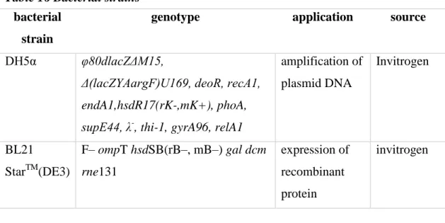
4.1.12 Bacterial strains
Bacterial strains were cultivated on agar plates at 37°C and selected via specific resistances.
Table 16 Bacterial strains bacterial
strain
genotype application source
DH5α φ80dlacZΔM15,
Δ(lacZYAargF)U169, deoR, recA1, endA1,hsdR17(rK-,mK+), phoA, supE44, λ-, thi-1, gyrA96, relA1
amplification of plasmid DNA
Invitrogen
BL21
StarTM(DE3)
F– ompT hsdSB(rB–, mB–) gal dcm rne131
expression of recombinant protein
invitrogen
4.1.13 Disposables
All used disposables listed in table 17 were disposed according to their usage via autoclaving or waste separation.
Table 17 Disposables
disposables manufacturer
96 Biosphere® Filter Tips, 0.1 – 10 µl Sarstedt 96 Biosphere® Filter Tips, 2 – 20 µl Sarstedt
Autoclave tape, 50 m Brand
Cell Scraper 25 cm Sarstedt
centrifuge tube, 15 ml Sarstedt
centrifuge tube, 50 ml Sarstedt
Cuvettes, 10 x 4 x 45 mm Sarstedt
Delicate Task Wipes – White Kimberly-Clark
Folded Filters (Qual.), Ø 240 mm Sartorius stedim
Micro tube, 1.5 ml Sarstedt
42 Microscope Cover Glasses, 24 x 50 mm Marienfeld
Microscope Cover Glasses, Ø 22 mm Thermo Scientific Microscope Slides, 25 x 75 x 1.0 mm Thermo Scientific
Multiply® -Pro cup, 0.2 ml Sarstedt
Nitril Extra-Sensitiv 24 cm NitriSense
Parafilm “M” Bemis
Pipette tip, 1000 µl Sarstedt
Pipette tip, 20 µl Sarstedt
Pipette tip, 200 µl Sarstedt
SafeSeal micro tube, 2 ml Sarstedt
Serological pipette, 10 ml Sarstedt
Serological pipette, 2 ml Sarstedt
Serological pipette, 5 ml Sarstedt
Super RX Fujifilm Fujifilm
TC-Dish 150, Standard Sarstedt
TC-Flask T25, Stand., Vent. Cap Sarstedt TC-Flask T75, Stand., Vent. Cap Sarstedt
TC-Plate 12 Well, Standard, F Sarstedt
TC-Plate 24 Well, Standard, F Sarstedt
TC-Plate 6 Well, Standard, F Sarstedt
Transfer pipette, 3.5 ml Sarstedt
Transfermembran, vliesvestärkte NC Hartenstein
WHATMAN Paper 460 x 570 VWR
4.1.14 Devices
Table 18 Devices
device application company
5050 ELV autoclave Tuttnauer
AxioCam MRc camera transmitted light microscopy Zeiss
Axiovert 200 microscope Zeiss
43
BL 1500 S chemical balance Sartorius
Bunsenbrenner LABOGAZ 470
flame treatment Roth
Centrifuge 5810R centrifuge Eppendorf
ChemiDocTM MP transilluminator Biorad
CT 15 RE table centrifuge Hitachi
Duomex 1030 platform shaker Heidolph
Eco-Mini System E SDS-PAGE Analytik Jena
Evolution™ 201/220 UV-Vis- Spektrophotometer
spectrophotometer Thermo Scientific
Femtotips® II microinjection capillary
Injection for germline transformation
Eppendorf
Fusion FX7 western blot developing Peqlab
Heraeus T 6200 drying chamber Thermo Scientific
Heraeus UT 6120 drying chamber Thermo Scientific
Light table Agarose gel analysis Danubia
LSM 710 Meta confocal microscope
confocal microscopy Zeiss
Mastercycler nexus gradient thermocycler Eppendorf Micromanipulator InjectMan
NI2
Injection for germline transformation
Eppendorf
Nanodrop1000 spectrometer (AG Meister) Thermo Scientific
Nanodrop2000 spectrometer Thermo Scientific
Pellet Mixers and Cordless Motor
homogenizing Drosophila embryos
VWR
Pipetboy electric pipetting aid Integra
Pipettes pipetting Eppendorf
Rotiphorese® PROclamp MINI Tank-Blotting-System
Western Blot Roth
Rotiphorese® Unit PROclamp MINI complete set
SDS-PAGE Roth
Sonopuls HD 2070/2200 protein purification Bandelin
44
Stereo zoom microscop binocular Motic
Thermomixer comfort heating block Eppendorf
Unitron incubator shaker Infors HT
UV slider transilluminator Intas
Vortex Genie 2 vortex mixer Intas
4.1.15 Data bases and software
Data bases and software used as a support for this work are shown in table 19.
Table 19 Data bases and software
data bases/software application
Adobe Photoshop image editing program
DNA Dynamo sequence analysis
Flybase database for Drosophila genetics
GIMP image editing program
IMAGEJ image editing program
NCBI databases for biomedical and genomic
information
ZEN 2010 Software microscope software