The significance of the laminin/nidogen-1 interaction for basement membrane formation and stability
in embryoid bodies
Inaugural-Dissertation zur
Erlangung des Doktorgrades
der Mathematisch-Naturwissenschaftlichen Fakultät der Universität zu Köln
vorgelegt von
Judith-Antonia Lummerstorfer aus Linz
Köln, 2001
Berichterstatter: Prof. Dr. M. Paulsson
Prof. Dr. M. Leptin
Tag der mündlichen Prüfung: 07.05.2001
Meinen Lieben
Die Entdeckungen der letzten Zeit lassen praktisch alles, was wir viele Jahre für richtig gehalten haben, als falsch oder nur bedingt richtig erscheinen. Meiner Meinung nach kann man heute nur noch eines mit Sicherheit sagen: Die Lichtgeschwindigkeit ist absolut das Schnellste, was es gibt. Möglicherweise.
Edward Teller
ABSTRACT
Basement membranes are thin layers of extracellular matrix which separate epithelial and endothelial cells from underlying connective tissue and surround nerve, muscle and fat cells.
Biochemical data indicate the important role of nidogen-1, a 150kD sulfated glycoprotein, in supporting basement membrane stability by connecting the laminin and type IV collagen network (Aumailley et al., 1989; Yurchenco and Schittny, 1990).
The aim of this study was to analyse the significance of the laminin/nidogen-1 interaction for basement membrane formation and stability in a more complex system. For that purpose F9 embryoid bodies, a cell culture model for basement membrane formation, were grown in the cell spin system after comparison with the conventional hanging drop method. F9 cells are mouse teratocarcinoma cells which differentiate and develop a basement membrane when cultured with retinoic acid.
Recombinant expression of the nidogen-binding site, located in LE module 4 of the γ 1 chain of the trimeric laminin molecule, as the γ1III3-5 fragment and its binding to free nidogen-1 molecules should interfere with the laminin/nidogen-1 interaction. As controls, F9 cells were transfected with the empty expression vector, the γ1V1-3 and the γ1III3-5mut fragment.
γ1V1-3 is similar to the γ1III3-5 fragment in secondary structure and size while γ1III3-5mut carries a point mutation drastically reducing its affinity to nidogen-1. For detection of recombinant expression and for purification purposes all constructs had been modified with the FLAG peptide at their N-terminal end.
Analysis of basement membrane formation by fluorescence microscopy revealed a different pattern in clones expressing the nidogen-binding site than in the controls. While cells carrying the empty expression vector and such expressing the γ 1V1-3 or the γ 1III3-5mut fragment developed embryoid bodies with completely or partially continuous basement membranes, only a punctate distribution of basement membrane components could be detected in cell aggregates expressing the nidogen-binding site. This indicated effective blocking of the laminin/nidogen-1 interaction by recombinant expression of the γ1III3-5 fragment. Additional studies on differentiation showed patchy distribution of cells expressing genes specific for visceral endoderm all over the embryoid body. In the controls these cells were organised into an peripheral epithelium which suggests a role of basement membranes in regulating the differentiation of these cells. To evaluate further functional aspects of the basement mem- brane discontinuity a diffusion assay was developed in which permeability properties could be tested. This revealed higher permeabilities among clones with comparable embryoid body morphologies when the basement membrane was disrupted.
To exclude the possibility that endogenous recombinant expression or the genetic mani-
pulation during transfection was responsible for the observed phenotypes, experiments were
also performed with the extraneous addition of the recombinant nidogen-binding site. Wild
type F9 cells were grown in suspension culture and treated with recombinantly expressed,
affinity purified nidogen-binding site fragment. Microscopy demonstrated a similar basement
membrane breakdown upon exogenous addition of the nidogen-binding site as observed be-
fore in embryoid bodies derived from F9 cells recombinantly expressing the γ 1III3-5 frag-
ment.
ZUSAMMENFASSUNG
Basalmembranen sind dünne Schichten extrazellulärer Matrix, die Epithel- und Endothel- zellen vom Bindegewebe trennen und Nerven-, Muskel- und Fettzellen umgeben. Eine auf biochemischen Daten (Aumailley et al., 1989) gestützte Hypothese erklärt die Stabilität der Basalmembran mit Nidogen-1, einem 150kD großen sulfatierten Glykoprotein, als Bindeglied zwischen dem Laminin- und Typ IV Kollagen- Netzwerk der Basalmembran (Yurchenco und Schittny, 1990).
Ziel war es, die Bedeutung der Laminin/Nidogen-1 Wechselwirkung für die Ausbildung und Stabilität von Basalmembranen in einem komplexeren System zu analysieren. Dafür wurde ein Zellkultur-Modell der Basalmembranbildung, das F9 Zellaggregatsystem, nach Ver- gleichen mit der "Hanging Drop"-Methode in Spinnerflaschen etabliert. Solche "Embryoid Bodies" aus F9 Maus Teratokarzinomazellen bilden bei Retinsäurebehandlung eine Basal- membran aus.
Rekombinante Expression der in LE Modul 4 der γ1 Kette des trimeren Laminin Moleküls lokalisierten Nidogenbindungsstelle im Fragment γ1III3-5 und dessen Bindung an freie Nidogen-1 Moleküle sollte das natürliche Gleichgewicht der Laminin/Nidogen-1 Interaktion stören. Als Kontrollen wurden neben dem leeren Expressionsvektor ein der Nidogen- bindungsstelle in Sekundärstruktur und Größe ähnliches Fragment der γ1 Kette, γ1V1-3, sowie eine mutierte Version der Nidogenbindungsstelle γ1III3-5mut in F9 Zellen transfiziert.
Zuvor waren alle Konstrukte zum Nachweis rekombinanter Expression und um eine Auf- reinigung zu ermöglichen mit einem N-terminalen FLAG Bindungsmodul versehen worden.
Die mikroskopische Untersuchung der die Nidogenbindungsstelle exprimierenden Klone ergab eine von den Kontrollen abweichende Ablagerung der Basalmembranproteine.
Während die nur resistente Kontrolle sowie die γ1III3-5mut und γ1V1-3 synthetisierenden
"Embryoid Bodies" entweder durchgehend oder wenigstens streckenweise Basalmembran ausbilden konnten, war in den die Nidogenbindungsstelle exprimierenden Klonen nur eine punktförmige Verteilung von Basalmembrankomponenten zu erkennen. Dies sprach für eine effiziente Blockierung der Laminin/Nidogen-1 Wechselwirkung durch die rekombinante Expression des γ1III3-5 Fragments. Die zusätzliche Bestimmung der Fähigkeit zur Differen- zierung zeigte, dass in dem Klon mit gestörter Basalmembranausbildung Zellen mit einer für das viscerale Endoderm spezifischen Genexpression über das ganze Zellaggregat verteilt waren. In den Kontrollen hingegen waren solche Zellen vorzugsweise an der äußeren Peripherie in einem Epithel organisiert, was eine Funktion der Basalmembran in der Steuer- ung der Zelldifferenzierung impliziert. Um die mikroskopisch erkennbare Zerstörung der Basalmembran auf ihre funktionelle Konsequenz hin zu testen, wurden ihre Permeabilitäts- eigenschaften mit der Diffusions-Messmethode bestimmt. Daraus ergab sich für "Embryoid Bodies" mit vergleichbarer Morphologie eine höhere Permeabiliät bei gestörter Basalmem- branausbildung.
Weil die Möglichkeit bestand, dass die den Zellmetabolismus belastende rekombinante Ex-
pression oder klonale Selektion ausschlaggebend für die beobachteten Phänotypen waren,
wurde außerdem in Suspensionskulturen herangezogene Wildtyp F9 "Embryoid Bodies" in
mit rekombinant erzeugten, affinitätsgereinigten Nidogenbindungsstellen versetztem Medium
differenziert. Deren mikroskopische Analyse zeigte bei Zugabe der Bindungsstelle von außen
dieselbe Störung der Basalmembranausbildung wie sie zuvor bei den Klonen mit endogener
rekombinanter Expression beobachtet worden war.
Lebenslauf
Persönliche Daten:
Name: Judith-Antonia Lummerstorfer Geburtsdatum: 17. Juli 1973
Geburtsort: Linz
Familienstand: ledig
Staatsangehörigkeit: österreichisch
Adresse: Höninger Weg 275, 50969 Köln
Schulausbildung:
79 - 83 Besuch der Volkschule Gramastetten
83 - 91 Besuch des Humanistischen Gymnasiums in Linz
06/91 Reifeprüfung
Hochschulausbildung:
10/91 - 02/97 Studium der Genetik an der Universität Wien, Österreich
07/93 erste Diplomprüfung
12/95 - 11/96 Diplomarbeit am Institut für Molekulare Pathologie, Wie n in der Arbeitsgruppe von Dr. A. Weith unter der Betreuung von Prof. Dr. D. Schweitzer
Thema: Regionale Feinkartierung der kritischen 1p36 Neuroblastom Konsensus Deletion mit DIRVISH
12/96 zweite Diplomprüfung
02/97 Sponsion (Verleihung des akad. Grades Mag. rer. nat.)
04/97 Beginn der Teilnahme am Graduiertenkolleg „Genetik zellulärer Systeme“
04/97 - 08/97 Rotationsperiode in den Arbeitsgruppen von Prof. Dr. B. Kemper, Prof. Dr. S.
Korsching, Prof. Dr. M. Leptin, Prof. Dr. M. Paulsson und Prof. Dr. K. Schnetz seit 09/97 Dissertation am Institut für Biochemie II, medizin. Fakultät der Universität zu
Köln. Betreuer: Prof. Dr. M. Paulsson
Thema: Bedeutung der Laminin/Nidogen-1 Wechselwirkung für
die Ausbildung und Stabilität von Basalmembranen in „Embryoid Bodies“
12/97 Verleihung des österr. Würdigungspreises durch den Bundesminister für Wissenschaft und Verkehr
04/97 - 03/00 Stipendiatin im Rahmen des DFG Graduiertenkollegs „Genetik zellulärer Systeme“
seit 04/00 wissenschaftliche Mitarbeiterin am Institut für Biochemie II
Contents Page
1. Introduction... 1
1.1. The evolution of extracellular matrices... 1
1.2. Basement membrane structure and function... 3
1.3. Basement membrane components... 3
1.3.1. Laminins... 3
1.3.1.1. Laminin isoforms... 3
1.3.1.2. Laminin receptors... 6
1.3.1.3. Laminin polymerisation... 8
1.3.2. Nidogens... 9
1.3.3. Type IV Collagen... 13
1.3.4. Perlecan, a heparan sulphate proteoglycan... 14
1.4. Basement membrane assembly and structure... 16
1.5. Embryoid bodies... 17
1.6. Aim of the present study... 19
2. Results... 20
2.1. Cloning of nidogen-binding site constructs and controls... 20
2.2. γ 1III3-5 expression in 293-EBNA cells... 22
2.3. Expression of γ1III3-5, γ1III3-5mut and γ1V1-3 in F9 and D3 cells... 24
2.4. Comparison of embryoid body phenotype in two different cell culture systems... 25
2.5. Comparison of F9 embryoid bodies expressing the laminin γ1 constructs... 28
2.5.1. Analysis of embryoid body growth rate and morphology... 28
2.5.2. Detection of basement membrane proteins in γ 1III3-5/A, γ 1III3-5/B, ... γ1III3-5mut and γ1V1-3 expressing embryoid bodies 30 2.5.3. Differentiation and permeability properties of F9 embryoid bodies expressing... γ1III3-5, γ1III3-5mut and γ1V1-3 35 2.6. Addition of affinity purified laminin g1 FLAG fusion protein γ1III3-5 to wild... type embryoid bodies 40 3. Discussion... 43
3.1. Establishment of cells expressing laminin γ1 FLAG fusion proeins... 43
3.1.1. Cloning of the constructs... 43
3.1.2. Promoter activity in different cell lines... 44
3.1.3. Protein expression in F9 mouse teratocarcinoma cells... 45
3.1.4. Expression and purification of the laminin γ 1III3-5 FLAG fusion protein from... 293-EBNA cells 47 3.2. Comparison of two different embryoid body cell culture systems... 47 3.3. The influence of laminin γ 1 FLAG fusion proteins on basement membrane...
formation and function
48 3.3.1. Changed expression and structural organisation of basement membrane...
components
48
3.3.2. Influence of the various polypeptides on differentiation... 52
3.3.3. Influence on the permeability properties of basement membranes... 55
3.4. Perspectives... 56
4. Materials and Methods... 57
4.1. Culture and maintenance of tissue culture cell lines... 57
4.1.1. Mouse teratocarcinoma cell line... 57
4.1.2. Human embryonic kidney cell line... 57
4.1.3. Establishment of stably transfected cell lines... 58
4.1.4. Thawing and freezing cells... 58
4.2. Molecular cloning... 58
4.2.1. Bacterial cell culture... 58
4.2.2. DNA preparation... 59
4.2.3. RNA preparation and northern blot analysis... 59
4.2.4. Labeling of DNA probes... 59
4.2.5. Polymerase chain reaction (PCR)... 60
4.2.6. Recombinant techniques... 60
4.3. Microscopy... 60
4.3.1. Preparation of cryosections... 60
4.3.2. Preparation of semithin sections... 61
4.3.3. Immunofluorescence staining... 61
4.3.4. Staining for cell death and viability... 62
4.4. Diffusion assay... 63
4.5. Protein chemistry... 64
4.5.1. Affinity chromatography... 64
4.5.2. Treatment of wild type F9 embryoid bodies with affinity purified FLAG fusion... protein γ 1III3-5 64 4.5.3. Immunoblot analysis... 64
5. Appendix... 66
5.1. Abbreviations... 66
5.2. Amino acid code... 67
5.3. Vectors... 68
6. References... 69
1. Introduction
1.1. The evolution of extracellular matrices
During the development from uni- to multi-cellularity novel structural requirements were placed upon the evolving organisms. A primary need was the maintenance of the body form. While in primitive organisms this could probably be met by receptor mediated cell-cell contacts, these became insufficient as size and complexity increased leading to the evolution of the extracellular matrix. Although it is uncertain which extracellular matrix function evolved first, it may be speculated that the underlying need was to provide physical support to maintain the integrity of the body and that this finally led to the production of a prototype connective tissue and, particularly, the evolution of the collagen family (van der Rest and Garrone, 1991; Garrone, 1998). Here great tensile strength is produced through the close intramolecular association supplied by the triple helix and intermolecular covalent bonds.
With specialisation of cells into tissues, it became necessary to evolve modes of compartmen-
talisation (Kleinman and Schnaper, 1993 ) . The physical barrier separating cell and tissue
types led to the formation of the architechtural framework of higher organisms. To transmit
this stabilisation from the extracellular scaffold to the cell, specialised areas may have
developed containing receptor molecules anchoring the intracellular skeleton to particular
molecules in the surrounding matrix. These receptors were possibly refined to also allow
signalling between the matrix and the cells and vice versa. So external events were not only
conveyed by soluble proteins, which may themselves be bound into the matrix, as is the case
with many cytokines, but also by the structural components themselves. This has led to the
extracellular matrix having key roles in the presentation of growth and guidance cues to cells,
which can in turn influence the secretion of extracellular matrix components. So, cell-matrix
contacts may determine cellular differentiation. This has become crucial for most normal de-
velopmental processes such as gastrulation (Czaker, 2000), neural crest formation (Poelmann
et al., 1990) and whether a cell or axon migrates, where it goes and when it stops migrating,
as well as for adult physiology and pathology (e.g. extravasation of white blood cells, meta-
stasis, wound healing). In addition to directing cellular motility, the extracellular matrix con-
trols the formation of tissues like bone, cartilage, and tendon with characteristic structural pro-
teins and growth factors. The latter contribute by establishing gradients of guidance cues in
which cells can orientate, differentiate and secrete those extracellular matrix components
serving the specific needs of each tissue.
Most studies of the extracellular matrix have been performed on vertebrate organisms, however investigations of invertebrates show that many extracellular matrix components are ancient and highly conserved proteins. For example, the fibril-forming collagens I, II, III and V , providing tensile strength, as well as the non-fibrillar collagen type IV of basement mem- branes are found in invertebrate organisms like sponges, worms and mussels (Coyne et al., 1997; Kramer et al., 1994; Garrone et al., 1993). However broad the spectrum of identified collagen homologs seems to be, little is known about the mechanisms behind their evolution (Engel, 1997). Ancestor proteins have also been found for molecules presenting information in the extracellular matrix. Perlecan (Laurie et al., 1986), a basement membrane proteoglycan of higher organisms, is also a cartilage component and plays an important role in chondro- genic differentiation probably by binding and presenting growth factors (French et al., 1999).
However, closely related proteoglycans are also present in primitive invertebrate organisms such as C. elegans and the fruit fly, where an ortholog of perlecan (Mullen et al., 1999;
Moerman et al., 1996) and a perlecan-like core protein sequence have been identified (Friedrich et al., 2000), respectively. Since primitive organisms do not have cartilage these findings are an example of how the roles of extracellular matrix proteins may be adapted to new functions during evolution.
Modern extracellular matrix proteins are mosaic proteins consisting of about 65 different domains (Bork, 2000). It has been suggested that they arose by exon shuffling (Patthy, 1996), a good example of which could be the mammalian nidogen-1. The earliest nidogen is seen in ascidians and contains three epidermal-growth-factor (EGF)-like motifs, three thyroglobulin-like motifs and five LDL-receptor YWTD domains (Nakae et al., 1993).
Through evolution three extra cysteine-rich epidermal-growth-factor (EGF)-like motifs have been added by exon shuffling and one thyroglobulin-like motif was achieved by combination of three ancestral ones. Further highly conserved homologs of nidogen are observed in fruit flies, nematodes and sea squirt (Mayer et al., 1998). Another example where gene duplication appears to have occurred is the laminin protein family, mainly present in basement mem- branes. Laminins are heterotrimeric glycoproteins consisting of three genetically different chains. Today eleven distinct chains and fifteen different laminin isoforms are known in mammals. Identification of two laminin chains in Drosophila (Fessler et al., 1987; Martin et al., 1999), four laminin chains (Hutter et al., 2000) and the laminin-related netrins (Wadsworth et al., 1996) in C.elegans strongly imply the existence of a single ancestral laminin chain which has duplicated further within the vertebrate lineage.
2
1.2. Basement membrane structure and function
Basement membranes are 40 to 120nm thick sheets of extracellular matrix found throughout the body. They line the basolateral membrane of all epithelial and endothelial cells, separate them from the underlying connective tissue and surround nerve, muscle and fat cells. Basement membranes may interpose between endothelial and epithelial cell sheets as in the lung alveoli and kidney glomeruli. In the kidney, the basement membrane functions as a porous filter, preventing the passage of macromolecules from blood into urine. Basement membranes beneath epithelia prevent the contact between fibroblasts and epithelial cells, but allow the passage of macrophages, lymphocytes or nerve processes. In addition, basement membranes play an important role during development, binding growth factors and hormones which influence cell metabolism, cell growth, cell polarisation, and differentiation (Streuli et al., 1991; Schuger et al., 1997). The ability of certain basement membrane molecules to inter- act with cell surface receptors allows it to direct cell migration during development or to help to reconstruct original tissue architecture (e.g. guidance of regenerating motor nerve terminals to neuromuscular junctions) after tissue injury. All these different basement membrane func- tions imply tissue- and time-specific expression of its components from embryogenesis to adulthood. Some of these basement membrane molecules will be discussed below in detail.
1.3. Basement membrane components
1.3.1. Laminins
1.3.1.1. Laminin isoforms
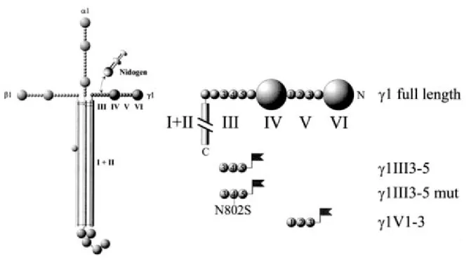
The laminins are large (600-800kD), cruciform glycoproteins (Tunggal et al., 2000)
consisting of three genetically different chains (α , β and γ) which share a common domain
structure (figure 1.1). The short arms formed by the amino-terminal regions of the β and γ
chains contain the domains III and IV instead of domains IIIa/b and IVa/b found in the α
chains (Sasaki and Yamada, 1987; Sasaki et al., 1987, 1988). These biologically active
domains are separated by flexible rows of EGF-like repeats, the LE motifs, which are stabi-
lised by disulfide bonds. The long arm results from oligomerisation through a triple helix of
domains I and II of the α, β and γ chain. At the carboxy-terminal end of the α chains the large G domain is formed by folding of the five tandemly arranged subdomains LG1-LG5 into separate globes.
Five α-, three β - and three γ- chains have been identified in mammals which could in theory be combined to give 45 cruciform heterotrimers. However due to additional assembly restrictions (e.g. γ2 is never seen connected with β1) only 15 laminin isoforms have been observed although more forms may well exist (Colognato and Yurchenco, 2000; Libby et al., 2000). Studies on the assembly of laminin chains indicate that certain sites within the carboxy-terminal α-helical region of the long arm are important for chain-specific assembly (Utani et al., 1994). Little is known about the mechanism of chain secretion in vivo, but cell culture studies demonstrate that the α chain can be secreted as a single subunit while β and γ can not (Yurchenco et al., 1997).
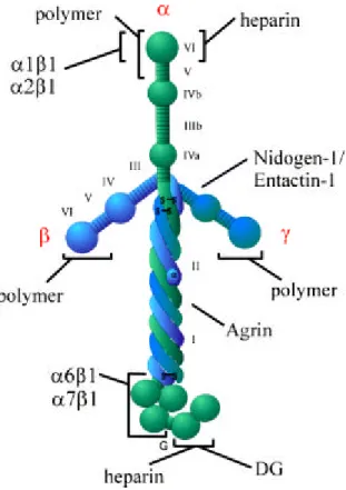
Figure 1.1: Schematic structure of the laminin molecule comprising an α, β and γ chain. The molecule has a cruci- form shape (Engel et al., 1981) with three short amino-terminal arms which are involved in polymerisation. The long carboxy -terminal arm consists of domain I and II of all three chains and forms a coiled-coil α-helix (Paulsson et al., 1985; Beck et al., 1993) which is terminated by a globular carboxy -terminal domain (G) contri- buted by the α chain only. The three-stranded coiled-coil domain is required for high-affinity binding to agrin (Kammerer et al., 1999). α1β1, α2β1, α6β1, and α7β1 indicate integrin binding sites. Other binding partners in- dicated are dystroglycan (DG), the polysaccharide heparin and nidogen-1/entactin-1.
4
An overall picture of the time- and tissue-specific deposition of laminin isoforms does not exist at the moment, because immunohistology and in situ hybridisations can only describe the distribution pattern of single laminin chains but not of the whole trimer. Still, three classes of epithelial laminins containing either the α 1, α 3 or α5 chain and the group of endothelial or mesenchymal laminins comprising the α2 chain or the α4 chain can be disting- uished (see table 1). So laminins-1, -3, -5, -6, -7, -10 and -11 underly epithelial structures while laminins-2, -4 and -12 are observed in the placenta (Paulsson et al., 1991), in basement membranes surrounding skeletal and cardiac muscles (Leivo and Engvall, 1988; Paulsson et al., 1991) and peripheral nerves (Uziyel et al., 2000). Laminins-8 and -9 are found in endo- thelial basement membranes of certain blood vessels, e.g. aorta (Frieser et al., 1997). The α 4 chain is also expressed by skeletal, cardiac muscle (Lefebvre et al., 1999; Liu and Mayne, 1996) as well as fat cells (Niimi et al., 1997a). Recent data indicate the existence of two addi- tional isoforms, laminin-14 and -15, present in the retinal matrix (Libby et al., 2000). The on- going identification of new laminin isoforms increases basement membrane complexity not only in tissue-specific composition, but also during development and repair (Erickson and Couchman, 2000).
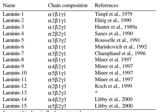
Name Chain composition References Laminin-1 α1β1γ1 Timpl et al., 1979 Laminin-2 α2β1γ1 Ehrig et al., 1990 Laminin-3 α1β2γ1 Hunter et al., 1989a Laminin-4 α2β2γ1 Sanes et al., 1990 Laminin-5 α3β3γ2 Rousselle et al., 1991 Laminin-6 α3β1γ1 Marinkovich et al., 1992 Laminin-7 α3β2γ1 Champliaud et al., 1996 Laminin-8 α4β1γ1 Miner et al. 1997 Laminin-9 α4β2γ1 Miner et al., 1997 Laminin-10 α5β1γ1 Miner et al., 1997 Laminin-11 α5β2γ1 Miner et al., 1997 Laminin-12 α 2 β 1 γ 3 Koch et al., 1999
Laminin-13 α3β2γ3 *
Laminin-14 α4β2γ3 Libby et al., 2000 Laminin-15 α5β2γ3 Libby et al., 2000
* not yet biochemically demonstrated
Table 1. Nomenclature of laminin isoforms (modified from Tunggal et al., 2000)
Basement membrane composition becomes even more complex when considering isoform expression changes during embryogenesis. Such switches can occur very rapidly as in the conversion of mesenchyme to epithelium in the developing kidney (Ekblom et al., 1998;
Miner and Li, 2000; Pedrosa-Domellof et al., 2000). The first sign of conversion into epithelium in vitro is the condensation of mesenchyme, followed by tubulogenesis with form- ation of an underlying basement membrane and cell polarisation. Formation of distinct tubules and cell polarisation correspond to the S-shaped stage of in vivo development. Studies of α chain expression during this process revealed that the α4 chain is transiently expressed in condensated mesenchyme, followed by α1 and α5 chain expression in S-shaped tubules and their elongations, respectively.
Laminin-1 is the major laminin essential for early embryogenesis (Dziadek and Timpl, 1985; Leivo et al., 1980; Cooper and MacQueen, 1983). The deletion of the LAMC1 gene encoding the γ1 chain results in absence of laminin-1 and embryonic lethality at day 5.5 post conceptum (Smyth et al., 1999). In its long arm structure (Aumailley et al., 1987) laminin-1 possesses cell-binding domains as well as sites for cell adhesion and stimulation of neurite- like outgrowth (Powell et al., 2000; Weston et al., 2000). The stimulatory effect of laminin on neurons is further supported by experimental data which demonstrated that laminin in neuron- extracellular matrix interaction prevents hippocampal cell death (Chen and Strickland, 1997).
In addition to its growth promoting activities laminin-1 was shown to modify the behavior of growth cones in Xenopus (Hoepker et al., 1999) possibly via induction of microtubular bund- ling in the growth cone of axons (Tang and Goldberg, 2000). Our understanding of laminin-1 as a basement membrane molecule specifically directing cell components is further supported by the discovery that it can organise acetylcholine receptors into clusters during synaptogen- esis at the neuromuscular junction (Sugiyama et al., 1997).
1.3.1.2. Laminin receptors
There appear to be two major sets of laminin receptors comprising α -dystroglycan and the integrins. The major binding sites for both receptors on the laminin molecule are located within the five subdomains of the G domain (Hohenester et al., 1999; Talts et al., 2000), however the ones for integrin are believed to also require the adjacent coiled-coil region consisting of the α 1, β1 and γ1 chain for proper folding (Aumailley et al., 1987; Goodman et al., 1987; Tisi et al., 2000).
6
α-Dystroglycan is part of a large transmembrane complex that plays an important role in muscle biology (Ervasti and Campbell, 1993; Hemler et al., 1999). It links laminin-2 to the myofiber cytoskeleton via indirect binding to dystrophin and blocking of this interaction with antibodies induces a dystrophic phenotype (Brown et al., 1999). Dystroglycan is expressed in developing and adult tissues by epithelial and neuronal cells which contact basement membranes (Matsumura et al., 1993; Durbeej et al., 1998) and is involved in kidney epithelial morphogenesis (Durbeej et al., 1995). The absence of dystroglycan leads to an embryonic lethality with structural and functional defects of one of the earliest basement membranes, the Reichert's membrane (Williamson et al., 1997), and analysis of dystroglycan null embryoid bodies shows disrupted basement membranes (Henry and Campbell, 1998).
Integrins are heterodimeric transmembrane molecules containing an α and a β subunit and are involved in signal transfer between the extracellular matrix and the cell interior (Hynes, 1992; Clark and Brugge, 1995). Only a subset of all known integrins, the α7, α6 and α3 integrins seem to specifically interact with the G domain (Belkin and Stepp, 2000). α6β4 integrin is found in hemidesmosomes whereas α6β1 integrins are often localised in focal contacts (Sonnenberg, 1992). For the integrins α1β1 and α2β1 the binding site was identified on the short arm of the laminin-1 molecule (Languino et al., 1989; Goodman et al, 1991), but it remains controversial if corresponding binding sites exist on other laminin isoforms (Pfaff et al., 1994; Colognato et al., 1997). α7β1 integrin is also laminin-specific and expressed highly in myoblasts (Von der Mark, 1991) together with α -dystroglycan. β1 integrin null embryoid bodies exhibit failures of basement membrane formation (Faessler and Meyer, 1995; Aumailley et al., 2000) a similar phenotype can also be observed in skin epithelium after conditional targeting of the β 1 integrin gene (Raghavan et al., 2000). Similar phenotypes can be caused by mutations in the α3, α6, β4 integrin subunits (George-Labouesse et al., 1996; Niessen et al., 1996; DiPersio et al., 1997) and in syndecan-2, a heparan sulphate proteoglycan (Klass et al., 2000).
Major functions of laminin receptors are in cell attachment and in providing signals
which are transferred to intracellular signal cascades. However, many results also indicate that
laminin receptors facilitate in situ laminin deposition on the cell surface as it has been demon-
strated for α-dystroglycan (Montanaro et al., 1999). Laminin polymerisation, a requirement
for basement membrane formation in turn induces redistribution of dystroglycan, α7β1
integrin and the cortical cytoskeleton of muscle cells (Colognato et al., 1999).
1.3.1.3. Laminin polymerisation
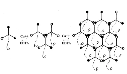
The three-arm interaction model of laminin polymerisation proposes that laminin molecules self-assemble above a critical concentration of 0.1µM through reciprocal, calcium- dependent binding at their amino-terminal short arms leading to the formation of large poly- mers (Yurchenco et al., 1985; Paulsson et al., 1988; Ancsin and Kisilevsky, 1996). This inter- action is conformation-dependent (Yurchenco and Cheng, 1994) and leaves the long laminin arms free for cell contacts. It is unknown whether in vivo laminin networks also contain heterogenous isoforms, but theoretically laminin-1 and -2 could co-polymerise (Cheng et al., 1997). Studies on the influence of lipid bilayers on laminin polymerisation suggest that plasma membranes could enhance aggregation of laminin molecules. The concentration critical for assembly was believed to be lowered by binding of laminins to lipid bilayers (Kalb and Engel, 1991). Although the association with cell surface receptors rather than exposed lipids was considered as a possible mechanism of enhancing polymerisation, recent in vitro evidence demonstrates that simple acidification of laminin monomer solution induces poly- merisation (Freire and Coelho-Sampaio, 2000).
Figure 1.2: Three-arm interaction model of laminin polymerisation (Yurchenco and Cheng, 1994). The short arms of the α, β and γ chain (continuous lines) are represented with globules at their amino-terminal ends. These can interact with each other in a calcium-dependent fashion in vitro. In this model the long arm (broken line) is free for interactions with cells.
8
1.3.2. Nidogens
Nidogen-1, a sulfated 150kD glycoprotein, was first isolated from F9 mouse terato- carcinoma cells and at that time called entactin (Hogan et al., 1980; Carlin et al., 1981).
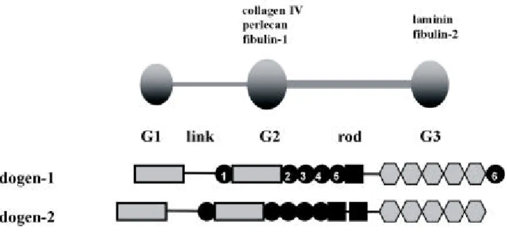
Differentiation of F9 cells with retinoic acid and dibutyryl cAMP induced synthesis of laminin-1 and nidogen-1. As shown by co-precipitation laminin-1 and nidogen-1 were in a complex that could be dissociated with sodium dodecyl sulfate. At the same time a protein which later proved to be identical with entactin was identified as a 80kD fragment in basement membranes of the Engelbreth-Holm-Swarm tumor and named nidogen (Timpl et al., 1983). The full-length, 150kD form of nidogen-1 could be purified with laminin-1 by EDTA extraction (Dziadek et al., 1985; Paulsson et al., 1987). Analysis of large amounts of the intact laminin-1/nidogen-1 complex purified from the extracellular matrix showed that the two proteins occur in an equimolar ratio (Paulsson et al., 1987). Data obtained from electron microscopy confirmed the interaction between laminin-1 and nidogen-1 (Paulsson et al., 1987) and binding studies with proteolytic fragments of the laminin-1/nidogen-1 complex indicated that the carboxy-terminus of nidogen-1 is responsible for binding laminin-1 (Mann et al., 1988). Electron microscopy of recombinant nidogen-1 showed three globular domains, G1 to G3, with G1 and G2 connected by a flexible link and G2 to G3 by a rod-like segment containing a series of EGF-like repeats (Mann et al., 1989; Durkin et al., 1995). The strong binding to laminin-1 with a K
Dof 0.5nM was found to be mediated by the C-terminal G3 domain (Fox et al., 1991).
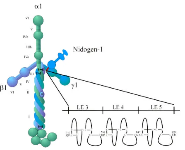
The nidogen-binding site on laminin-1 was localised to LE module 4 within domain III of the laminin γ1 chain (Gerl et al., 1991; Mayer et al., 1993; Poeschl et al., 1996). This part of the γ1 chain could be crystallised (Stetefeld et al., 1996) and structurally analysed (Baumgartner et al., 1996).
A typical EGF-like LE module forms a rigid structure. The rigidity is given by eight
non-contiguously arranged cysteines which interact via disulfide bridges (figure 1.3). This
leads to formation of four loops as observed in the nidogen-binding site module γ 1III4
between the 1st and 3rd cysteine, the 2nd and 4th, and two further between the 5th and 6th
and the 7th and 8th. The heptapeptide sequence Asn-Ile-Asp-Pro-Asn-Ala-Val located within
the first loop was found to be crucial for binding to nidogen-1 (Poeschl et al., 1994) in
combination with hydrogen bonding between the first and third loop providing binding struc-
tures and conformational stability (Stetefeld et al., 1996). Exchange of the central Asn and Val by Ser leads to a 100.000 fold lower binding affinity. Such an exchange has occurred in the corresponding domain γ2III4 of laminin-5 (α3β3γ2) despite the high overall sequence identity between the γ1III4 and γ2III4 motifs (Mayer et al., 1995) and laminin-5 can accordingly not bind nidogen-1.
Figure 1.3: The interaction between laminin-1 and nidogen-1. Nidogen-1 binds with its globular domain G 3 to the short arm of the γ1 chain. The nidogen-binding site, LE module 4 of the γ1 chain, is shown enlarged together with the neighbouring LE mocules. LE modules contain eight cysteines residues which upon disulfide bonding form loops similar to those of EGF (Cooke et al., 1987).
In vitro binding assays indicate the ability of nidogen-1 to mediate complex formation between laminin and collagen type IV and also between laminin-1 and the proteoglycan perlecan (Aumailley et al., 1993; Hopf et al., 1999). Nidogen-1 can further bind fibulins, extracellular matrix proteins composed of multiple arrays of epidermal growth-factor like modules similar to the LE modules of laminin (Adam et al., 1997). If the interaction between laminin and nidogen-1 is blocked by antibodies raised against the nidogen-binding site, in vitro epithelial development in organ cultures of embryonic kidney and lung gets disrupted (Ekblom et al., 1994). Similar in vitro experiments performed with submandibular gland
10
organ cultures lead to disruption of the basement membrane between epithelium and mesenchyme and reduced branching epithelial morphogenesis (Kadoya et al., 1997). These results suggest that the formation of the laminin/nidogen-1 complex may be a key event during epithelial development. Detailed analysis of the sites of laminin-1 and nidogen-1 gene expression by in situ hybridisation (Dong and Chung, 1991; Thomas and Dziadek, 1993;
Ekblom et al., 1994, Fleischmajer et al., 1995) have revealed that laminin-1 is predominantly produced by epithelial cells while nidogen-1 is secreted by mesenchymal cells. The binding of mesenchymal nidogen-1 to epithelial laminin-1 is believed to occur at the interface between epithelial and mesenchymal tissues where basement membrane formation could be dependent on complex formation between laminin-1 and nidogen-1 (Dziadek et al., 1995).
Additional evidence for nidogen-1 production by mesenchymal cells was obtained in coculture experiments with rat mesenchymal peritubular and epithelial-like Sertoli cells (Konrad et al., 2000). Even though nidogen-1 was transcribed in peritubular cells and Sertoli cells, its mRNA was only translated by peritubular cells. Antibody perturbation experiments could further show that nidogen-1 is required for cell adhesion of peritubular cells. The fin- ding that nidogen-1 is secreted by mesenchymal cells was also confirmed using a culture microecosystem of mammary epithelium where nidogen-1 is produced by mesenchymal cells but deposited between epithelial cells. Analysis of this in vitro system initially designed to investigate the role of nidogen-1 in gene regulation showed that nidogen-1 can regulate β - casein expression in cooperation with laminin-1 (Pujuguet et al., 2000).
The susceptibility of uncomplexed nidogen-1 to proteolytic degradation (Dziadek et al., 1988) allows speculation about such degradation playing a role during embryogenesis, where basement membrane assembly and degradation are tightly regulated (Dziadek, 1995).
The growing knowledge about the action of matrix metalloproteases (Werb, 1997; Giannelli et al., 1997) will lead to a better understanding of the mechanisms behind this balance which is crucial for normal growth, morphogenesis and tissue repair.
Nidogen-2 was recently isolated as osteonidogen or entactin-2 from an osteoblast-like
cell line and shown to have 27.4% identity to nidogen-1 on the amino acid level (Kimura et
al., 1998). Recombinant nidogen-2, a highly glycosylated 200kD protein, has a shape similar
to nidogen-1. Electron microscopy shows that it also consists of three globular domains con-
nected by two threads although it is somewhat different in length (figure 1.4). Immuno-
fluorescence and northern blots revealed coexpression and colocalisation of both nidogens in vessel walls and other basement membrane zones, but differences in heart and skeletal muscle (Kohfeldt et al., 1998). Nidogen-2 can bind to the nidogen-binding site γ1III4 of the laminin γ1 chain but with a 100 to 1000 fold lower affinity than nidogen-1 and interacts with a second binding-site of laminin-1 unrelated to the γ1III4 module. Deletion of LE module 4 abolishes binding of both nidogens to the recombinant γ1III3-5 fragment. In addition, nidogen-2 binds type IV and type I collagen and perlecan. However it does not interact with fibulin-1 or -2 like nidogen-1 (Kohfeldt et al., 1998).
Figure 1.4: The domain organisation and modular structures of nidogen-1 and nidogen-2. Both nidogens comprise three globular domains. G1 and G2 are joined by a link region while G2 and G3 are connected by a rod-like structure. Different basement membrane proteins binding to G2 and G3 domain of nidogen-1 are in- dicated. Legend: dark circles represent epidermal growth factor (EGF) like motifs, which are numbered in nidogen-1, dark boxes represent thyroglobulin (TG) like motifs and hexagons represent low density lipoprotein (LDL) receptor YWTD motifs (partially taken from Murshed, 2001).
To understand the in vivo role of the nidogens in basement membranes, the mouse and worm model were studied. The lack of nidogen-1 in mice does not affect basement membrane formation, instead nidogen-1 -/- mice develop seizures and other neurological defects later in life (Murshed et al., 2000). This phenotype could be explained by the existence of the struc- turally related nidogen-2/entactin-2 (Kohfeldt et al., 1998; Kimura et al., 1998) and a possible compensatory effect of nidogen-2 in basement membrane assembly of nidogen-1 deficient mice. Although immunostaining for nidogen-2 is stronger in distinct basement membranes, it does not appear to be transcriptionally upregulated in nidogen-1 -/- mice. Nidogen-2 null mutant mice also appear normal. The production of nidogen-1/-2 double null-mutant mice is currently underway (Smyth, personal communication). This will help to explain the import- ance of nidogen-1 and -2 and their contribution to basement membrane stability. Recent results obtained from loss of function experiments in C. elegans support the neurological
12
phenotype found in nidogen-1 -/- mice. Introducing a stop codon into the homologous C.
elegans nidogen gene causes irregular neuronal migration (Kim et al., 2000). Deletions in NID-1 however do not affect type IV collagen assembly into basement membranes (Kang and Kramer, 2000).
1.3.3. Type IV Collagen
The collagens are a glycoprotein family with at least 19 genetically distinct types (Prockop and Kivirikko, 1995) which can be divided into fibril-forming and non-fibrillar molecules. Typically, collagens consist of a ropelike superhelix comprising three polypeptide α chains wound round each other. These α chains contain a series of Gly-X-Y repeats. In 20- 22% of all triplets the positions X and Y are occupied by proline and hydroxyproline, re- spectively. The non-fibrillar collagens can form three-dimensional networks (type IV), beaded filaments (type VI), anti-parallel dimers (type VII) or a hexagonal lattice (type VIII).
7S Triple Helix NC1
NC1
7S NC1
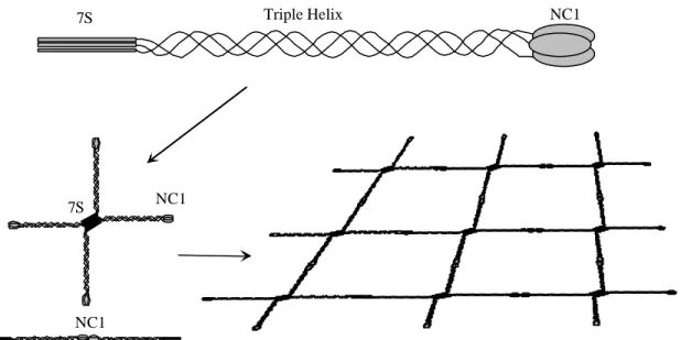
Figure 1.5: Schematic representation of the type IV collagen network. Three α chains interact and form a triple helix with amino-terminal 7S and carboxy -terminal NC1 domains. Covalent interactions between four 7S and two NC1 domains and additional lateral interactions (not shown) result in a lattice which provides mechanical strength to the basement membrane (taken from Tunggal, 2000).
Type IV collagen contains a carboxy-terminal noncollagenous domain (NC-1), a
350nm long triple-helical domain and an amino-terminal 7S domain. It is only present in
basement membranes, where it can self-assemble into a network via anti-parallel interactions
between the 7S domains of four molecules (Risteli et al., 1980; Glanville et al., 1985;
Tsilibary and Charonis, 1986), the interaction of two molecules at their carboxy-terminal NC1 domains and by lateral aggregation (Timpl et al., 1981; Yurchenco and Furthmayr, 1984).
This network (figure 1.5) provides structural support to the basement membrane, facilitates assembly of laminin, nidogen-1/entactin-1 and perlecan (Laurie et al, 1986) and functions as a size-selective filter unit.
Type IV collagen α chains are encoded by six genes, COL4A1-COL4A6 which are arranged in pairwise head-to-head organisation (Soininen et al., 1988; Mariyama et al., 1992;
Zhou et al., 1994). Since bidirectional regulatory elements have been identified between each pair, coordinate transcription and expression of COL4A1-COL4A2, COL4A3-COL4A4 and COL4A5-COL4A6 was suggested. This assumption was confirmed by the finding that the most abundant type IV collagen with ubiquitous expression in basement membranes (Hudson et al., 1993) has the chain composition [ α1(IV)]
2α2 (IV). In addition, colocalisation of the α3 and α 4 chains (Kleppel et al., 1989; Miner and Sanes, 1994) as well as the α5 and α6 chains could be demonstrated, although the α 5 chain is also expressed without the α 6 chain in the glomerular basement membrane (Peissel et al., 1995).
Mutations in the COL4A3, COL4A4 or COL4A5 gene result in a progressive heredi- tary disease of the glomerular basement membrane, Alport's syndrome (Kashtan and Michael, 1993). In Alport's patients the basement membranes of the kidneys are disorganised and fragile and less resistant to the high hydrostatic pressure. Mouse models which either lack the α3(IV) chain (Cosgrove et al, 1996; Miner and Sanes, 1996) or both the α3(IV) and the α 4(IV) chain undergo fibrosis leading to a glomerulonephrosis. This is possibly due to the failure in switching from α 1, α 2 expression to α3, α 4 and α5 expression which occurs during glomerular development (Lu et al., 1999).
1.3.4. Perlecan, a heparan sulphate proteoglycan
Proteoglycans are very heterogenous in composition and are present in the extra- cellular matrix, on cell surfaces (Bernfield et al., 1999) or in intracellular granules (Burditt et al., 1985). They consist of a protein core and glycosaminoglycan (GAG) chains (Prydz and Dalen, 2000). These long, unbranched, strongly anionic polysaccharide chains can bind
14
cations as well as H
2O. Thereby hydrated GAG gels are able to resist pressure changes in tissues, a property that is particularly important for joint function. In vitro GAGs can bind growth factors, matrix components, enzymes, enzyme inhibitors and cell adhesion molecules.
The core proteins themselves may show particular biological activities (Iozzo, 1998). Insights gained from in vivo studies in Drosophila and mice imply specific functions for heparan sulphate proteoglycans in cell differentiation and morphogenesis (Perrimon and Bernfield, 2000 ) .
In mammals three proteoglycans are found in basement membranes: Perlecan (Murdoch et al.,1994), agrin (Groffen et al., 1998) and bamacan (Couchman et al., 1996). Per- lecan, the most widespread and highly expressed proteoglycan of basement membranes and cartilage, comprises a 470kD core protein with a beads-on-a-string-like appearance and often carries three heparan sulphate side chains at its amino-terminal end (Costell et al., 1996;
Groffen et al., 1996; Schulze et al., 1996; Friedrich et al., 1999). Domain III of its protein core interacts with cell surfaces (Chakravati et al., 1995, Peng et al., 1998; Hohenester et al., 1999;
Talts et al., 1999) while domain IV binds nidogens, the laminin-1/nidogen-1 complex (Battaglia et al., 1992), type IV collagen (Laurie et al., 1986, Villar et al., 1999), fibronectin, fibulin-2, and heparin (Hopf et al., 1999).
Perlecan occurs in preimplantation embryos prior to basement membrane formation
(Dziadek et al., 1985; Smith et al., 1997) and inside the blastocyst on the outer surface of
trophectoderm cells (Carson et al., 1993). Interestingly, the distribution pattern of perlecan is
similar to that of fibroblast growth factor (FGF)-2 in various basement membranes of the
mouse embryo (Friedl et al., 1997) which led to speculations about an involvement of per-
lecan in growth control (Klein et al., 1995; Weiser et al., 1997). Perlecan expression was also
demonstrated on the surface of vascular endothelial cells, possibly concentrating thromo-
spondin 1 (Vischer et al., 1997), and in mesenchymal tissues like cartilage where it persists
into adulthood (Handler et al., 1997). Mice lacking perlecan show defects in cartilage and
cephalic development (Arikawa-Hirasawa et al., 1999) and while basement membranes form
they become disrupted with increasing mechanical stress (Costell et al., 1999) which suggests
a role of perlecan in regulating basement membrane integrity and permeability (Murdock et
al., 1993; Gauer et al., 1996; Groffen et al., 1997; Groffen et al., 1999).
1.4. Basement membrane assembly and structure
Although many different basement membrane components and numerous isoforms of these have been identified, our understanding how molecules become deposited, locally concentrated and organised into basement membranes after secretion is comparatively poor.
In vitro experiments demonstrate that dermal fibroblasts can produce type IV collagen, perlecan and nidogen, but they need keratinocyte integrins to bind type IV collagen in order to initiate basement membrane formation (Fleischmajer et al., 1998). Keratinocytes are also necessary for relocalisation of nidogen, which is of dermal origin during early basement membrane formation of in vitro skin models (Fleischmajer et al., 1995). Retinal basement membrane instead seems to require collagens for reassembly of neuroepithelia (Halfter et al., 2000) while alveolar epithelial cells can synthesise all major basement membrane components but still need exogenous laminin-1 for complete basement membrane assembly (Furuyama and Mochitate, 2000).
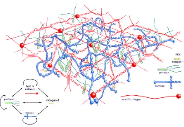
Figure 1.6: Molecular model of the basement membrane based on in vitro data (modified from Yurchenco and Schittny, 1990).The two major basement membrane networks are formed by laminin (blue) and type IV collagen (red) and are bridged by nidogen-1 (yellow). Perlecan (green) is a heparan sulphate proteoglycan which can interact with itself, type IV collagen and laminin.
16
Most of these studies on basement membrane formation are descriptive or in vitro experiments. In an in vivo approach the deletion of nidogen-binding module γ 1III4 interferes with the formation of the laminin-1/nidogen-1 complex (Mayer et al., 1998). Although embryonic stem cells homozygous for this defect secrete mutant laminin-1 which fails to associate with nidogen-1, they are still able to form embryoid bodies with a differentiation pattern similar to wild type. However complex the mechanisms of basement membrane formation might be, the self assembly of laminin and type IV collagen (described above) are still considered the main principles of basement membrane formation and the proposed function of nidogen-1 as the linker module between the type IV collagen and laminin networks has dominated the model of basement membrane structure (Aumailley et al., 1989;
figure 1.6).
1.5. Embryoid bodies
Embryoid bodies are cellular aggregates derived from either embryonic stem cells or embryonic carcinoma cells (Martin, 1980), which resemble early stages of mouse develop- ment (figure 1.7). The pluripotent F9 cells (Bernstine et al., 1973; Alonso et al., 1991) have a limited differentiation repertoire forming endoderm-like structures which develop only upon treatment with e.g. retinoic acid (Strickland and Mahdavi, 1978) or dibutyryl cyclic AMP (cAMP; Hogan et al., 1983). In F9 derived embryoid bodies treated with retinoic acid cells are found on the outer surface which are morphologically similar to visceral endoderm while F9 monolayers treated with retinoic acid and dibutyryl cAMP instead differentiate into parietal endoderm (Strickland et al., 1980; Damjanov et al., 1994). Parietal and visceral endoderm are two distinct populations of extra-embryonic endoderm found in the normal mouse embryo shortly after implantation (Hogan et al., 1994).
Since embryonic carcinoma cell lines can participate in the formation of chimeric mice, they were an important tool for the first gene transfer experiments in mouse before it was possible to establish totipotent embryonic stem cell lines in vitro (Martin et al., 1980).
Today embryoid bodies grown from F9 cells represent a well studied in vitro system of early
mouse embryogenesis which can help to understand the nature of endodermal and epithelial
differentiation (Coucouvanis and Martin, 1995). For example, it has been shown that Indian
hedgehog, which is upregulated during extra-embryonic endoderm differentiation in F9 embryoid bodies is also increased in 6.5 day old mouse embryos (Becker et al., 1997).
Similarly, α-Fetoprotein is synthesised by visceral endoderm (Hogan et al., 1981; Grover et al., 1983) in F9 embryoid bodies and it is restricted to the visceral endoderm during post- implantation development in mouse (Dziadek and Adamson, 1978). Differentiation of F9 cells into parietal or visceral endoderm upon treatment with retinoic acid and cAMP or retinoic acid alone, respectively, is accompanied by increasing synthesis of basement membrane proteins. At the transcriptional level a coordinate increase of the laminin α1, β1 and γ1 chains and the type IV collagen α1 chain (Durkin et al., 1986; Kleinman et al., 1987) has been demonstrated, whereas at the protein level the synthesis of the laminin β1 and γ1 chains is first induced followed by the α 1 chain of type IV collagen and α -fetoprotein (Rogers et al., 1990). Analysis of the gene regulation of the β 1 and γ1 chains revealed that both contain DNA regulatory elements which are activated during F9 induction (Chang et al., 1996; Li and Gudas, 1996). Differentiation into visceral or parietal endoderm leads to a 5-10 fold and 15- 20 fold increase in the synthesis of basement membrane proteins (Howe and Solter, 1980;
Prehm et al., 1982; Cooper et al., 1983) and in parietal endoderm the production of laminin and nidogen-1 occurs independently. Laminin and nidogen-1 are secreted to the medium and deposited at the cell surface and at cell junctions (Carlin et al., 1983; Chung et al., 1993).
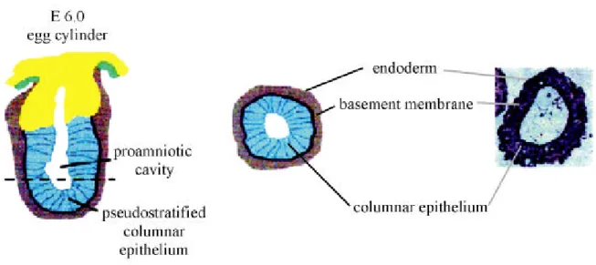
Figure 1.7: Embryoid bodies undergo differentiation processes analogous to early mouse development. On the left a saggital section through a mouse embryo at embryonic day 6.0, when the proamniotic cavity has been formed, is depicted. To the right a cross section (dashed line) through the egg cylinder is shown and compared to an embryoid body after cavitation. (modified from Coucouvanis and Martin, 1995).
18
1.6. Aim of the present study
The purpose of this study was to analyse the importance of the laminin/nidogen-1 interaction for proper basement membrane formation and stability. The contribution of nidogen-2 to stabilisation of major basement membrane networks is not clear at the moment.
Based on the basement membrane model designed by Yurchenco and Schittny (1990) nidogen-1 links the laminin lattice to the type IV collagen network. In order to test this hypothesis, I planned to disturb the laminin/nidogen-1 interaction by introducing an excess of recombinantly expressed nidogen-binding sites. In theory these additional binding-sites should compete with intrinsic laminin molecules presenting their binding-sites to nidogen molecules.
For time-specific interference with the laminin/nidogen-1 interaction at different
stages of basement membrane development inducible expression systems such as the
ecdysone-inducible (Invitrogen) or the retroviral tet on/off system (Hofmann et al., 1996)
could be applied. It was also decided to use the EF1α promotor for constant protein
expression which was previously shown to work well in F9 cells (Niimi and Kitagawa,
1997b). F9 and D3 (Evans and Kaufman, 1981) cells were chosen as they represent an in vitro
system of basement membrane formation very similar to early stages of mouse development
where the basement membrane separates the inner ectodermal cell mass from the outer
endodermal epithelium. To rule out that any phenotype might be caused by artefacts due to
clonal selection of overexpressing cells, the laminin γ1III3-5 FLAG fusion protein was also
added extraneously to developing wild type embryoid bodies. This required the nidogen-
binding site to be expressed in 293-EBNA cells using the CMV promotor, affinity-purified
and added to differentiating embryoid bodies derived from wild type F9 cells. For analysis of
basement membrane formation and stability molecular biological, biochemical, microscopi-
cal, and physiological techniques were used and the expression of markers for cell differen-
tiation were determined in the embryoid bodies.
2. Results
2.1. Cloning of nidogen-binding site constructs and controls
All constructs (figure 2.1) were derived by PCR from full-length mouse laminin γ1 cDNA which was obtained by reverse transcription of mouse kidney total RNA. Considering the complex secondary structure of LE 4, which contains the nidogen-binding site, it was expressed together with its neighbouring LE modules 3 and 5 (amino acid position 771-932).
Previous work had shown that such a polypeptide folds correctly (Stetefeld et al., 1996) and binds to nidogen-1 in a similar manner to native laminin (Mayer et al., 1993). For control purposes, the construct γ1III3-5mut with the point mutation N802S in LE 4, which decreases binding affinity for nidogen-1 by 46,000 fold (Poeschl et al., 1996) and the construct γ 1V1-3 containing the LE modules 1-3 (amino acid position 340-492) of domain V of the γ1 chain were also expressed. To enable secretion of the expressed proteins to the extracellular space and their detection and purification, sequences encoding the the BM40 signal peptide and FLAG tag (Hopp et al., 1988), respectively, were added to the N-terminus of the polypeptides.
Figure 2.1: Production of nidogen-binding site constructs and controls: The site for interaction between laminin- 1 and nidogen-1 has been localised to LE module 4 of domain III of the laminin γ1 chain (Mayer et al., 1993).
Three different FLAG fusion proteins ( ) were constructed and modified with the BM40 signal peptide to ensure secretion to the extracellular space, γ1III3-5 coding for LE modules 3-5, γ1III3-5mut which differs from γ1III3-5 in a point mutation of amino acid 802 from N to S a mutation causing a 46000 fold loss of nidogen binding activity (Poeschl et al., 1996) and γ1V1-3 comprising 3 LE modules of domain V.
20
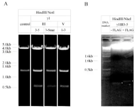
Although LE modules 3-5 had earlier been cloned into pCEP-Pu (Kohfeldt et al., 1997) which added the BM40 signal peptide, to insert the FLAG sequence the LE 3-5 coding sequence had to be subcloned into NheI/NotI restricted CMV-NFlag (a pCEP-Pu based vector with the FLAG tag placed 3 prime to the BM40 signal peptide sequence). To obtain LE modules 1-3 a PCR was performed on the γ1 cDNA using the sense primer AAT T GC TAG CCC TTC CTT GTG ACT GCA ATG GC and the antisense primer AAT A GC GGC CGC CTA GGG TGT GCA GCC CTT AGG containing a NheI and NotI site, respectively. After digestion the PCR product was cloned into the restriction digested CMV-NFlag plasmid. To mutate amino acid 802 in γ1III3-5 from an asparagine to a serine, the sequence for the FLAG tagged LE modules 3-5 was subcloned into pBluescript KS (+) by HindIII/XhoI digestion.
Then site-directed mutagenesis was performed using the Transformer Site-Directed Muta- genesis Kit (Clontech Laboratories Inc.) following the manufacturer's instructions with TransSCA (5' GTG ACT GGT GAG GCC TCA ACC AAG TC 3') as the selection primer and the primer N802S (5' GTG TAA CGA CAA TAT TGA CCC CAG CGC GGT TGG C 3') as the mutagenic primer. Constructs were verified by DNA sequencing and correspond to γ1 sequences available in the SWISS-PROT data base (primary accession number P02468).
Figure 2.2: Restriction digest on nidogen-binding site constructs and controls cloned into pBKEF -5 (A) and pCEP-Pu (B). A) HindIII/NotI restriction of the empty pBKEF-5 expression vector yields two fragments of 4.0kb and 1.4kb separated on 0.7% agarose gel. Upon insertion of the nidogen-binding site constructs γ1III3-5, γ1III3-5mut and the control region γ1V1-3 an additional 0.6kb band appears. Lane γ1 III3-5mut is derived from a later experiment. B) HindIII/NheI restriction of γ1III3-5 cloned into pCEP-Pu excises the BM40 signal peptide while a slightly shifted fragment coding for the BM40 signal peptide plus the FLAG tag is obtained from γ1III3- 5 inserted into CMV-NFlag. Restriction digests were separated on a 1.5% agarose gel and for better comparison the fragments of interest are marked with white dots.
The DNA coding for γ1III3-5mut, γ1III3-5 and γ1V1-3 were excised with HindIII/XhoI from pBluescript KS (+) or CMV-NFlag respectively and inserted into pBKEF- 5 linearised with HindIII/SalI. This vector is a fusion plasmid formed of pBK-CMV and pEF BOS (Mizushima and Nagata; 1990) and contains the EF1α promotor which drives protein expression in F9 cells. For expression of the nidogen-1 binding site in 293-EBNA cells (Smyth et al., 2000) γ1III3-5 cloned into CMV-NFlag was used (figure 2.2).
The constructs described above were expected to lead to constitutive protein ex- pression. For inducible expression with the mammalian ecdysone-inducible system (Invitro- gen) and the retroviral tet on/off system (Hofmann et al., 1996), γ1III3-5 was also cloned into the expression vectors pIND and pGEM-IRES respectively. To test the ecdysone system, F9 cells were transfected with pIND γ1III3-5 and pVgRXR, the transactivator encoding plasmid.
Resistant F9 cells were grown either under differentiating or non-differentiating conditions and protein expression induced with muristerone A, a synthetic analog of the steroid hormone ecdysone. Cell supernatants and extracts were tested for γ 1III3-5 expression by immuno- blotting but no recombinant protein was detected. Cloning of the γ1III3-5 construct into pGEM-IRES was stopped when control infections with pGEM-IRES lacZ containing viruses showed extremely low efficiency (results not shown).
2.2. γγ 1III3-5 expression in 293-EBNA cells
293-EBNA cells were electroporated with γ1III3-5 CMV-NFlag, selected for puromycin resistance and grown to confluency. Serum-free supernatant was collected, TCA precipitated and tested for expression of the recombinant protein. Polyacrylamide gel electrophoresis showed that the fusion protein was expressed without degradation (figure 2.3).
Immunoblotting with the anti-FLAG Bio M2 antibody on the reduced supernatant of trans- fected 293-EBNA cells identified a single, 25kD band of a FLAG tagged protein. Cells ex- pressing the γ1III3-5 construct were grown in large scale and 500ml supernatant was collected for affinity purification. The supernatant was centrifuged and after overnight dialysis into TBS, pH 7.4 was loaded on to a anti-FLAG M2 agarose affinity column. The column was washed four times with TBS, pH 7.4 to remove unspecifically bound proteins and the FLAG tagged protein was eluted with 100µg/ml FLAG peptide.
22
Figure 2.3: Expression of γ1III3-5 in 293-EBNA cells. A) Coomassie staining of a 15% polyacrylamide gel loaded with reduced supernatants from transfected and untransfected cells reveals an additional band of about 25kD in the medium of resistant cells while no recombinant protein is found in the cell extracts. All lanes are excised from one gel. B) Western blot analysis with the anti-FLAG Bio M2 antibody under reducing conditions identifies a FLAG tagged protein of 25kD in the supernatant of transfected cells. Representative lanes are derived from one experiment.
Figure 2.4: Affinity purification of recombinantly expressed nidogen-binding site construct γ1III3-5. Collected supernatants from untransfected (control) and transfected cells, flow-through and wash fractions were TCA precipitated and separated on a 15% polyacrylamide gel after addition of 5% ß-mercaptoethanol. The gel elution is documented with 1/50 of each fraction. Lanes are derived from two gels.
Comparison of the flow-through with the supernatant loaded onto the column showed that not all protein bound to the affinity matrix (figure 2.4). Therefore the purification was repeated after regeneration of the anti-FLAG M2 agarose gel. In total 400µg protein could be purified from 500ml of cell supernatant. Before use in cell culture experiments the protein was dialysed against unsupplemented DMEM F9 growth medium.
2.3. Expression of γγ 1III3-5, γγ 1III3-5mut and γγ 1V1-3 in F9 and D3 cells
F9 cells were electroporated with the FLAG fusion constructs γ1III3-5, γ1III3-5mut and γ 1V1-3 and selected for resistance with G418. A total of 25 clones per construct were picked and expanded. Expressing clones (six for γ1III3-5, nine for γ1III3-5mut and four expressing γ1V1-3) were identified by immunoblotting with the Bio M2 monoclonal antibody against the FLAG epitope. For comparison of the levels of protein expression in these cell clones, loading was standardised with the detection of BM40, a calcium binding extracellular matrix protein (Nischt et al., 1991), also produced by undifferentiating F9 cells (figure 2.5).
Figure 2.5: Levels of exogenous protein expression: Western blot analysis on supernatants of stably transfected F9 teratocarcinoma cells with a mouse monoclonal antibody detecting the FLAG tag, shows approximately equal expression of all three constructs, γ1III3-5, γ1III3-5mut and γ1V1-3. In case of the γ1III3-5mut construct γ1III3- 5mut/B cells were choosen for further studies. As a control F9 cells carrying the empty expression vector were analysed. For normalisation of loading a rabbit polyclonal antibody against the mouse BM40 protein was used.
24
Electroporation of the embryonic stem cell line D3 with the same constructs used for the transfection of F9 cells gave many G418 resistant clones, but none expressed the FLAG tagged recombinant proteins (results not shown).
2.4. Comparison of embryoid body phenotype in two different cell culture systems
Two systems, the hanging drop method and the cell spin system have been established as embryoid body culture techniques. However direct comparison, especially for the pro- duction of F9 derived embryoid bodies has not been carried out. To test which one was the most appropriate, F9 cells which carry the empty expression vector and are G418 resistant, were cultured as embryoid bodies in both systems. These cells are designated the control clone below.
The drop method uses 20µl of a cell suspension containing 1.5x10
5cells/ml hanging on an inverted petri-dish. After two days in culture each drop contained a small aggregate of cells which were released from the drops and kept in suspension cultures of 10ml in a bac- terial petri-dish until harvest. During this period, medium was changed every second day.
The cell spin system in contrast started with a density of 1.2x10
5cells/ml. 100ml of this cell suspension was maintained in siliconated glass bottles overnight under continuous gentle stirring. The next day when small aggregates had formed, 200ml of fresh medium was added and thereafter changed daily.
The control clone appeared to grow equally well in both systems (data not shown) and embryoid body morphology showed similar results. In both systems, bright central areas indi- cative of cavity formation could be observed (figure 2.6A).
To analyse the expression of basement membrane components induced by retinoic
acid in both systems, total homogenates were prepared from differentiated control embryoid
bodies after 12 days culture. Polyacrylamide gel electrophoresis of these homogenates and
analysis for laminin-1 and nidogen-1 by immunoblotting was performed with a rabbit
polyclonal antibody raised against laminin-1/nidogen-1 complexes isolated from the EHS
tumor. Bands were seen for the β1 (220kD) and γ1 (210kD) chains of laminin-1 as well as
nidogen-1 (150kD) with similar expression levels for all three proteins in both systems. No band corresponding to the α 1 laminin chain (400kD) was seen (figure 2.6B). Basement mem- brane structure was analysed by immunohistochemistry on embryoid body cryosections with the same antibody (figure 2.6C). This revealed continuous stretches of laminin and nidogen-1 present directly basal to the most peripheral cells in the embryoid body. These signals were interpreted as intact basement membranes and occurred in both systems.
Figure 2.6: Comparison of embryoid bodies formed by the hanging drop technique or in the cell spin system. F9 cells carrying the empty expression vector represent the control clone and were raised for 12 days with both methods in culture with retinoic acid. Analysis by transmission light microscopy is shown in representative images in A (Bar = 100µm). Immunoblotting for laminin-1 and nidogen-1 with a rabbit polyclonal antibody on total homogenates prepared from 12 day old embryoid bodies and standardisation with a mouse monoclonal antibody directed against actin are depicted in B. C shows immunohistochemical localisation of laminin-1 in cryosections of 12 day old cell aggregates (Bar = 100µm). In D similar cryosections were stained for TROMA-1.
(Bar = 50µm).