Inaugural-Dissertation zur
Erlangung des Doktorgrads
der Mathematisch-Naturwissenschaftlichen Fakult¨at der Universit¨at zu K¨oln
vorgelegt von Qingfen Yu aus Guangdong, China
J¨ ulich 2017
Prof. Dr. Jan K. G. Dhont
Tag der m¨ undlichen Pr¨ ufung: 15 Dezember 2017
Biologische Membranen sind d¨ unne, fl¨ ussige Filme, die aus Lipidmolek¨ ulen, Proteinen, und Zuckern aufgebaut sind. Vesikelbildung ist wichtig f¨ ur den Materialtransport zwischen den Organellen und der Plasmamembran in biolo- gischen Zellen. Nanopartikel sind eine spezielle Art von Material, das mit kon- trollierter Struktur, Zusammensetzung und physikochemikalischen Oberfl¨ach- eneigenschaften hergestellt werden kann. Nanoteilchen k¨onnen ¨ uber Membra- nen durch Endozytose und Exozytose transportiert werden. Die genauen mech- anischen Mechanismen hierbei sind jedoch noch nicht komplett verstanden.
In der Literatur ist bekannt, dass das Einwickeln von Nanoteilchen durch Lipidmembranen durch Gr¨oße, Form und den Oberlfl¨acheneingenschaften der Teilchen, sowie durch die biophysikalischen Eigenschaften der Membran bes- timmt wird. F¨ ur gekr¨ ummte Membranstrukturen, z. B. Vesikel und R¨ohren, wird das Einwickeln auch stark durch die Membrankr¨ ummung vor der Wech- selwirkung mit dem Teilchen beeinflußt. In ¨ahnlicher Weise beeinflußt die Membrandeformation durch teilweise eingewickelte Teilchen in der N¨ahe das Einwickeln eines Nanoteiclhens. Desweiteren ist in der Literatur bekannt, dass teilweise eingewickelte Nanoteilchen sich, durch die Membrandeformation ver- mittelt, gegenseitig anziehen.
In dieser Arbeit wird die Membran durch eine mathematische Fl¨ache besch- rieben, deren kr¨ ummungselastische Eigenschaften mit Hilfe des Helfrich-Modells berechnet werden. Die Biegesteifigkeit, das Gauß’sche Biegemodul, und die spontane Membrankr¨ ummung sind die Materialparameter der Membran. Das Modell kann Membranen auf Mikrometerskalen beschreiben, auf denen die molekularen Eigenschaften der Lipidmolek¨ ule nicht mehr direkt ber¨ ucksichtigt werden m¨ ussen. In dieser Arbeit wird die Deformationsenergie numerisch mit Hilfe von triangulierten Fl¨achen berechnet, auf der Grundlage der beiden Hauptkr¨ ummungen in jedem Punkt der Fl¨ache.
F¨ ur Nanoteilchen an nicht kugelf¨ormigen Vesikeln wurde die Rolle der
Teilchen-gr¨oße, der Vesikelgr¨oße, der Vesikelform, und der spontanen Kr¨ ummung
der Membran f¨ ur das Einwickeln der Teilchen und die Vesikelform systema-
tisch untersucht. F¨ ur die Stomatozyten, Oblaten und Prolaten ist nicht nur die
lokale Membran-kr¨ ummung an dem Punkt wichtig an dem das Teilchen bindet,
sondern auch die Kr¨ ummungsenergie der freien Membran. Bei gegebenem
flussen, beobachtet. Insbesondere stabilisieren teilweise eingewickelte Teilchen oblate und stomatozyte Vesikelformen f¨ ur Teilchen, die von außen an die Mem- bran binden, und prolate und stomatozyte Formen f¨ ur Teilchen, die von innen an die Membran binden. Wenn sich das Vesikelvolumen w¨ahrend des Ein- wickeln von Teilchen ver¨andern kann, f¨ uhrt das Vorhandensein von gel¨osten Stoffen sowohl im innern als auch außerhalb des Vesikel zu einem Kompres- sionsenergiebeitrag zur Vesikeldeformationsenergie. Die Kompressionsenergie stabilisiert teilweise eingewickelte Teilchenzust¨ande, sowohl f¨ ur Nanoteilchen innerhalb als auch außerhalb der Vesikel. Bei hohen Konzentrationen der L¨osungen wride der ¨ Ubergang zwischen dem teilweise und dem vollst¨andig eigewickeltem Zustand diskontinuerlich. Abschließend werden Systeme mit Teilchen und Membranr¨ohren untersucht. In diesem Fall werden sowohl die Einwickel- ¨ Uberg¨ange, als auch die membran-vermittelte Wechselwirkung zwis- chen zwei teilweise eingewickelten Teilchen berechnet. Im Gegensatz zur Lit- eratur wird sowohl eine anziehende als auch eine abstoßende Wechselwirkung zwischen den Teilchen gefunden, abh¨angig vom Teilchenabstand und von der Adh¨asionsst¨arke zwischen den Teilchen und der Membran.
Die Ergebnisse dieser Arbeit tragen zum besseren Verst¨andnis der mech-
anischen Mechanismen beim Einwickeln von Nanoteilchen durch Lipidmem-
branen bei. Insbesondere wurden die Einwickel¨ uberg¨ange der Teilchen, die
Formver¨anderungen der Vesikel und membran-vermittelte Wechselwirkungen
zwischen Teilchen f¨ ur stark gekr¨ ummte Membranen berechnet. Solche Struk-
turen sind inzwischen durch verbesserte Mikroskopietechniken experimentell
beobachtbar und wurden in verschiedenen Bereichen biologischer Zellen ge-
funden.
Biological membranes are fluid thin films composed of lipids, proteins and sugars. They participate in cargo trafficking between membrane-bounded or- ganelles and the plasma membrane via budding and vesicle formation. Nanopar- ticles are one specific nano-scale cargo that can be engineered with controlled structure, composition, and physicochemical surface properties. They can be transported across a membrane by endocytosis and exocytosis. However, the mechanical mechanisms for nanoparticle-membrane interactions are still de- bated. Previous studies show that nanoparticle wrapping by membranes de- pends on nanoparticle size, shape, and surface functionalisation, as well as the membranes’ biophysical properties. For highly-curved membrane structures, such as vesicles and tethers, nanoparticle wrapping is strongly dependent on the membrane curvature prior to wrapping. Similarly, wrapping is affected by the membrane deformations due to nearby partial-wrapped nanoparticles, and partial-wrapped nanoparticles have been reported to mutually attract each other via membrane deformation.
The membrane is described as a mathematical surface and use the contin- uum membrane model based on the Helfrich Hamiltonian to characterize its mechanical properties. Here, the curvature-elastic properties of the membrane are characterized using three material parameters, the bending rigidity, the spontaneous curvature, and the Gaussian saddle-splay modulus. The model can describe membranes on large scales up to micrometers, where molecular details can be neglected. In this thesis, the membrane deformation energy for nanoparticle-wrapping is calculated numerically using triangulated membranes based on the principal curvatures of the surface at every point.
For the interaction of nanoparticles with non-spherical vesicles, the role of
particle size, vesicle size and shape, and membrane spontaneous curvature on
both nanoparticle wrapping and vesicle shape is studied. For non-spherical
vesicle shapes, such as stomatocytes, oblates, and prolates, not only the lo-
cal curvature at the point where the particle attaches but also the deforma-
tion energy of the free membrane is important. For fixed vesicle volume and
membrane area, complex wrapping behavior, where particle wrapping tran-
sitions and vesicle shape transitions can be coupled, is found. Furthermore,
partial-wrapped membrane-bound particles impose boundary conditions for
upon nanoparticle wrapping, the presence of solute inside the vesicle gives rise to a compression energy contribution to the vesicle deformation energy.
For the first time, an osmotic pressure contribution is taken into account for nanoparticle-wrapping calculations. The deformation-induced osmotic pres- sure difference stabilizes partial-wrapped states for both nanoparticles enter- ing and exiting vesicles. For high solute concentrations, the transition between the partial-wrapped and the complete-wrapped state becomes discontinuous.
Finally, wrapping of nanoparticles at membrane tubes is investigated. Here, both wrapping transitions and membrane-mediated particle-particle interac- tions are studied. Contrary to the literature, both mutual attraction and repulsion between nanoparticles are observed, depending on their separation and on the adhesion strength between the nanoparticle and the membrane.
The results presented in this thesis contribute to the understanding of me-
chanical mechanisms for membranes wrapping nanoparticles. In particular,
wrapping transitions, shape transitions, and membrane-mediated iterations
between partial-wrapped nanoparticles are predicted for highly-curved mem-
brane structures. Such structures recently became experimentally observable
due to improved microscopy techniques, and have been found to be abundant
in biological cells.
1 Introduction 1
1.1 Motivation . . . . 1
1.2 Membranes . . . . 3
1.2.1 Composition . . . . 3
1.2.2 Structure . . . . 6
1.2.3 Elastic properties . . . . 8
1.2.4 Function . . . 10
1.3 Nanoparticles . . . 13
1.3.1 Types . . . 13
1.3.2 Mechanical properties . . . 16
1.3.3 Applications and toxicological risks . . . 16
2 Methods 19 2.1 Membrane models for different length and time scales . . . 19
2.2 Continuum model . . . 23
2.2.1 Monge gauge . . . 23
2.2.2 Implicit model . . . 26
2.2.3 Helfrich Hamiltonian . . . 28
2.2.4 Gauss-Bonnet theorem . . . 31
2.3 Energy minimization . . . 32
3 Nanoparticle wrapping by non-spherical vesicles 34 3.1 Introduction . . . 35
3.2 Model and calculation technique . . . 37
3.2.1 Calculating deformation and adhesion energies . . . 37
3.2.2 Calculating and characterizing wrapping and shape tran- sitions . . . 39
3.3 Results . . . 42
3.3.1 Wrapping diagrams . . . 42
3.3.2 Wrapping transitions . . . 43
3.3.3 Shape transitions . . . 46
3.4 Discussion . . . 50
3.5 Conclusions . . . 52
4 Osmotic pressure matters for nanoparticle-vesicle interactions 55
4.1 Introduction . . . 56
4.2 Model and calculation technique . . . 57
4.2.1 Membrane deformation and osmotic pressure energies . . 57
4.2.2 Triangulated membranes . . . 59
4.2.3 Energy contributions for wrapping at low and high os- motic pressures . . . 59
4.2.4 Wrapping transitions . . . 61
4.3 Results . . . 63
4.3.1 Wrapping energies . . . 63
4.3.2 Wrapping and shape transitions . . . 65
4.3.3 Energy barriers . . . 67
4.3.4 Membrane tension and vesicle lysis . . . 68
4.4 Conclusions . . . 71
5 Tether-mediated nanoparticle interaction 73 5.1 Introduction . . . 73
5.2 Model and calculation technique . . . 76
5.2.1 Energy contributions . . . 76
5.3 Results . . . 80
5.3.1 Wrapping transitions . . . 80
5.3.2 Energy barriers . . . 83
5.3.3 Membrane-mediated nanoparticle interactions . . . 85
5.3.4 Contact-line deformations . . . 87
5.4 Conclusions . . . 87
6 Conclusions 91 A Appendix 93 A.1 Volume and area constraints . . . 93
A.2 Reduced volumes after complete wrapping . . . 94
A.3 Spinodals for wrapping transitions . . . 94
B Appendix 96 B.1 Binding and envelopment transitions . . . 96
B.2 Binding-envelopment transition . . . 97
C Appendix 98 C.1 Minimal energies and wrapping fractions for specific adhesion strengths . . . 98
C.2 Contact line deformations for nanoparticles . . . 98
References 101
Introduction
1.1 Motivation
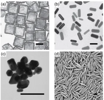
Nanoparticles are colloidal particles with sizes on the nanometer scale. They are ubiquitous in both natural and artificial environments. A lot of familiar examples of nanoparticles are found around us, including viruses and proteins [1], inorganic nanoparticles (e.g., titanium dioxide (TiO
2)) in sunscreens and paints [2], and silica nanoparticles used as solid lubricants [2]. In many cases, the shapes or structures are single crystals with platonic shapes (cubes, octa- hedra, and spheres), and spherical or ellipsoidal. They also have more compli- cated architectures, including the so-called “lamellar twinned particles” (LTPs) or “Multiply-Twinned Particles” (MTPs) containing two, five or twenty single crystal units, and icosahedral, decahedral, and more complex “polyparticles”
with polyicosahedral structures [3]. In addition to different components, sizes, and shapes, specific physicochemical properties of nanoparticles including op- tical, electronic, mechanical and surface properties play an important role in their applications [4].
With their well-controlled properties, engineered nanoparticles can be used
for diagnostic and therapeutic applications, for example, they are candidates
for drug delivery systems, as well as for tumor-cell targeting [4–6]. The first
essential step for nanoparticles function in human bodies is the interactions of
nanoparticles with the membranes of vesicles and cells. Biological membranes
are very fluid films with thicknesses of 4 − 5 nm. For cellular membranes, such
fluid films are composed of amphipathic lipid molecules, various proteins, and
carbohydrates. These components assemble according to the so-called “Fluid-
Mosaic Membrane Model (F-MMM)” [7] for plasma membranes, in which the
proteins are either embedded in or floated on the amphipathic lipid bilayer,
and carbohydrates facilitate the connections of lipids and proteins to cellular
matrix and cytoskeleton. Biological membranes display specific mechanical
properties, such as bending, stretching, and shape elasticity [8]. The important
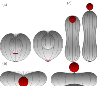
Figure 1.1: Spherical nanoparticles (red) entering and exiting vesicles (gray) via endocytosis and exocytosis. The shapes for the vesicles from left to right are stomatocyte, oblate, stomatocyte, and prolate.
functions for biological membranes are to separate different environment and to control the cargo trafficking across the membranes to maintain the balance of cellular environments. In general, cargoes like drug-loaded nanoparticles can translate through membranes via endocytosis and exocytosis [9, 10], see Fig. 1.1. These processes in biological cells are usually very complicated, and can be affected by a large numbers of factors including active processes.
For large nanoparticles with sizes above 15 nm, a thorough investigation of
nanoparticle translocation by wrapping is required for understanding membrane-
associated nanoparticle transport. A large amount of theoretical [11–13], com-
putational [14–16], and experimental [17, 18] studies have focused on this chal-
lenging topic, but the mechanical mechanisms are still not well understood, as
wrapping and adhesion of nanoparticles at membrane depend on a multitude
parameters, such as components and the structure of the membrane, as well
as on the solutions properties. Here we use the continuum model based on the
Helfrich Hamiltonian [19, 20] to investigate the engulfment of nanoparticles by
membranes, for which a large number of biophysical details can be ignored. We
consider the elastic energies and deformation shapes associated with nanopar-
ticle uptake by membranes, in order to reveal the mechanical mechanisms for
nanoparticle-membrane interactions. We aim at providing some meaningful
predictions for more efficient and less toxic nanoparticle transport via mem-
brane budding.
1.2 Membranes
Biological membranes are extremely fascinating soft matter systems. Different from the rigid surfaces, they are fluid and can form specific structures, such as enclosed vesicles with different shapes, see Fig. 1.1. The shapes result from the biochemical components of the membrane and properties of its environ- ment. For membrane formation, lipid molecules aggregate spontaneously into
“leaflets” where the molecules are jointed together by the weak non-covalent forces. These bilayers are able to extend in the lateral dimensions of up to 10 micrometers, which is considerably larger than the thickness of the mem- brane of only a few nanometers [7, 21, 22]. The big variation in the length scale allows us to regard the biological membrane as two-dimensional surface embed- ded in three-dimensional space. Here, detailed biophysical characteristics are negligible. These flexible membranes are controlled by their properties on the mesoscale; we particularly focus on their elastic properties for the membrane deformations, such as thickness change, shearing, stretching, bending. Other important physical aspects regarding the geometric shapes of the membranes are also introduced briefly.
1.2.1 Composition
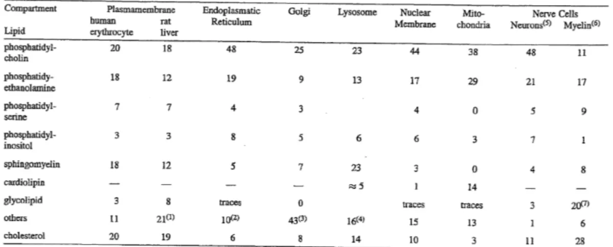
Biological membranes are composed of lipids and proteins [7, 23]. The lipids are amphiphilic molecules that consist of a polar hydrophilic head group and a non-polar hydrophobic tail. In cell membranes, different lipids are found, such as phospholipids, glycolipids, and other sterols. Depending on the types of cells and organelles, the fractions of each lipid can be varied. The most abun- dant lipid constituents are phospholipids in most biological membranes. The phospholipids in a cell include phosphatidic acid (phosphatidate) (PA), phos- phatidylcholine (lecithin) (PC), phosphatidylethanolamine (cephalin) (PE), phosphatidylserine (PS), phosphatidylinositol (PI), sphingomyelin (SM), car- diolipin (CL) [23, 24], see Tab. 1.1.
The phospholipids are different from each other relying upon the hydrocar- bon tail length, i.e., the number of carbon atoms in tails, and the saturation, i.e., the presence of carbon-carbon double bonds [25]. PC is a lipid with a cylindrical shape, for which the volumes for the head and the tail groups are approximately the same. For PE, the tail part area is larger compared to the head part, therefore the lipid displays a conical shape. PA has an inverse conical shape as the lipids have large tail and small head moieties.
Furthermore, phospholipids with different head groups are able to influence
the chemical properties of the leaflet surface, and to execute various functions
in the cells [23–25]. For instance, PC is a key component for membrane-
mediated cell signaling and molecule transport; PE plays an important role
in membrane fusion, as well as in regulating the membrane curvature. PA
Table 1.1: A summary of lipid compositions of various organelles, plasma membranes, and cells. Reprinted from Ref. [23].
is important for both membrane signaling and regulating curvature. Similar to the heads, hydrocarbon tails with different structures can also change the membrane properties. One example is unsaturated lipids that increase the fluidity of lipid bilayers [26].
In addition to lipid molecules, biological membranes contain a variety of proteins. Around 20 − 30% of the biological genes encode such membrane pro- teins [27, 28]. The considerable advancements in the experimental methodol- ogy for structural biology, such as X-ray diffraction, solution nuclear magnetic resonance (NMR), and, more recently, single-particle cryo-electron microscopy (cryo-EM), or electron cryomicroscopy, lead to high-resolution structures of the membrane proteins [29, 30]. There are over 2, 500 protein structures stored in the protein data base (PDB), and they are divided into over 700 different protein species [30].
One important species of the membrane proteins are those that gener- ate and regulate membrane curvature, and they are therefore involved in the changes of membrane shapes and even in topological changes like creation of the pores [31, 32]. These proteins can be classified into two different groups;
one group can change the lipid composition of the two leaflets to induce
the asymmetry of the membrane, such as the phospholipid flippase and the
lipid modifying enzymes [31, 33]. The other group generates and sustains the
membrane curvature by the mechanical interactions between the proteins and
membrane monolayer [31]. Relying on the diverse qualitative features, the
curvature-associated mechanisms [31, 32, 34], which are not mutually exclu-
sive, include:
(a) protein scaffolding [35]: the protein domain, the protein monomer or oligomer, and even multiple proteins with an intrinsic curvature aggregate on the membrane and bend the underlying membrane surfaces facing them;
(b) hydrophobic insertion [36]: the protein stabilizes the membrane cur- vature by virtue of the insertion of the hydrophobic residues into the local domains in amphipathic lipid molecules. The insertion process of the proteins are also known as “wedging”;
(c) oligomerization [35, 36]: specific proteins, instead of generating and regulating the membrane curvature individually, can oligomerize to form the polymerized coat proteins linked to the outer monolayer. The cooperativity of the oligomerized proteins can amplify curvature sorting on membranes with different curvatures.
(d) steric effects [37]: the steric protein-protein interactions are found for proteins which have no intrinsic curvature effects, such as scaffolding effects, membrane-inserting domains, and ligomerization properties. They are crowed in the local membrane and induce the curvature by mutual steric interactions.
A large variety of membrane curvature-associated proteins are found exper- imentally. The most-widely studied proteins are Bin-Amphiphysin-RVs (BAR) domain-containing proteins [35–37]. BAR domains have banana shapes and bind to the membrane through the concave surface of the lipid-binding regions.
They are found to induce the cylindrical curvature. The BAR protein family is further subclassified into classical BAR, N-BAR, F-BAR, and other BAR proteins relying upon the detailed structures. N-BAR proteins contribute to the membrane-curvature via the scaffolding mechanism, while F-BAR pro- teins are characterized by the hydrophobic insertion region and can induce membrane tubulation upon binding. Classical BAR proteins, such as arfaptin, can also form the highly-curved tubular membrane without hydrophobic inser- tion. Furthermore, both N-BAR and F-BAR domain proteins can oligomerize on curved membranes and result in the striated, attice-like protein coats on the bilayers. For crowding eripheral proteins, they can sence and regulate membrane bending via the nonspecific steric interactions. Other membrane- curvature associated proteins include coat protein complex I (COPI) and II (COPII), epsin N-terminal homology (ENTH) and AP180 N-terminal homol- ogy (ANTH) domain-containing proteins, small G proteins such as Arf1, and C2 domain proteins [35–37].
In addition to the mechanisms mentioned above, the interactions between
membrane and specific lipids, the “packing defects” in curved lipid bilayers [38],
the demixing of lipid molecules with various spontaneous membrane curvatures
[39], and cytoskeletal polymerization and motor proteins pulled tubules are
suggested to explain the formation and stability of the membrane curvatures
[36]. Despite the various studies and theories on membrane curvature, the
relative significance of the potential contributions from different mechanisms is still hotly debated.
A third important component is the carbohydrates. Membrane carbohy- drates contribute 5 − 10% to the membrane mass. Most of them are located on the the extracellular side of the cell membrane and bind to the lipids and proteins via glycosylation [40]. Glycosylation is a common chemical reaction in cell in which a carbohydrate is covalently linked to the functional group of the membrane molecules. After such a chemical modification, the resulting com- ponents are called glycoconjugate, including oligosaccharides, proteoglycans, glycolipids and glycoproteins [41]. The neutral sugars in cells are glucose, galactose, mannose, fucose, as well as N-acetyl galactosamine, and they are abundant in the glycoconjugates [41, 42].
1.2.2 Structure
After the comprehensive understanding in the membrane components, a nat- ural question is what a whole structure these different component can arrange into. The structure must be capable to explain the existing research data, such as fluidity and dynamics of the membrane, and to predict the outcomes for future experiments. In 1972, S. J. Singer and G. L. Nicolson first introduced the Fluid-Mosaic Membrane Model (F-MMM) of biological membrane struc- ture [7]. The basic micro-structure of the F-MMM was based on the concept of hydrophobic interaction and its effect on the thermodynamics of the protein structures. Hydrophobic interaction is the tendency of hydrophobic moieties of molecules to self-aggregate to exclude water molecules, and the tendency of hydrophilic moiety to associate with the aqueous solution. The lipid molecules, the most important constituent of the biological membrane, are amphiphilic substances. They assemble to lipid bilayers driven by the hydrophobic effect as well as van der Waals forces in aqueous environment. For membrane proteins, their attachment or insertion into lipid bilayers also result from the hydropho- bic effect between the hydrophobic tail groups of lipids and the hydrophilic acid residues of proteins. Meanwhile, the hydrophilic groups of the proteins protrude into the aqueous environment around the lipid bilayers
1.
Based on the consideration on the hydrophobic interactions between lipids and proteins, the F-MMM model states that the cell membrane is a completely fluid two-dimensional lipid bilayer that contains globular integral proteins and specific integral protein complexes which are indiscriminately scattered in the membrane. In 1976, the original F-MMM was improved by reflecting the con- straints from extracellular and intracellular substances on the lateral spreading and moving of membrane constituents, in particular integral proteins, glyco-
1
This claim for the protein-lipid arrangement holds true only for some membrane proteins,
including integral or intrinsic proteins.
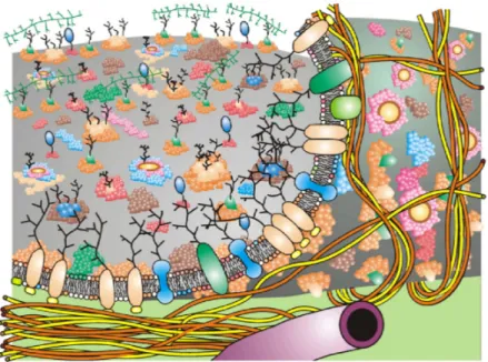
Figure 1.2: Updated Fluid-Mosaic Membrane Model contains lipid bilayers, proteins, gloy- coproteins, cytoskeletal and extracellular matrix. Different molecules are labeled by different colors. Reprinted from Ref. [7].
proteins, and protein complexes [43]. Some restricted and semi-restricted re- gions are from the specific interactions of lipid-lipid, and lipid-protein on the membrane, which can advance the development of protein complex and lipid rafts. The reduced mobility of the integral transmembrane glycoproteins and protein complexes are related to link of the cell membrane with the cytoskele- ton filaments as well as the extracellular matrix.
Many other important descriptions for the thermodynamics associated with
the membrane structure were introduced since the F-MMM has been proposed
in 1970s. One special characteristic are the deformation of the membrane. For
instance, the elastic modulus depends on the weak energies of lipid tilting and
splaying [21, 22]. How the deformation energy result in different membrane
structures on large length scales will be discussed further in the subsequent
Chapter “Methods”. In addition, the plasma membrane is, often, asymmetric
with thousands of different lipid molecules or proteins heterogeneously dis-
tributed in the two monolayers. For example, PC is abundant in the extracel-
lular monolayer, whereas PE and PS are exclusively localized in the intracel-
lular monolayer [7, 23]. The generation and maintenance of the asymmetry of
the lipid bilayers can be mediated with the passive transbilayer motion (flip-
flop) and active energy-consuming bilayer scrambling [44]. The transbilayer
motion, or flip-flop, occurs when single lipid molecules spontaneously flip be-
tween the monolayers of the bilayer. Bilayer scrambling refers to the behavior
of a bundle of lipid molecules, and translocations are less frequent as large free
energy change is required. As a result, a perfectly symmetric structure of the cell membrane is rarely found. A third critical consideration for refining the F-MMM is taking into account “hydrophobic mismatch” [45]. The thickness of the intermediate hydrophobic regions of the transmembrane proteins can be (approximately) equal, longer, and shorter than the biological membranes’
thickness. In order to prevent the unfavorable contact of the hydrophobic re- gions of lipids and proteins with the hydrophilic regions of lipids, proteins, or the aqueous environment, the biological membranes can make a few adapta- tions to the hydrophobic mismatch. The mattress model proposed by Mourit- sen and Bloom in 1984 [46] was used to describe the phase diagrams of the mixing of amphiphilic lipids and proteins in aqueous environment. The elastic properties, and the indirect or direct interactions, such as van der Waals-like interactions, for the cell membrane in included in the model. The mismatch effect can influence the protein aggregation, local orientational changes, and conformational adjustments of proteins and lipids in the membrane.
Biological membranes, such as the cell membrane, are made up of a large number of different lipids and proteins (see the updated F-MMM presented in the Fig. 1.2). Model lipid bilayer membranes consisting of lipid molecules only are also found. Common examples are lipid bilayers, vesicles, and micelles [47, 48]. Lipid bilayers is the thin, flat, and fluid two-monolayer membrane, and is the basic structure of cellular membranes. Vesicles are structures with an encapsulating ‘bag’ shape, and are in biological systems naturally produced for material-transport associated processes, e. g. exocytosis and endocytosis.
A variety of vesicles are present within a cell, including exosomes, microvesi- cles, apoptotic blebs or vesicles, phagosomes, and COPI and COPII coated vesicles. If vesicles are created artificially via lipid molecules, they are known as liposomes. Liposomes that are enclosed by many phospholipid bilayer, are multilamellar liposomes; with one layer only they are called unilamellar. Mi- celles, or micellae, are aggregates of phospholipids in aqueous solutions. They exist in equilibrium with spherical shapes. For non-polar solvents, the hy- drophobic tail groups of lipids are pointing to the external solvent, while the hydrophilic head groups are gathered in the core. In this case, the structures are called inverse or reverse micelles. Some typical lipid structures are shown in Fig. 1.3.
1.2.3 Elastic properties
Biological and physical properties are important for membrane-mediated in- teractions, deformations, and shape fluctuations [8, 21, 22], they include:
(a) thickness change: biological membranes contain many embedded pro-
teins and glycoproteins, which can be related to the compressibility of the
membrane. Furthermore, the heterogeneous compositions of membranes in-
Figure 1.3: Representative structures for lipid bilayer and vesicles (liposome and micelle).
Taken from public domain: Wikipedia.
fluence their peristaltic motions, or thickness changes, of the lipid bilayers.
However, the fluctuations of the thickness are limited because of the small compressibility of lipid membrane. The amplitude of the thickness change for the lipid membrane is determined by the experiment, the value is as small as 4 A [49]. The thickness change is essential for many biological events, such as the shape change for the erythrocytes with high flexibility.
(b) stretching: stretching the lipid bilayer can change the area per lipid, which may lead to the exposure of the hydrophobic regions like the lipid chains to the aqueous environment. The contacts for hydrophobic groups and water molecules are energy unfavorable, thus stretching is sub-dominant to bending for membrane deformations.
(c) shearing: the lipid bilayer is a fluid thin film, for which any types of shear forces are ruled out in the lipid membrane. The deformation of the shearing is likely to occur if the lipid membrane is linked to an external scaffold structure, such as the lattice structure and the cytoskeleton.
(d) bending: for the shape fluctuations of the lipid membrane, bending is the most important contribution to the deformation of the membrane. The bending energy is associated with the bending rigidity and membrane curva- tures. For some particular systems, other factors such as surface tension and geometric contributions (van der Waals or Coulomb) are also significant for the membrane deformation.
(e) compression: instead to describe the biological membrane as a perfect
single sheet structure (for instance the midplane), we can add the microscopic details regarding the membrane compression to the membrane structure. For the lipid membranes which are bent, the physical properties of the two leaflet are different. For the upper leaflet, the stretching dominants the membrane changes; for the lower leaflet, the compression of the surface occurs. For incom- pressible membranes, the equilibrium shapes have minimal bending energy; for compressible membranes, the combined bending and stretching energy deter- mine the stable states for the membrane.
(f) tilting: the lipid molecules are not necessarily perpendicular to the neutral plane
2of the membrane. Experiments show that the angle of tilt of the lipid molecule on the dipalmitoylphosphatidyl-choline (DPPC) bilayer can be as large as 30
◦(degree) [50] with respect to the normal vector of the neutral plane. Tilting of the lipid molecules is also found for the membranes in the fluid lamellar phase via X-ray scattering. The lipid tilting is associated with a variety of biological processes, including the membrane fusion and inverted amphiphilic mesophases. For low temperatures, the lipid tilting is not relied upon the membrane curvature. Thus it can be coupled into the description of membrane shape as an independent degree of freedom with additional physics implications.
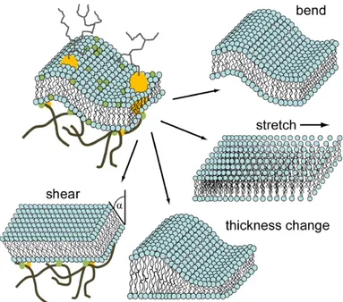
The important structures of the biological membranes include lipid mono- layer and bilayer with different biophysical properties. Both of them can be mathematically represented as a thin two dimensional flat sheet embedding in the space for large length scale, refer to the following Chapter “Methods” for further discussions. For lipid bilayers, if the lipid molecules cannot exchange from different leaflets. The lipid area difference for two monolayers is constant, as a result, the associated large-scale bending dominant Hamiltonian is same for both monolayer and bilayer structures. Figure 1.4 shows the four major biophysical properties contributing to membrane deformation. The structures and fluctuations of the lipid membranes are common in a cell, and play a key role in various membrane functions.
1.2.4 Function
Biological membranes, e.g., cell membrane or plasma membrane, surrounds the local spaces or compartments in which different chemical or biochemical substances are contained. In the cytoplasm of a cell, membranes define spe- cialized subunits that are individually enclosed by the lipid bilayers, so-called
“membrane-bound” organelles. The cellular transport of molecules and parti- cles among different organelles and plasma membrane is essential for diverse functions in a cell. Two fundamental biological processes for cellular trafficking
2
A neutral plane is the unstrained surface in the middle of the bent lipid bilayers with a
constant area.
Figure 1.4: Elastic properties contribute to membrane deformations: thickness change, shearing, stretching and bending. Reprinted from Ref. [8].
are endocytosis and exocytosis [9, 10, 51]. In the following sections, we will introduce the main pathways for endo- and exocytosis, and their underlying roles for cell functions.
Endocytosis is a process through which molecules, proteins, and particles
are engulfed by the cell membrane [9, 10]. Usually, it is energy-consuming for
a cell to absorb and internalize biological substances. Three typical pathways
are found for endocytosis: classical endocytosis [52], kiss-and-run endocyto-
sis [53], and bulk endocytosis [54]. Classical endocytosis includes clathrin-
and caveolae-dependent and -independent pathways. Clathrin-dependent en-
docytosis is a receptor-mediated pathway, which is the predominant process in
most cells. Caveolae-dependent endocytosis is a common way of cells to up-
take small biomolecules, and the invaginations are flask shapes. The clathrin-
and caveolae-independent endocytosis have endocytic-like membrane interme-
diates, such as irregular depth inclusions, and coated or uncoated pits. Ex-
amples for this endocytosis are lipid raft and ARF6 (a small molecular weight
GTPase) associated pathways. In kiss-and-run endocytosis, a vesicle (e.g. a
synaptic vesicle) from extracellular environment contacts the plasma mem-
brane in a short time (kiss), and releases the contained biomolecules by the
small transiently-open fusion pores. The vesicle is not collapsed on the mem-
brane and can be retrieved from the plasma membrane after the fusion pore is
closed (run). For bulk endocytosis, large endosome-like structures or cisternae
are found in a cell, which are deep invaginations internalized from a large area
of plasma membrane. Multiple vesicles can bud off from the membranes of
the large structures. Endocytosis plays a key role in mediating cell functions, such as membrane curvature generation, vesicle formation and maintenance, as well as cell movement and division.
Exocytosis, the counterpart of endocytosis, is an active energy-cost process in which biological material is transported out the cell when a vesicle fuses with the plasma membrane [9, 10]. Exocytosis includes three important pathways:
full-collapse fusion [55], kiss-and-run exocytosis [53], and compound exocytosis [56]. Kiss-and-run exocytosis is similar to kiss-and-run endocytosis, the main differences is in exocytosis, a domestic vesicle expels the biomolecules out of the plasma membrane. Full-collapse fusion, different from the kiss-and-run endocytosis where a vesicle is in contact, a vesicle completely fuses with the plasma membrane, the whole vesicle disappears as the large fusion pore leads to flattening into the plasma membrane. Compound exocytosis refers to the fusion of small (20 − 50 nm) or large (300 − 2, 000 nm) [57] vesicles which come from the fusion of multiple vesicles, or vesicles with preformed giant vesicles with fused but not collapsed membranes. Exocytosis is important for synaptic strength and plasticity, vesicle replenishment, and multivesicle release.
For cellular trafficking in both endocytosis and exocytosis, the membrane or protein mediated budding from a donor membrane to an acceptor membrane is generally the first important step [58]. For protein-driven membrane budding, one most significant process is the creation of the coated vesicle-like structure.
Clathrin, an important protein in membrane budding, can generate different types of basket-shape or lattice-shape coated vesicles with radii of 30 − 50 nm [59]. On plasma membranes, clathrin binds to the flat platform-shape adaptor proteins and facilitates the budding of protein-attached membrane in clathrin- dependent endocytosis. The coat protein complex I (COPI) and II (COPII) [60, 61] are responsible for the membrane budding for the coated vesicle car- rier from the endoplasmic reticulum (ER) to the Golgi apparatus. Similar to clathrin, vesicle-like basket or lattice invaginations are formed, which are not strongly dependent on the lipid composition.
While protein-dominant membrane budding is characterized by the forma- tion of the coated vesicles, membrane-mediated budding is related to the phase separation in heterogeneous lipid bilayers synthesized in micron length scale.
Depending on lipid composition and temperature, the lipid mixtures can exist
in either the liquid or the solid (“gel”) phase. At given temperature, both
liquid and solid phases can be coexistent for spatially separated different lipid
clusters [62]. Furthermore, the unsaturated lipids are abandon in liquid disor-
dered (L
d) phase, while the liquid ordered (L
o) phase contains more saturated
lipids [63]. As a result, the thickness of the lipid membrane in L
ophase is
larger than that in L
dphase. The energetically unfavorable hydrophobic con-
tacts between the amphipathic lipid molecules for the two liquid states lead to
line tension, which is the energy cost per length. In order to minimize the line
tension energy, the membrane domains tend to come together to from some circular zones, which are known as “microdomains”. Most microdomains in membrane budding are rich in sterol- and sphingolipid molecules, or the so- called “lipid rafts” [58]. When the sizes of the microdomains are large enough for which the line tension energies exceed the Helfrich deformation energies (see the further introduction in Chaper “Methods”), the membrane will bud out and form distorted vesicles to reduce the contact sizes, and the vesicles can even detach from the membrane as the line tension energies are extremely high [64]. For some viruses toxins, such as shiga toxin B [65], can give rise to the arrangements of L
omicrodomians, which leads to the formation of negative- curvature favorable tubular vesicles.
1.3 Nanoparticles
Depending on the specific applications of nanoparticles, the definition and viewpoint of them can vary. The meanings of nanoparticles and nanomateri- als are different for different organizations [66]. The common characteristic of nanoparticles is that they are materials (natural or artificial) in nanoscale (e.g., 1 nm = 10
−9m). They have different components and shapes, see Figs. 1.5 and 1.6. Such small particles can enter the cells via endocytosis. After exert- ing their functions, they can be expelled out by virtue of exocytosis. A large effort has been devoted to the studies of the interactions between nanoparti- cles and membranes, like the budding mechanism [67, 68], to understand how nanoparticles are wrapped by the membranes. However, owing to the complex- ity of biological membranes in structures, components and elastic properties, the elucidations of specific mechanisms for nanoparticle wrapping are still big challenges. For the following sections, we will introduce some typical types of nanoparticles, as well as their important roles in different potential applica- tions.
1.3.1 Types
Nanoparticles are made from a variety of materials including, but no limited to, lipids (e.g. phospholipids and chelesterol), polymers and inorganic materials (e.g. metals, composites, and ceramics), see Figs. 1.5 and 1.6. Nanoparti- cles gained from these three types of materials is enormous; just list some of them: liposomes, dendrimers, micelles, polymeric drug conjugates, carbon based structures, and metallic nanoparticles [5, 6, 66]. In the following parts, I will introduce one or two important nanoparticles from these three types, as well as their potential applications.
(a) liposomes: the phospholipid molecules of liposomes can self-assemble
to closed vesicles in aqueous environments, which can be used to encapsu-
late biotech drug molecules. Liposomes are a leading and most intelligent drug delivery system for both active and passive membrane transport [69, 70].
Many liposome-based drug carriers used for clinical applications are already in market, such as ambisome, daunoXome, inflexal V. Liposomes have enor- mous advantages rendering them as suitable drug delivery systems. First, liposomes are made of lipid molecules with good biocompatibility, which are easily available for formulating and can display specific biological, chemical, and mechanical aspects. Second, the amphiphilic character of liposomes allows to capture both hydrophilic and hydrophobic agents in their structures. Last, the modifications of the physicochemical properties (size, charge, and surface ligands) of liposomes lead to more functionally favorable carriers. For instance, the stealth liposomes with attached polyethylene glycol-units (PFG) are able to survive longer in the body.
(b) micelles: in micelles, water-insoluble drugs can be linked covalently to their hydrophobic inner cores shielded by amphiphilic groups of the component molecules [4, 70]. Such spheroidal shapes of micelles can also be “core-shell”
structures with radii 5 − 50 nm [5], see Fig. 1.3. A hydrophilic shell can protect the micelles from phagocytosis by macrophages or uptake by reticuloendothe- lial systems (RES), as the water-bound regions form “splayed” structures such as “polymer brushes”. As a result, micelle drug delivery systems can have a prolonged lifetime. Another good aspect of micelle drug-delivery systems is their high efficiency in accumulation at target drug sites [6, 66]. One common example is polymeric micelles, see Fig. 1.5.
Figure 1.5: Representative structures for polymeric nanoparticles for (a) oblate, disk shape,
(b) bullet shape, and (c) pill shape, and (d) dumbbell shape. Reprinted from Ref. [71].
(c) silicon: silicon nanoparticles are engineered by the methods that are used in semiconductors and micro-electromechanical systems (MEMS) [5].
Typical techniques are photolithography, etching, and deposition [72]. Pop- ular silicon-based materials are porous silicon and silica, and silicon dioxide.
The structures include calcified nanopores, porous nanoparticles, and nanonee- dles. By controlling the nanopore diameter and density, the substances (e.g., biomolecules and drugs) stored in pores can pass through the pores with a specific rate. One of the important silicon-based materials is porous hollow silica nanoparticles, which can deliver cargoes in cells.
(d) metal: metallic nanoparticles are fabricated from different metals, such as gold (Au), silver (Ag), platinum (Pt), and palladium (Pd) [4, 5], see Fig. 1.6.
The metallic nanoparticles have been used for a long time. For example, in 4th century AD, Lycurgus cups were fabricated with many small-sized ( ∼ 20 nm) nanoparticles [66], which reflect special optical characteristics. Nowadays, one important application of metallic nanoparticles is in biomedicine and biomem- brane. Usually, metal covers a core material (e.g. a silica particle) and form a very thin shell around the core. On these hollow metallic covers, a large number of ligands, for instance, nucleic acids, lipid molecules, small proteins, as well as sugars, can be easily anchored to the nanoparticles. The connections between metallic nanoparticles and biological materials can be implicated by a variety of conjugation means, such as bifunctional linkages, lipophilic interac- tion, silanization (self-assembly with organofunctional alkoxysilane molecules), electrostatic attraction, and nanobead interactions.
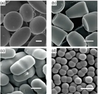
Figure 1.6: Representative structures for (a) cube-like and (b) rod-like gold nanoparticles,
(c) irregular silver nanoparticles, and (d) hematite nanospindles. Reprinted from Ref. [71].
1.3.2 Mechanical properties
Nanoparticles have many distinct mechanical properties, which are associated with interfacial forces, including hardness, elastic modulus, stress and strain, adhesion and friction, and movement [4, 73]. An better understanding of these properties of nanoparticles provides significant aids in nanotechnology, and the evaluations for the functions of nanoparticles.
(a) hardness and elastic modulus: the hardness and elastic properties of nanoparticles are tested by microindentation and nanoindentation techniques.
In particular, atomic force microscopy (AFM) is helpful to characterize the deformability of nanoparticles. For nanoparticles with sizes smaller or ap- proximate to hundreds of nanometers, the hardness and elastic modulus can vary, depending on the components and shapes of nanoparticles. For instance, for silicon nanoparticles, the elastic modulus increase with decreasing sizes of nanoparticles. For metallic nanoparticles, the dislocations inside the nanopar- ticles are found during deformations.
(b) adhesion and friction of nanoparticles: in addition to vertical deflection of AFM cantilever, the torsional deflection of cantilever and colloidal-probe techniques are used for investigating the adhesion and friction of nanoparticles on the substance. Research shows that the adhesion force is linearly depen- dent on the radii of nanoparticles, and the hydrophobic interfaces strongly decrease the adhesion for particles and substances. Meanwhile, the friction forces between nanoparticles and substances increase with particle radius, and is proportional to the contact areas.
(c) movement: the movement of nanoparticles due to different forces such as Brownian motion force, medium and environment is difficult to quantify.
Recently, two main methods are used to study the movements of nanoparti- cles, one is particle tracking with the fluorescence technique, the other one is dynamic light scattering (DLS) technique. Study shows that during move- ment, the rotating speeds of nanoparticles is much slower than velocities.
Nanoparticles with sizes comparable to the length of liquid molecules tend to self-assemble in the medium.
1.3.3 Applications and toxicological risks
Relying upon the components, sizes, shapes, and mechanical properties of nanoparticles, they can be used in various fields, such as nanomedicine, food sciences, chemical and cosmetics. Here we focus on the applications of nanopar- ticles in the fields of biology, biochemistry, and medicine. We also discuss potential negative side effects related to their applications, such as toxicities.
The most important function for nanoparticles in biological cell is they can
serve as delivery systems. As mentioned above, both liposomes [69, 70] and
polymeric micelles [5] are suitable drug delivery systems as they have specific
lipid components, vesicular structures, and biochemical characteristics, which guarantees unique advantages in drugs delivery. Among these drug-delivery systems, the nanoparticle-based structures carrying drugs for brain or the cen- tral nervous system (CNS) need more attention [5]. The difficulty for brain drug delivery is due to the blood-brain barrier (BBB), an extremely compli- cated structure composed of impermeable endothelial cells with tight junctions, enzyme catalysis, and efficient efflux-transport. As a result, only a few small molecules (ions and peptides) and a very limited number of macromolecules are able to pass the barrier. It has been shown that poly(butylcyanoacrylate) based nanoparticles can transport several agents, such as dalargin, hexapep- tide, doxorubicin, into the brain. These drug-carrying agents are transported across BBB by through of protein-mediated endocytosis [74, 75]. In addi- tion to drugs, nanoparticle delivery systems for peptides, proteins, and genes [76, 77] have been reported. For instance, polymer-based nanoparticles, such as polymeric micelles, encapsulate insulin in the hydrophobic core and pre- serve the activity of insulin from hydrolytic and enzymatic degradation [78].
Poly(DL-lactide-co-glycolide) (PLGA) nanoparticles are used to transport the therapeutic genes for bone healing [79].
Another significant function for nanoparticles is efficient targeting [80, 81].
The special optical properties of nanoparticles, e.g. metallic nanoparticles, are used as markers for distinct biological tissues and cells, as well as for photo- targeting thermal therapy [82]. Nanoparticles can also be used for tumor targeting, either by passive targeting or active targeting [5, 80]. In passive targeting, an enhanced vascular permeability and retention (EPR) effect are found in tumors, which result from aggregation of therapeutic agents in tu- mor cells. The accurate targeting is possible due to the high selectivity for agents and ligands that can be linked to nanoparticles. Nanoparticles with sizes of 50 − 500 nm, such as liposomes, micelles, and dendrimers, can deliver drugs to tumor tissue in this way. In active targeting, the ligands (peptides, growth factors, transferrin, antibodies and small compounds) of drug-loaded nanoparticles are directly recognized by the receptors expressed on tumor cells, which initiates the active ligand-receptor binding mediated endocytosis. For example, folate, a small compound, is easily conjugated to the nanoparticles and selectively bind to the carcinomas of the ovary, breast, lung, kidney and brain. The accuracy and efficiency of nanoparticle targeting depends also on their physiochemical properties, such as size, shape and surface charge.
In addition, nanoparticles have potential applications in stem cell therapy,
and stimulate immune response. However, besides the rapid developments
and wide applications of nanoparticles, toxicity and side effects of nanoparti-
cles are also found [4, 5]. It has been reported that they affect lungs, skin,
intestinal tract, and other organs. Adverse health risks of nanoparticles are as-
sociated with the binding and wrapping by both targeted and normal cells. For
the nanoparticles in lungs, the poisonous biological outcomes depend strongly
on the size of the nanoparticles. Ultrafine nanoparticles, contrary to larger
nanoparticles, are able to induce more severe adverse effects, including the
development of inflammation and even tumors [83].
Methods
2.1 Membrane models for different length and time scales
Biological membranes are very thin fluid layers which are composed of hun- dreds of amphiphilic phospholipids and/or embedded proteins. The typical thickness of a lipid-bilayer membrane is d ≈ 5 nm. This is by far smaller compared to the lateral dimension of the membrane, which can be as large as 10 µm. Also, the biophysical processes associated with lipid membranes vary in time scale. For instance, small molecules (ions and water molecules) diffuse across the membrane as rapid as a few nanoseconds [84], and membrane fusion of giant lipid vesicles is shown to occur on a longer time-scale as microsecond [85]. Thus a variety of membrane models covering a wide range of dimensions, from nanometer to micronmeter, are required.
Depending on the question concerned, one can choose the most appropri- ate computing models to describe the biological phenomena on different length and time scales. Generally speaking, the details of the membrane structure and molecular interactions are usually investigated on nanometer scale, while the morphologies of biological vesicles and membrane self-assemblies are mi- crometer scales. Membrane models corresponding to different scales from mi- croscopic to be macroscopic are classified to quantum models, all-atom, coarse- grained models, supra-coarse-grained models, and continuum models such as triangulated membrane and mesh-less models [86–90], as shown in Fig. 2.1.
Quantum, atomistic and coarse-grained models, and the continuum models, in particular the triangulated membrane model, will be introduced in the follow- ing.
Quantum models
On the quantum mechanics level, electronic structure and atomic nuclei are
explicitly included in the theoretical models. It is feasible to obtain information
(f )
Figure 2.1: Different length-scale representations for (a) all atom molecular dynamics sim- ulation, (b) coarse-grained (CG) model in explicit solvent, (c) coarse-grained model (CG) in implicit solvent, (d) meshless model, (e) triangulation of membrane. Reprinted from Ref. [86], and (f) different length- and time-scale representations from small to large for quantum mechanics, all-atom, coarse-grained, supra-coarse-grained simulations, and a cell.
Reprinted from Ref. [87].
about the interaction between electrons, charge transfer, chemical bonds, as well as the band structure of materials with chemical accuracy [89, 91, 92].
The most accurate technique to calculate the electronic structure is wave
function-based calculations. However, the computational costs for dealing with
wave functions are huge for large molecules [91, 92]. Instead, for large-scale
biological systems, the quantum chemical properties can be obtained using
density functional theory (DFT) with high accuracy and lower computational
costs [89, 91, 92]. The chemical information for small molecules, obtained using quantum mechanical calculations, can help for atomistic molecular dynamic simulations [87, 89, 91, 92].
All-atom models
A great number of biophysical processes can be understood at an atomistic level, such as receptor-ligand binding [93, 94], protein-membrane interactions [95, 96], protein-nucleic acid interactions [97, 98], as well as protein folding [99]. Atomistic molecular dynamics simulations can be divided into two differ- ent categories, depending on whether each atom is independently included in the models [100, 101]. One category is all-atom simulations, where all atoms are simulated in explicit-solvent systems. The other category is united-atom simulations, where a few atoms are integrated into one united-atom, also called pseudo-atom, which can approximately represent the chemical properties of a group of atoms and which is only slightly larger than a single atom.
In molecular dynamic simulations, the dependence of the free energy of the biological system on the coordinates of the molecules is often described by potentials, usually referred to as force field [90, 92, 102]. The mathematical parameters for the (inter-)atomic potential energy can be obtained from ab initio or quasi-quantum mechanics calculations, or from experimental meth- ods like X-ray diffraction, nuclear magnetic resonance (NMR), and neutron spectroscopy. A typical empirical force field has two different energy contri- butions, one component is the intramolecular or local energy including bond stretching, dihedral angles, and angle bending; the other one is intermolecular interactions, which can be hydrophobic repulsion and van der Waals interac- tions.
Coarse-grained models
For coarse-grained molecular dynamics simulations, at least three heavy atoms are integrated into one pseudo-atom [88]. A biological molecule in a coarse-grained model can be represented by a hybrid of individual atoms and pseudo-atoms, or by pseudo-atoms alone. As a result, a large number of atoms
“have vanished”, and the number of degrees of freedom for calculating in- tramolecular, intermolecular interaction potentials is reduced. Because of the high speed of the hydrogen bond vibrations, the simulated time step for all- atom molecular dynamics simulation is limited to 1 − 2 fs. For coarse-grained molecular dynamics simulations, the time step is about 10 − 1000 times longer than than all-atom molecular dynamics simulations [103–106].
Generally speaking, there are two main philosophies to design coarse-grained models for different molecular systems
1: bottom-up and top-down approaches
1
The other approaches are ‘knowledge-based’ model, which rely on experimentally deter-
mined structures.
[107–109]. For bottom-up approaches, the molecular descriptions for the coarse- grained models are obtained from a more detailed model, for instance, a clas- sical, empirical atomistic model. For top-down approaches,the molecular de- scriptions are based on experimental data, usually thermodynamic or macro- scopic properties.
Coarse-grained molecular dynamics simulations have been used to investi- gate the biophysical processes on a larger scales, such as the protein aggregation mediated by lipid molecules [110, 111], membrane fusion and fission [112, 113], and protein-protein interactions [114]. In addition, the information obtained using coarse-grained models can help to bridge the gap between micro- and macroscopic descriptions.
Supra coarse-grained models
In ‘supra’ coarse-grained models, also referred to as ‘highly’, ‘ultra’, and
‘shaped-based’ coarse-grained models, much more atoms, molecules, and even some parts of proteins and membranes are integrated into a single pseudo- molecule [115, 116]. Therefore, they are even more coarse models compared to coarse-grained models. Supra coarse-grained systems can be combined with particle-based hydrodynamic methods, such as multiparticle collision (MPC), dissipative particle dynamics (DPD), and Brownian dynamics (BD) [86, 87, 117, 118]. Therefore, supra coarse-grained models can treat large of biological molecules in implicit-solvent simulations with increased speed, but decreased accuracy. Examples are large-scale membrane-mediated Bin-Amphiphysin- Rvs proteins (BAR) interactions [119, 120], and aggregated amyloid fibrils in Alzheimer’s disease [121, 122].
Triangulated-membrane models
A mesh model made up of several polygon meshes (vertices, edges, and faces) and is typically used to describe the geometric surface and membrane modeling [123–125], a common example is the dynamics triangulated mem- brane model, which is based on the continuum model for which detailed de- scriptions can be found in the following Sec. 2.2.
Meshless membrane models
The meshless membrane model is also dependent on the continuum model.
For such model, the potential interactions, instead of the meshes, control the
assembly of the particles for the fluid membranes. There are three main types
of potentials, including soft-core repulsion, anisotropic attractions, and hy-
drophobic muti-body interactions [126]. Most of the meshless models are sim-
ulated in solvent-free environment, and are used to study the bending defor-
mation of membranes [86, 127], as well as the membrane dynamics in flows
[128, 129].
2.2 Continuum model
From the previous introduction, we know that lipid-bilayer can laterally extend up to the micrometer length scale, which is significantly larger than the bi- layer thickness. Therefore, lipid-bilayer membranes can be described as a two- dimensional fluid surface embedded in the three-dimensional space. We first look back to important concepts and definitions of differential geometry that are used to describe the curves and surfaces for the membranes parametrized in Monge gauge, and extend the geometric description to the implicit model.
Subsequently, we will introduce and discuss the Helfrich Hamiltonian in some depth, followed by the Gauss-Bonnet theorem that is usually exploited to deal with the integral over the Gaussian curvatures. Finally, we will discuss the methods used for energy minimization.
2.2.1 Monge gauge
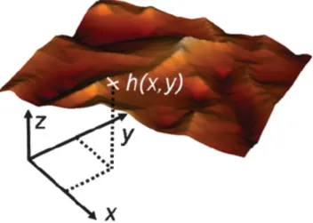
We start to consider the membrane as an one-sheet model where the lipid bilayers is regarded as a mathematical surface [21, 22]. In Monge gauge, the surface can be described using a height function h(x, y) in a three-dimensional Cartesian coordinate system. A simple case is the spherical surface shape, it can be parametrised in Monge gauge as
z = ± h(x, y) = ± p
R
2− x
2− y
2, (2.1) where the radius of the sphere is R. The plane { x, y } is the reference surface, and z = h(x, y) is the height function. The sign ± refers to the upper (positive) and below (negative) half of the sphere.
Figure 2.2: A surface parametrized in by Monge gauge. The membrane is defined mathe-
matically as { x, y, z } such that z = h(x, y). Reprinted from Ref. [130].
For each point on the reference plane, we can find a specific height function h(x, y), which maps each pair of the reference coordinates { x, y } to a point in the embedding three-dimensional space, see Fig. 2.2. For a specific geometric surface parametrized by Monge gauge,
r(x, y) =
x y h(x, y)
, (2.2)
where the coordinate pairs (x, y) ∈ R
2, and r refers to a vector for the point in R
3.
Followed by the parametrized surface, the tangent vectors along the coor- dinate system axes can be obtained by
e
x=
1 0 h
x
, e
y=
0 1 h
y
, (2.3)
where the partial derivatives of the height field in the x and y directions are h
x= ∂h/∂x and h
y= ∂h/∂y, respectively. The vector set { e
x, e
y} are tangent to the geometric surface, and the directions of these two vectors are along the axes of the coordinate system. Both vectors, e
xand e
y, span a local tangent surface space for every point of the surface.
The two vectors e
xand e
yare tangent to the surface represented by the Monge gauge parameterization, but they are not always perpendicular to each other. We use the “metric tensor” or “metric” of the surface to reflect if they are orthogonal in the embedding space,
g
xy= e
x· e
y=
1 + h
2xh
xh
yh
xh
y1 + h
2y. (2.4)
Here, the metric tensor g
xyis not a 2 × 2 matrix, but a twofold covariant vector, and if it contracts with the corresponding contravariant vector g
xy, gives rise to the unit matrix. The g
xyis found to be
g
xy= 1 1 + h
2x+ h
2y1 + h
2y− h
xh
y− h
xh
y1 + h
2x. (2.5)
The tensor g
xyis also referred as the first fundamental form of the surface.
As the two vectors, e
xdx and e
ydy, describe an immeasurably small local tangent part of to the surface, the area element in Monge gauge is
dA = | e
xdx × e
ydy | = q
e
2xe
2y− (e
x· e
y)
2dxdy
= q
| g
xy| dxdy ,
(2.6)
where | g
xy| is the determinant of the metric tensor g
xy,
| g
xy| = 1 + h
2x+ h
2y= 1 + ∇
2h, (2.7) with Laplace operator ∇
2= (∂
x2, ∂
y2).
From the two tangent vectors, e
xand e
y, we can define the surface normal vector as
n = e
x× e
y| e
x× e
y| = e
x× e
yp | g
xy|
= 1
p 1 + ( ∇ h)
2
− h
x− h
y1
(2.8)
to get a local three-dimensional coordinate system. The normal vector n is normalized to unit length. The second fundamental form K
xyof the surface is obtained from the normal vector n as
2K
xy= − e
y· ∂
xn = n · ∂
xe
y= n · ∂
xyr
= 1
1 + ( ∇ h)
2h
xxh
xyh
xyh
yy. (2.9)
The tensor K
xyis also known as the “extrinsic” curvature tensor, as it requires the normal vector, which can embed the surface into a higher-dimensional space. According to the definition, the extrinsic tensor K
xyessentially mea- sures the changes of the local curvature when the normal vector walks along the surface.
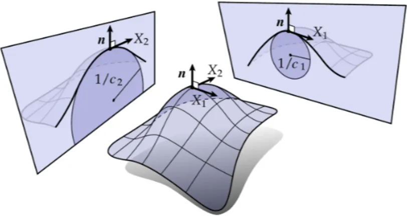
The curvature tensor K
xyis a quadratic form, two nonzero eigenvalues and eigenvectors can be found. In fact, two eigenvectors are known as the principle directions and point into the directions along which the minimal and maximal curvatures of the surface are found. The corresponding eigenvalues are called as principle curvatures, denoted as c
1and c
2, see Fig. 2.3. The trace of the curvature tensor K
xyis a constant scalar variable, which gives rise to the mean curvature H of the surface,
H = g
xyK
xy2 = c
1+ c
22
= ∇ · ∇ h
p 1 + ( ∇ h)
2!
= (1 + h
2x)h
yy+ (1 + h
2y)h
xx− 2h
xh
yh
xy2(1 + h
2x+ h
2y)
3/2.
(2.10)
2