AUS DER FAKULTÄT FÜR MEDIZIN DER UNIVERSITÄT REGENSBURG
PD DR. WOLFGANG HARTUNG INNERE MEDIZIN
VERGLEICH VON ULTRASCHALL UND PROJEKTIONSRADIOGRAPHIE IN DER DARSTELLUNG VON OSTEOPHYTEN UND EROSIONEN IM MITTELFUSS
Inaugural – Dissertation zur Erlangung des Doktorgrades
der Medizin
der
Fakultät für Medizin der Universität Regensburg
vorgelegt von Mathis Camerer
2018
Dekan: Prof. Dr. Dr. Torsten E. Reichert 1. Berichterstatter: PD Dr. Wolfgang Hartung 2. Berichterstatter: Prof. Dr. Dr. Joachim Grifka Tag der mündlichen Prüfung: 14.02.2019
VERGLEICH VON ULTRASCHALL UND
PROJEKTIONSRADIOGRAPHIE IN DER DARSTELLUNG VON OSTEOPHYTEN UND EROSIONEN IM MITTELFUSS
Inhalt
1. Zusammenfassung ... 2
1.1. Einleitung ... 2
1.2. Material und Methoden ... 4
1.3. Ergebnisse ... 6
1.4. Diskussion ... 8
1.6. Literaturverzeichnis ... 13
2. Originalpublikation ... 15
3. Danksagung ... 29
1. Zusammenfassung
1.1. Einleitung
Der Ultraschall ist ein etabliertes Bildgebungsverfahren mit hoher Validität und Reliabilität in der Beurteilung von Knochenläsionen, namentlich Osteophyten und Erosionen [1]. Verglichen mit der Magnetresonanztomographie (MRT) bietet der Ultraschall in der Befundung von Osteophyten und Erosionen in vielen Gelenkregionen eine gute Sensitivität und Spezifität [2-4]. In der klinischen Praxis ist der Ultraschall zudem kostengünstig, schnell durchführbar und ubiquitär verfügbar.
Neben dem Ultraschall steht die Projektionsradiographie (auch “konventionelles Röntgen“) zur Darstellung von Osteophyten und Erosionen zur Verfügung.
Erosionen sind hierbei insofern von klinischer Bedeutung, als sie bei Patienten mit rheumatoider Arthritis mit einem schlechten funktionellen Outcome assoziiert sind [5].
Es besteht inzwischen hinreichend Evidenz, dass der Ultraschall, insbesondere im Frühstadium der rheumatoiden Arthritis [6], verglichen mit dem Goldstandard MRT mit höherer Sensitivität und diagnostischer Genauigkeit als die Projektionsradiographie Erosionen in rheumatypischen Gelenken detektiert [3, 4].
Während sich ein Großteil der Studien bisher mit den häufig betroffenen Gelenken der oberen Extremität beschäftigte, wurden die Mittelfußgelenke bislang nur in einer Studie näher behandelt [7].
Neben Erosionen können auch Osteophyten sowohl mit dem Ultraschall, als auch mittels Projektionsradiographie evaluiert werden. Osteophyten sind ein Hauptcharakteristikum der Osteoarthrose und korrelieren mit der strukturellen Progression der Erkrankung [8]. Klinisch konnte für Knie- und Fingergelenke ein Zusammenhang zwischen dem Vorkommen von Osteophyten und Schmerzsymptomatik nachgewiesen werden [9-11]. Darüberhinaus sind Osteophyten ein Prädiktor für den Funktionsverlust des Kniegelenks nach 3 Jahren [12]. Was die Detektionsraten von Osteophyten in Ultraschall und Projektionsradiographie angeht, fand der Ultraschall in Patienten mit Hand- und Fingergelenksarthrose mehr Osteophyten als die Projektionsradiographie [2, 13, 14].
Jedoch fehlen bisher vergleichbare Studien für die Fußgelenke.
So war das Hauptziel unserer Studie, den diagnostischen Wert von Ultraschall und Projektionsradiographie in der Detektion von Osteophyten und Erosionen in den Mittelfußgelenken zu vergleichen. Dazu wurden sowohl Patienten mit entzündlichen, als auch mit nicht-entzündlichen Gelenkerkrankungen untersucht.
Darüberhinaus sind bislang die Häufigkeiten von im Ultraschall nachweisbaren Gelenkveränderungen im Mittelfuß noch unvollständig erfasst. Daher war unser Sekundärziel die Bestimmung der Häufigkeit von Osteophyten, Erosionen, Gelenkergüssen, und Power-Doppler-Signalen im Mittelfuß.
1.2. Material und Methoden
Patienten Es wurden stationäre Patienten der Klinik und Poliklinik für Rheumatologie und Klinische Immunologie des Asklepios Klinikums Bad Abbach ausgewählt, welche über aktuelle Röntgenbilder beider Füße verfügten. Nachfolgend wurden die Patienten in eine Gruppe mit entzündlichen bzw. nicht-entzündlichen Gelenkerkrankungen eingeteilt. Patienten mit der Diagnose einer entzündlichen Gelenkerkrankung, die potentiell die Fußgelenke befallen kann, wurden entsprechend in die Gruppe „entzündliche Gelenkerkrankung“ stratifiziert.
Ultraschallbefunde und Röntgenbilder wurden unabhängig voneinander von einem Ultraschallexperten respektive Radiologen beurteilt, die jeweils zum anderen Verfahren verblindet waren.
Die Studie wurde von der Ethikkommission der medizinischen Fakultät des Universitätsklinikums Regensburg bewilligt. Die Patienten wurden über die Untersuchung aufgeklärt und gaben ihre schriftliche Zustimmung zur Studienteilnahme.
Ultraschalluntersuchung Der Ultraschall wurde innerhalb einer Woche nach der Projektionsradiographie von einem der Autoren der Studie (W.H.) mit langjähriger Erfahrung im Ultraschall des Bewegungsapparates durchgeführt. Dabei wurde ein Logiq E 9 Gerät von GE (GE Healthcare, Buckinghamshire, UK) mit einem L8-18i Linearschallkopf und einer Frequenz von 8-18 MHz verwendet. Für die Untersuchung nutzten wir eine Frequenz von 18 MHz, eine Pulsrepetitionsfrequenz von 11.9 MHz mit dem höchsten „Gain“ ohne Hintergrundrauschen und einen niedrigen Wandfilter.
Die standardisierte Ultraschalluntersuchung wurde beidseits in dorsal-longitudinaler Schnittführung durchgeführt, wobei folgende Gelenke des Mittelfußes untersucht wurden: Articulatio talonavicularis (TN), calcaneocuboidea (CC), naviculocuneiforme mediale/intermedium/laterale (NCM/NCI/NCL), und die erste bis fünfte Articulatio tarsometatarsalis (TMT 1-5) (siehe Bild 1). In jedem Gelenk wurden Osteophyten (corticale Protrusionen > 1mm), Erosionen (corticale Einbrüche > 1 mm gemäß der OMERACT-Definition [15]) und Gelenkergüsse (Gelenkkapselabhebung > 1mm) dichotomisiert erfasst (Vorhandensein ja/nein). Die Power-Doppler (PD)-Aktivität wurde semiquantitativ auf einer Skala von 0 bis 3 bewertet [16]. Ein Wert größer als 0 galt dabei als positives Power-Doppler-Signal.
Projektionsradiographie Röntgenbilder (posterior-anterior und oblique) jedes Fußes wurden von einem Experten in Muskuloskelettaler Radiologie bewertet (P.H.).
Die Aufnahmen wurden mit einem Fokus-Film-Abstand von 105 cm angefertigt mit Expositionsfaktoren von 52 kV und 3.2 mAs. Für jedes Gelenk wurde das Vorliegen von Osteophyten und Erosionen (jeweils Läsionen > 1mm) dichotomisiert erfasst (Vorhandensein ja/nein).
Statistische Analyse Die statistische Auswertung erfolgte auf Gelenkebene mithilfe der SPSS Statistiksoftware Version 22.0. Für quantitative Parameter wurde jeweils der Mittelwert (± Standardabweichung) bestimmt. Zusätzlich wurde für jedes Gelenk die Prävalenz von in der Bildgebung nachgewiesenen Pathologien berechnet. Der Chi-Square-Test und Fisher´s-Exact-Test wurden zum Vergleich von kategorialen Variablen herangezogen. Der Grad der Übereinstimmung wurde mittels κ-statistic ermittelt. Die statistische Signifikanz wurde auf p < 0.05 festgesetzt.
1.3. Ergebnisse
Insgesamt 124 Patienten (davon 58% weiblich), 90 mit entzündlicher und 34 mit nicht-entzündlicher Gelenkerkrankung wurden in diese Studie eingeschlossen, und Gelenke von 246 Füßen wurden untersucht. Die Patientencharakteristika sind in Tabelle 1 aufgeführt. In der Auswertung konnten 2 Füße nicht berücksichtigt werden, ein Fuß aufgrund einer Vorfußamputation, ein anderer aufgrund mutilierender Schäden bei rheumatoider Arthritis. Zusätzlich konnten wegen eingeschränkter Darstellbarkeit in der Projektionsradiographie 2 Gelenke, im Ultraschall 13 Gelenke nicht beurteilt werden. So wurden letztendlich insgesamt 2445 Gelenke in die Untersuchung eingeschlossen.
Prävalenz von Osteophyten in Ultraschall und Projektionsradiographie Im Ultraschall wurden signifikant mehr Osteophyten (n = 344; 14.1% der Gelenke) als in der Projektionsradiographie (n = 13; 0.5%) detektiert (siehe Tabelle 2 und Bild 2).
Nur 8 Osteophyten konnten mit beiden Bildgebungsverfahren dargestellt werden. 5 Osteophyten waren nur in der Projektionsradiographie sichtbar, 3 davon im TMT-1- und 2 im TN-Gelenk (p < 0.001).
Während im Ultraschall Osteophyten am häufigsten im NCI- (19.5%; 48/246) und NCL-Gelenk (17.3%; 42/243) (p < 0.01) auftraten, konnten in der Projektionsradiographie dort keine Osteophyten dargestellt werden. Zwischen den beiden Verfahren ergab sich in Bezug auf Osteophyten eine nur schwache Übereinstimmung (κ-statistic 0.035).
Prävalenz von Erosionen in Ultraschall und Projektionsradiographie Im Ultraschall konnten mehr Erosionen (2.5%; 60/2445) als in der Projektionsradiographie (0.1%; 3/2445) nachgewiesen werden. Nur eine Erosion im NCM-Gelenk wurde in beiden Methoden dargestellt (p = 0.072). Erosionen im Ultraschall waren am häufigsten im NCI- (4.5%; 11/246) und TMT-4-Gelenk (4.5%;
11/246) detektierbar, während die Projektionsradiographie keine Erosionen in diesen Gelenken nachweisen konnte. So ergab sich auch in Bezug auf Erosionen eine nur schwache Übereinstimmung zwischen Ultraschall und Projektionsradiographie (κ- statistic 0.029).
Prävalenz von Gelenkergüssen und positiven Power-Doppler-Signalen in Ultraschall und Projektionsradiographie Ein Gelenkerguss war in 8.8%
(215/2445), ein positives PD-Signal in 8.3% (204/2445) aller Gelenke zu beobachten.
Gelenkergüsse traten am häufigsten in CC- (24.4%; 59/242), TN- (21.9%; 53/242) und NCI-Gelenken (9.8%; 24/246) auf, wohingegen die TMT-Gelenke 1-4 selten betroffen waren (p < 0.001). Ein positives PD-Signal wurde am häufigsten in NCL- (13.6%; 33/243), TMT-4- (11.1%; 27/244), TMT-3- und TMT-5-Gelenken (jeweils 10.2%; 25/245) nachgewiesen (p < 0.05).
Ultraschall bei entzündlichen und nicht-entzündlichen Gelenkerkrankungen Im Ultraschall wurden signifikant mehr Erosionen bei entzündlichen (2.9%; 51/1767) als bei nichtentzündlichen (1.3%; 9/678) (p < 0.05) Gelenkerkrankungen entdeckt. Im Gegensatz dazu ergab sich eine nicht signifikante Differenz zwischen entzündlichen und nichtentzündlichen Gelenkerkrankungen im Hinblick auf sowohl Osteophyten als auch Gelenkergüsse (14.7%; 260/1767 vs. 12.4%; 84/678 und 9.2%; 163/1767 vs.
7.7%; 52/678). Im Power-Doppler-Ultraschall zeigte sich keine signifikante Differenz bei der Hyperperfusion zwischen entzündlichen (8.5%; 151/1767) und nichtentzündlichen Gelenkerkrankungen (7.8%; 53/678). Jedoch wurde bei 5 Patienten mit entzündlicher Gelenkerkrankung (5,6%; 5/90 Patienten mit entzündlichen Gelenkerkrankungen) und nur einem Patienten mit nichtentzündlicher Gelenkerkrankung (2.9%; 1/34 Patienten mit nichtentzündlicher Gelenkerkrankung) ein PD-Score von 3 nachgewiesen.
1.4. Diskussion
Der Großteil der Studien hat sich bis dato mit Pathologien des Vor- und Rückfußes beschäftigt [17], obwohl Läsionen des Mittelfußes häufig sind [17, 18]. Während man ursprünglich von einer seltenen Entität ausging, zeigte kürzlich eine Bevölkerungsstudie in England, dass die Prävalenz von symptomatischer Osteoarthrose des Mittelfußes bei über 50-Jährigen bei 12% liegt [19]. Da die Gelenke des Mittelfußes eine entscheidende Rolle bei der Lastübertragung zum Vorfuß während des Gangs spielen, können Läsionen des Mittelfußes zu erheblichen Funktionsbeeinträchtigungen und Einschränkungen der Mobilität führen [17].
Neben der Osteoarthrose ist auch die Rheumatoide Arthritis der Fußgelenke ein häufiges Krankheitsbild. So ist hier - neben der Hand - der Fuß der am häufigsten betroffene Gelenkkomplex, sowohl in frühen als auch späten Krankheitsstadien. In etwa ¾ der Fälle geht die rheumatische Fußbeteiligung dabei mit Gehbeschwerden einher [20].
Unsere Studie schloss sowohl Patienten mit entzündlichen wie auch nichtentzündlichen Gelenkerkrankungen des Mittelfußes ein. Es handelt sich um die erste Studie, welche alle Gelenke des Mittelfußes berücksichtigt. So konnte erstmals die Prävalenz von im Ultraschall nachweisbaren Osteophyten und Erosionen im Mittelfuß in unterschiedlichen Populationen bestimmt werden.
Außerdem wurden erstmalig alle Gelenke des Mittelfußes sowohl mittels Ultraschall, als auch mittels Projektionsradiographie auf das Vorliegen von Osteophyten und Erosionen evaluiert. Wir konnten zeigen, dass der Ultraschall mehr Osteophyten darzustellen vermag als die Projektionsradiographie. Hierbei ergab sich eine nur schwache Übereinstimmung zwischen den beiden Verfahren.
Unsere Ergebnisse stehen im Einklang mit Studien bei Patienten mit Fingerpolyarthrose, die ebenfalls eine höhere Sensitivität des Ultraschalls zeigten. In unserer Studie war die Überlegenheit des Ultraschalls aufgrund der niedrigen Rate an gefundenen Osteophyten in der Projektionsradiographie sogar noch deutlicher [2, 13, 14]. In der Projektionsradiographie wurden Osteophyten und Erosionen in den peripher gelegenen Gelenken des Mittelfußes detektiert, aber keine Läsionen in den Artt. NCI und NCL im Zentrum des Mittelfußes. Im Gegensatz dazu wurde im
Ultraschall im zentralen Mittelfuß eine große Anzahl an Knochenläsionen nachgewiesen. In der Projektionsradiographie könnte die komplexe Anatomie vieler kleiner Knochen es aufgrund von Summationseffekten erschwert haben, Knochenläsionen im Zentrum des Mittelfußes nachzuweisen. Ein weiterer Grund für niedrige Detektionsraten in der Projektionsradiographie ist vermutlich, dass nur Läsionen tangential zum Röntgenstrahl nachgewiesen werden können. Insgesamt könnten die niedrigen Detektionsraten in der Projektionsradiographie die schwache Übereinstimmung zwischen den beiden Verfahren erklären. Um einen besseren Einblick in diese Thematik zu bekommen, wären jedoch Computer- oder Magnetresonanztomographie-Untersuchungen notwendig.
Nach unserem Kenntnisstand existiert momentan nur eine Studie, welche Ultraschall und Projektionsradiographie bezüglich der Detektion von Erosionen vergleicht:
Petterle et al. [7] untersuchten 50 Patienten mit Rheumatoider Arthritis (mittlere Krankheitsdauer: 9.81 Jahre), die zum Zeitpunkt der Erhebung keine Symptomatik in den Fußgelenken hatten. Es wurden Sprunggelenke, Metatarsophalangealgelenke und 4 der 10 Mittelfußgelenke untersucht: TN-, CC-, NCM- und TMT-5-Gelenk. Wie auch in unserer Studie wurden die Mittelfußgelenke im Ultraschall in dorsal- longitudinaler Schnittführung evaluiert. Neben Röntgenaufnahmen des Fußes (a.p.
und Profil) wurden auch Aufnahmen des Knöchels (a.p. und Profil) angefertigt. In Übereinstimmung mit unserer Studie entdeckte der Ultraschall mehr Erosionen als die Projektionsradiographie. Im Gegensatz zu unseren Ergebnissen ermittelte Petterle eine gute Übereinstimmung zwischen den beiden Verfahren für die TN-, CC- und NCM-Gelenke. Ursächlich dafür könnte sein, dass unterschiedliche Röntgenebenen verwendet wurden und zusätzlich Röntgenaufnahmen des Fußknöchels miteinbezogen wurden. Darüberhinaus war die Krankheitsdauer der Patienten mit rheumatoider Arthritis bei Petterle et al. im Mittel länger als bei Patienten in unserer Studie. Dies könnte die höheren Detektionsraten von Erosionen in der Projektionsradiographie und somit eine bessere Korrespondenz mit den Sonographiebefunden bei Petterle et al. erklären.
Wir entschieden uns dagegen, neben Osteophyten und Erosionen auch Gelenkspaltverschmälerungen mittels Ultraschall und Projektionsradiographie zu evaluieren, da Zweifel an einer reliablen Definition und Akquisition bestanden.
der Computertomographie, validiert worden. Jedoch wäre die Durchführung einer Computertomographie zusätzlich zur Projektionsradiographie ohne medizinische Indikation aus Strahlenschutzgründen nicht durch das Ethikkomitee akzeptiert worden.
Momentan ist die Projektionsradiographie das Standardverfahren zur Diagnose der Osteoarthrose und ein Screening-Instrument für rheumatoide Arthritis der Fußgelenke. Jedoch zeigen unsere Ergebnisse, dass der Ultraschall des Mittelfußes eine nützliche Alternative zur Projektionsradiographie in der klinischen Praxis und in der Forschung darstellt. Aufgrund der hohen Detektionsraten von Osteophyten und Erosionen könnte man den Ultraschall beispielsweise auch bei Patienten mit Mittelfußbeschwerden in Betracht ziehen, bei denen das Röntgenbild keine Gelenkdestruktion zeigt. Ein weiterer Vorteil des Ultraschalls ist die Fähigkeit entzündliche Veränderungen wie eine Synovitis nachzuweisen. Auf diese Weise können entzündliche Gelenkerkrankungen wie rheumatoide Arthritis früher diagnostiziert, die Krankheitsaktivität überwacht und das Risiko der Krankheitsprogression abgeschätzt werden [21]. In Zukunft könnten weiterführende Ultraschallstudien, die sich mit der Frühdiagnose und dem Monitoring von Osteoarthrose und rheumatoider Arthritis im Mittelfuß befassen, zur Verbesserung der Patientenversorgung beitragen.
1.5. Anhang
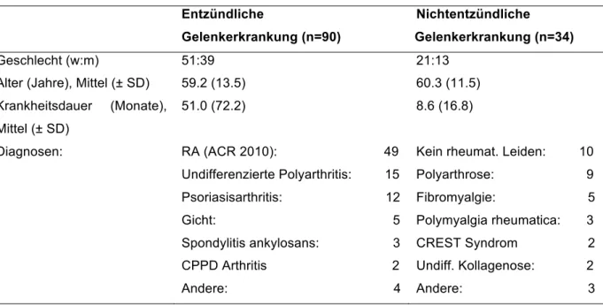
Tabelle 1. Patientencharakteristika *
Entzündliche
Gelenkerkrankung (n=90)
Nichtentzündliche Gelenker Gelenkerkrankung (n=34)
Geschlecht (w:m) 51:39 21:13
Alter (Jahre), Mittel (± SD) 59.2 (13.5) 60.3 (11.5)
Krankheitsdauer (Monate), Mittel (± SD)
51.0 (72.2) 8.6 (16.8)
Diagnosen: RA (ACR 2010): 49 Kein rheumat. Leiden: 10 Undifferenzierte Polyarthritis: 15 Polyarthrose: 9
Psoriasisarthritis: 12 Fibromyalgie: 5 Gicht: 5 Polymyalgia rheumatica: 3 Spondylitis ankylosans: 3 CREST Syndrom 2
CPPD Arthritis 2 Undiff. Kollagenose: 2 Andere: 4 Andere: 3
* Die Zahlenwerte entsprechen der Anzahl der Patienten, wenn nicht anders angegeben.
RA (ACR 2010): Rheumatoide Arthritis nach den ACR Kriterien von 2010;
Tabelle 2. Prävalenz von Osteophyten und Erosionen in Ultraschall und Projektionsradiographie*
Gelenk US Osteophyten PR Osteophyten US Erosionen PR Erosionen
TN 16.9% (41/242) 1.7% (4/242) 2.1% (5/242) 0% (0/242)
CC 8.7% (21/242) 0.8% (2/242) 0.4% (1/242) 0% (0/242)
NCM 10.6% (26/246) 0% (0/246) 1.6% (4/246) 0.4% (1/246)
NCI 19.5% (48/246) 0% (0/246) 4.5% (11/246) 0% (0/246)
NCL 17.3% (42/243) 0% (0/243) 2.9% (7/243) 0% (0/243)
TMT 1 8.9% (22/246) 2.0% (5/246) 0% (0/246) 0% (0/246)
TMT 2 15.0% (37/246) 0% (0/246) 1.6% (4/246) 0.4% (1/246)
TMT 3 16.3% (40/245) 0% (0/245) 2.9% (7/245) 0.4% (1/245)
TMT 4 13.9% (34/244) 0% (0/244) 4.5% (11/244) 0% (0/244)
TMT 5 13.5% (33/245) 0.8% (2/245) 4.1% (10/245) 0% (0/245)
Total 14.1% (344/2445) 0.5% (13/2445) 2.5% (60/2445) 0.1% (3/2445)
* Die Zahlenwerte sind Prozentzahlen (Anzahl der betroffenen Gelenke/Anzahl der untersuchten Gelenke).
US: Ultraschall; PR: Projektionsradiographie; TN: Art. talonavicularis; CC: Art. calcaneocuboidea;
NCM: Art. naviculocuneiforme mediale; NCI: Art. naviculocuneiforme intermedium; NCL: Art.
naviculocuneiforme laterale; TMT: Art. tarsometatarsalis; Total: Alle Gelenke
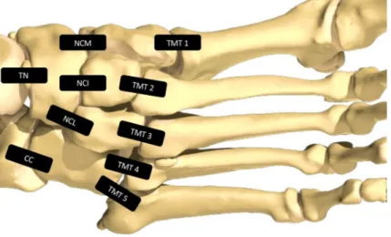
Bild 1. Sonographische Schnittebenen. TN: Art. talonavicularis; CC: Art. calcaneocuboidea; NCM:
Art. naviculocuneiforme mediale; NCI: Art. naviculocuneiforme intermedium; NCL: Art.
naviculocuneiforme laterale; TMT: Art. tarsometatarsalis
Bild 2. Patient mit nichtentzündlicher Gelenkerkrankung und Osteophyten in den Gelenken TMT 2-4, NCL, NCI, und NCM links. a) In der Projektionsradiographie zeigt sich keiner der sonographisch dargestellten Osteophyten b) Ultraschall des NCM-Gelenks mit 2 großen Osteophyten
1.6. Literaturverzeichnis
1. Keen HI, Wakefield R, Conaghan PG (2014) Optimising ultrasonography in rheumatology. Clin Exp Rheumatol 32:13-16.
2. Mathiessen A, Haugen IK, Slatkowsky-Christensen B, Boyesen P, Kvien TK, Hammer HB (2013) Ultrasonographic assessment of osteophytes in 127 patients with hand osteoarthritis: exploring reliability and associations with MRI, radiographs and clinical joint findings. Ann Rheum Dis 72:51-6.
3. Szkudlarek M, Narvestad E, Klarlund M, Court-Payen M, Thomsen HS, Ostergaard M (2004) Ultrasonography of the metatarsophalangeal joints in rheumatoid arthritis: comparison with magnetic resonance imaging, conventional radiography, and clinical examination. Arthritis Rheum 50:2103-12.
4. Wakefield RJ, Gibbon WW, Conaghan PG, O'Connor P, McGonagle D, Pease C, et al (2000) The value of sonography in the detection of bone erosions in patients with rheumatoid arthritis: a comparison with conventional radiography. Arthritis Rheum 43:2762-70.
5. Bombardier C, Barbieri M, Parthan A, Zack DJ, Walker V, Macarios D, et al (2012) The relationship between joint damage and functional disability in rheumatoid arthritis: a systematic review. Ann Rheum Dis 71:836-844.
6. Backhaus M, Burmester GR, Sandrock D, Loreck D, Hess D, Scholz A, et al (2002) Prospective two year follow up study comparing novel and conventional imaging procedures in patients with arthritic finger joints. Ann Rheum Dis 61:895- 904.
7. Sant'Ana Petterle G, Natour J, Rodrigues da Luz K, Soares Machado F, dos Santos MF, da Rocha Correa Fernandes A, et al (2013) Usefulness of US to show subclinical joint abnormalities in asymptomatic feet of RA patients compared to healthy controls. Clin Exp Rheumatol 31:904-12.
8. Barr AJ, Campbell TM, Hopkinson D, Kingsbury SR, Bowes MA, Conaghan PG (2015) A systematic review of the relationship between subchondral bone features, pain and structural pathology in peripheral joint osteoarthritis. Arthritis Res Ther 17:228.
9. Cicuttini FM, Baker J, Hart DJ, Spector TD (1996) Association of pain with radiological changes in different compartments and views of the knee joint.
Osteoarthritis Cartilage 4:143–7.
radiographically diagnosed osteophytes and magnetic resonance detected cartilage defects in the tibiofemoral joint. Ann Rheum Dis 57:401–7.
11. Kortekaas MC, Kwok WY, Reijnierse M, Huizinga TW, Kloppenburg M (2011) Osteophytes and joint space narrowing are independently associated with pain in finger joints in hand osteoarthritis. Ann Rheum Dis 70:1835–7.
12. Muraki S, Akune T, Nagata K, Ishimoto Y, Yoshida M, Tokimura F (2015) Does osteophytosis at the knee predict health-related quality of life decline? A 3-year
follow-up of the ROAD study. Clin Rheumatol 34:1589–97.
13. Keen HI, Wakefield RJ, Grainger AJ, Hensor EM, Emery P, Conaghan PG (2008) Can ultrasonography improve on radiographic assessment in osteoarthritis of the hands? A comparison between radiographic and ultrasonographic detected pathology. Ann Rheum Dis 67:1116-20.
14. Wittoek R, Carron P, Verbruggen G (2010) Structural and inflammatory sonographic findings in erosive and non-erosive osteoarthritis of the interphalangeal finger joints. Ann Rheum Dis 69:2173-6.
15. Wakefield RJ, Balint PV, Szkudlarek M, Filippucci E, Backhaus M, D'Agostino MA, et al (2005) Musculoskeletal ultrasound including definitions for ultrasonographic
pathology. J Rheumatol 32:2485-7.
16. Backhaus M, Ohrndorf S, Kellner H, Strunk J, Backhaus TM, Hartung W, et al (2009) Evaluation of a novel 7-joint ultrasound score in daily rheumatologic practice:
a pilot project. Arthritis Rheum 61:1194-201.
17. Chan PS, Kong KO (2013) Natural history and imaging of subtalar and midfoot joint disease in rheumatoid arthritis. Int J Rheum Dis 16:14-8.
18. Patel A, Rao S, Nawoczenski D, Flemister AS, DiGiovanni B, Baumhauer JF (2010) Midfoot arthritis. J Am Acad Orthop Surg 18:417-25.
19. Thomas MJ, Peat G, Rathod T (2015) The epidemiology of symptomatic midfoot osteoarthritis in community-dwelling older adults: cross-sectional findings from the
Clinical Assessment Study of the Foot. Arthritis Res Ther 17:178.
20. Grondal L, Tengstrand B, Nordmark B, Wretenberg P, Stark A (2008) The foot:
still the most important reason for walking incapacity in rheumatoid arthritis. Acta
Orthopaedica 79:257–261.
21. Colebatch AN, Edwards CJ, Ostergaard M, Van der Heijde D, Balint PV, D'Agostino MA, et al (2013) EULAR recommendations for the use of imaging of the joints in the clinical management of rheumatoid arthritis.Ann Rheum Dis 72:804–814.
2. Originalpublikation
This is a post-peer-review, pre-copyedit version of an article published in Clinical Rheumatology. The final authenticated version is available online
High resolution ultrasound of the midfoot: Sonography is more sensitive than conventional radiography in detection of osteophytes and erosions in inflammatory and non-inflammatory joint disease.
Mathis Camerer
mathis.camerer@stud.uni-regensburg.de Boris Ehrenstein
b.ehrenstein@asklepios.com Patrick Hoffstetter
p.hoffstetter@asklepios.com Martin Fleck
m.fleck@asklepios.com
Wolfgang Hartung (corresponding author) w.hartung@asklepios.com
Asklepios Medical Center, Department of Rheumatology and Clinical Immunology, Kaiser-Karl-V-Allee 3, D-93077 Bad Abbach, Germany.
Abstract
This study aimed to compare the diagnostic value of ultrasonography to conventional radiography in detecting osteophytes and erosions in the midfoot joints in patients suffering from inflammatory and non-inflammatory joint disease. Patients with current foot radiographs were included and stratified in two cohorts: inflammatory and non- inflammatory joint disease. The ten midfoot joints of each foot were evaluated by conventional radiography assessing the presence of osteophytes and erosions and by ultrasonography determining the presence of osteophytes, erosions, and joint effusion. Power Doppler activity was scored semi-quantitatively from 0 to 3. A total of 2445 joints in 124 patients (90 with inflammatory joint disease, 34 with non- inflammatory joint disease) were assessed. Ultrasonography detected significantly more osteophytes than conventional radiography (344; 14.1% vs. 13; 0.5%), as well as more erosions (60; 2.5% vs. 3; 0.1%). There was weak agreement between the two modalities (κ- statistic 0.029–0.035). Power Doppler ultrasonography demonstrated no significant difference in hyperperfusion comparing patients with inflammatory joint disease and non-inflammatory joint disease. Ultrasonography of the midfoot is more sensitive than conventional radiography in the detection of osteophytes and erosions in patients suffering from inflammatory and non- inflammatory joint disease. Thus, midfoot ultrasonography may be a useful tool in the diagnosis of joint diseases as rheumatoid arthritis and osteoarthritis.
Keywords: midfoot, foot, ultrasonography, conventional radiography, osteophyte, erosion.
Introduction
Ultrasonography (US) and conventional radiography (CR) are established imaging modalities for the assessment of cortical bone lesions including erosions and osteophytes. When compared to MRI, US has good sensitivity and specificity in the detection of osteophytes and erosions in many joint regions [1–3], rendering it an inexpensive and readily available alternative in clinical practice.
In recent years, there has been a strong effort in the assessment of erosions to better identify RA patients with poor outcome. Thus, now there is good evidence that US is more effective in detecting erosions than CR in joints affected by RA [2, 3], especially in the early course of the disease [4]. Whereas the majority of existing studies refer to the frequently involved upper limb joints of RA patients, the midfoot joints were so far only partly addressed in one study [5]. Beside erosions, also osteophytes can be assessed by both US and CR. Osteophytes are the main feature of osteoarthritis, and while largely unstudied in the foot, presence of osteophytes in the knee is associated with structural progression [6] and predicts functional disability after 3 years [7]. In patients with hand osteoarthritis, US detected more osteophytes than CR [1, 8, 9], but comparable imaging studies for the foot joints have not been reported yet. Therefore, the aim of our study was to compare the diagnostic value of US and CR in the detection of osteophytes and erosions in the midfoot joints in patients suffering from inflammatory (IJD) and non-inflammatory joint disease (NIJD).
Our secondary goal was to evaluate the prevalence of ultrasonographic osteophytes, erosions, joint effusion, and power Doppler signal in the midfoot in IJD and NIJD.
Materials and Methods
Patients Inpatients of our tertiary rheumatology center with current foot radiographs were included and stratified in two cohorts: IJD and NIJD. The criterion for assignment to the IJD group was the presence of an established inflammatory joint disease, which could potentially affect the foot joints.
All patients were investigated in both feet by an ultrasonography expert and a radiologist who were blinded to the results of the other modality.
Ultrasonographic assessment US was performed within a week after radiography by one author (W.H.) with longstanding experience in musculoskeletal US. A Logiq e 9 device from GE (GE Healthcare, Buckinghamshire, UK) with a L8-18i linear hockey stick probe with 8–18 MHz frequency was used. The technical parameters of the examination included a gray scale frequency of 18 MHz, a pulse repetition frequency of 900 Hz, and a power Doppler (PD) frequency of 11.9 MHz with highest gain level without background noise and low wall filter. Standardized US assessment was performed bilaterally by a dorsal longitudinal scan of the following midfoot joints:
talonavicular, calcaneocuboid, medial/intermediate/lateral naviculocuneiform (NCM/NCI/NCL), and 1st to 5th tarsometatarsal joint (TMT 1–5) (see Fig. 1). In each joint, osteophytes (cortical protrusions >1 mm), erosions (cortical breaks >1 mm according to the OMERACT definition [10]), and joint effusion (joint capsule distension of >1 mm) were assessed dichotomously (presence/absence). PD activity was scored semi-quantitatively from 0 to 3 [11]. Any score >0 was defined as
„positive PD signal“.
Radiographic evaluation CR images (posterior-anterior and oblique view) of each foot were evaluated by an expert in musculoskeletal radiology (P.H.). The
of 52 kV and 3.2 mAs.
For each joint, osteophytes and erosions (lesions >1 mm) were scored dichotomously as present or absent.
Statistical analysis Statistical analysis was performed on joint level using SPSS statistical software, version 22.0. For quantitative parameters, the mean ± SD were determined for each. In addition, the prevalence of imaging findings was calculated for each joint. The chi-square test and Fisher’s exact test were used to compare categorical variables. Levels of agreement were calculated by κ-statistic. Statistical significance was set at p < 0.05.
Results
A total of 246 feet of 124 patients (58% female), 90 with IJD and 34 with NIJD, were assessed. Patients’ characteristics are shown in Table 1. One foot was excluded due to a forefoot amputation; in another patient’s foot, the joints could not be differentiated because of mutilating joint damage by RA. Two joints were not assessed by CR and 13 joints could not be examined by US due to poor visibility.
Thus, in total, 2445 joints were assessed.
Prevalence of osteophytes in US and CR US detected significantly more osteophytes (n = 344; 14.1% of joints) than CR (n = 13; 0.5%) (cf. Table 2 and Fig.
2). Only eight osteophytes were observed by both methods. Five osteophytes were found only by CR, three of them in the TMT1 and two of them in the talonavicular joints (p < 0.001). Whereas ultrasonographic osteophytes were most frequently detected in the NCI (19.5%; 48/246) and NCL joints (17.3%; 42/243) (p < 0.01), CR could not identify any osteophytes there. There was weak agreement between the
two modalities in detecting osteophytes (κ-statistic 0.035).
Prevalence of erosions in US and CR US (2.5%; 60/2445) identified more erosions than CR (0.1%; 3/2445); only one erosion in the NCM joint was detected by both methods (p = 0.072). Ultrasonographic erosions were most common at the NCI (4.5%; 11/246) and TMT4 joints (4.5%; 11/244) (p < 0.01), while CR could not detect any erosion in these joints (Table 2). Thus, there was poor agreement between US and CR in detecting erosions (κ-statistic 0.029).
Prevalence of joint effusion and PD signal in US Effusion and positive PD signal were present in 8.8% (215/2445) and 8.3% (204/2445) of all joints, respectively. Joint effusion was most frequent in the calcaneocuboid (24.4%; 59/242), talonavicular (21.9%; 53/242), and NCI joints (9.8%; 24/246), whereas the TMT 1–4 joints were rarely affected (p < 0.001). Positive PD signal was most commonly found in the NCL (13.6%; 33/243), TMT4 (11.1%; 27/244), TMT3, and TMT5 joints (10.2%; 25/245 each) (p < 0.05).
US findings in IJD and NIJD US observed significantly more erosions in IJD (2.9%;
51/1767) than in NIJD (1.3%; 9/678) (p < 0.05). By contrast, there was a non- significant difference between IJD and NIJD regarding the detection of osteophytes and joint effusion (14.7%; 260/1767 vs. 12.4%; 84/678 and 9.2%; 163/1767 vs. 7.7%;
52/678).
Power Doppler ultrasonography (PDUS) also demonstrated no significant difference in hyperperfusion between IJD (8.5%; 151/1767) and NIJD patients (7.8%; 53/678).
However, there were five IJD patients (5.6%; 5/90 IJD patients) versus only one NIJD patient (2.9%; 1/34 NIJD patients) (having polymyalgia rheumatica) with a PD score of 3.
Discussion
To date, the majority of studies assessing the foot have focused on forefoot and hindfoot pathology [12], although lesions in the midfoot are quite common [12, 13].
Originally thought to be rare, a recent population study in England revealed a prevalence of 12% of symptomatic midfoot OA in people aged over 50 years [14]. As midfoot joints play a crucial role in transferring the load to the forefoot during gait, their affection by IJDs or NIJDs can cause significant foot dysfunction resulting in mobility impairment [12].
This is the first study examining all the midfoot joints with comparison of osteophytes and erosions investigated by US and CR. We could show that US detects more osteophytes and erosions than CR. Correlation between the modalities was poor.
Aside from that, for the first time, the prevalence of ultrasonographic osteophytes and erosions in different populations was determined by investigating all the midfoot joints. While comparable studies of osteophytes examined by US and CR are still missing, our results were in accordance with studies in hand osteoarthritis showing a higher sensitivity of US [1, 8, 9]. In our study, this result was enhanced by the small number of osteophytes found by CR. CR detected osteophytes and also erosions on the midfoot „edges“, but no bony lesions in the NCI and NCL joints in the midfoot
„center“. In contrast, US found a large number of bony lesions in the center. Due to summation effects, the complex anatomy of many small bones may have made it difficult for CR to detect bony lesions there. A further limiting factor of CR detection rates may have been that it only shows lesions tangentially to the beam. Low detection rates by CR may explain the weak agreement between the two modalities.
However, to get a better insight into this question, CT studies would be needed. To our knowledge, only one study with comparison of erosions detected by US and CR
has been reported to date: Petterle et al. [5] investigated 50 RA patients with asymptomatic feet (mean disease duration, 9.81 years). Assessment included the ankle, metacarpophalangeal, and four of the ten midfoot joints: talonavicular, calcaneocuboid, NCM, and TMT5. As in our study, the midfoot joints were evaluated by dorsal US scan. Beside foot radiographs (AP and profile), also ankle radiographs (AP and profile) were obtained. Consistent with our study, US detected more erosions than CR. Contrary to our results, Petterle found good correlations between the two modalities for the talonavicular, calcaneocuboid, and NCM joints. One reason may be the different radiographic planes used plus the additional evaluation of ankle radiographs. Furthermore, Petterle included only RA patients with long disease duration, whereas in our study, patients with IJD and NIJD with shorter disease duration were investigated. This might explain the higher detection rate of erosions by CR in Petterle’s study and thus may have delivered results more corresponding to US. In addition to osteophytes and erosions, we decided not to compare joint space narrowing examined by US and CR due to concerns about reliable definition and acquisition.
Ideally, our results from US and CR would have been validated with a gold standard such as computed tomography (CT). Though performing a CT scan additionally to CR without medical indication would not have been accepted by the ethics committee for reasons of radiation protection.
Currently, CR is the standard method in the diagnosis of OA and a screening tool in RA of the foot. However, our results demonstrate that midfoot US may be a useful alternative to CR in clinical practice and research. Due to the high detection rate of osteophytes and erosions, one may for example consider US in patients with midfoot
ability to detect inflammatory signs as synovitis, which may be used to diagnose IJDs as RA earlier, monitor disease activity, and predict disease progression [15].
In the future, further ultrasonographic studies addressing the earlier diagnosis of OA and RA in the midfoot may result in better patient management.
Tables and Figures
Table 1. Patients' characteristics
IJD group (n=90) NIJD group (n=34)
Gender (f:m) 51:39 21:13
Age (years), mean (± SD) 59.2 (13.5) 60.3 (11.5)
Disease duration (months), mean (± SD)
51.0 (72.2) 8.6 (16.8)
Diagnoses: RA (ACR 2010): 49 No rheumatic disease: 10 Undifferentiated polyarthritis: 15 Polyosteoarthritis: 9 Psoriatic arthritis: 12 Fibromyalgia: 5 Gout: 5 Polymyalgia rheumatica: 3
Ankylosing spondylitis: 3 CREST syndrome 2 CPPD arthritis 2 UCTD: 2 Others: 4 Others: 3
Values are the number unless otherwise indicated
IJD inflammatory joint disease, NIJD non-inflammatory joint disease, RA (ACR 2010) Rheumatoid arthritis according to the ACR 2010 criteria, UCTD Undifferentiated connective tissue disease
Table 2. Prevalence of osteophytes and erosions detected by ultrasonography or conventional radiography
Joint US osteophytes CR osteophytes US erosions CR erosions
TN 16.9% (41/242) 1.7% (4/242) 2.1% (5/242) 0% (0/242)
CC 8.7% (21/242) 0.8% (2/242) 0.4% (1/242) 0% (0/242)
NCM 10.6% (26/246) 0% (0/246) 1.6% (4/246) 0.4% (1/246)
NCI 19.5% (48/246) 0% (0/246) 4.5% (11/246) 0% (0/246)
NCL 17.3% (42/243) 0% (0/243) 2.9% (7/243) 0% (0/243)
TMT 1 8.9% (22/246) 2.0% (5/246) 0% (0/246) 0% (0/246)
TMT 2 15.0% (37/246) 0% (0/246) 1.6% (4/246) 0.4% (1/246)
TMT 3 16.3% (40/245) 0% (0/245) 2.9% (7/245) 0.4% (1/245)
TMT 4 13.9% (34/244) 0% (0/244) 4.5% (11/244) 0% (0/244)
TMT 5 13.5% (33/245) 0.8% (2/245) 4.1% (10/245) 0% (0/245)
Total 14.1% (344/2445) 0.5% (13/2445) 2.5% (60/2445) 0.1% (3/2445) Values are the percentage (number of affected joints/number of joints examined)
US ultrasonography, CR conventional radiography, TN talonavicular, CC calcaneocuboid, NCM medial naviculocuneiform, NCI intermediate naviculocuneiform, NCL lateral naviculocuneiform, TMT tarsometatarsal, Total Total number of joints affected
Figure 1. Ultrasound scanning planes. TN talonavicular, CC calcaneocuboid, NCM/NCI/NCL medial/intermediate/lateral naviculocuneiform, TMT tarsometatarsal
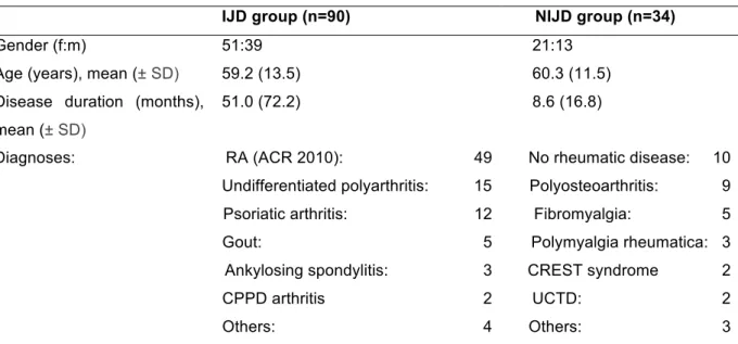
Figure 2. Patient with NIJD revealing osteophytes at the left TMT II–IV, NCL, NCI, and NCM. a CR showing none of the osteophytes seen by sonography. b Ultrasonography of the NCM depicting two clear osteophytes*
Acknowledgements
The authors would like to thank all participating patients who made this study possible.
Compliance with ethical standards
The study has been approved by the local ethics committee and has been performed in accordance with the ethical standards laid down in the 1964 Declaration of Helsinki and its later amendments. Furthermore, all patients gave written informed consent prior to their inclusion in the study
Conflict of interest
The authors declare they have no conflicts of interest.
References
1. Mathiessen A, Haugen IK, Slatkowsky-Christensen B, Boyesen P, Kvien TK, Hammer HB (2013) Ultrasonographic assessment of osteophytes in 127 patients with hand osteoarthritis: exploring reliability and associations with MRI, radiographs and clinical joint findings. Ann Rheum Dis 72:51-6.
2. Szkudlarek M, Narvestad E, Klarlund M, Court-Payen M, Thomsen HS, Ostergaard M (2004) Ultrasonography of the metatarsophalangeal joints in rheumatoid arthritis: comparison with magnetic resonance imaging, conventional radiography, and clinical examination. Arthritis Rheum 50:2103-12.
3. Wakefield RJ, Gibbon WW, Conaghan PG, O'Connor P, McGonagle D, Pease C, et al (2000) The value of sonography in the detection of bone erosions in patients with rheumatoid arthritis: a comparison with conventional radiography. Arthritis Rheum 43:2762-70.
4. Backhaus M, Burmester GR, Sandrock D, Loreck D, Hess D, Scholz A, et al (2002) Prospective two year follow up study comparing novel and conventional imaging procedures in patients with arthritic finger joints. Ann Rheum Dis 61:895- 904.
5. Sant'Ana Petterle G, Natour J, Rodrigues da Luz K, Soares Machado F, dos Santos MF, da Rocha Correa Fernandes A, et al (2013) Usefulness of US to show subclinical joint abnormalities in asymptomatic feet of RA patients compared to healthy controls. Clin Exp Rheumatol 31:904-12.
6. Keen HI, Wakefield RJ, Grainger AJ, Hensor EM, Emery P, Conaghan PG (2008) Can ultrasonography improve on radiographic assessment in osteoarthritis of the hands? A comparison between radiographic and ultrasonographic detected pathology. Ann Rheum Dis 67:1116-20.
7. Wittoek R, Carron P, Verbruggen G (2010) Structural and inflammatory sonographic findings in erosive and non-erosive osteoarthritis of the interphalangeal finger joints. Ann Rheum Dis 69:2173-6.
8. Wakefield RJ, Balint PV, Szkudlarek M, Filippucci E, Backhaus M, D'Agostino MA, et al (2005) Musculoskeletal ultrasound including definitions for ultrasonographic pathology. J Rheumatol 32:2485-7.
9. Backhaus M, Ohrndorf S, Kellner H, Strunk J, Backhaus TM, Hartung W, et al (2009) Evaluation of a novel 7-joint ultrasound score in daily rheumatologic practice:
a pilot project. Arthritis Rheum 61:1194-201.
10. Beitinger N, Ehrenstein B, Schreiner B, Fleck M, Grifka J, Luring C, et al (2013) The value of colour Doppler sonography of the knee joint: a useful tool to discriminate inflammatory from non-inflammatory disease? Rheumatology (Oxford) 52:1425-8.
11. Chan PS, Kong KO (2013) Natural history and imaging of subtalar and midfoot joint disease in rheumatoid arthritis. Int J Rheum Dis 16:14-8.
12. Patel A, Rao S, Nawoczenski D, Flemister AS, DiGiovanni B, Baumhauer JF (2010) Midfoot arthritis. J Am Acad Orthop Surg 18:417-25.
3. Danksagung
Meinem Betreuer PD Dr. med. Wolfgang Hartung gilt ein besonderer Dank für seine Unterstützung während des gesamten Entstehungsprozesses meiner Doktorarbeit und für die große Geduld, gerade in der Phase der Datenerhebung.
Des Weiteren möchte ich mich bei PD Dr. med. Boris Ehrenstein, PD Dr. med.
Patrick Hoffstetter und Prof. Dr. med. Martin Fleck bedanken, die die Arbeit in dieser Form ermöglicht haben.