Characterisation of the lectin microvirin from Microcystis aeruginosa PCC 7806 and new
insights into the role of microcystin
Dissertation
zur Erlangung des akademischen Grades doctor rerum naturalium (Dr. rer. nat.)
im Fach Biologie
eingereicht an der
Mathematisch-Naturwissenschaftlichen Fakultät I der Humboldt-Universität zu Berlin
von
Jan-Christoph Kehr
geboren am 21.02.1977 in Osnabrück
Präsident der Humboldt-Universität zu Berlin Prof. Dr. Christoph Markschies
Dekan der Mathematisch-Naturwissenschaftlichen Fakultät I Prof. Dr. Lutz-Helmut Schön
Gutachter: 1. Prof. Dr. Elke Dittmann
2. Dr. Nicole Tandeau de Marsac
3. PD Dr. Hans von Döhren
C ONTENTS
CONTENTS ... 2
ZUSAMMEN FASSU NG... 5
ABSTRACT ... 6
1 INTRODUCTION ... 7
1.1 CYANOBACTERIA...7
1.1.1 Microcystis aeruginosa ...7
1.1.2 Microcystin ...11
1.2 MICROVIRIN...15
1.3 MRPC ...17
1.4 LECTINS...18
1.4.1 Mechanism of carbohydrate binding ...19
1.4.2 Functions of lectins ...20
1.4.3 Animal lectins ...20
1.4.4 Plant lectins ...21
1.4.5 Bacterial lectins ...22
1.4.6 Cyanobacterial lectins ...23
1.4.7 Microcystis lectins...23
1.4.8 Cyanovirin-N...24
1.5 AIM OF THIS WORK...26
2 M ATERIALS AND METHOD S ...28
2.1 MATERIALS...28
2.1.1 Bacterial strains...28
2.1.2 Kits ...30
2.1.3 Chemicals...30
2.1.4 Enzymes ...32
2.1.5 Antibodies ...32
2.1.6 Nucleic acids...33
2.1.7 Filters and Membranes ...34
2.1.8 List of Manufacturers ...34
2.2 METHODS...36
2.2.1 Microbiological methods...36
2.2.2 Molecular biological methods ...36
2.2.3 Protein biochemical methods ...40
3 RESU LTS ...50
3.1 CHARACTERISATION OF MICROVIRIN...51
3.1.1 Characterisation of heterologously expressed His-MVN ...51
3.1.2 Mass spectrometric analysis of His-Mvn...52
3.1.3 Assigment of disulphide bonds in His-Mvn ...52
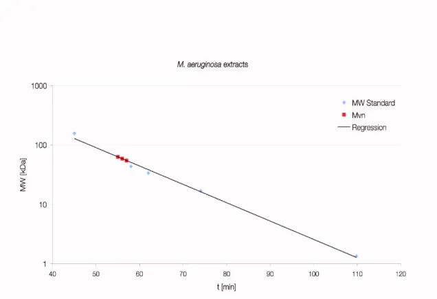
3.1.4 Size determination of native Mvn from M. aeruginosa ...54
3.1.5 Carbohydrate specificity of Mvn ...55
3.1.6 Anti-HIV activity of Mvn...58
3.1.7 Detection of mannan moieties on the M. aeruginosa cell surface ...59
3.1.8 In situ detection of Mvn in M. aeruginosa PCC 7806 ...63
3.1.9 Microvirin binds to LPS ...65
3.1.10 The loss of Mvn leads to a reduced production of microcystin...67
3.2 DIRECT INTERACTION OF MICROCYSTIN AND MICROVIRIN...69
3.2.1 Binding of microcystin to Mvn ...69
3.2.2 Microcystin binding can be suppressed by blocking of thiol groups...70
3.2.3 Microcystin binds to cysteines via N-methyl-dehydroalanine ...72
3.2.4 Influence of microcystin binding on the oligomerisation of Mvn ...74
3.3 INFLUENCE OF OXIDATIVE STRESS CONDITIONS ON MICROCYSTIN-PROTEIN INTERACTIONS...77
3.4 OCCURENCE AND EVOLUTION OF MVN...81
3.4.1 Distribution of mvn among different Microcystis species...81
3.4.2 Evidence for the loss of Mvn in M. aeruginosa NIES 843 ...83
3.4.3 Comparison of Mvn sequences from various M. aeruginosa strains...84
3.4.4 Phylogeny of CV-N domains...85
3.5 FIELD STUDIES...89
3.5.1 Morphotype diversity and sample quality...89
3.5.2 Immunofluorescence detection of Mvn ...94
3.5.3 LBA on field samples ...96
3.5.4 Immunofluorescence detection of MrpC ...98
3.5.5 The MrpC antibody recognises accompanying bacteria ...100
4 DISCU SSION ... 102
4.1 LIMITATIONS...102
4.2 GENERAL CHARACTERISATION OF MICROVIRIN...103
4.3 MICROCYSTIN BINDS TO PROTEINS...104
4.3.1 Microcystin binds covalently to cysteine SH-groups of MVN ...104
4.3.2 General considerations on in vivo microcystin binding ...105
4.4 IMPACT OF MICROCYSTIN ON MICROVIRIN...109
4.4.2 Influence of microcystin-binding on disulphide bond formation ...110
4.5 IMPLICATIONS ON MICROCYSTIN DETECTION AND RISK ASSESSMENT...111
4.6 IMPLICATIONS FROM FIELD STUDIES...112
4.7 HYPOTHESIS FOR THE FUNCTION OF MICROVIRIN...114
4.7.1 Mvn in intraspecies interactions and morphotype determination ...114
4.7.2 Mvn and buoyancy regulation ...116
4.7.3 Microcystin and Mvn in stress adaptation...116
4.8 PHYLOGENETIC ASPECTS OF MVN...117
4.9 CONCLUDING REMARKS AND OUTLOOK...120
REFERENCES ... 122
SUPPLEMENT ... 142
ABBREVI ATI ONS ... 144
PUBLICATIONS ... 147
SELBSTÄNDIGKEI TSERKLÄRU NG ... 149
Z USAMMEN FASSUNG
Sowohl in Süßwasserseen als auch in marinen Gewässern kommt es in den Sommermonaten immer wieder zu Massenentwicklungen von Cyanobakterien, sogenannten “Blüten”. In Seen werden diese oftmals von Cyanobakterien der Gattung Microcystis dominiert, deren Arten häufig Toxine bilden und somit eine Gefahr für Menschen und Tiere darstellen. Die verbreitesten dieser Toxine sind die leberschädigen Microcystine, die eine Klasse nichtribosomal synthetisierter Peptide darstellen. Nachdem die toxische Wirkung der Microcystine bisher als deren Hauptfunktion angesehen wurde, deuten neuere Forschungsergebnisse darauf hin, dass Microcystine eine andere Primärfunktion für die Produzenten besitzen. Es wird unter anderem angenommen, dass Mircocystine eine Rolle in der Antwort auf oxidativen Stress, wie er z.B.
durch Starklicht ausgelöst wird, spielen.
Im Rahmen dieser Studie wurde Microvirin (Mvn), ein putatives Lektin aus Microcystis aeruginosa PCC 7806, von dem angnommen wurde, dass es funktional mit Microcystin assoziiert ist, charakterisiert. Zunächst konnte gezeigt werden, dass Mvn tatsächlich zuckerbindende Aktivität besitzt und spezifisch Mannan, ein Oligosaccharid aus Mannoseuntereinheiten, erkennt. Bindestudien mit fluoreszenzmarkiertem Mvn und Antikörpern zeigten, dass Zucker dieses Typs auf der Zelloberfläche von M. aeruginosa PCC 7806 lokalisiert sind und eine Bindestelle für das sekretierte Mvn darstellen.
Mit Hilfe fluoreszenzmikroskopiebasierender Methoden wurde gezeigt, dass sowohl Mvn als auch das korrespondierende Mannanoligosaccharid stammspezifisch sind. Weiterhin konnte durch PCR gezeigt werden, dass das mvn-Gen in allen getesteten Microcystis-Stämmen vorkommt, die auch Gene für die Microcystinbiosynthese besitzen.
Eine direkte Interaktion von Microcystin und Mvn konnte in vitro bestätigt werden. Microcystin bindet dabei über seinen N-Methyl-Dehydroalaninrest kovalent an die reduzierten Cysteinreste des Proteins. Ein Einfluss auf die Oligomerisierung des Proteins wurde festgestellt. Microcystin scheint unspezifisch Cysteinreste von Proteinen zu binden, und es konnte gezeigt werden, dass dies besonders unter oxidativen Stressbedingungen wie Eisenmangel und Starklichtexposition verstärkt geschieht. Die Daten liefern somit weitere Indizien für eine Rolle von Microcystin in der Stressadaptation.
A BSTRACT
Cyanobacteria frequently appear as so-called “water-blooms” during summer months.
Cyanobacteria of the genus Microcystis, whose species often dominate freshwater lakes, produce toxins that represent a potential threat for humans and animals. The most prominent toxins are the non-ribosomally synthesised hepatotoxic microcystins. Toxicity has been considered the main function of these peptides, but recent studies propose different primary functions of microcystins for their producers. The involvement of microcystins in the response to oxidative stress was proposed recently.
Within this study the putative lectin microvirin (Mvn), which was suggested to be functionally related to microcystin, was characterised. Initially it was shown that Mvn does indeed possess a carbohydrate binding activity, and specificity for mannan, an oligosaccharide made of mannose subunits, was proven. Binding studies using fluorescence-labelled Mvn and antibodies identified carbohydrates of this type at the cell surface of M. aeruginosa being a binding site for the secreted Mvn.
Fluorescence microscopy techniques were employed to show that Mvn as well as the corresponding mannan oligosaccharide are strain-specific. Additionally it was shown by PCR that the mvn gene is present in all tested Microcystis strains possessing microcystin biosynthesis genes.
A direct interaction of microcystin and Mvn was confirmed in vitro. Microcystin covalently binds to the reduced cysteine residues of the protein via its N-methyl-dehydroalanine moiety. An impact on the oligomerisation state of Mvn was observed. Microcystin seems to bind cysteine residues in an unspecific manner in vivo, and it was shown that this occurs especially under conditions of oxidative stress such as iron depletion and exposition to high light. Hence, the data provide further evidence for an involvement of microcystins in stress adaptation.
1 I NTRODUCTION 1.1 Cyanobacteria
Cyanobacteria, also referred to as blue-green algae, are oxygenic photolithotrophic prokaryotes capable of CO2 assimilation through photosynthesis using the reducing power derived from the light-driven water cleavage (Lengeler et al., 1999). They are the only group among bacteria that perform an oxygenic photosynthesis and they have contributed essentially to the formation of the oxygen atmosphere of the earth (Dismukes et al., 2001). Fossil cyanobacteria findings have been dated to 2.5 to 3.5 billion years (Schopf and Packer, 1987). Due to their photosynthesis activity cyanobacteria are important primary producers (Liu et al., 1998). They occupy a wide range of habitats reaching from aquatic – both marine and freshwater – to terrestrial. These include extreme habitats like hot springs and deserts (Whitton and Potts, 2000). Other species are able to fix molecular nitrogen in heterocysts - specialised cells that are impervious for oxygen, which is toxic for the nitrogenase enzyme (Rippka, 1988). Other species can live in symbiosis with plants or fungi (DePriest, 2004). The progenitor of today’s chloroplasts was an ancient cyanobacterium-like endosymbiont that was engulfed by a heterotrophic eukaryote (McFadden, 1999).
Cyanobacteria show considerable morphological diversity and have been classified into five sections based on morphological features (Rippka et al., 1979). Representatives of section I and section II are unicellular and divide by binary fission or multiple fission, respectively. Section III comprises filamentous nonhetercystous cyanobacteria. Members of section IV and V are also filamentous but are capable of cell differentiation. The strains of section V can be distinguished from section IV members by their ability to form branched filaments.
Cyanobacteria are a rich source of secondary metabolites, in particular non-ribosomal peptides (NRPS) and polyketides (PKS) (Welker and von Döhren, 2006). By using degenerate primers Christiansen et al. have demonstrated that NRPS genes are present in 75% of 146 axenic strains of the Pasteur Culture Collection, which included members of all cyanobacterial sections (Christiansen et al., 2001).
1.1.1 M
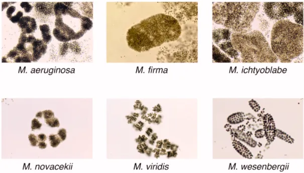
ICROCYSTIS AERUGINOSAforming genus and its different species are determined by distinct colony morphology (FFig. 1).
However, this taxonomy is not supported by phylogenetic analyses. Comparisons of 16S to 23S ribosomal DNA internal transcribed spacer sequences from five morphospecies revealed a high degree of sequence similarity. Clusters in a phylogenetic tree generated from this data did not correspond to morphological characteristics of the examined strains (Otsuka et al., 1999).
Therefore, these strains should be considered as one species regarding them as morphological variants (Otsuka et al., 2001).
Microcystis species produce a variety of secondary metabolites, mainly NRPS (Welker et al., 2004). Because of its toxic impact on eukaryotes the hepatotoxic heptapeptide microcystin (see 1.1.2) is the most prominent.
Fig. 1: A selection of comm on Microcystis colony morphotypes (pictures from:
http://research.kahaku.go.jp/botany/aoko/aokokids/mycro-pictures.html).
1.1.1.1 H
ABITATMicrocystis inhabits freshwater lakes all over the world. In contrast to e.g. Planktothrix species that are mainly found in shallow lakes, Microcystis commonly occupies deep lakes with a stable stratification, because it performs an extensive buoyancy regulation. During warm summers
Microcystis occurs in so called “blooms“ - mass developments of cyanobacteria. Bloom formation is promoted by eutrophication of lakes.
1.1.1.2 C
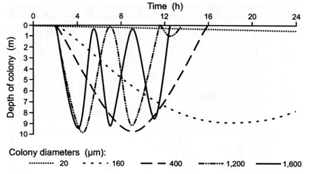
OLONY FORMATION AND BUOYANCY REGULATIONA key feature of Microcystis spp. is the ability to form characteristic colonies, which seems to play an important role in buoyancy regulation. In stratified lakes Microcystis is able to perform vertical migration in the water body. The buoyancy is regulated through gas vesicles and carbohydrate content of the cells. Microcystis continues to form gas vesicles at high irradiance (150 μmol m-2s-1), but buoyancy is reduced due to the accumulation of carbohydrates (Thomas and Walsby, 1985). Field studies have confirmed that the relative gas vesicle volume does not change significantly during the day. The loss of buoyancy is mainly driven by the accumulation of carbohydrate and protein ballast (Ibelings et al., 1991). The cell density increases as a result of enhanced photosynthesis activity at the lake surface and the cells become less buoyant. The carbohydrate ballast is reduced as a consequence of respiration in deeper layers of the lake and the cell density decreases again. Under light–limited conditions M. aeruginosa was shown to be always buoyant. A loss of buoyancy was only observed when energy was generated faster than it could be utilised for growth (Kromkamp et al., 1988). Furthermore, the gas vesicle volume decreased during phosphorus-limited and nitrogen-limited growth, whereby the role of carbohydrate accumulation was emphasised (Chu et al., 2007). Since nutrient limitation in stratified lakes often occurs in the surface layer, the cells might migrate to deeper layers to meet their nutrient requirements. A further parameter of vertical migration is the colony size. Though it does not influence the buoyancy itself colony size contributes by modulating velocity of the sinking and rising, respectively. Several models of the vertical migration (FFig. 2) were computed and showed that, according to Stoke’s law, large colonies migrate faster than smaller ones (Rabouille et al., 2005). Hence, colony diameter determines the time the cells are exposed to light or nutrition rich dark layers (Rabouille et al., 2003). The influence of colony size has been verified by field observations where small colonies were most abundant in deeper water (Ibelings et al., 1991).
Fig. 2: A model describing the correlation between colony size and vertical migration.
According to Stoke’s law an increase in colony diameter results in an accelerated movement.
Therefore bigger colonies can adjust their position in the water column far more rapidly than smaller ones. From Chorus & Bartram, 1999.
1.1.1.3 C
HEMOTYPES OFM
ICROCYSTISNumerous Microcystis strains produce a variety of secondary metabolites and the chemotype is defined as the entirety of peptides produced by a particular strain. Although the morphotype of Microcystis cannot be deduced from the genotype (Otsuka et al., 2000; Otsuka et al., 1999), several studies tried to find a correlation between the morphotype and the chemotype. A study conducted by Fastner et al. (2001) in Lake Wannsee (Berlin, Germany) revealed a correlation between certain peptide combinations and morphotypes (Fastner et al., 2001). Microcystins were chiefly found in M. aeruginosa, while colonies of M. ichtyoblabe and M. wesenbergii did not contain microcystins but anabaenopeptins, microginins and cyanopeptolins. Additionally, the occurrence of microcystin in combination with anabaenopeptins and microginins was mutually exclusive. A second study in lakes around Berlin could not find a clear correlation of morphotype and chemotype, although the production of microcystins was mainly attributed to M. aeruginosa while M. ichtyoblabe did not produce microcystins (Welker et al., 2004). A survey on distribution of microcystin-producing Microcystis in European freshwater bodies revealed that M. aeruginosa and M. botrys morphospecies have a higher proportion of mc-producers (<70%) than M. flos-aquae and M. ichtyoblabe, while M. wesenbergii did not contain
microcystin at all (Via-Ordorika et al., 2004). In 2000 a PCR-based study on the distribution of microcystin genotypes in Lake Wannsee led to similar results (Kurmayer et al., 2002).
1.1.2 M
ICROCYSTINMicrocystins (FFig. 3) are a group of hepatotoxic heptapeptides that are predominantly produced by strains of the genera Anabaena, Microcystis and Planktothrix. In addition, occurence of microcystins has been reported for single strains of Anabaenopsis, Hapalosiphon and Nostoc (Sivonen and Jones, 1999). Microcystins (mc) share the common structure of cyclo(-Adda-D-Glu-Mdha-D-Ala-L-X-D-MeAsp-L-Z) where X and Z are variable L-amino acids, Adda is 3-amino-9-methoxy-2,6,8-trimethyl-10-phenyl-4,6-decadienoic acid, D-MeAsp is 3- methylaspartic acid and Mdha is N-methyl-dehydroalanine (Botes et al., 1984; Sivonen and Jones, 1999). Modifications occur at all seven amino acid residues and over 70 variants have been described to date.
Fig. 3: Microcystin- LR
The toxicity of microcystins originates from their ability to bind eukaryotic protein phosphatases of type 1 and 2A (MacKintosh et al., 1990; Runnegar et al., 1995; Runnegar et al., 1993;
Toivola et al., 1994). Protein phosphatases are the antagonists of protein kinases and their inhibition causes a hyper-phosphorylation of their target proteins. Microcystin is taken up by the organic anion transport system of hepatocytes and induces excessive phosphorylation of cytoskeletal filaments in the liver, which can lead to a hemorrhagic shock (Falconer and Yeung,
health were reported. In Brazil, unsufficient treatment of water contaminated with microcystin- LR caused the death of 40 dialysis patients (Carmichael et al., 2001; Jochimsen et al., 1998).
1.1.2.1 M
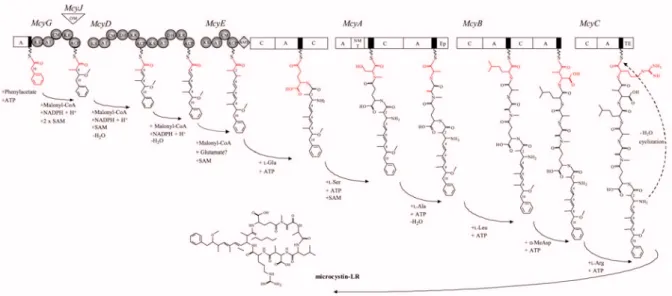
ICROCYSTIN BIOSYNTHESISMicrocystin is synthesised by a large multienzyme complex (Dittmann et al., 1997; Tillett et al., 2000) comprising nonribosomal peptide synthetases (NRPS), polyketide synthases (PKS) and additional modifying enzymes (FFig. 4). This enables the incorporation of non-proteinogenous amino acids and the introduction of modification like epimerisations and methylations.
NRPS incorporate each single amino acid by the “multiple-carrier-thio-template”-mechanism into the growing peptide chain. The NRPS comprise a linear order of modules and each module performs the recognition and the activation of a single amino acid and the subsequent linkage to the peptide chain. The sequence of the peptide is directly determined by the arrangement of the modules (Finking and Marahiel, 2004; Schwarzer et al., 2003).
The Adda moiety in microcystin is synthesised by PKS. PKS catalyse the sequential condensation of short chain carbonic acids. Similar to NRPS, PKS are modular and the structure of the product can be deduced from the number and the arrangement of the modules.
Fig. 4: M odel for t he formation of microcystin and predicted domain struct ures of the six m ultienzymes (McyA-E, G) involved in microcystin biosynt hesis. White rectangles represent peptide synthetase domains (NRPS). The NRPS thiolation motif is shown as a black bar.
Grey circles represent polyketide synthase domains (PKS). The stepwise incorporation of the individual microcystin moieties is indicated in red. KS, h-ketoacyl synthase; AT, acyltransferase; ACP, acyl carrier protein; KR, ketoacyl reductase; DH, dehydratase; CM, C-methyltransferase; OM; O- methyltransferase, A, aminoacyl adenylation; C, condensation; AMT, aminotransferase. Precursors are indicated below the arrows. From Dittmann & Börner, 2005.
The genes encoding the microcystin biosynthesis are organised in a bidirectional operon with mcyABC transcribed in one direction and mcyD-J in the opposite direction. In addition to the biosynthesis genes a putative ABC-transporter McyH is encoded, which may facilitate the proposed export of microcystin (Pearson et al., 2004).
1.1.2.2 F
UNCTION OF MICROCYSTINSThe synthesis of microcystin involves the expression of a large enzyme complex and 48 single reaction steps (Tillett et al., 2000), which implies that it is of significant benefit to the orgnanism justifying this great effort. Several studies tried to answer the question of microcystin function, but no primary function could be assigned beyond doubt so far. A variety of studies have analysed the factors that stimulate the production of microcystin. Multiple impact like nutrients in particular phosphorus, nitrogen and inorganic carbon and environmental factors like light and
2005; Hesse and Kohl, 2001; Kaebernick et al., 2000; Lee et al., 2000; Long et al., 2001; Lyck et al., 1996; Song et al., 1998; Watanabe and Oishi, 1985; Wiedner et al., 2003). However, none of these parameters could be identified as the dominating criterion. Nevertheless, most studies found an influence of the growth phase and microcystin production was highest during exponential growth. Thus the function could not be inferred from the knowledge of parameters driving microcystin production.
In the following the most discussed hypotheses shall be introduced. Different modes of microcystin action were suggested which can be classified as interspecies, intraspecies or intracellular function.
1.1.2.2.1 I
NTERSPECIES FUNCTIONDue to its toxicity microcystin was considered a protection against ingestion by Daphnia galeata (Christoffersen, 1996). Nevertheless, other studies contradict by pointing out that Daphnia cannot distinguish between toxic and nontoxic strains (Rohrlack et al., 2001) and beyond this they are not able to ingest the large colonies usually formed by microcystin producers.
Additionally, the feeding inhibition observed on Daphnia fed with toxic and non-toxic Microcystis strains could not be attributed to microcystin (Kaebernick et al., 2001). It was inferred that reduced growth of Daphnia fed with non-toxic strains compared to a Scenedesmus diet can be attributed to general avoidance of Microcystis (Lurling, 2003).
Furthermore, allelopathic interactions among cyanobacteria and other lake species such as Daphnia, green algae or dinoflagellates are discussed. However, the significance of these laboratory experiments is unclear, because the microcystin concentrations used exceeded those that can be found in the natural environment. Allelopathic interactions between toxic and non-toxic strains were disproven by competition experiments (Kardinaal et al., 2007; Takeya et al., 2004)
1.1.2.2.2 I
NTRASPECIES FUNCTIONSome reports propose a role of microcystin as an infochemical, similar to that of homoserine lactones in quorum sensing. Two proteins, MrpA and MrpB (mmicrocystin-rrelated pprotein), that exhibit significant similarity to the quorum-sensing regulated proteins RhiA and RhiB from Rhizobium leguminosarum were shown to be less expressed in the mcyB mutant. Thus
accumulation of microcystin in the surrounding media might trigger a coordinated response of the whole M. aeruginosa community (Dittmann et al., 2001). Others report that microcystin released by cell lysis stimulates expression of the microcystin synthesis proteins and propose that the enhanced production of microcystin enhances the fitness of the remaining cells (Schatz et al., 2007).
1.1.2.2.3 I
NTRACELLULAR FUNCTIONSThe major part of microcystin is usually cell-bound and not released to the medium. Some studies showed that microcystin is located at distinct sites within the cell. Immunogold localisation experiments (Gerbersdorf, 2006; Young et al., 2005) found microcystin to be bound to thylakoids, phosphate bodies and carboxysomes. A thylakoid localisation was further supported by the isolation of these (Jüttner and Luthi, 2008).
Zilliges compared the proteome of M. aeruginosa PCC 7806 wild type and microcystin-deficient mcyB mutant by 2D gel electrophoresis and could show that differences are often found in proteins related to CO2 fixation (Zilliges et al., 2008). Most proteins of the Calvin cycle including RubisCO were differentially expressed. Furthermore, the direct binding of microcystin to RubisCO was proposed and confirmed in vitro. The author postulated a role of microcystin in redox-regulated processes similar to that of thioredoxins. The influence of carbon availability on microcystin production was previously described (Jähnichen et al., 2001). As mentioned above, carbon fixation and accumulation of intracellular carbohydrates are key factors of buoyancy regulation, as is the colony size. Two extraccellular proteins were identified that might contribute to colony formation, which both seemed to be affected by microcystin. These proteins – microvirin (Mvn) and MrpC – will be introduced in the following.
1.2 Microvirin
Microvirin (Mvn) was identified during the analysis of the partial genome sequence of M.
aeruginosa PCC 7806 available at that time (Kehr, 2003). The aim was to identify genes that might be related to microcystin. The microvirin gene (mvn) was chosen because it is located downstream of a 84 bp sequence (mcy-Box, FFig. 5), which displayed 84% identity to a nucleotide region that overlaps one of the transcriptional start points of the mcyA gene
(http://www.ncbi.nlm.nih.gov/blast/Blast.cgi) and the only protein exhibiting significant similarity (above 20%) in 2003 was cyanovirin-N (see chapter 1.4.8). Thus the protein was named microvirin. Cyanovirin-N (CV-N) is a oligomannose-binding lectin. Lectins bind oligosaccharides and are usually involved in cell-cell recognition and attachment (see chapter 1.4).
In order to analyse the expression of Mvn in M. aerugiosa PCC 7806 and its microcystin- deficient mutants, the protein was expressed heterologously and an antibody was raised against the purified recombinant protein. It was shown that the expression increased at higher culture densities. Additionally, the expression was delayed in the mcyB mutant providing first evidence of an influence of microcystin on Mvn.
Fig. 5: Schematic representation of the conserved nucleotide box (mcy-box) upst ream of A) the microvirin (m vn) gene and of B) microcystin biosynthesis genes (mcy). Arrows indicate distances from translational and transcriptional start sites. C) Sequence comparision of the nucleotide regions adjacent to the mvn and mcy genes, respectively.
It was proposed that Mvn might play a role in microcystin modulated colony formation in M.
aeruginosa. A mutant was generated by insertional mutagenesis, but initial investigations did not reveal a peculiar phenotype. However, the laboratory cultured strain M. aerugniosa PCC 7806 is unable to form colonies anymore and therefore the hypothesis could not be proven by a knockout phenotype.
1.3 MrpC
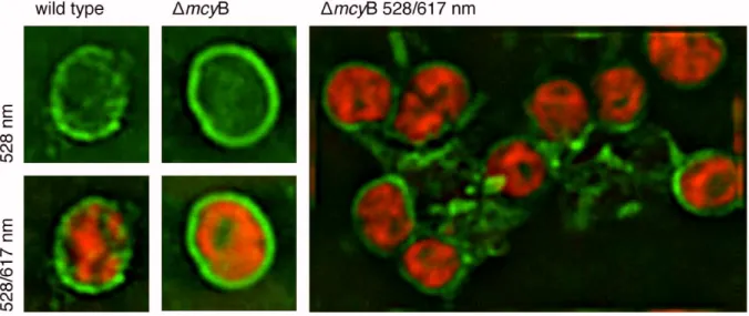
In addition to Mvn a second extracellular protein called MrpC that is strongly affected by microcystin was found. The MrpC protein was identified by Zilliges et al. and characterised as the most abundant extracellular protein from the supernatant of a M. aeruginosa PCC 7806 culture (Zilliges et al., 2008). The expression of MrpC was strongly increased in the microcystin- deficient mcyB mutant compared to the wild type under all conditions tested. The distribution of the MrpC encoding gene showed a correlation with a microcystin genotype and together with the expression studies the results implied a strong relation to microcystin. Further biochemical characterisation proved MrpC to be the target of O-glycosylation, presumably by a glycosyltransferase encoded downstream of the mrpC. Presence of a transit peptide indicated a transmembrane localisation via the Sec pathway. Immunofluorescence microscopy confirmed the increased abundance of the protein in the mcyB mutant and showed that MrpC covers the whole cell surface of M. aeruginosa (FFig. 6). Additionally, MrpC was shown to form connections between individual cells of the mcyB mutant. Therefore, the protein was implicated in microcystin dependent colony formation in M. aeruginosa PCC 7806.
Fig. 6: IImm unofluorescence micrographs of the mcyB mutant and WT st rains of M.
aeruginosa PCC 7806 obtained by using t he antibody against MrpC and a FITC- coupled secondary antibod y. The green fluorescence represents the MrpC protein, which is distributed over the cell surface of M. aerugiosa in a sphere-like manner. The protein is much more abundant in the mutant and seems to establish connection between individual cells. Images were recorded showing only the antibody fluorescence (528 nm) or additionally the chlorophyll autofluorescence (528/617 nm). From Zilliges et al., 2008.
1.4 Lectins
Lectins are a heterogeneous group of proteins that reversibly and with high specificity bind to carbohydrates. Lectins are found in virtually all organisms ranging from bacteria to fungi and to plants and animals. The first description of a lectin refers to Stillmark (1888) who observed the erythrocyte-agglutinating effect of a protein isolated from the castor tree (Ricinus communis) which he named ricin. Until the second half of the 20th century lectins were called hemagglutinins. Various lectins showed different hemagglutinating activity on red blood cells from different animals, an observation that led Karl Landsteiner to discover the human A, B, 0 blood type system in 1900. In 1954 Boyd and Shapleigh introduced the term “lectin“ derived form the Latin word “legere“ (to choose) to summarise plant agglutinins. This concept was later extended to embrace all sugar-specific agglutinins of nonimmune origin, irrespective of source and blood type specificity (Sharon and Lis, 1972).
Although lectins from different organisms display no or only little similarity on the sequence level, they share some important structural features. A striking feature shared by many lectins is the proportion of -sheets in the tertiary structure (Sharon and Lis, 1990).
Lectins mediate biological recognition by detecting specific carbohydrate moieties inside cells, on cell surfaces and physiological fluids (Sharon and Lis, 1989). High specificity is achieved by storing information in carbohydrates. In contrast to amino acids or nucleotides, carbohydrates bear a high coding capacity. Monosaccharides contain several approximately chemically equivalent sites for chain elongation. Chemically distinct compounds can be generated by linking one unit at the reducing end to different hydroxy groups of the second unit, also allowing branched structures. Furthermore, the occurrence of different anomeric variants at each linkage has to be taken into account. With a set of 20 different monosaccharide building blocks as many as 1.44x10
15hexasaccharides are possible regarding the considerations mentioned above. The number of possible permutations for a hexapeptide from a set of 20 amino acids is only 6.4x10
7(Gabius et al., 2004). The level of diversity can be further extended by the introduction of substituents like sulfation and epimerisation of D- to L-forms.
It was estimated that over the half of all proteins occuring in nature is glycosylated (Apweiler et
al., 1999) and additionally cell membranes are decorated with glycolipids. This emphasises theimportance of glycoproteins and lectins that are involved in numerous and diverse biological
processes (Gabius et al., 2002). Indeed glycosylation is very common in bacteria (Hitchen and
Dell, 2006) and defects in glycosylation led to impaired pathogenicity and attachment of
mucosal pathogens to host cells (Szymanski and Wren, 2005). The majority of glycosylated bacterial proteins identified in pathogens are implicated in interactions with the host (Benz and Schmidt, 2002).
1.4.1 M
ECHANISM OF CARBOHYDRATE BINDINGThe carbohydrate binding sites of lectins exhibit a relatively low affinity to monosaccharides with dissociation constants in the millimolar range, whereas the oligosaccharides are bound with nanomolar affinity. This arises from the structure of the binding pocket which is a rather shallow depression on the protein surface in contrast to e.g. the sugar-binding bacterial periplasmic receptors for glucose or galactose that bind the ligand in the interior of the protein (Lis and Sharon, 1998). Lectins interact with carbohydrates primarily through hydrogen bonds and hydrophobic interactions. Charge-charge interactions are not relevant because most saccharides are uncharged. The hydrogen bonds are formed between hydroxyl groups of the carbohydrate which interact with amino and hydroxyl groups and oxygen atoms of the protein.
The hydroxyl groups of sugars enable cooperative hydrogen bonding where an OH group can act as a donor of one hydrogen bond and an acceptor of two hydrogen bonds simultaneously (Weis and Drickamer, 1996).
Although carbohydrates are highly polar, they have significant nonpolar patches formed by aliphatic protons and carbons at the epimeric centers. These patches interact with aromatic residues like tryptophan or phenylalanine in the protein. In addition, several classes of lectins require divalent cations like Ca2+ or Mn2+ to be functional. Two modes of interaction, direct and indirect, were described. Direct interactions occur in C-type lectins where a Ca2+ is required to form direct coordination bonds with the sugar ligand. However, indirect interactions are given if a cation interacts only with amino acid side chains to stabilise the interaction with the ligand.
Most lectins contain two or more carbohydrate binding sites, which is the reason for their hemagglutinating activity. One lectin can bind to at least two carbohydrate moieties on different cells, which results in the precipitation of the cells. Additionally, many lectins form multimers, which further increases the number of binding sites compared to the monomer. The resulting multivalency employed by many if not all lectins is considered to substantially enhance the selectivity and affinity of lectin interactions (Rini, 1995).
1.4.2 F
UNCTIONS OF LECTINSLectins are involved in many processes in and between organisms and although they play diverse roles the recognition of specific carbohydrates is a common prerequisite to accomplish their function. Biological processes that require a specific targeting are e.g. defense, innate immunity, symbiosis or glycoprotein trafficking (TTa b. 1, (Sharon and Lis, 2004)). In addition to the examples listed in TTab. 1 some well-characterised lectins from plants, animals and bacteria are introduced in the following and known cyanobacterial lectins are described.
Tab. 1: Function of lectins. Adapted from Sharon and Lis, 2004.
LLectin Role in
Microorganisms Amoeba Bacteria Infuenza virus
Infection Infection Infection
Plants Various
Legumes
Defence Symbiosis Animals Calnexin, calreticulin, ERGIC-53
Collectins Dectin-1 Galectins
Macrophage mannose receptor
Man-6-P receptors L-selectin
E- and P-selectin Siglecs
Spermadhesin
Control of glycoprotein biosynthesis Innate immunity
Innate immunity
Regulation of cell growth and apoptosis;
regulation of the cell cycle; modulation of cell- cell and cell-substratum interactions
Innate immunity; clearance of sulfated glycoprotein hormones
Targeting of lysosomal enzymes Lymphocyte homing
Leukocyte trafficking to sites of inflammation Cell-cell interactions in the immune and neural system
Sperm-egg interaction
1.4.3 A
NIMAL LECTINSThe examples listed in TTab. 1 show that lectins have manifold functions in mammals, but the majority are innate immune molecules. Many lectin-like innate immune proteins and receptors that interact with bacteria are known today (Palaniyar et al., 2002). The pulmanory surfactant proteins SP-A and SP-D are C-type (Ca2+-dependent) lectins that maintain surfactant
homeostasis were additionally shown to be important host defense components (Holmskov et al., 2003; Wright, 2005). Both proteins bind to bacteria causing agglutination and thus hinder their entrance into the host cell and dissemination. In addition they enhance the phagocytosis by macrophages. Several pathogens, among them Staphylococcus aureus, Pseudomonas aeruginosa, Escherichia coli, Mycobacterium tuberculosis, Haemophilus influenzae and yeast and fungi are recognised (Kishore et al., 2006). Another important lectin in the innate immunity is the mannose-binding lectin (MBL), which binds to a large number of pathogens. MBL was shown to activate a novel pathway of complements (lectin pathway) that is independent from antibodies or C1 protease (Dommett et al., 2006).
The lectins calnexin and calreticulin localised in the endoplasmic reticulum (ER) are involved in glycoprotein folding. They serve as molecular chaperones, recognising newly synthesised and glycosylated polypeptide chains and preventing aggregation and export of incompletely folded proteins from the ER (Helenius and Aebi, 2004).
Lectins can also play a role in symbioses between animals and bacteria. The marine nematode Laxus oneistus secretes a mannose-specific lectin onto the posterior region of its cuticle to facilitate attachment of its sulfur-oxidising bacterial symbiont (Bulgheresi et al., 2006).
1.4.4 P
LANT LECTINSLegume lectins that are involved in Rhizobia-legume symbiosis are the best-investigated plant lectins. According to the lectin recognition hypothesis proposed by Hamblin and Kent (1973), Bohlool and Schmidt (1974), Dazzo and Hubbell (1975) and reviewed by Hirsch (Hirsch, 1999), legume lectins mediate the specificity between the legume host and the Rhizobium symbiont.
The bacteria expose carbohydrates on their cell surface, e.g. lipopolysaccharides, capsular polysaccharides or acidic exopolysaccharides that can be bound by plant lectins. Several studies showed that mutants in the production of these polysaccharides are often unable to establish symbiosis.
Another function frequently described for plant lectins is the defense against pathogens like fungi, insects, and bacteria. Several chitin-binding lectins (chitin is a long-chain polymer of N- acetylglucosamine) and chimeric proteins comprising a chitin-binding domain as well as a chitinase domain exhibit a protective activity against fungi (Peumans and Van Damme, 1995).
Many plant lectins are toxic to insects and mammals and are stored in vacuoles in plant seeds or adult plants. Upon ingestion of the plant by herbivores they are released and deploy their toxicity. The wheat germ agglutinin (WGA) from wheat (Triticum aestivum) e.g. is toxic to weevils, and the common bean’s phytohemagglutinin (PHA) is toxic to mammals (Chrispeels and Raikhel, 1991). It was shown that PHA causes lesions in the intestinal mucosa.
1.4.5 B
ACTERIAL LECTINSBacterial lectins are often involved in infection and mediate the initial step of host-cell- recognition and attachment. The human pathogen Helicobacter pylori attaches to human salvary glycoproteins (Prakobphol et al., 2005) and expresses several lectins and lectin-like proteins (Hynes et al., 2003).
A group of chimeric proteins carrying a lectin domain includes the AB–toxins of Enterobacteria.
These toxins comprise two domains, the A and B domain, which are made of two polypeptide chains usually linked by two disulphide bonds. The B domain (binding domain) is a lectin and responsible for receptor recognition, whereas the A domain (activity domain) causes the cytotoxic effects. One group of AB-toxins are the Shiga and Shiga-like toxins from Shigella sp.
and E. coli. They bind to the glycolipid Gb3 on the host-cell-surface and are then internalised by endocytosis (Sandvig, 2001). Glycolipids are also the attachment site for a Porphyromonas gingivalis adhesin (Hellstrom et al., 2004).
Bacterial lectins also play an important role in communal behaviour. The dental pathogen Streptococcus mutans expresses several secreted glucan-binding proteins (Gbp) during biofilm formation. Deletion of the respective genes significantly altered the physical properties of the biofilm produced. Especially a reduction in thickness of the biofilm and an impaired ability of cell-aggregation was observed (Banas et al., 2007; Lynch et al., 2007).
The involvement of lectins in biofilm formation was also described for the biofilm model organism Pseudomonas aeruginosa. The quorum-sensing regulated LecA lectin was detected and the mutants showed a reduced surface coverage and no evidence of microcolony formation. Furthermore, the biofilm development was impaired by the preincubation with hydrophobic galactosides, which are the type of sugars specifically recognised by LecA (Diggle et al., 2006).
1.4.6 C
YANOBACTERIAL LECTINSVery little is known about lectins and their role in cyanobacteria. In recent years few lectins from cyanobacteria were isolated starting with the anti-HIV protein cyanovirin-N from Nostoc ellipsosporum (Gustafson et al., 1997) (see chapter 1.4.8). The developmental potential of cyanovirin-N as anti-HIV drug led to screenings for substances with similar activity in cyanobacteria. Hence, lectins have been isolated from different cyanobacterial genera that all exhibit a more or less strong affinity to oligomannose, the same oligosaccharide that is recognised by cyanovirin-N (Ziókowska and Wlodawer, 2006). Namely these are the Oscillatoria agardhii agglutinin (OAA) and scytovirin from Scytonema varium (Bokesch et al., 2003;
McFeeters et al., 2007; Sato et al., 2007). Both proteins comprise two domains that show sequence homology to each other and contain internal disulphide bonds. Nevertheless, information about their in vivo function is lacking so far.
1.4.7 M
ICROCYSTIS LECTINSTwo lectins, MAL and MVL, were isolated from Microcystis strains. The Microcystis viridis lectin (MVL) agglutinates rabbit erythrocytes and hemagglutinating activity could be inhibited by yeast mannan. It consists of 113 aa and is composed of two tandemly repeated homologous domains of 54 aa. There are currently no homologous sequences to be found in the databases.
Expression studies revealed that the MVL is expressed in cultures cultivated without aeration at the stationary phase. Therefore the authors consider MVL a stress protein that may be involved in adaptation to an unfavourable growing environment (Yamaguchi et al., 1999). MAL has been isolated from M. aeruginosa M228. The 55.2 kDa protein comprises three tandemly repeated homologous domains of 61 aa which show partial similarity to the -amylase from Clostridium beijerinckii. The genes adjacent to the mal gene display homology to a cytochrome P-450 and a polyketide synthase. The hemagglutinating activity of MAL could be inhibited by N-acetyl-D- galactoseamine and lactose. The expression of the protein was highest at low temperature (15°C) and low light (12 μEm-2) (Jimbo et al., 2000; Yamaguchi et al., 1998; Yamaguchi et al., 2000).
1.4.8 C
YANOVIRIN-N
Cyanovirin-N (CV-N) is a lectin isolated from the filamtenous cyanobacterium Nostoc ellipsosporum during a screening for compounds with anti-HIV activity. It is a small 11 kDa lectin with specifity for 1,2-mannan. The protein was found to bind to such carbohydrate epitopes present in the gp120 envelope protein of the HI-virus. Through binding to gp120 CV-N disturbs the interaction with the cell surface receptor CD4 and thus prevents the virus from infecting the host cell (Bewley and Otero-Quintero, 2001; Boyd et al., 1997; Chang and Bewley, 2002; Dey et al., 2000; Esser et al., 1999; Mori and Boyd, 2001; O'Keefe et al., 2000; Shenoy et al., 2001).
Due to its potential use as an HIV therapeutic detailed analysis focussed on the biochemical and structural properties of CV-N are available in the literature, but yet no information on the role of this lectin in Nostoc ellipsosporum exists. The most important features will be outlined in the following.
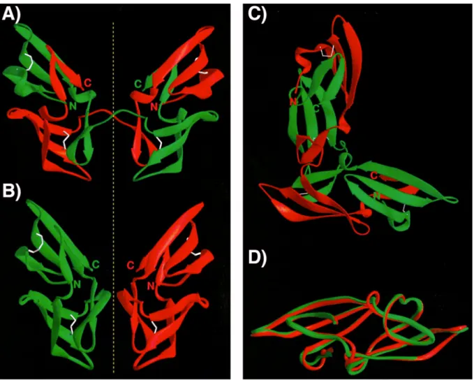
CV-N comprises two domains that share 32% sequence identity to each other and that may result from a duplication event during evolution. Several structures were obtained from cyanovirin-N (FFig. 7) by NMR as well as by X-ray crystallography (Bewley, 2001; Bewley et al., 2002; Bewley and Otero-Quintero, 2001; Botos et al., 2002; Botos et al., 2002; Fromme et al., 2007; Sandstrom et al., 2004; Shenoy et al., 2002; Yang et al.). The protein exists as a monomer in solution and is made of two distinct symmetric domains A and B that are formed by strand exchange of the sequence repeats. This means that domain A is made of residues 1- 39 and 90-101 and domain B is formed by residues 39-90. The protein contains four cysteine residues and an intra-domain disulphide bond is present in each domain. The whole structure is dominated by anti-parallel -sheets (Bewley et al., 1998). The crystallised protein was shown to form domain-swapped dimers (FFig. 7A-C), in which domain A of one monomer interacts with domain B of the second monomer and vice versa (Yang et al., 1999). Mutants were created that formed stable dimers in solution (Han et al., 2002).
Fig. 7: Crystal struct ure on CV- N: A-C) Different views of the domain swapped dimer of CV-N.
Individual monomers are shown in green and red. The yellow dashed line indicates the symmetry. D) Structure of a CV-N monomer. The high proportion of anti-parallel -sheets and the pronounced symmetry of multimers are frequently found in lectins. From Yang et al., 1999.
1.4.8.1 C
YANOVIRIN-N D
OMAINSIn recent years several cyanovirin-N-like proteins have been identified, mostly in filamentous ascomycetes. The European Bioinformatics Instiute (EBI) contains an entry in its InterPro
database under the accesion number IPR011058 (http://www.ebi.ac.uk/interpro/IEntry?ac=IPR011058) that lists all proteins containing a CV-N
domain. Apparently some of these proteins contain two CV-N domains, while very few only contain half a domain that represents just one of the internal repeats of cyanovirin-N. Percudani and coworkers analysed the CV-N homologues concerning their secondary structure and their evolutionary relationship. The major findings will be summarised in the following (Percudani et
LysM domain that is inserted into the hinge region between the two CV-N repeats. Furthermore, signal-peptides for membrane translocation were identified in the CV-N homologues of the fern Ceratopteris richardii indicating an extracellular function. The cysteine residues involved in disulphide bond formation in cyanovirin-N are conserved among the homologues that posses a signal peptide while they are replaced in most of the intracellular proteins.
The overall tertiary structure of the CV-N domain family members seems to be conserved across the whole family, because hydrophobic residues crucial for protein folding are conserved, while e.g. the amino acids that form the carbohydrate binding pocket are rather variable.
The rather patchy organism distribution of the CV-N domain implies that the organisms acquirred the protein after the separation of the lineages by horizontal gene transfer (HGT) events. However, the internally repeated structure of cyanovirin-N arose from a unique ancient duplication.
1.5 Aim of this Work
Many reports published recently suggest functions for microcystin that do not focus on its toxicity but rather postulate a physiological relevance of the peptide. Proteomic studies have shown a major impact of the absence of microcystin on the abundance of a number of proteins.
Among these, the extracellular glycoprotein MrpC as well as glycosyltransferases were identified, which proposed an influence of microcystin on cell surface composition.
The objective of this study is the characterisation of microvirin (Mvn), which previously was shown to be influenced by the presence of microcystin. Homology based prediction implied the secretion of Mvn and a carbohydrate binding capability. Thus the subcellular localisation should be examined and the carbohydrate binding activity and specificity should be elucidated. In order to confirm the proposed functional relationship of microvirin and microcystin further, the distribution of the mvn gene among laboratory and field strains of Microcystis should be examined.
Several publications have shown the ability of microcystin to bind to proteins. Therefore, it has to be tested if the relationship of Mvn and microcytin becomes manifested in an interaction of both. Thus, binding of microcystin to the heterologously expressed Mvn should be tested. If a
binding of microcystin is observed the conditions that promote the binding as well as the mechanism should be investigated.
2 M ATERIALS AND M ETHODS 2.1 Materials
2.1.1 B
ACTERIAL STRAINS2.1.1.1 M
ICROCYSTIS AERUGINOSAThe strain Microcystis aeruginosa PCC 7806 used in this study was obtained from the “Pasteur Culture Collection“ (Paris, France). The mutants used in this study were derived from this strain.
M. aeruginosa mcyB (Dittmann et al., 1997) and mcyH mutants (Pearson et al., 2004) generated by insertional mutagenses are deficient in microcystin production. The mvn mutant was produced by disruption of the mvn gene (Kehr, 2003). All mutants were selected on chloramphenicol.
Additionally, a set of M. aeruginosa strains was used to investigate the distribution of the mvn gene within the species (TTab. 2).
Tab. 2: List of M. aeruginosa st rains used to investigate the distribution of the mvn gene.
M. aeruginosa strain
Source
PCC 7005 PCC 7820 PCC 7941 PCC 9354 PCC 9355 PCC 9432 PCC 9622 PCC 9624 PCC 9701 PCC 9717 PCC 9804 PCC 9805 PCC 9806 PCC 9807 PCC 9808 PCC 9809 PCC 9812 PCC 9905 PCC 100-24 PCC 100-25
“Pasteur Culture Collection”, Paris, France
HUB 5.3 Humboldt University,
Berlin, Germany NIES 44
NIES 89 NIES 100 NIES 104 NIES 299
National Institute for Envrionmental Studies, Tsukuba, Japan
UWOCC CBS UWOCC MRC UWOCC MRD
University of Wisconsin at OshKosh Culture Collection, Madison, USA
2.1.1.2 O
THER CYANOBACTERIANostoc punctiforme ATCC 29133 from the “American Tissue and Culture Collection” and Synechocystis PCC 6803 from the “Pasteur Culture Collection” were used as controls in immunofluorescence microscopy.
2.1.1.3 E
SCHERICHIA COLIFor cloning purposes the strain E. coli XL-1 blue (Stratagene) was used, heterologous expression was performed in the strain E. coli BL21 (DE3) (Novagen).
2.1.2 K
ITSBio-Rad Protein Assay Biorad
FluoroTagFITC Conjugation Kit Sigma-Aldrich Jetsorb „Gel Extraction Kit“ Genomed Nickel-NTA-Superflow Qiagen PCR Purification Kit Qiagen Plasmid Mini Prep Qiagen Taq DNA-Polymerase PCR Kit Qiagen SuperSignal West Pico Pierce
2.1.3 C
HEMICALS2-mercaptoethanol C. Roth
Acetone C. Roth
Acrylamide/Bisacrylamide (37.5:1) C. Roth Acetonitril “HPLC Gradient Grade“ C. Roth
Agar, washed Difco
Agarose Biozym Diagnostik
Ampicillin Roche Diagnostics
APS C. Roth
Bacto-Agar Difco
Bacto-Trypton Difco Boric acid C. Roth Bovine serum albumin, fraction V C. Roth
Bromphenol blue SERVA Feinbiochemika Chloramphenicol Roche Diagnostics Coomassie staining “Roti-Blue” C. Roth
Dithiothreitol C. Roth
dNTP MBI Fermentas
DTNB C. Roth
EDTA C. Roth
Ethanol C. Roth
Ethidium bromide C. Roth Formaldehyde 37% C. Roth GelCode Blue Stain Reagent Pierce
Glycerol C. Roth
Glycine C. Roth
HEPES Amersham Pharmacia Hydrochloric acid C. Roth
IPTG C. Roth
Isopropanol C. Roth Magnesium chloride C. Roth
Methanol C. Roth
PMSF Serva Potassium chloride C. Roth
N-Propylgallate C. Roth
Sodium acetate C. Roth Sodium chloride C. Roth Sodium dihydrogenphosphate C. Roth
Sodium dodecylsulfate (SDS) SERVA Feinbiochemika Sodium hydrogenphosphate C. Roth
Sodium hydroxide C. Roth
Tris C. Roth
Tween 20 Sigma
Urea ICN Biochemicals
X-Gal C. Roth
Yeast extract Difco
2.1.4 E
NZYMESLysozyme GERMED
Restriction Endonucleases MBI Fermentas New England Biolabs T4-DNA-Ligase MBI Fermentas Taq-Polymerase Qiagen
2.1.5 A
NTIBODIESThe following antibodies were used in this study (The used titer is given for all antibodies. In some cases antibodies were applied in a different titer for immunoblotting and immunofluorescence microscopy (IFM).
Tab. 3: Antibodies used in t his st ud y.
Antibody Antigen Source Titer Reference
Primary antibodies
Anti-Microcystin Adda moiety of microcystin from
M. aeruginosa
Mouse, polyclonal
1:10,000 Alexis
Anti-MrpC MrpC from
M.aeruginosa
Guinea pig, polyclonal
1:5000 or 1:500
Zilliges et al., 2008
Anti-Mvn Microvirin from
M.aeruginosa
Rabbit, polyclonal
1:10,000 or 1:500 (IFM)
Kehr, 2003
Anti-Poly-Histidin Poly-Histidin Mouse, monoclonal
1:10,000 Sigma
Secondary antibodies Anti-Guinea Pig IgG FITC conjugate
Mouse IgG Goat 1:100 (IFM) Sigma
Anti-Mouse IgG
Horseradish Peroxidase conjugate
Mouse IgG Sheep 1:10,000 Amersham Pharmacia
Anti-Rabbit IgG FITC conjugate
Rabbit IgG Goat 1:100 (IFM) Sigma
Anti-Rabbit IgG
Horseradish Peroxidase conjugate
Rabbit IgG Goat 1:10,000 Sigma
2.1.6 N
UCLEIC ACIDS2.1.6.1 P
LASMIDSpDrive Qiagen
pet15b Novagen
2.1.6.2 P
RIMERName Sequence Comment
mvn_fw ATGCCTAATTTTTCGCACACTTGTAG mvn_rv TCCAATTTCCAGTTGGCTGTCGTT
Screening, amplification and sequencing of mvn genes from various Microcystis strains
mcyE_fw TTCCCCTTAACTCGACATGG mcyE_rv TAAAGTCGCCAATCCAGCAA
Detection of mcy genotypes among Microcystis strains. The primers were deduced from conserved regions in the mcyE gene.
2.1.7 F
ILTERS ANDM
EMBRANES3MM Filter-Paper Whatman
Hyperfilm MP X-ray detection film Amersham Pharmacia PVDF-Blotting-Membrane Hybond C Amersham Pharmacia
2.1.8 L
IST OFM
ANUFACTURERSCompany Based in
Agilent Technologies Darmstadt, Germany
Ambion, Inc. Austin, USA
Amersham Biosciences Europe GmbH Freiburg, Germany Applied Biosystems Weiterstadt, Germany Applied Precision Issaquah, USA
Biolabs Frankfurt am Main, Germany Bio-Rad Laboratories Richmond, USA
Boehringer GmbH Mannheim, Germany C. Roth GmbH & Co. KG Karlsruhe, Germany
Difco Detroit, USA
DuPont de Nemours GmbH Bad Homburg, Germany
Eppendorf Hamburg, Germany
Eurogentech Seraing, Belgien Fluka-Biochemika Steinheim, Germany
GERMED Berlin, Germany
Gibco/ BRL Life Technologies New York, USA
Heraeus Hanau, Germany
ICN Biochemicals/ MP Biomedicals Irvine, USA
Invitrogen GmbH Karlsruhe, Germany
MBI Fermentas GmbH St. Leon-Rot, Germany
Merck Darmstadt, Germany
Millipore Cooperation Bedford, USA
New England Biolabs Schwalbach, Germany
Novagen Nottingham, UK
PerkinElmer Instruments Shelton, USA
Philips Instruments Eindhoven, Netherlands
Promega Cooperation Madison, USA
Qiagen Hilden, Germany
Roche Diagnostics GmbH Mannheim, Germany Schleicher & Schüll Dassel, Germany Serva Feinbiochemika & Co. KG Heidelberg, Germany
Shimadzu Kyoto, Japan
Sigma Chemical Company St. Louis, USA
Waters Eschborn, Germany
Whatman Paper Ldt. Maidstone, UK
Zeiss Jena, Germany
2.2 Methods
2.2.1 M
ICROBIOLOGICAL METHODS2.2.1.1 C
ULTIVATION OF CYANOBACTERIACyanobacteria were cultivated in BG11-medium in Erlenmeyer flasks or on plates containing BG11-medium and 0.7% agar (Rippka et al., 1979). Stock cultures were grown in liquid at 23°C and continuous illumination (~20-30 μEm-2s-1). Exposure experiments were conducted using aliquots of a preculture grown at 16 μEm-2s-1 and constant aeration in a batch vessel. The culture aliquots were transferred to sterile batch vessels and subjected to the desired light conditions under constant aeration for two hours. The following light conditions were applied:
High light (70 μEm-2s-1), low light (16 μEm-2s-1) and darkness. External microcystin was added in some case (for details refer to the results section).
2.2.1.2 C
ULTIVATION OFE
SCHERICHIA COLIEscherichia coli cells were cultivated under standard conditions either in liquid LB medium or on LB agar in petri dishes (Sambrook and Russel, 2001). Cultures for preparation of plasmid vector DNA were incubated in 3-4 ml liquid LB medium at 37°C and shaking at 220 rpm. Proper antibiotics were added in the appropiate concentrations.
2.2.2 M
OLECULAR BIOLOGICAL METHODS2.2.2.1 P
REPARATION OF GENOMICDNA
FROMM
ICROCYSTIS AERUGINOSAThe isolation of genomic DNA from Microcystis aeruginosa was performed as described previously (Hisbergues et al., 2003). Cyanobacterial cells were harvested by centrifugation (4000 x g, 10 min) and the pellet was washed twice in TE-buffer. The pellet was resuspended in 0.5 ml TES-buffer and incubated on ice for 1 h. Afterwards lysozyme was added to a final concentration of 2 mg/ml and the cells were incubated at 37°C in a waterbath for 1 h. Then SDS (2% final concentration) and ProteinaseK (50 μg/ml final concentration) were added and the mixture was incubated at 50°C for 2-3 h. Following one volume of phenole/chloroform (1:1) was added, the tubes were shaken and centrifuged at 13,000 rpm for 10 min. The supernatant was transferred to a fresh tube and the extraction step was repeated. After centrifugation the
supernatant was mixed with chloroform/isoamylalcohol (24:1) and centrifuged as before. The upper aqueous phase was mixed with 0.7 vol isopropanol to precipitate the DNA and centrifuged at 13,000 rpm for 30 min. The resulting pellet was washed with 70% ethanol and air-dried after centrifugation. The DNA was resuspended in water or TE-buffer. Finally, 1 μl of an RNase-Mix was added and the tubes were incubated at 37°C for 1h.
TE buffer 10 mM Tris-HCl, 1 mM EDTA
TES buffer 25% w/v Saccharose, 50 mM Tris-HCl, 100 mM EDTA, pH 8.0
2.2.2.2 P
REPARATION OF PLASMIDDNA
FROME
SCHERICHIA COLIPlasmid DNA from E. coli was isolated according to the method of alkaline lysis (Sambrook and Russel, 2001). Cells from a 3 ml culture grown overnight in LB medium supplemented with the appropriate antibiotic were harvested by centrifugation and resuspended in 300 μl buffer P1. An equal volume of buffer P2 was added after incubation at RT for 5 min and the tubes were flipped 4-6 times. The cells were incubated at RT for 5 min before 300 μl of buffer P3 were added. The tubes were flipped again and kept on ice for at least 5 min. Cellular debris were pelleted by centrifugation in a table centrifuge (13,000 rpm, 4°C, 30 min). The supernatant was transferred to a fresh tube and the DNA was precipitated with 0.7 vol 2-propanol. After centrifugation (13,000 rpm, 4°C, 30 min) the pellet was washed with 70% ethanol and centrifuged again. The pellet was air-dried and resuspended in water or TE-buffer.
P1 50 mM Tris-HCl pH 8.0; 10 mM EDTA P2 200 mM NaOH; 1% SDS
P3 3 M Potassium acetate, pH 5.0
2.2.2.3 Q
UANTIFICATION OFDNA
DNA concentrations were determined by measuring the absorption at 260 nm in a photometer.
The absorption of one arbitrary unit corresponds to 50 μg/μl double-stranded DNA.
2.2.2.4 A
GAROSE GEL ELECTROPHORESIS OFDNA
DNA fragments were separated on 0.5%-1.2% (w/v) agarose gels by electrophoresis (Sambrook and Russel, 2001). Ethidium bromide was added to the gels in final concentration of 0.05 μg/ml to visualise the DNA under UV illumination. The gels were run in TBE running buffer at a constant voltage of 100 V. DNA samples were mixed with DNA loading dye prior to application onto the gel. An aliquot from a PstI digest of phage DNA was applied as size marker on each gel.
TBE running buffer 45 mM Tris-Borate, pH 8.5; 1.8 mM EDTA
5 x DNA loading dye 50 % Ficoll; 1 mM EDTA, pH 8.0; 0.05 % (w/v) Bromophenole blue; 0.05 % (w/v) Xylene cyanol
2.2.2.5 E
LUTION OFDNA
FRAGMENTS FROM AGAROSE GELSDNA fragments were eluted from agarose gels using the Jetsorb “Gel Extraction Kit” (Genomed) according to the manufacturer’s manual.
2.2.2.6 P
OLYMERASE CHAIN REACTIONAmplification of DNA fragments was performed using the Taq DNA-Polymerase PCR Kit (Qiagen). The web-based software “Primer3” (http://frodo.wi.mit.edu/cgi- bin/primer3/primer3_www.cgi) was used to design primers. Reactions were carried out in a volume of 20 μl containing the Taq-buffer, 2 μl MgCl2, 10 μmol of each primer, 10 μmol dNTPs, 1 U Taq-polymerase and 1 μl template DNA. The reaction was run in a thermocycler using a suitable amplification program. Common steps were the initial denaturation at 95°C for 5 min followed by 35 cycles of denaturation at 95°C for 30 s, a primer annealing step at the primer specific temperature for 30 s and the sequence elongation at 72°C. The time of elongation was chosen depending on the expected length of the PCR product assuming an amplification speed of 1 kb/min. The PCR reaction was completed with a final elongation step at 72°C for 10 min.
Different template DNA was used depending on the particular analysis. Purified genomic DNA was applied in a final concentration of 50-200 ng while plasmid DNA was used in a concentration of ~10 ng. Colony PCR was performed for quick analysis of a large set of
bacteria or clones. A bacterial suspension was diluted 20-100fold and boiled for 10 min for this before 1μl was applied to the PCR reaction mix.
2.2.2.7 P
URIFICATION OFDNA
Primers, nucleotides and other impurities were removed from PCR reactions by purification with the Qiaquick PCR Purification Kit (Qiagen) following the manufacturer’s instructions.
2.2.2.8 D
IGESTION OFDNA
WITH RESTRICTION ENDONUCLEASESDNA was cleaved with the restriction endonuclease of choice in a reaction volume of 20-50 μl according to manufacturer’s instructions.
2.2.2.9 L
IGATION OFDNA
FRAGMENT IN PLASMID VECTORSPurified PCR products were directly ligated into the pDrive vector (Qiagen) following the company’s protocol.
Directed cloning of DNA fragments into the pet15b expression vector was achieved by digesting the vector with NdeI and BamHI restriction endonucleases. Afterwards, the DNA fragment of choice was cut out from the pDrive cloning vector using the same enzymes, purified by gel extraction (see 2.2.2.5) and subsequently ligated into the linearised pet15 vector using the T4 DNA ligase (Fermentas) in the recommended buffer. The ligation mix was incubated at 4°C overnight.
2.2.2.10 S
EQUENCING OFDNA
DNA was cloned into the pDrive vector and the column purified (Qiagen) vector DNA was delivered for automatic sequencing to “SMB Service in Molecular Biology” (Berlin). Raw sequence files were viewed and edited with “4Peaks” software (http://mekentosj.com/4peaks) on a Macintosh computer running MacOS X.
2.2.2.11 T
RANSFORMATION OFE
SCHERICHIA COLICells of E. coli were transformed utilising the CaCl2-chemically induced competence (Sambrook and Russel, 2001) of the XL-1 strain (Stratagene). A volume of 200 μl of XL-1 cell culture was mixed with the respective ligated plasmid vector and kept on ice for 20 min. The mixture was then subjected to a heat shock in a water bath at 42°C for 1 min. Afterwards, 500 μl of SOC medium were added and cells were incubated for 1 hour at 37°C and shaking at 220 rpm. Cells were spread on LB agar plates containing the appropriate antibiotic. In case of cloning into the pDrive vector (Qiagen) 40 μl X-Gal solution (20 mg/ml in DMF) and 40 μl of IPTG solution (0.1 M) were added to the agar to facilitate blue-white selection of positive clones. The plates were incubated at 37°C overnight.
Positive clones were picked and used to inoculate 3-4 ml LB medium containing the proper antibiotic. The cells were grown shaking at 220 rpm and 37°C overnight and plasmids were isolated (see chapter 2.2.2.2).
2.2.3 P
ROTEIN BIOCHEMICAL METHODS2.2.3.1 H
ETEROLOGOUS EXPRESSIONFor the expression of recombinant His-Mvn 25 ml of LB-Medium supplemented with 100 μg/ml Ampicillin were inoculated with E. coli cells containing the expression construct and grown overnight at 37°C on a shaker (220 rpm). The following day, the overnight culture was used to inoculate the large-scale expression culture in a dilution of 1/50. The culture was grown under conditions mentioned above until an OD600=0.6 was reached. The expression was induced by addition of IPTG to a final concentration of 1 mM and the cells were grown for 3 h. Finally the cells were harvested by centrifugation at 4000 x g for 20 min. The pellets were either frozen or stored at -20°C or subsequently subjected to protein extraction.
2.2.3.2 P
URIFICATION OFH
IS-M
VN FROME.
COLIThe recombinant His-Mvn was purified from the periplasmic fraction. The pellet from an expression culture was resuspended in 40 ml osmotic shock buffer (30 mM Tris-HCl, 20%
sucrose, pH 8.0) supplemented with EDTA to a final concentration of 1 mM and incubated on ice for 10 min with gentle shaking. The cells were centrifuged (6000 x g, 20 min, 4°C), the
supernatant was removed and the pellet was resuspended in the same volume of an ice-cold 5 mM MgSO4 solution and shaken for 10 min on ice. The cells were centrifuged again as described above and the supernatant was retained and extensively dialysed against lysis buffer (50 mM NaH2PO4, 300 mM NaCl, pH 8.0) with three exchanges of the buffer at 4°C.
Afterwards, the lysate was transferred to a fresh 50 ml tube and imidazol was added to a final concentration of 10 mM followed by the addition of 4 ml Ni-NTA slurry (Qiagen). Then the tube was mounted on a rotary shaker and incubated for 1 h at 4°C. Subsequently the mixture was applied to the column with the bottom outlet capped and the Ni-NTA was allowed to settle before the bottom cap was removed. The column flow-through was discarded and the column was washed with 40 ml wash buffer (lysis buffer containing 30 mM imidazol). Finally the column was eluted with 8 x 0.5 ml elution buffer (lysis buffer containing 250 mM imidazol). The purity was estimated from SDS-PAGE gel and highly concentrated fractions were pooled.
2.2.3.3 D
ETERMINATION OF PROTEIN CONCENTRATIONProtein concentrations were determined using the Bio-Rad Protein Assay according to the manufacturer’s instructions. Briefly, 1-5 μl of protein solution was added to a mixture of 0.2 ml dye reagent and 0.8 ml H2O, mixed and incubated for 10 min at RT. Absorbance was measured at 595 nm and the concentrations were calculated using standard curve generated by dilutions of BSA.
2.2.3.4 SDS-P
OLYACRYAMIDE GEL ELECTROPHORESIS(SDS-PAGE)
Proteins were separated by the method of discontinuous gel electrophoresis (Laemmli, 1970).
The gels consisted of a separating gel containing 10-15% acrylamide depending on the protein size range that was to be examined and a stacking gel of 4% acrylamide. Samples were mixed with 5x loading buffer and heated at 95°C for 10 min. Depending on the application a reductant was added to the loading buffer or not (see results). The gels were run at a constant current of 25 mA per gel in the Mini Protean II system (Bio-Rad). Afterwards the gels were washed in distilled water for 15 min and stained with GelCode Blue Stain Reagent (Pierce).
Separating gel 10-15% (v/v) acrylamide/bisacrylamide 37.5:1 (v/v); 375 mM Tris-HCl, pH